Introduction
Spindle cell melanoma (SCM) is a rare subtype of
malignant melanoma composed of spindled neoplastic cells arranged
in sheets and fascicles (1). The
diagnosis of SCM is challenging, as SCM may occur anywhere on the
body and frequently mimics amelanotic lesions, including scarring
and inflammation (2–4). Histologically, cytologic features of SCM
are indistinct and often confused with those of other epithelial
neoplasms, including sarcomas and lymphomas (5–8).
Immunohistochemistry is a helpful tool in distinguishing SCM from
other sarcomas and carcinomas (9,10).
However, diagnosis remains a challenge as a number of sarcomas
share some morphological and immunohistochemical features with SCM
(5,11). Differentiation of SCM from
desmoplastic melanoma is difficult because both melanomas are
characterized by atypical, spindled, malignant melanocytes.
However, the size of spindle cell collagen areas and the
immunohistochemical markers, S100, MelanA and Tyrosinase, allow
differential diagnosis (10).
Therefore, the integration of clinical and histological assessment
is essential for the diagnosis of SCM (2,8). Diagnosis
of SCM is often delayed until patients exhibit advanced-stage
disease, typically with widespread metastasis and poor treatment
outcomes (3,6,12,13).
A limited number of case reports and incomplete
retrospective case studies of the differential diagnostic
viewpoints of SCM exist (3,4,11–15). To the best of our knowledge, few
studies have reported SCM incidence, clinicopathologic features,
treatment, treatment outcome and disease-specific independent
prognostic factors. Thus, the present study performed a
retrospective analysis of a series of clinical cases using data
from the Surveillance, Epidemiology and End Results (SEER)
Program.
Materials and methods
Data collection
In the present study, data was analyzed from the
SEER Program, National Cancer Institute Public Use Dataset, which
contains publically available records of 18 population-based cancer
registries, which together represent 28% of the USA population.
Data were extracted regarding patients with a primary diagnosis of
SCM, according to the International Classification of Diseases for
Oncology, Third Edition (ICD-O-3), using histology codes: 8772/3
(16). Cases were excluded if
treatment or outcome data were unavailable for survival analysis.
The data extraction was carried out with the official software
SEER*Stat, version 8.3.4. (URL: http://seer.cancer.gov/data/).
Statistical analyses
Overall Statistical analysis was accomplished using
Statistical Package for Social Sciences (SPSS; version 23.0, for
Windows; IBM Corp., Armonk, IL, USA). χ2 test or
Fisher's exact test was used to analyze associations among baseline
parameters. The primary endpoint in the present study was
considered to be the date of SCM-associated mortality. The time
point between the date of diagnosis and the date of SCM-associated
mortality was defined as disease-specific survival (DSS).
Mortalities associated with SCM were considered to be events, while
deaths attributed to other causes were considered to be ‘censored
observations’. In terms of overall survival (OS) and DSS rates,
Kaplan-Meier method and the log-rank test were utilized and
multivariate Cox proportional hazard models were used to identify
significant risk factors for survival outcomes. All statistical
tests were two-sided, and P<0.05 was considered to indicate a
statistically significant difference.
Results
Study population and
characteristics
The following demographic and clinicopathological
characteristics were selected for analysis: Age at diagnosis,
ethnicity, primary tumor location, Tumor-Node-Metastasis (TNM)
stage, American Joint Committee on Cancer (AJCC) stage,
pathological grade (the American Joint Committee on Cancer/Union
for International Cancer Control staging system), SEER historic
stage, treatment modalities, vital status and follow-up time
(17). Unfortunately, complete data
was not available for all cases.
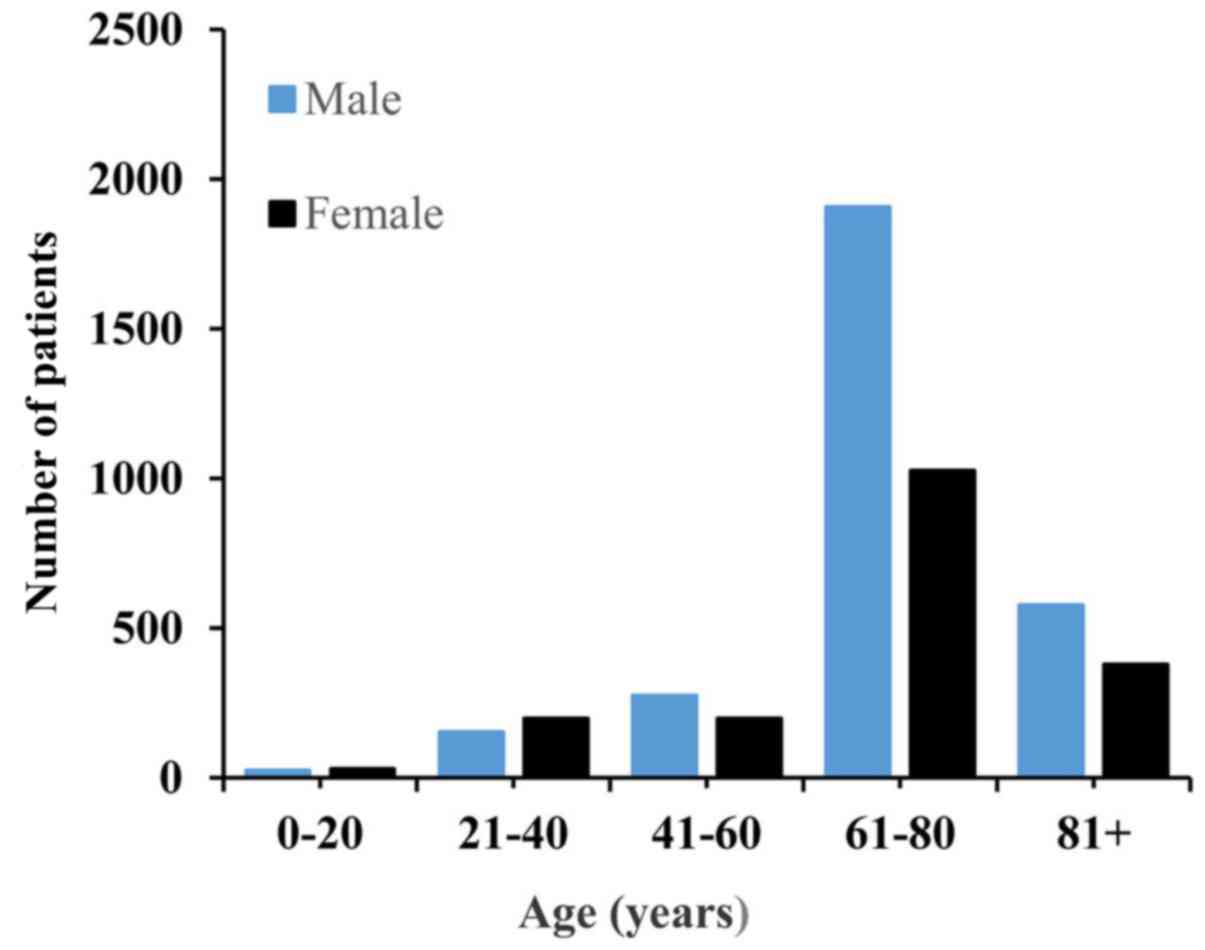
Data from 4,761 patient diagnosed with SCM between
1973 and 2017 was retrieved from the SEER database. The total
cohort consisted of 1,829 women and 2,932 men, with a female:male
ratio of 0.62:1. The patients' age ranged from 3–101 years and a
median age of 66 years. The age and sex distributions are presented
in Fig. 1. The median follow-up time
was 53 months (range, 0–500 months). Regarding ethnicity, Caucasian
people accounted for 96.7% of the study population. The majority of
the cases of SCM had originated from the skin, and the eye and bony
orbits were the second-most affected tumor site. Surgical resection
was performed in 88.7% cases. The basic demographic and
clinicopathologic characteristics of the whole patients are
summarized in Table I. Kaplan-Meier
analysis was utilized for time-to-event analysis. Statistically
significant differences in OS rate were identified depending on age
(P<0.001), sex (P<0.001), tumor location (P<0.001),
ethnicity (P=0.008), AJCC stage (P<0.001), T stage (P<0.001),
pathological grade (P<0.001), N stage (P<0.001), treatment
modalities (P<0.001) and M stage (P<0.001) (Fig. 2). Univariate Cox regression analysis
analysis revealed that age, ethnicity, sex, tumor location,
pathological grade, AJCC stage, T stage, N stage, M stage, SEER
historic stage and treatment modalities were associated with OS
(Table II). Multivariate Cox
regression analysis revealed that positive N stage, age >66
years and SEER historic stage of regional and distant metastasis
were factors independently associated with worse OS (Table III).
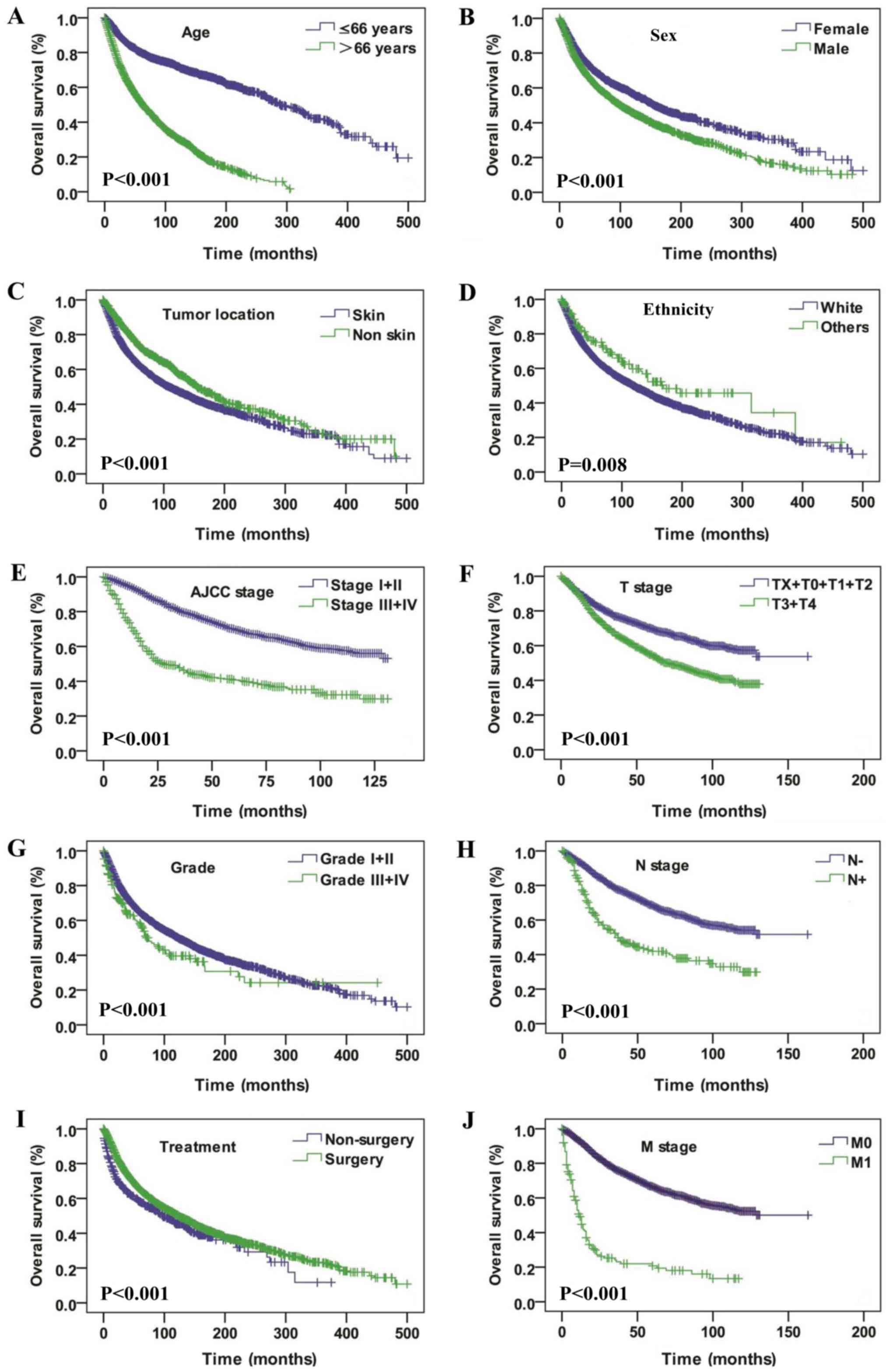 | Figure 2.Overall survival curves of patients
with spindle cell melanoma compared according to (A) age, (B) sex,
(C) tumor location, (D) ethnicity, (E) AJCC stage, (F) T stage, (G)
pathological grade, (H) N stage, (I) treatment, and (J) M stage.
The log-rank test was utilized to compare the curves. AJCC,
American Joint Committee on Cancer; T, tumor; N, node; M,
metastasis. |
 | Table I.The baseline characteristics of the
SCM cases extracted from the SEER database. |
Table I.
The baseline characteristics of the
SCM cases extracted from the SEER database.
|
| DSS | OS |
|---|
|
|
|
|
|---|
| Parameters | Alive | Dead | P-value | Alive | Dead | P-value |
|---|
| Age |
| ≤66
years | 1,333 | 361 | <0.001 | 1,512 | 621 | <0.001 |
| >66
years | 710 | 406 |
| 1,095 | 1,533 |
|
| Sex |
|
Female | 878 | 274 | <0.001 | 1,066 | 763 | <0.001 |
|
Male | 1,165 | 493 |
| 1,541 | 1,391 |
|
| Ethnicity |
|
White | 1,947 | 745 | 0.009 | 2,506 | 2,098 | 0.008 |
|
Black | 16 | 9 |
| 17 | 17 |
|
|
Others | 80 | 13 |
| 84 | 39 |
|
| Tumor location |
| Eyes
and bony orbits | 383 | 118 | <0.001 | 433 | 294 | <0.001 |
|
Internal organs | 7 | 14 |
| 9 | 21 |
|
| Nose
and mouth | 18 | 20 |
| 19 | 29 |
|
|
Skin | 1,624 | 587 |
| 2,131 | 1,766 |
|
| Other
site | 11 | 28 |
| 15 | 44 |
|
| Grade |
| I | 26 | 4 | <0.001 | 30 | 8 | <0.001 |
| II | 16 | 1 |
| 20 | 8 |
|
|
III | 16 | 20 |
| 20 | 40 |
|
| IV | 12 | 16 |
| 15 | 31 |
|
|
Unknown | 1,973 | 726 |
| 2,522 | 2,067 |
|
| AJCC stage |
| I | 552 | 42 | <0.001 | 745 | 184 | <0.001 |
| II | 533 | 118 |
| 729 | 371 |
|
|
III | 138 | 57 |
| 176 | 115 |
|
| IV | 36 | 73 |
| 50 | 115 |
|
| T stage |
| T0 | 22 | 26 | <0.001 | 30 | 40 | <0.001 |
| T1 | 394 | 38 |
| 524 | 148 |
|
| T2 | 345 | 44 |
| 451 | 154 |
|
| T3 | 244 | 60 |
| 347 | 178 |
|
| T4 | 278 | 117 |
| 375 | 284 |
|
| TX | 99 | 42 |
| 144 | 125 |
|
| N stage |
| N0 | 1,226 | 210 | <0.001 | 1,656 | 683 | <0.001 |
| N1 | 57 | 46 |
| 77 | 79 |
|
| N2 | 35 | 16 |
| 46 | 38 |
|
| NX | 65 | 55 |
| 93 | 129 |
|
| M stage |
| M0 | 1,324 | 246 | <0.001 | 1,796 | 779 | <0.001 |
| M1 | 34 | 73 |
| 48 | 114 |
|
| MX | 25 | 8 |
| 28 | 36 |
|
| SEER stage |
|
Localized | 1,499 | 305 | <0.001 | 1,893 | 1,133 | <0.001 |
|
Regional | 419 | 263 |
| 547 | 632 |
|
|
Distant | 8 | 9 |
| 14 | 18 |
|
|
Unknown | 70 | 53 |
| 88 | 143 |
|
| Treatment |
|
Non-surgery | 208 | 109 | 0.011 | 266 | 255 | 0.195 |
|
Surgery | 1,827 | 655 |
| 2,332 | 1,891 |
|
|
Unknown | 8 | 3 |
| 9 | 8 |
|
| Table II.Univariate Cox regression analysis in
terms of DSS and OS rates of SCM |
Table II.
Univariate Cox regression analysis in
terms of DSS and OS rates of SCM
|
| DSS | OS |
|---|
|
|
|
|
|---|
| Parameters | HR (95% CI) | P-value | HR (95% CI) | P-value |
|---|
| Age |
| ≤66
years | 1.0
(reference) | <0.001 | 1.0
(reference) | <0.001 |
| >66
years | 2.372
(2.054–2.739) |
| 3.594
(3.257–3.965) |
|
| Ethnicity |
|
Caucasian | 1.0
(reference) |
| 1.0
(reference) |
|
|
Black | 1.430
(0.741–2.760) | 0.286 | 1.345
(0.835–2.169) | 0.223 |
|
Others | 0.497
(0.287–0.861) | 0.013 | 0.602
(0.439–0.827) | 0.002 |
| Sex |
|
Female | 1.0
(reference) | <0.001 | 1.0
(reference) | <0.001 |
|
Male | 1.403
(1.210–1.627) |
| 1.355
(1.240–1.481) |
|
| Tumor location |
| Eye and
bony orbits | 1.0
(reference) |
| 1.0
(reference) |
|
|
Internal organs | 5.461
(3.132–9.522) | <0.001 | 3.817
(2.449–5.948) | <0.001 |
| Nose
and mouth | 3.824
(2.377–6.151) | <0.001 | 3.231
(2.204–4.738) | <0.001 |
|
Skin | 1.413
(1.159–1.722) | 0.001 | 1.642
(1.450–1.860) | <0.001 |
| Other
site | 5.735
(3.790–8.679) | <0.001 | 3.877
(2.821–5.329) | <0.001 |
| Grade |
| I | 1.0
(reference) |
| 1.0
(reference) |
|
| II | 0.247
(0.028–2.209) | 0.211 | 0.864
(0.324–2.304) | 0.771 |
|
III | 4.496
(1.536–13.154) | 0.006 | 3.405
(1.593–7.278) | 0.002 |
| IV | 5.303
(1.773–15.867) | 0.003 | 3.923
(1.802–8.537) | 0.001 |
|
Unknown | 1.508
(0.564–4.209) | 0.413 | 1.781
(0.889–3.566) | 0.104 |
| AJCC stage |
| I | 1.0
(reference) |
| 1.0
(reference) |
|
| II | 2.892
(2.033–4.113) | <0.001 | 2.072
(1.736–2.474) | <0.001 |
|
III | 5.966
(4.002–8.895) | <0.001 | 2.896
(2.293–3.658) | <0.001 |
| IV | 25.917
(17.641–38.075) | <0.001 | 10.091
(7.968–12.781) | <0.001 |
| T stage |
| T0 | 1.0
(reference) |
| 1.0
(reference) |
|
| T1 | 0.087
(0.053–0.143) | <0.001 | 0.214
(0.151–0.304) | <0.001 |
| T2 | 0.113
(0.069–0.183) | <0.001 | 0.267
(0.189–0.378) | <0.001 |
| T3 | 0.217
(0.137–0.344) | <0.001 | 0.392
(0.278–0.552) | <0.001 |
| T4 | 0.391
(0.256–0.599) | <0.001 | 0.609
(0.437–0.848) | 0.003 |
| TX | 0.378
(0.232–0.617) | <0.001 | 0.627
(0.439–0.895) | 0.010 |
| N stage |
| N0 | 1.0
(reference) |
| 1.0
(reference) |
|
| N1 | 4.273
(3.103–5.883) | <0.001 | 2.320
(1.837–2.929) | <0.001 |
| N2 | 3.448
(2.072–5.737) | <0.001 | 2.296
(1.656–3.185) | <0.001 |
| NX | 4.812
(3.572–6.483) | <0.001 | 3.306
(2.737–3.994) | <0.001 |
| M stage |
| M0 | 1.0
(reference) |
| 1.0
(reference) |
|
| M1 | 10.339
(7.923–13.491) | <0.001 | 5.559
(4.556–6.783) | <0.001 |
| MX | 1.337
(0.661–2.705) | 0.419 | 1.783
(1.276–2.491) | 0.001 |
| SEER stage |
|
Localized | 1.0
(reference) |
| 1.0
(reference) |
|
|
Regional | 2.914
(2.469–3.439) | <0.001 | 1.921
(1.742–2.119) | <0.001 |
|
Distant |
20.058(10.243–39.278) | <0.001 | 8.328
(5.204–13.328) | <0.001 |
|
Unknown | 2.828
(2.112–3.786) | <0.001 | 1.791
(1.505–2.132) | <0.001 |
| Treatment |
|
Non-surgery | 1.0
(reference) |
| 1.0
(reference) |
|
|
Surgery | 0.671
(0.548–0.822) | <0.001 | 0.761
(0.667–0.867) | <0.001 |
|
Unknown | 0.649
(0.206–2.044) | 0.460 | 0.679
(0.336–1.374) | 0.282 |
| Table III.Multivariate Cox regression analysis
of SCM for DSS and OS rates. |
Table III.
Multivariate Cox regression analysis
of SCM for DSS and OS rates.
|
| DSS | OS |
|---|
|
|
|
|
|---|
| Parameters | HR (95% CI) | P-value | HR (95% CI) | P-value |
|---|
| Age |
| ≤66
years | 1.0
(reference) | <0.001 | 1.0
(reference) | <0.001 |
| >66
years | 2.502
(1.900–3.296) |
| 3.799
(3.160–4.568) |
|
| Ethnicity |
|
Caucasian | 1.0
(reference) |
| 1.0
(reference) |
|
|
Black | 2.286
(0.727–7.196) | 0.157 | 2.664
(1.187–5.980) | 0.018 |
|
Others | 1.564
(0.690–3.544) | 0.284 | 1.297
(0.731–2.303) | 0.374 |
| T stage |
|
TX+T0+T1+T2 | 1.0
(reference) | <0.001 | 1.0
(reference) | <0.001 |
|
T3+T4 | 2.113
(1.567–2.847) |
| 1.485
(1.261–1.749) |
|
| N stage |
|
Negative | 1.0
(reference) | <0.001 | 1.0
(reference) | 0.001 |
|
Positive | 2.437
(1.617–3.673) |
| 1.564
(1.204–2.031) |
|
| SEER stage |
|
Localized | 1.0
(reference) |
| 1.0
(reference) |
|
|
Regional | 1.682
(1.199–2.361) | 0.003 | 1.458
(1.208–1.760) | <0.001 |
|
Distant | 57.206
(22.241–147.138) | <0.001 | 18.856
(10.145–35.047) | <0.001 |
Significant differences in the DSS analysis were
also identified depending on age (P<0.001), sex (P<0.001),
pathological grade (P<0.001), AJCC stage (P<0.001), ethnicity
(P=0.009), T stage (P<0.001), tumor location (P<0.001), M
stage (P<0.001), treatment modalities (P<0.001), and SEER
historic stage (P<0.001) (Fig. 3).
Univariate Cox regression analysis demonstrated that age,
ethnicity, sex, tumor location, pathological grade, AJCC stage, T
stage, N stage, M stage, SEER historic stage and treatment
modalities were associated with DSS (Table II). The multivariate Cox regression
model revealed that age >66 years T3+T4 stage, positive N-stage
and SEER historic stage of regional and distant metastasis were
independently associated with a poor OS rate (Table III).
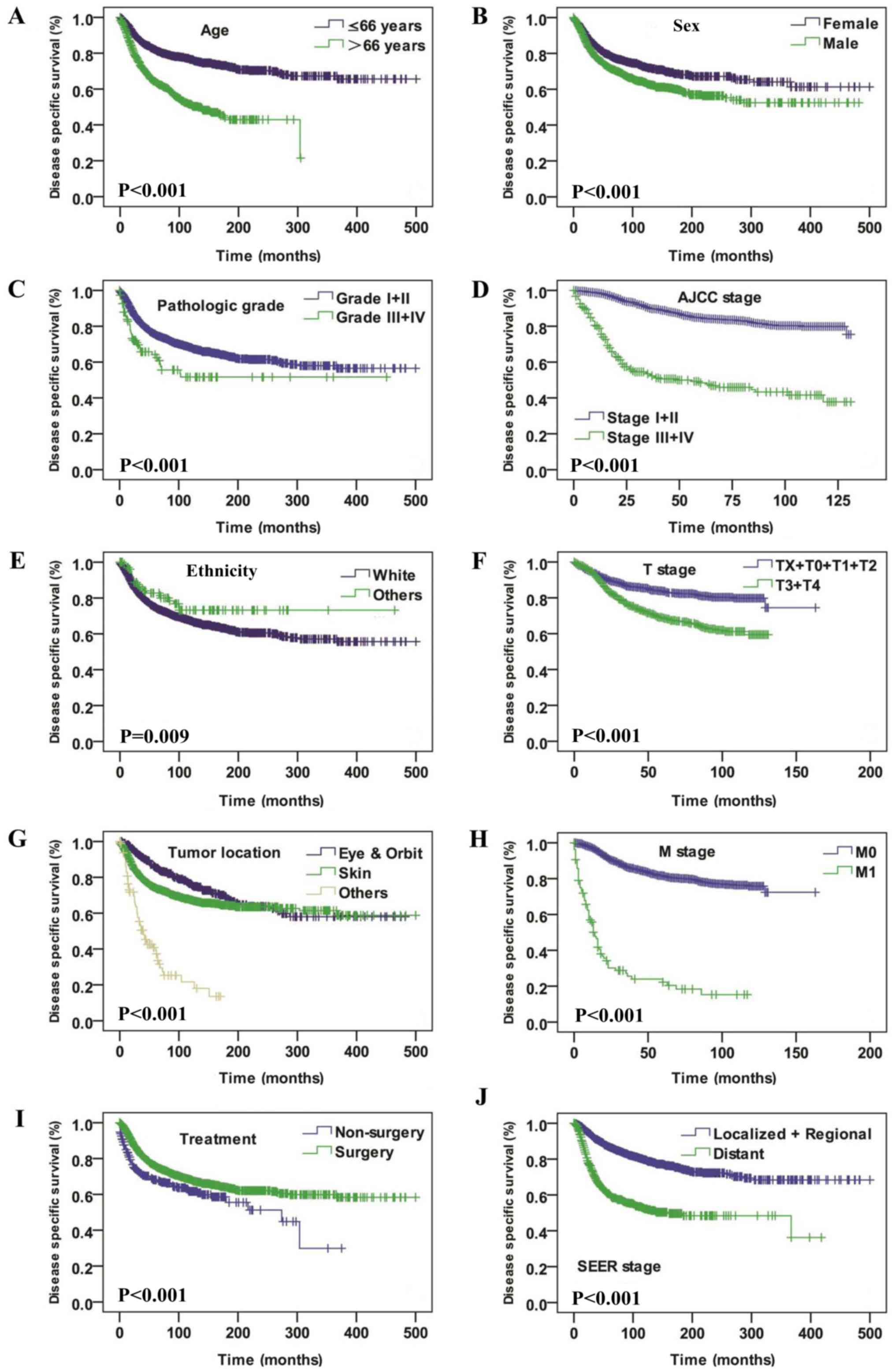 | Figure 3.Disease specific survival curves of
patients with spindle cell melanoma compared according to (A) age,
(B) sex, (C) pathological grade, (D) AJCC stage, (E) ethnicity and
(F) T stage, (G) tumor location, (H) M stage, (I) treatment and (J)
SEER stage. The log-rank test was utilized to compare curves. AJCC,
American Joint Committee on Cancer; T, tumor; N, node; M,
metastasis; SEER, Surveillance, Epidemiology and End Results. |
Discussion
As a morphological variant of melanoma, SCM is rare
and its incidence has been variably reported between 3 and 14% of
all melanoma cases (including desmoplastic melanoma) (15,18,19).
Diagnosis of SCM is challenging and awareness of its clinical and
cytological features as well as immunohistochemical markers are
essential to reach the correct diagnosis (9,10,15). Due to the rarity of SCM, its clinical
and prognostic characteristics remain to be fully elucidated. To
the best of our knowledge, the present study is the first to
investigate SCM incidence as well as survival analysis on a large
scale.
The present study demonstrates that the incidence of
SCM was highest in the 6–8th decade of life in males. Caucasian
people accounted for the majority of the study population. SCM
lesions originated most commonly from the skin and eyes, and the
bony orbits were the second-most affected tumor site. SCM shares
various features with conventional melanoma. Previous studies have
demonstrated that melanomas arise from the melanocytes of the skin
and eyes in response to intrinsic and extrinsic stimuli, including
pro-inflammatory signals, oncogenes and UV radiation (12,20,21). Human
pigmentation is a polygenic quantitative trait with high
heritability and it is modulated by estrogen and androgens via
regulation of melanin synthesis (22). This may explain why SCM mainly
originates from skin and eyes and has a predilection of male and
Caucasian people.
According to previous studies, it appears that age,
sex, ethnicity and tumor location are important prognostic factors
for patients with melanoma (22–24). The
present study indicates that patients with SCM who were male, aged
>66 years, Caucasian, or with tumors located in the skin were
associated with poor OS and DSS rates. In the multivariate Cox
regression analysis, age >66 years was independently associated
with poor OS and DSS rates, which was in consistence with previous
studies on melanoma (24–27). Seeing as it was demonstrated that SCM
was more likely to occur in patients >66 years of age, potential
poor tolerance of complications and common comorbidities of elderly
patients should be also taken into consideration when making
treatment protocol.
Pathological grade has been demonstrated to be an
important prognostic factor for estimation of survival outcome in
melanoma (17). The results of the
present study indicate that well-differentiated SCM was associated
with a relatively good outcome both in terms of OS and DSS rate,
compared with poorly differentiated SCM. Advanced stage SCM was
associated with a relatively poor OS and DSS rates compared with
early-stage tumors. Cox multivariate regression also suggested that
pathology was an important consideration, with advanced T stage,
positive N stage and SEER historic stage of metastasis being
independently associated with poor OS and DSS rates. This
emphasizes that a full awareness of the morphological and clinical
presentations of SCM are required for the accurate diagnosis of
SCM. However, due to the unspecific clinical presentation of SCM,
early detection is often delayed (4,12–14). Metastatic SCM from an unknown primary
site should be taken into consideration when diagnosing SCM lesions
(15,18,28).
Fine-needle aspiration may be a rapid and effective tool for
surveillance of recurrent and metastatic cases of SCM, however,
accurate diagnosis is challenging owing to the varied cytologic
morphologic appearances (6,29–31).
In the present study, surgery was the only
identified treatment modality for SCM. The results indicate that
surgery was favorable for OS and DSS rates. A previous study
demonstrated that wide local excision with clear margins, sentinel
node biopsy and regular follow-up examinations were crucial in the
management of SCM (4). However, SCM
cases should be monitored carefully as metastasis is possible fir
multiple years after surgery (32).
For tumors in complex anatomic regions, including the head and
neck, the treatment of elderly patients or those at an advanced
stage by radical surgery with clear margins is difficult (3). Under these circumstances, therapeutic
planning is challenging (33).
Therefore, multi-cancer randomized clinical trials are urgently
required to improve the available treatment for this melanoma
subtype.
While the SEER Program is a comprehensive and
geographically representative registry, a few limitations of the
present study should be noted. Due to alterations of the criteria
for the histological diagnosis of SCM the diagnosis of SCM patients
in the past may be inconsistent with more recent diagnoses. Another
limitation is that complete data was not available for all cases. A
number of important prognostic data, including surgical types,
margin status and adjuvant therapies were either absent or
incomplete in the available SEER data, and therefore there
influence on prognosis could not be assessed. In addition, the
results suggest that SCM patients may represent an older
population, and data on comorbidities that may affect treatment
protocols and outcomes is lacking. To the best of our knowledge,
this is the first large population study of SCM with a robust
long-term follow-up survival assessment provided by SEER, which
will improve the existing knowledge of the demographic of SCM, its
clinicopathological features and disease-specific prognostic
factors.
Overall, a large scale report of SCM demographic
trends, clinicopathological features, treatment outcomes and
disease-specific independent prognostic factors was presented. The
results demonstrate that SCM mostly occurred in Caucasians and
males, and the highest incidence occurred in the 6–8th decades of
life. Age, ethnicity, T stage, N stage and SEER historic stage were
independent prognostic factors of DSS and OS rates.
Acknowledgements
Not applicable.
Funding
The present study was supported by The Scientific
Research Foundation of Shanghai Stomatological Hospital, Fudan
University (grant no. SSDCZ-2016-01).
Availability of data and materials
The datasets generated during the present study are
available in the official software SEER*Stat, v.8.3.4 repository,
[https://seer.cancer.gov/data/], and
analyzed in Statistical Package for Social Sciences (SPSS; v.23.0,
for Windows; IBM Corp., Armonk, IL, USA).
Authors' contributions
ZX and PS were major contributors in the writing of
the manuscript. FY and LF collected and collated the patient data.
HZ analyzed and the interpreted patient data. AW was responsible
for planning and organizing the study and checking the data and
manuscript. All authors read and approved the final manuscript.
Ethics approval and consent to
participate
Due to the retrospective nature of this study, it
was granted an exemption in writing by the University of Fudan
institutional review board (IRB).
Patient consent for publication
Not applicable.
Competing interests
The authors declare that they have no competing
interests.
Glossary
Abbreviations
Abbreviations:
|
SCM
|
spindle cell melanoma
|
|
OS
|
overall survival
|
|
DSS
|
disease specific survival
|
|
SEER
|
the surveillance, epidemiology and end
results
|
|
AJCC stage
|
American Joint Committee on Cancer
stage
|
|
HR
|
hazard ratio
|
|
CI
|
confidence interval
|
References
|
1
|
Winnepenninckx V, De Vos R, Stas M and van
den Oord JJ: New phenotypical and ultrastructural findings in
spindle cell (desmoplastic/neurotropic) melanoma. Appl
Immunohistochem Mol Morphol. 11:319–325. 2003. View Article : Google Scholar : PubMed/NCBI
|
|
2
|
Diaz A, Valera A, Carrera C, Hakim S,
Aguilera P, García A, Palou J, Puig S, Malvehy J and Alos L:
Pigmented spindle cell nevus: Clues for differentiating it from
spindle cell malignant melanoma. A comprehensive survey including
clinicopathologic, immunohistochemical, and FISH studies. Am J Surg
Pathol. 35:1733–1742. 2011. View Article : Google Scholar : PubMed/NCBI
|
|
3
|
Dainichi T, Kobayashi C, Fujita S,
Shiramizu K, Ishiko T, Kiryu H, Urabe K, Tsuneyoshi M and Furue M:
Interdigital amelanotic spindle-cell melanoma mimicking an
inflammatory process due to dermatophytosis. J Dermatol.
34:716–719. 2007. View Article : Google Scholar : PubMed/NCBI
|
|
4
|
Sheff JS and Pane TA: Spindle cell
melanoma arising from decades-old burn scar. Plast Reconstr Surg.
124:274e–275e. 2009. View Article : Google Scholar : PubMed/NCBI
|
|
5
|
Jackson CR, Minca EC, Kapil JP, Smith SC
and Billings SD: Superficial malignant peripheral nerve sheath
tumor with overlying intradermal melanocytic nevus mimicking
spindle cell melanoma. J Cutan Pathol. 43:1220–1225. 2016.
View Article : Google Scholar : PubMed/NCBI
|
|
6
|
Walia R, Jain D, Mathur SR and Iyer VK:
Spindle cell melanoma: A comparison of the cytomorphological
features with the epithelioid variant. Acta Cytol. 57:557–561.
2013. View Article : Google Scholar : PubMed/NCBI
|
|
7
|
Falconieri G, Bacchi CE and Luzar B:
Cutaneous clear cell sarcoma: Report of three cases of a
potentially underestimated mimicker of spindle cell melanoma. Am J
Dermatopathol. 34:619–625. 2012. View Article : Google Scholar : PubMed/NCBI
|
|
8
|
Yeh I, Vemula SS, Mirza SA and McCalmont
TH: Neurofibroma-like spindle cell melanoma: CD34 fingerprint and
CGH for diagnosis. Am J Dermatopathol. 34:668–670. 2012. View Article : Google Scholar : PubMed/NCBI
|
|
9
|
Tacha D, Qi W, Ra S, Bremer R, Yu C, Chu
J, Hoang L and Robbins B: A newly developed mouse monoclonal SOX10
antibody is a highly sensitive and specific marker for malignant
melanoma, including spindle cell and desmoplastic melanomas. Arch
Pathol Lab Med. 139:530–536. 2015. View Article : Google Scholar : PubMed/NCBI
|
|
10
|
Weissinger SE, Keil P, Silvers DN, Klaus
BM, Möller P, Horst BA and Lennerz JK: A diagnostic algorithm to
distinguish desmoplastic from spindle cell melanoma. Mod Pathol.
27:524–534. 2014. View Article : Google Scholar : PubMed/NCBI
|
|
11
|
Stowman AM, Mills SE and Wick MR: Spindle
cell melanoma and interdigitating dendritic cell sarcoma: Do they
represent the same process? Am J Surg Pathol. 40:1270–1279. 2016.
View Article : Google Scholar : PubMed/NCBI
|
|
12
|
Rawandale NA and Suryawanshi KH: Primary
spindle cell malignant melanoma of esophagus: An unusual finding. J
Clin Diagn Res. 10:OD03–OD04. 2016.PubMed/NCBI
|
|
13
|
Sundersingh S, Majhi U, Narayanaswamy K
and Balasubramanian S: Primary spindle cell melanoma of the urinary
bladder. Indian J Pathol Microbiol. 54:422–424. 2011. View Article : Google Scholar : PubMed/NCBI
|
|
14
|
Agostini P, Rivero A, Parra Martin JA and
Soares-de-Almeida L: Pedunculated polypoid melanoma. A case report
of a rare spindle-cell variant of melanoma. Dermatol Online J J.
21:pii2015.
|
|
15
|
Piao Y, Guo M and Gong Y: Diagnostic
challenges of metastatic spindle cell melanoma on fine-needle
aspiration specimens. Cancer. 114:94–101. 2008. View Article : Google Scholar : PubMed/NCBI
|
|
16
|
Fritz A, Percy C and Jack A: International
classification of diseases for oncology: ICD-O-3. World Health
Organization; 2000
|
|
17
|
Elder DE: Pathological staging of
melanoma. Methods Mol Biol. 1102:325–351. 2014. View Article : Google Scholar : PubMed/NCBI
|
|
18
|
Murali R, Doubrovsky A, Watson GF,
McKenzie PR, Lee CS, McLeod DJ, Uren RF, Stretch JR, Saw RP,
Thompson JF and Scolyer RA: Diagnosis of metastatic melanoma by
fine-needle biopsy: Analysis of 2,204 cases. Am J Clin Pathol.
127:385–397. 2007. View Article : Google Scholar : PubMed/NCBI
|
|
19
|
Gupta SK, Rajwanshi AK and Das DK: Fine
needle aspiration cytology smear patterns of malignant melanoma.
Acta Cytol. 29:983–988. 1985.PubMed/NCBI
|
|
20
|
Soengas MS and Patton EE: Location,
Location, Location: Spatio-temporal cues that define the cell of
origin in melanoma. Cell Stem Cell. 21:559–561. 2017. View Article : Google Scholar : PubMed/NCBI
|
|
21
|
Kim J, Lazar AJ, Davies MA, Homsi J,
Papadopoulos NE, Hwu WJ, Bedikian AY, Woodman SE, Patel SP, Hwu P
and Kim KB: BRAF, NRAS and KIT sequencing analysis of spindle cell
melanoma. J Cutan Pathol. 39:821–825. 2012. View Article : Google Scholar : PubMed/NCBI
|
|
22
|
Hernando B, Ibarrola-Villava M, Fernandez
LP, Peña-Chilet M, Llorca-Cardeñosa M, Oltra SS, Alonso S, Boyano
MD, Martinez-Cadenas C and Ribas G: Sex-specific genetic effects
associated with pigmentation, sensitivity to sunlight, and melanoma
in a population of Spanish origin. Biol Sex Differ. 7:172016.
View Article : Google Scholar : PubMed/NCBI
|
|
23
|
Voinea S, Blidaru A, Panaitescu E and
Sandru A: Impact of gender and primary tumor location on outcome of
patients with cutaneous melanoma. J Med Life. 9:444–448.
2016.PubMed/NCBI
|
|
24
|
Kadakia S, Chan D, Mourad M and Ducic Y:
The prognostic value of age, sex, and subsite in cutaneous head and
neck melanoma: A clinical review of recent literature. Iran J
Cancer Prev. 9:e50792016.PubMed/NCBI
|
|
25
|
Balch CM, Soong SJ, Gershenwald JE,
Thompson JF, Coit DG, Atkins MB, Ding S, Cochran AJ, Eggermont AM,
Flaherty KT, et al: Age as a prognostic factor in patients with
localized melanoma and regional metastases. Ann Surg Oncol.
20:3961–3968. 2013. View Article : Google Scholar : PubMed/NCBI
|
|
26
|
Stokes WA and Lentsch EJ: Age is an
independent poor prognostic factor in cutaneous head and neck
melanoma. Laryngoscope. 124:462–465. 2014. View Article : Google Scholar : PubMed/NCBI
|
|
27
|
Arce PM, Camilon PR, Stokes WA, Nguyen SA
and Lentsch EJ: Is sex an independent prognostic factor in
cutaneous head and neck melanoma? Laryngoscope. 124:1363–1367.
2014. View Article : Google Scholar : PubMed/NCBI
|
|
28
|
Kobayashi G and Cobb C: A case of
amelanotic spindle-cell melanoma presenting as metastases to breast
and axillary lymph node: Diagnosis by FNA cytology. Diagn
Cytopathol. 22:246–249. 2000. View Article : Google Scholar : PubMed/NCBI
|
|
29
|
Lindsey KG, Ingram C, Bergeron J and Yang
J: Cytological diagnosis of metastatic malignant melanoma by
fine-needle aspiration biopsy. Semin Diagn Pathol. 33:198–203.
2016. View Article : Google Scholar : PubMed/NCBI
|
|
30
|
Mayayo Artal E, Gomez-Aracil V, Mayayo
Alvira R, Azua-Romeo J and Arraiza A: Spindle cell malignant
melanoma metastatic to the breast from a pigmented lesion on the
back. A case report. Acta Cytol. 48:387–390. 2004. View Article : Google Scholar : PubMed/NCBI
|
|
31
|
Arora SK, Gupta N, Kang M and Rajwanshi A:
Fine-needle aspiration cytology in a case of metastatic spindle
cell melanoma in liver. Diagn Cytopathol. 38:425–426.
2010.PubMed/NCBI
|
|
32
|
Santeusanio G, Ventura L, Mauriello A,
Carosi M, Spagnoli LG, Maturo P, Terranova L and Romanini C:
Isolated ovarian metastasis from a spindle cell malignant melanoma
of the choroid 14 years after enucleation: Prognostic implication
of the keratin immunophenotype. Appl Immunohistochem Mol Morphol.
8:329–333. 2000. View Article : Google Scholar : PubMed/NCBI
|
|
33
|
Gladfelter P, Darwish NHE and Mousa SA:
Current status and future direction in the management of malignant
melanoma. Melanoma Res. 27:403–410. 2017. View Article : Google Scholar : PubMed/NCBI
|