Introduction
Endometrial carcinoma is the most frequently
diagnosed gynecologic malignancy in industrialized countries. The
incidence of endometrial carcinoma is ascribed to the rising
prevalence of nulliparity and metabolic diseases such as diabetes
mellitus type 2 and obesity (1).
Aromatization, a process in which androstenedione is converted to
estrone in peripheral fat, is thought to be the main contributor to
high levels of estrogen measured in obese women and significantly
increases the risk of developing endometrial cancer (2). Almost 80% of diagnosed endometrial
carcinomas are endometrioid adenocarcinomas (also known as type I
cancers), which are strongly dependent on the stimulatory effects
of estrogen (3,4). A large proportion of these tumors are
diagnosed in their early stages, and hysterectomies are the primary
curative treatment modality (5).
Although surgical management offers a favorable prognosis, advanced
stage or recurrent disease responds poorly to standard therapies
and thus has a poor prognosis (4).
High-dose medroxyprogesterone acetate (MPA) has been approved for
the treatment of type I endometrial carcinoma. However, up to 30%
of patients with endometrial hyperplasia and endometrioid carcinoma
are resistant to progestin therapy (6).
Our group and others previously reported on the
frequency of mutated genes within the
phosphatidylinositol-4,5-bisphosphate 3-kinase/phosphatase and
tensin homolog/protein kinase B (PI3K/PTEN/AKT) survival pathway
(7,8).
17β-Estradiol [E2; (17β)-estra-1,3,5 (10)-triene-3,17-diol] induces development of
the majority of endometrial carcinomas, reportedly by activating
phosphoinositide-3-kinase catalytic alpha (PIK3CA) polypeptides,
suppressing PTEN, and upregulating survivin through estrogen
receptor-α (ERα) signaling (9).
Survivin, the smallest member of the inhibitor of apoptosis (IAP)
protein family, is upregulated almost exclusively in malignant
cells and is barely detectable in adult terminally-differentiated
tissues. However, survivin is highly expressed in most human
malignant tumors, including colorectal, esophageal, pancreatic,
gastric, and others (10–12). E2 selectively upregulates survivin in
hormone receptor-positive, but not receptor-negative cancer cells
(13). Higher expression levels of
survivin have been detected in over 83% of clinical endometrial
cancer samples (14). We recently
reported that survivin was overexpressed in over 87% of sixteen
endometrial carcinoma cell lines tested and demonstrated that high
expression of the BIRC5 survivin-encoding gene is an
independent poor prognostic factor for endometrial carcinoma
(15).
Kaempferol
[3,5,7-trihydroxy-2-(4-hydroxyphenyl)-4H-1-benzopyran-4-one] is a
dietary bioflavonoid with anticancer, anti-inflammatory, and
anti-oxidant properties that suppresses cell proliferation in human
cancers through various mechanisms, including induction of tumor
suppressor p53 and inhibition of ERα (16). Further, kaempferol reportedly binds to
ERα, preventing its interaction with coactivator peroxisome
proliferator-activated receptor gamma coactivator (PGC)-1 alpha.
Antitumor effects associated with kaempferol have been reported in
various human cancers, including osteosarcoma, breast, and ovarian
cancers (17–19). However, the effects of kaempferol on
endometrial carcinoma cells and whether the inhibitory effects of
kaempferol against ERα affect estradiol-induced survivin expression
remain unclarified. Thus, we aimed to evaluate the antitumor
effects of kaempferol on endometrial carcinoma cells, as well as
its effect on survivin protein expression following suppression of
ERα.
Materials and methods
Endometrial cancer cell lines
The estrogen receptor-positive Ishikawa cell line
was kindly offered by Dr. Masato Nishida (Kasumigaura Medical
Center, Ibaraki, Japan). The HEC-265 endometrial cancer cell line,
also positive for estrogen receptors, and the HEC108 and HEC180
estrogen receptor-negative endometrial cancer cell lines were
previously established by our co-author, Prof. Hiroyuki Kuramoto
(20). The cell lines were maintained
in Eagle's minimum essential medium (EMEM) containing 10% fetal
bovine serum (FBS) and antibiotics. Phenol red-free medium was used
when estradiol was applied. Kaempferol was purchased from
Sigma-Aldrich; Merck KGaA (Darmstadt, Germany) and stored as a 10
mM stock solution at −20°C in the dark.
Cell viability assay
Cells were cultured in 96-well plates
(2×10³ cells per well) in an appropriate medium for 24 h
in a humidified incubator (37°C and 5% CO2) to allow for
attachment. The medium was then removed and fresh medium containing
an increasing concentration of kaempferol was added, after which
the cells were incubated for another 72 h. To perform a MTT assay,
10 µl of Cell Count Kit-8 solution (Dojindo Molecular Technologies,
Inc., Kumamoto, Japan) was added to each well, and the cells were
incubated for 3 h before being analyzed. At that point, a
microplate reader (BioTek Instruments, Inc., Winooski, VT, USA) was
used to measure the change in absorbance at 450 nm. Measurements of
cells treated only with dimethyl sulfoxide (DMSO) were used for
normalization. This experiment was performed at least three
times.
Cell cycle analysis
Cells were cultured in 6-cm dishes (4×10⁵
cells per dish) for 24 h, after which the medium was replaced with
fresh medium containing DSMO (control), 36 µM kaempferol, or 72 µM
kaempferol and further incubated at 37°C and 5% CO2 for
48 h. Cells were then collected and processed as previously
described (21,22). Cell cycle progression was performed
via fluorescence-activated cell sorting (FACS) with an Epics XL
instrument (Beckman Coulter, Inc., Brea, CA, USA) and CellQuest Pro
v.3.1 software (BD Biosciences, Franklin Lakes, NJ, USA). This
experiment was performed at least three times.
Apoptosis evaluation
Cells were plated in 60-mm dishes (4×10⁵
cells per dish) and incubated for 24 h. The medium was then
replaced with fresh medium containing DMSO (control), 36 µM
kaempferol, or 72 µM kaempferol and incubated at 37°C and 5%
CO2 for another 48 h. Estradiol (10 nM final
concentration) was added to the cells 3 h prior to harvesting.
Cells were collected and processed as discussed elsewhere (23). Annexin V-fluorescein isothiocyanate
(FITC)/propidium iodide (PI) double-positive cells are expressed as
a percentage of apoptotic cells, as determined via flow cytometry.
The experiment was repeated at least three times.
Western blotting
Western blots (Bio-Rad Laboratories, Inc., Hercules,
CA, USA) were used to analyze protein samples extracted from cells
that were treated with DMSO (control), 36 µM kaempferol, or 72 µM
kaempferol for 48 h and then 10 nM of E2 for 3 h. A ProteoExtract
subcellular proteome extraction kit (Calbiochem; EMD Biosciences,
Inc., Merck KGaA, Darmstadt, Germany) was used to differentially
extract proteins according to their subcellular (i.e., membrane,
cytosol, or nuclear) localization. Primary antibodies for the
following compounds were used to probe the membranes: ERα (D-12),
p53 (Santa Cruz Biotechnology, Inc., Dallas, TX, USA), survivin
(71G4B7), B-cell lymphoma 2 (Bcl-2) protein, and cleaved poly
adenosine diphosphate-ribose polymerase (PARP; Cell Signaling
Technology, Inc., Danvers, MA, USA). The antibody-detecting
housekeeping protein β-actin (Sigma-Aldrich; Merck KGaA) was used
as a loading control. All antibodies were used in compliance with
manufacturers' recommendations after protein bands were enhanced
with ECL Select Solutions A and B (GE Healthcare Life Sciences,
Piscataway, NJ, USA).
Statistical analysis
Statistical significance of differences between
groups was evaluated with one-way analysis of variance with Tukey's
post hoc test using JMP Pro. v.12 (SAS Institute, Inc., Cary, NC,
USA) and GraphPad Prism 6 (GraphPad Software, Inc., La Jolla, CA,
USA) software. ImageJ v.1.48 software (NIH, Bethesda, Maryland,
USA) was used to quantify protein expression bands. P<0.05 was
considered to indicate a statistically significant difference.
Results
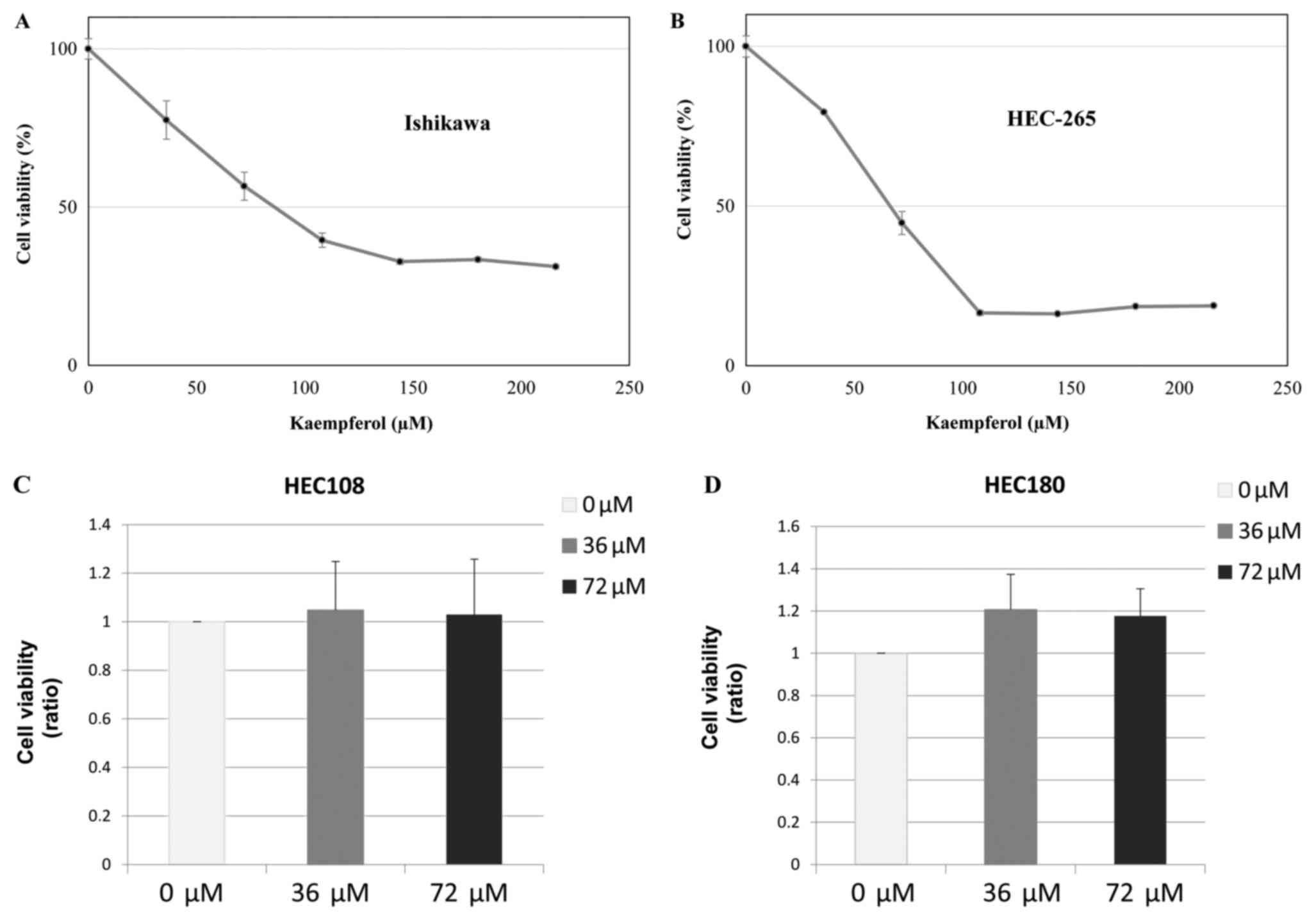
Kaempferol suppresses viability of
Ishikawa and HEC-265 cells
Cell viability assays were performed to assess the
growth inhibitory effects of kaempferol against Ishikawa and
HEC-265 estrogen receptor-positive endometrial cancer cells.
Kaempferol successfully suppressed the growth of Ishikawa and
HEC-265 cells, with IC50 values of 83 and 65 µM,
respectively (Fig. 1A and B). We
further examined the anti-tumor effects of kaempferol in HEC108 and
HEC180 estrogen receptor-negative endometrial cancer cells.
Kaempferol did not suppress the growth of HEC108 and HEC180 cells
(Fig. 1C and D).
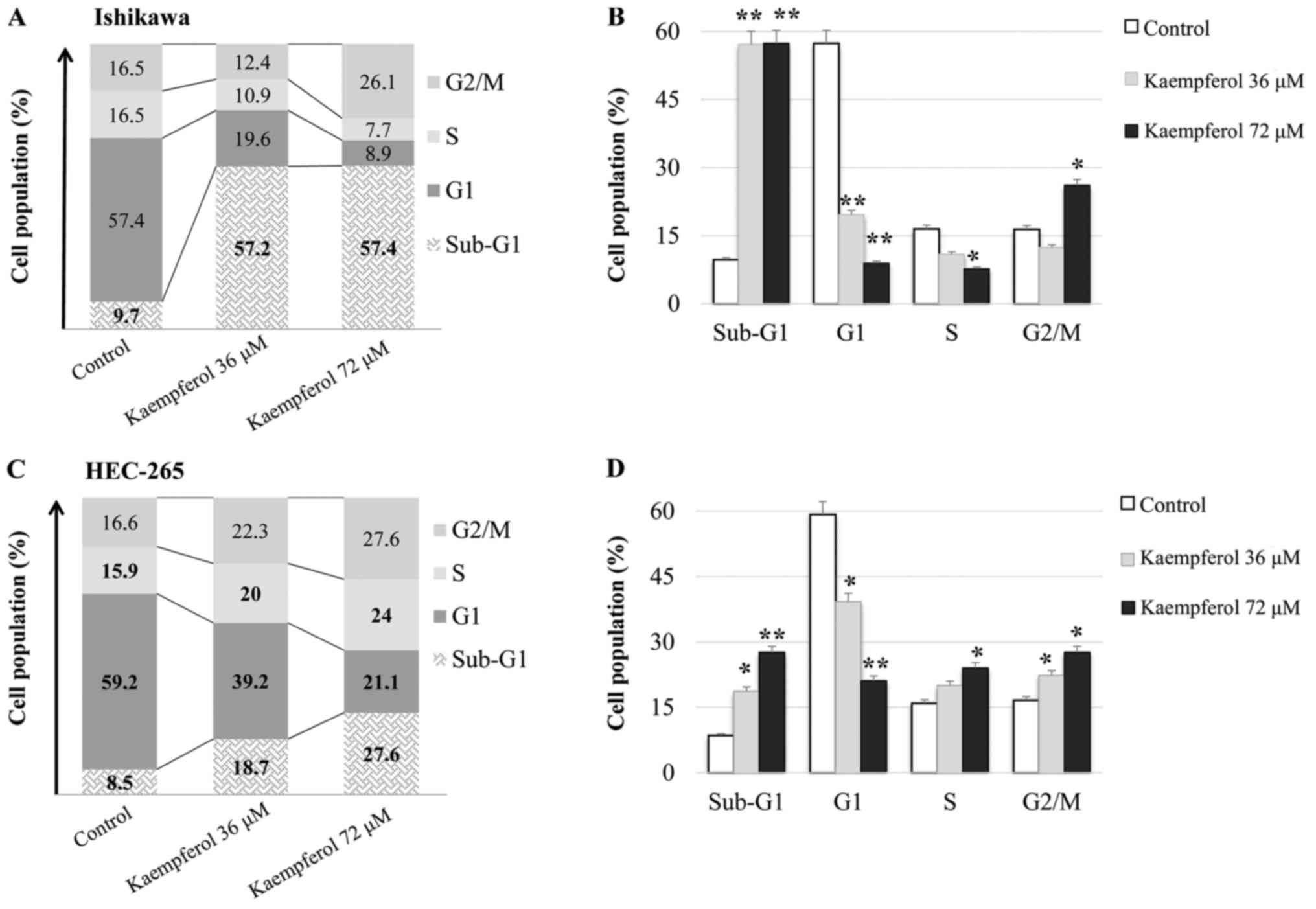
Kaempferol induces sub-G1 and G2/M
phases of the cell cycle
To understand the mechanisms by which kaempferol
suppresses cell growth in endometrial carcinoma, cells treated with
varying concentrations of kaempferol were incubated for 48 h and
then evaluated via FACS for cell cycle progression. We found that
kaempferol induced cell accumulation in the sub-G1 and G2/M phases
in Ishikawa and HEC-265 endometrial cancer cells, while
significantly suppressing the G1 phase (P<0.01; Fig. 2A and B).
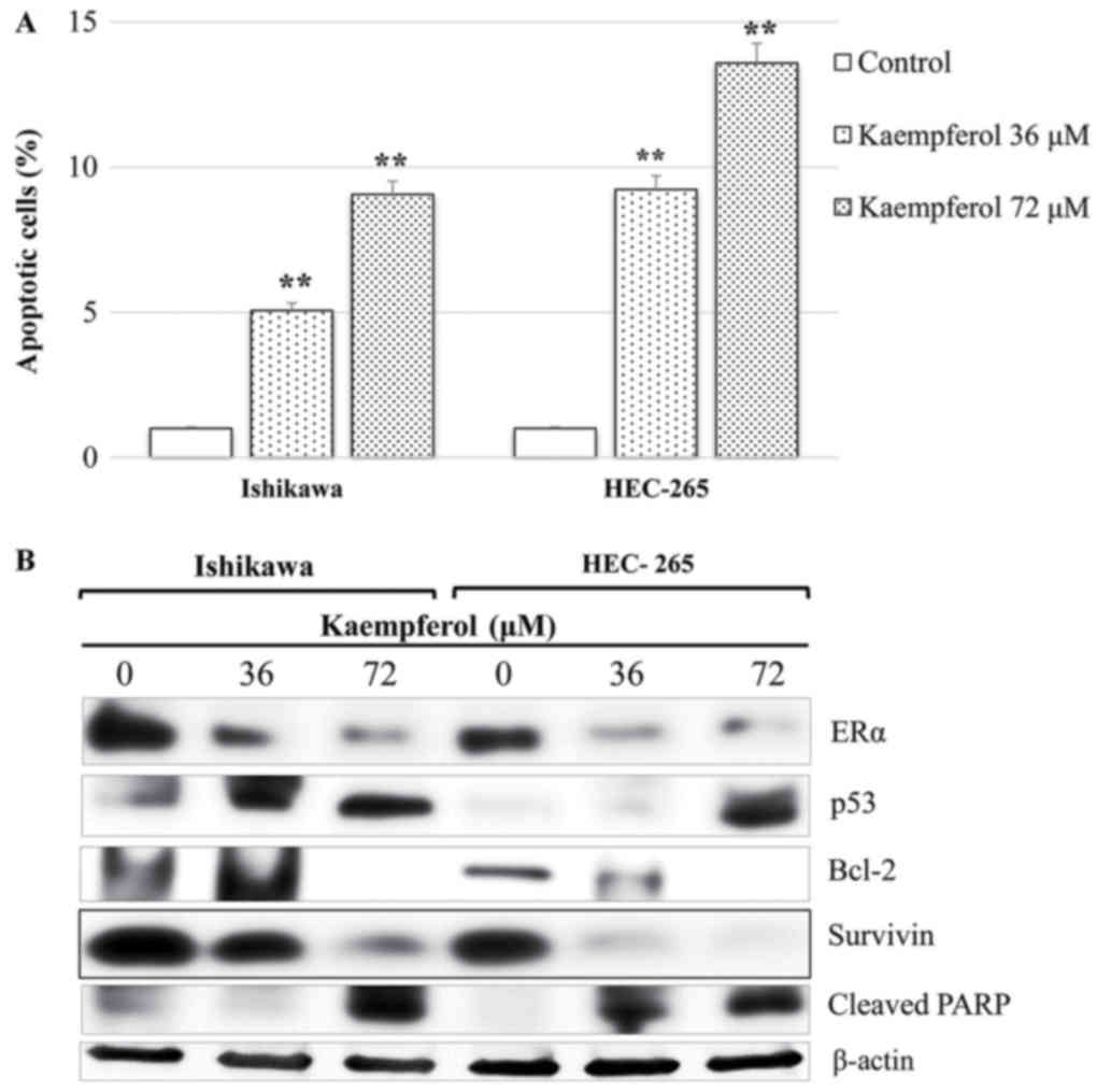
Kaempferol induces apoptotic cell
death in endometrial cancer by suppressing ERα, survivin and
Bcl-2
We evaluated the cytotoxicity of kaempferol against
endometrial cancer cells and possible mechanisms by analyzing the
induction of apoptosis using an annexin V-FITC/PI assay. We found
that kaempferol significantly (P<0.01) induced apoptotic cell
death in Ishikawa and HEC-265 cells (Fig.
3A). Further analysis via western blot revealed that kaempferol
induced apoptosis largely by suppressing ERα and the antiapoptotic
proteins survivin and Bcl-2 and by inducing p53 and PARP cleavage
(Fig. 3B).
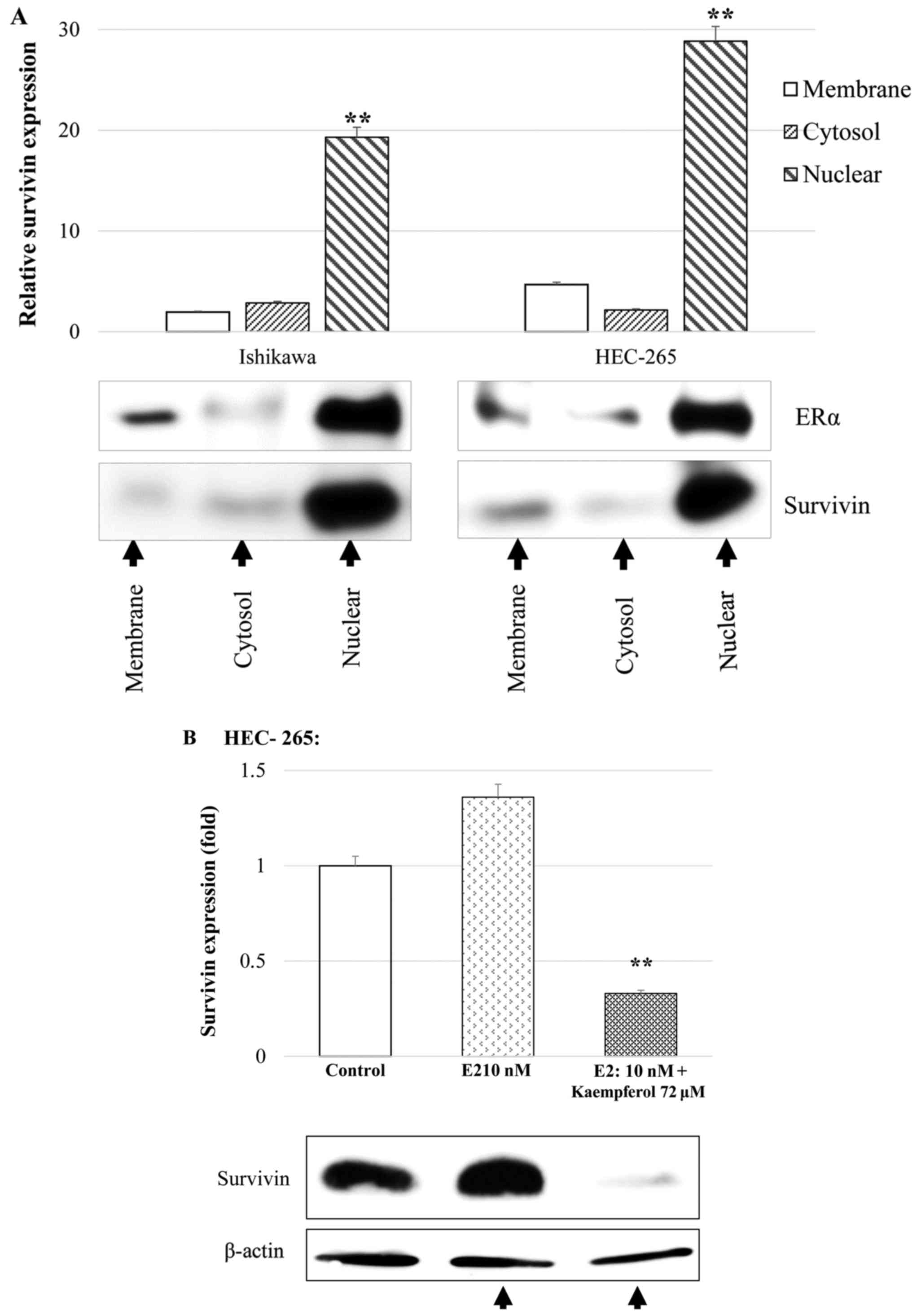
E2 significantly induces nuclear
co-expression of ERα and survivin in endometrial cancer cells
To investigate the site of action of E2 and its role
in ERα and survivin activity in subcellular compartments,
subcellular protein samples were extracted from Ishikawa and
HEC-265 cells treated with E2 and/or kaempferol and analyzed via
western blot. E2 significantly induced co-expression of nuclear ERα
and survivin (Fig. 4A). This result
suggests that E2 primarily induces nuclear ERα and survivin,
consequently preventing apoptosis. Treatment of cells with
kaempferol inhibited estradiol-induced upregulation of survivin and
led to apoptotic cell death (Fig.
4B).
Discussion
Targeting ERα in endometrial cancer using kaempferol
may be a feasible therapeutic strategy, as ERα-mediated oncogenic
effects, including upregulation of Nogo-B receptor (NgBR) and
survivin, as well as activation of the PI3K/mTOR/Akt pathway have
been established (13,24). Moreover, ERα is known to interact with
survivin and p53. Studies have demonstrated that ERα interacts
in vivo with p53 bound to promoters of survivin and
multidrug resistance gene-1 (MDR1), both of which are
p53-transcriptional repression targets (24,25).
Additionally, ERα can directly bind to p53, which plays an
important role in mediating apoptosis and leads to downregulation
of p53-mediated transcriptional activation.
Survivin interacts with p53 such that wild-type p53
suppresses the expression of survivin by blocking transcription of
the BIRC5 survivin gene (26).
Furthermore, the resistance of cancer cells to apoptosis is
attributable to upregulation of survivin via its promoter, which
results from loss of the p53 tumor suppressor. Survivin inhibits
apoptosis through several mechanisms, including direct binding and
inhibition of caspases-3 and −9, synergizing with the
X-chromosome-linked inhibitor of apoptosis protein (XIAP), binding
to the pro-apoptotic protein secondary mitochondria-derived
activator of caspase/direct inhibitor of apoptosis-binding protein
with low pI (SMAC/DIABLO), and by preventing activation of the
procaspases (26,27). Steroids are known to trigger
translocation of cytoplasmic ERs to the nucleus (28); however, there is limited knowledge
about the mechanisms involved and the resulting effects.
The Wnt signaling pathway plays an important role in
stem cells and is responsible for regulation of survivin
expression. Thus, survivin antagonists may affect cancer stem cells
(29). Although there are various
apoptosis-based cancer therapies (30), targeting survivin provides several
advantages. Suppressing survivin compromises not only the
anti-apoptotic cascade, but also the multiple cellular signaling
networks required to maintain tumors and their microenvironments
(31). Additionally, targeting
survivin does not affect normal cells or tissues. For instance, a
phase I clinical trial of a survivin-based vaccination has been
completed using immunologic antigen-specific responses in which no
side effects were reported (32,33). Thus,
survivin-based therapeutics might possess more favorable toxicity
profiles than other treatment options.
The findings of this study provide a novel
therapeutic strategy for endometrial cancer treatment using
kaempferol, a natural dietary flavonoid. This approach offers
additional merits attributable to the readily availability and
affordability of kaempferol and because kaempferol effectively
targets E2-induced/ERα-mediated oncogenic signaling pathways, which
play a crucial role in tumorigenesis of most endometrial
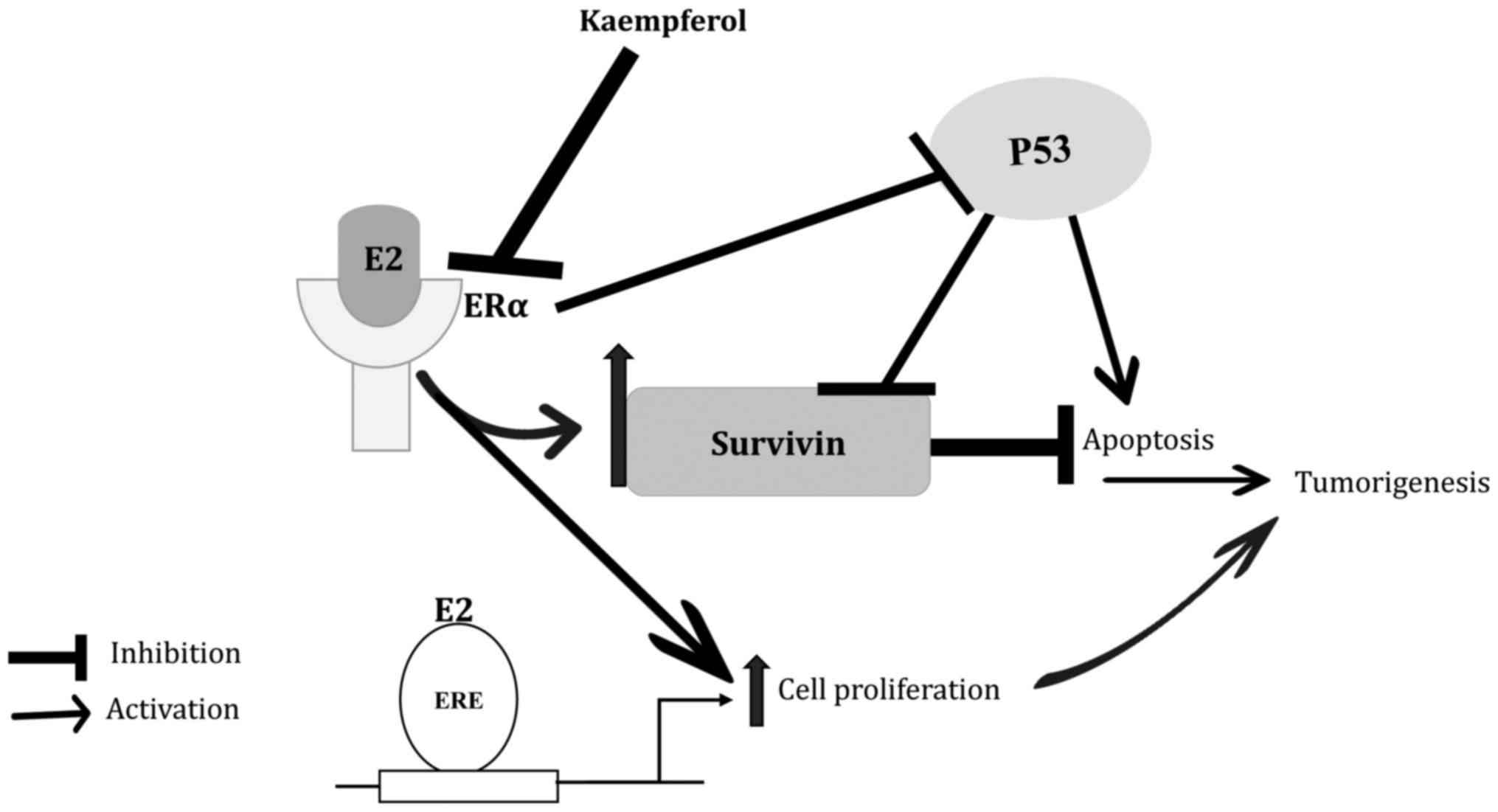
carcinomas. As we proposed in Fig. 5,
kaempferol inhibits ERα, leading to suppression of survivin and
apoptotic cell death, in addition to inducing p53, which was
reported previously (34–36). Results show that kaempferol had no
antitumor effects against estrogen receptor-negative endometrial
cancer cells. These results further support our proposed mechanism.
While other survivin antagonists have been described, kaempferol
provides added benefits because ERα is upstream of survivin and can
activate unrelated pathways. Furthermore, as a phytoestrogen,
kaempferol may offer a more favorable toxicity profile than other
survivin-based therapeutics.
This study has some limitations. First, there are no
biomarkers for predicting the sensitivity of endometrial carcinoma
to kaempferol. Second, ex-vivo experiments may be required
to further clarify the antitumor effects of kaempferol in
endometrial cancer. Finally, the effectiveness and tolerability of
kaempferol must be assessed in patients with endometrial carcinomas
in clinical trials.
The present study demonstrates the potential
antitumor effects of kaempferol against endometrial carcinoma
cells. Therefore, further research into its development as a novel
molecularly-targeted agent against endometrial carcinoma is
recommended.
Acknowledgements
The authors would like to thank Miss Kaori Tomita
(Department of Obstetrics and Gynecology, Faculty of Medicine,
University of Tokyo, Tokyo, Japan) for providing support and
assistance; Dr. Masato Nishida (Kasumigaura Medical Center,
Ibaraki, Japan) and Professor Satoru Kyo (Shimane University
Faculty of Medicine, Shimane, Japan) for providing Ishikawa and
endometrial immortalized cells, respectively.
Funding
The present study was financially supported by a
Grant-in-Aid for Scientific Research (grant nos. 26462515, 17K11269
and 15K10705), and Grants-in-Aid for Young Scientific Research from
the Ministry of Education, Culture, Sports, Science and Technology
of Japan (grant nos. 15K20128, 16H06757, 16K21330, 17K16832 and
16K20176); the present study was also partially supported by AMED
(grant no. JP17cm0106502).
Availability of data and materials
The datasets used and/or analyzed during the current
study are available from the corresponding author on reasonable
request.
Authors' contributions
AHC, KS and KO conceived and designed the study. SO,
TF, MT, AM, TK and YI designed the experiments. AK and MK performed
the cell viability assay, and all of the other experiments were
performed by AHC. AHC and KS acquired the data. The data were
analyzed and interpreted by AHC, KS, KO, MT, KN, YM, OH, KK, YO and
TF. AHC and KS prepared the manuscript and figures. AHC, KS, KO, YO
and TF reviewed and revised the manuscript for important
intellectual content. Technical and material support was provided
by AK, MK, MU, TF and HK. HK and MU reviewed the manuscript for
important intellectual content. MU analyzed and interpreted the
data in additional experiments. HK established and provided
endometrial cancer cell lines used in the study. All of the authors
approved the final version of this manuscript.
Ethics approval and consent to
participate
Not applicable.
Patient consent for publication
Not applicable.
Competing interests
The authors declare that they have no competing
interests.
Glossary
Abbreviations
Abbreviations:
|
Akt
|
protein kinase B
|
|
Bcl-2
|
B-cell lymphoma 2
|
|
CHIP
|
chromatin immunoprecipitation
|
|
DMSO
|
dimethyl sulfoxide
|
|
E2
|
17β-estradiol
|
|
EMEM
|
Eagle's minimum essential medium
|
|
ERα
|
estrogen receptor-α
|
|
FACS
|
fluorescence-activated cell
sorting
|
|
FBS
|
fetal bovine serum
|
|
FITC
|
fluorescein isothiocyanate
|
|
IAP
|
inhibitor of apoptosis
|
|
IC50
|
half maximal inhibitory
concentration
|
|
MDR1
|
multidrug resistance gene-1
|
|
MPA
|
medroxyprogesterone acetate
|
|
NgBR
|
Nogo-B receptor
|
|
PARP
|
poly adenosine diphosphate-ribose
polymerase
|
|
PGC
|
proliferator-activated receptor γ
co-activator
|
|
PI
|
propidium iodide
|
|
PI3K
|
phosphatidylinositol-4,5-bisphosphate
3-kinase
|
|
PTEN
|
phosphatase and tensin homolog
|
|
XIAP
|
X-chromosome linked inhibitor of
apoptosis protein
|
References
|
1
|
Bokhman JV: Two pathogenetic types of
endometrial carcinoma. Gynecol Oncol. 15:10–17. 1983. View Article : Google Scholar : PubMed/NCBI
|
|
2
|
Schenker JG, Weinstein D and Okon E:
Estradiol and testosterone levels in the peripheral and ovarian
circulations in patients with endometrial cancer. Cancer.
44:1809–1812. 1979. View Article : Google Scholar : PubMed/NCBI
|
|
3
|
Yang HP, Gonzalez Bosquet J, Li Q, Platz
EA, Brinton LA, Sherman ME, Lacey JV Jr, Gaudet MM, Burdette LA,
Figueroa JD, et al: Common genetic variation in the sex hormone
metabolic pathway and endometrial cancer risk: Pathway-based
evaluation of candidate genes. Carcinogenesis. 5:827–833. 2010.
View Article : Google Scholar
|
|
4
|
Hernandez E: Pathological findings and
prognosis from uterine malignancy. Curr Opinion Obstet Gynecol.
5:480–485. 1993. View Article : Google Scholar
|
|
5
|
Lachance JA, Darus CJ and Rice LW:
Surgical management and postoperative treatment of endometrial
carcinoma. Rev Obstet Gynecol. 1:97–105. 2008.PubMed/NCBI
|
|
6
|
Chaudhry P and Asselin E: Resistance to
chemotherapy and hormone therapy in endometrial cancer. Endocr
Relat Cancer. 16:363–380. 2009. View Article : Google Scholar : PubMed/NCBI
|
|
7
|
Oda K, Stokoe D, Taketani Y and McCormick
F: High frequency of coexistent mutations of PIK3CA and PTEN genes
in endometrial carcinoma. Cancer Res. 65:10669–10673. 2005.
View Article : Google Scholar : PubMed/NCBI
|
|
8
|
Chen J, Zhao KN, Li R, Shao R and Chen C:
Activation of PI3K/Akt/mTOR pathway and dual inhibitors of PI3K and
mTOR in endometrial cancer. Curr Med Chem. 21:3070–3080. 2014.
View Article : Google Scholar : PubMed/NCBI
|
|
9
|
Scully MM, Palacios-Helgeson LK, Wah LS
and Jackson TA: Rapid estrogen signaling negatively regulates PTEN
activity through phosphorylation in endometrial cancer cells. Horm
Cancer. 5:218–231. 2014. View Article : Google Scholar : PubMed/NCBI
|
|
10
|
Jaiswal PK, Goel A and Mittal RD:
Survivin: A molecular biomarker in cancer. Indian J Med Res.
141:389–397. 2015. View Article : Google Scholar : PubMed/NCBI
|
|
11
|
Ryan BM, O'Donovan N and Duffy MJ:
Survivin: A new target for anti-cancer therapy. Cancer Treat Rev.
35:553–562. 2009. View Article : Google Scholar : PubMed/NCBI
|
|
12
|
Altieri DC: Targeting survivin in cancer.
Cancer Lett. 332:225–228. 2013. View Article : Google Scholar : PubMed/NCBI
|
|
13
|
Wang B, Zhao B, North P, Kong A, Huang J
and Miao QR: Expression of NgBR is highly associated with estrogen
receptor alpha and survivin in breast cancer. PLoS One.
8:e780832013. View Article : Google Scholar : PubMed/NCBI
|
|
14
|
Erkanli S, Kayaselcuk F, Kuscu E, Bagis T,
Bolat F, Haberal A and Demirhan B: Expression of survivin, PTEN and
p27 in normal, hyperplastic, and carcinomatous endometrium. Int J
Gynecol Cancer. 16:1412–1418. 2006. View Article : Google Scholar : PubMed/NCBI
|
|
15
|
Chuwa AH, Sone K, Oda K, Ikeda Y, Fukuda
T, Wada-Hiraike O, Inaba K, Makii C, Takeuchi M, Oki S, et al:
Significance of survivin as a prognostic factor and a therapeutic
target in endometrial cancer. Gynecol Oncol. 141:564–569. 2016.
View Article : Google Scholar : PubMed/NCBI
|
|
16
|
Luo H, Rankin GO, Li Z, DePriest L and
Chen YC: Kaempferol induces apoptosis in ovarian cancer cells
through activating p53 in intrinsic pathway. Food Chem.
128:513–519. 2011. View Article : Google Scholar : PubMed/NCBI
|
|
17
|
Hung H: Inhibition of estrogen receptor
alpha expression and function in MCF-7 cells by kaempferol. J Cell
Physiol. 198:197–208. 2004. View Article : Google Scholar : PubMed/NCBI
|
|
18
|
Luo H, Rankin GO, Liu L, Daddysman MK,
Jiang BH and Chen YC: Kaempferol inhibits angiogenesis and VEGF
expression through both HIF dependent and independent pathways in
human ovarian cancer cells. Nutr Cancer. 61:554–563. 2009.
View Article : Google Scholar : PubMed/NCBI
|
|
19
|
Chen HJ, Lin CM, Lee CY, Shih NC, Peng SF,
Tsuzuki M, Amagaya S, Huang WW and Yang JS: Kaempferol suppresses
cell metastasis via inhibition of the ERK-p38-JNK and AP-1
signaling pathways in U-2 OS human osteosarcoma cells. Oncol Rep.
30:925–932. 2013. View Article : Google Scholar : PubMed/NCBI
|
|
20
|
Kuramoto H, Nishida M, Morisawa T, Hamano
M, Hata H, Kato Y, Ohno E and Lida T: Establishment and
characterization of human endometrial cancer cell lines. Ann N Y
Acad Sci. 622:402–421. 1991. View Article : Google Scholar : PubMed/NCBI
|
|
21
|
Inaba K, Oda K, Aoki K, Sone K, Ikeda Y,
Miyasaka A, Kashiyama T, Fukuda T, Makii C, Arimoto T, et al:
Synergistic antitumor effects of combination PI3K/mTOR and MEK
inhibition (SAR245409 and pimasertib) in ovarian carcinoma cells by
fluorescence resonance energy transfer imaging. Oncotarget.
7:29577–29591. 2016. View Article : Google Scholar : PubMed/NCBI
|
|
22
|
Fukuda T, Oda K, Wada-Hiraike O, Sone K,
Inaba K, Ikeda Y, Makii C, Miyasaka A, Kashiyama T, Tanikawa M, et
al: Autophagy inhibition augments resveratrol-induced apoptosis in
Ishikawa endometrial cancer cells. Oncol Lett. 12:2560–2566. 2016.
View Article : Google Scholar : PubMed/NCBI
|
|
23
|
Stoica GE, Franke TF, Moroni M, Mueller S,
Morgan E, Iann MC, Winder AD, Reiter R, Wellstein A, Martin MB and
Stoica A: Effect of estradiol on estrogen receptor-alpha gene
expression and activity can be modulated by the ErbB2/PI 3-K/Akt
pathway. Oncogene. 22:7998–8011. 2003. View Article : Google Scholar : PubMed/NCBI
|
|
24
|
Sayeed A, Konduri SD, Liu W, Bansal S, Li
F and Das GM: Estrogen receptor alpha inhibits p53-mediated
transcriptional repression: Implications for the regulation of
apoptosis. Cancer Res. 67:7746–7755. 2007. View Article : Google Scholar : PubMed/NCBI
|
|
25
|
Hoffman WH, Biade S, Zilfou JT, Chen J and
Murphy M: Transcriptional repression of the anti-apoptotic survivin
gene by wild-type p53. J Biol Chem. 277:3247–3257. 2002. View Article : Google Scholar : PubMed/NCBI
|
|
26
|
Johnson ME and Howerth EW: Survivin: A
bifunctional inhibitor of apoptosis protein. Vet Pathol.
41:599–607. 2004. View Article : Google Scholar : PubMed/NCBI
|
|
27
|
Mita AC, Mita MM, Nawrocki ST and Giles
FJ: Survivin: Key regulator of mitosis and apoptosis and novel
target for cancer therapeutics. Clin Cancer Res. 14:5000–5005.
2008. View Article : Google Scholar : PubMed/NCBI
|
|
28
|
Raam S, Richardson GS, Bradley F,
MacLaughlin D, Sun L, Frankel F and Cohen JL: Translocation of
cytoplasmic estrogen receptors to the nucleus: Immunohistochemical
demonstration utilizing rabbit antibodies to estrogen receptors of
mammary carcinomas. Breast Cancer Res Treat. 3:179–199. 1983.
View Article : Google Scholar : PubMed/NCBI
|
|
29
|
Reya T, Morrison SJ, Clarke MF and
Weissman IL: Stem cells, cancer, and cancer stem cells. Nature.
414:105–111. 2001. View Article : Google Scholar : PubMed/NCBI
|
|
30
|
Reed JC: Apoptosis-targeted therapies for
cancer. Cancer Cell. 3:17–22. 2003. View Article : Google Scholar : PubMed/NCBI
|
|
31
|
Sawyers C: Targeted cancer therapy.
Nature. 432:294–297. 2004. View Article : Google Scholar : PubMed/NCBI
|
|
32
|
Hirschowitz EA, Foody T, Kryscio R,
Dickson L, Sturgill J and Yannelli J: Autologous dendritic cell
vaccines for non-small-cell lung cancer. J Clin Oncol.
22:2808–2815. 2004. View Article : Google Scholar : PubMed/NCBI
|
|
33
|
Otto K, Andersen MH, Eggert A, Keikavoussi
P, Pedersen LØ, Rath JC, Böck M, Bröcker EB, Straten PT, Kämpgen E
and Becker JC: Lack of toxicity of therapy-induced T cell responses
against the universal tumour antigen survivin. Vaccine. 23:884–889.
2005. View Article : Google Scholar : PubMed/NCBI
|
|
34
|
Li W, Du B, Wang T, Wang S and Zhang J:
Kaempferol induces apoptosis in human HCT116 colon cancer cells via
the Ataxia-Telangiectasia Mutated-p53 pathway with the involvement
of p53 upregulated modulator of apoptosis. Chem Biol Interact.
177:121–127. 2009. View Article : Google Scholar : PubMed/NCBI
|
|
35
|
Kim SH and Choi KC: Anti-cancer effect and
underlying mechanism(s) of kaempferol, a phytoestrogen, on the
regulation of apoptosis in diverse cancer cell models. Toxicol Res.
29:229–234. 2013. View Article : Google Scholar : PubMed/NCBI
|
|
36
|
Lee CF, Yang JS, Tsai FJ, Chiang NN, Lu
CC, Huang YS, Chen C and Chen FA: Kaempferol induces
ATM/p53-mediated death receptor and mitochondrial apoptosis in
human umbilical vein endothelial cells. Int J Oncol. 48:2007–2014.
2016. View Article : Google Scholar : PubMed/NCBI
|