Introduction
Hepatocellular carcinoma (HCC), which accounts for
70–85% of the primary liver cancers (1), is the fifth most common cancer and
second most common cause of cancer death in men worldwide (2). In 2012 alone, an estimated 782,500 new
liver cancer cases were diagnosed, and there were approximately
745,500 deaths due to liver cancer (2). Newly-developed therapeutics using
direct-acting antivirals are eradicating most HCVs (3). However, the prognosis of HCC remains
poor owing to tumor invasiveness, intra- and extra-hepatic
metastasis, multicentric carcinogenesis, and resistance to
chemotherapy (4,5). The identification of novel biomarkers
for HCC is therefore of great importance in improving the outcome
of patients with HCC.
Cellular adhesion molecules, interacting cellular
communications, are divided into four groups according to their
molecular structures: Cadherins, selectins, integrins, and an
immunoglobulin superfamily (6).
Intercellular adhesion molecule (ICAM)-1, a member of the
immunoglobulin superfamily, is broadly expressed on the membrane of
normal tissues, and is selectively expressed in human malignancies
(7–10). ICAM-1 is the ligand for the
β2-integrins, lymphocyte function-associated antigen (LFA)-1, and
Mac-1 (11,12). The expression of ICAM-1 is regulated
by locally produced inflammatory cytokines such as IL-1β, tumor
necrosis factor α, interleukin (IL)-6, and interferon-γ (13,14).
Interestingly, the soluble form of ICAM-1 (sICAM-1) has also been
reported to have angiogenic activity (15).
To elucidate the mechanisms of tumor progression in
HCC, and to establish certain prognostic markers, we investigated
the serum concentration of sICAM-1 and its relationships with
inflammatory and nutritional parameters.
Materials and methods
Patients
Thirty-six patients with HCC were enrolled (30 men
and six women; mean age, 70.5 years; range, 34 to 84 years) in a
prospective setting. In addition, samples from 27 healthy
volunteers (10 males and 17 females, mean age, 54.3 years; range 35
to 84 years) were used as controls. Blood samples were collected
from the patients between February 2011 and August 2013, before
initiation of treatment. Sera from patients were stored at −80°C
until use. All of the patients underwent curative-intent surgery at
our department. Following surgery, each patient's final cancer
stage was determined pathologically according to the 8th edition of
the TNM classification system of malignant tumors published by the
Union for International Cancer Control (16). Liver fibrosis stage was determined
according to the METAVIR score (17).
In addition, the Child-Pugh score and indocyanine green retention
rate at 15 min (ICGR15) were examined to evaluate liver function.
The study protocol was approved by the ethics committee of
Fukushima Medical University, and written informed consent was
obtained from all enrolled patients and healthy volunteers. Thus,
it was designed and conducted in accordance with Good Clinical
Practice Guidelines and the latest revision of the Declaration of
Helsinki.
Measurements of parameters
The serum concentrations of IL-6, vascular
endothelial growth factor, and sICAM-1 were measured using an
enzyme-linked immunosorbent assay (ELISA; R&D Systems,
Minneapolis, MN, USA) according to the manufacturer's instructions.
Each sample was used only once after thawing, and not all blood
samples were of sufficient volume for all measurements. Patient
nutritional status was determined by measuring the serum
concentrations of total protein, albumin, retinol binding protein
(RBP), transthyretin (TTR), and transferrin, as well as body mass
index (BMI) at diagnosis. These parameters were measured at the
Central Clinical Laboratory of Fukushima Medical University
Hospital. As for the inflammatory parameters, C-reactive protein
(CRP), white blood cell count, neutrophil and lymphocyte counts,
and the neutrophil-to-lymphocyte ratio (NLR), were used.
Statistical analysis
Data are presented as frequencies or percentages for
categorical variables and mean ± standard error for continuous
variables, unless otherwise indicated. For categorical clinical
variables, differences between the groups were evaluated using
Fisher's exact test. The differences in mean values between the
groups were analyzed using the Mann-Whitney U test. A receiver
operating characteristic (ROC) curve was used to evaluate the
usefulness of the examined parameters as a prognostic factor, and
associations between two variables were quantified using Spearman's
rank correlation coefficient. The mean observation period was 68.5
months (median: 68.7, range: 45.3–83.9), and the final assessment
of disease status was made on December 28, 2017. Overall survival
(OS) and recurrence-free survival (RFS) were calculated using the
Kaplan-Meier method, and differences between the groups were
assessed by using the log-rank test. Factors found to be
significant in the univariate analysis were subjected to
multivariate analysis using a Cox proportional hazard model to
identify independent predictors of prognosis. A two-sided P-value
of <0.05 was considered statistically significant. All
statistical calculations were performed using SPSS®
version 24 (IBM Japan, Tokyo, Japan).
Results
Analysis using an ROC curve
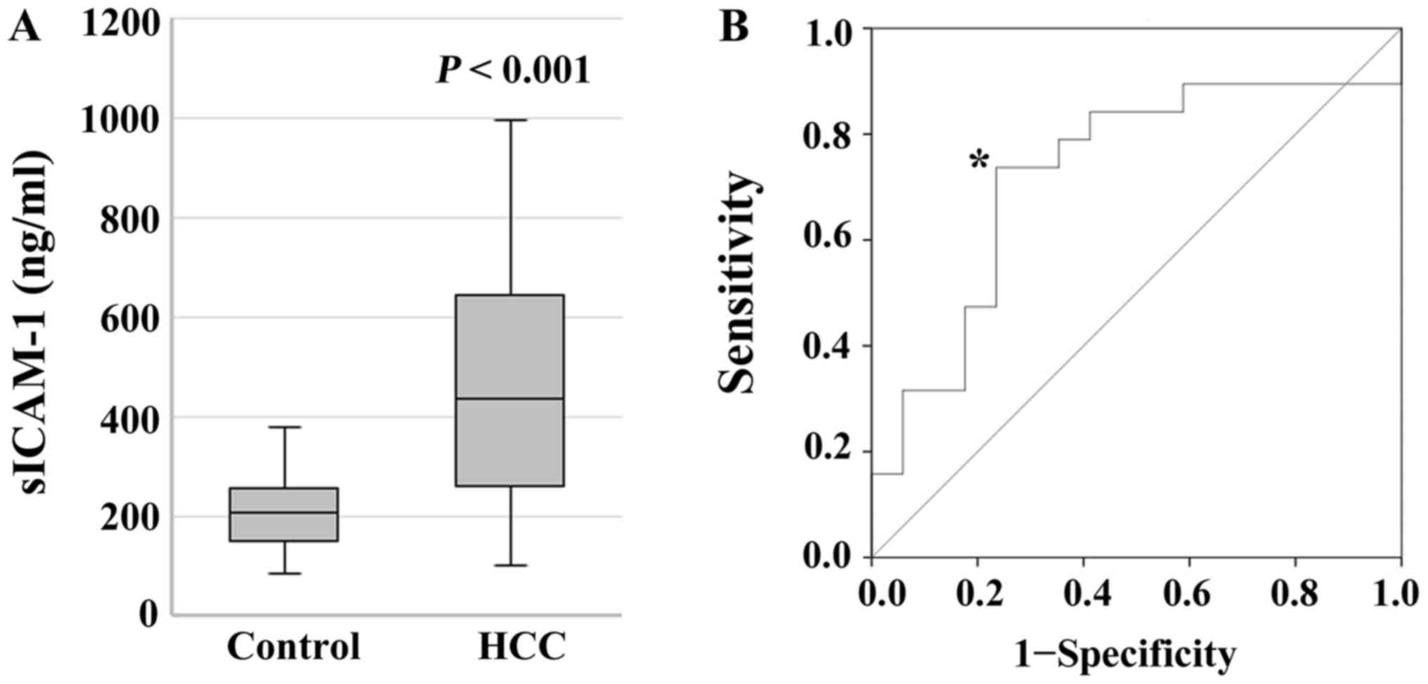
Patient characteristics are summarized in Table I. The sICAM-1 serum levels of the HCC
patients (median: 438.9 ng/ml, range: 101.1–994.0 ng/ml) were
higher than those of the healthy volunteers (median: 207.6 ng/ml,
range: 87.8–381.2 ng/ml) (P<0.001; Fig. 1A). In an analysis using a ROC curve
(Fig. 1B), the serum sICAM-1 was
evaluated as a useful biomarker to predict patient survival
(P=0.022), and a sICAM-1 level of 440 ng/dl was determined as the
cutoff value. At this cutoff value, sensitivity was 0.737 and
specificity was 0.706. Table II
shows the patient characteristics according to serum sICAM-1 level.
The incidence of ICGR ≥15 was statistically higher in the patients
with sICAM-1 ≥440 than in those with sICAM-1 <440
(P<0.001).
 | Table I.Patient demographics. |
Table I.
Patient demographics.
| Category | N | (%) |
|---|
| Age |
|
|
|
<75 | 24 | 66.7 |
| ≥75 | 12 | 33.3 |
| Sex |
|
|
| Male | 30 | 83.3 |
|
Female | 6 | 16.7 |
| T |
|
|
| T1a | 8 | 22.2 |
| T1b | 16 | 44.4 |
| T2 | 12 | 33.3 |
| N |
|
|
| N0 | 33 | 91.7 |
| N1 | 3 | 8.3 |
| M |
|
|
| M0 | 36 | 100.0 |
| M1 | 0 | 0.0 |
| Stage |
|
|
| IA | 8 | 22.2 |
| IB | 16 | 44.4 |
| II | 10 | 27.8 |
| III | 0 | 0.0 |
| IVA | 2 | 5.6 |
| Operation |
|
|
|
Partial | 10 | 27.8 |
|
Segmentectomy | 5 | 13.9 |
|
Sectionectomy | 7 | 19.4 |
|
Lobectomy | 8 | 22.2 |
| Extended
lobectomy | 6 | 16.7 |
| Table II.Patient demographics according to
sICAM-1 level. |
Table II.
Patient demographics according to
sICAM-1 level.
| Characteristics | sICAM-1 <440
(n=18) | sICAM-1 ≥440
(n=18) | P-value |
|---|
| Age |
|
| 1.000 |
|
<75 | 12 | 12 |
|
| ≥75 | 6 | 6 |
|
| Sex |
|
| 0.658 |
|
Male | 14 | 16 |
|
|
Female | 4 | 2 |
|
| T |
|
| 0.075 |
| T1 | 15 | 9 |
|
| T2 | 3 | 9 |
|
| N |
|
| 0.229 |
| N0 | 18 | 15 |
|
| N1 | 0 | 3 |
|
| Stage |
|
| 0.229 |
| Stage
I–III | 18 | 15 |
|
| Stage
IV | 0 | 3 |
|
| Virus |
|
| 1.000 |
| − | 7 | 8 |
|
| + | 11 | 10 |
|
| ICGR15 |
|
|
<0.001a |
|
<15 | 18 | 7 |
|
|
≥15 | 0 | 11 |
|
| PT |
|
| 0.603 |
|
≥70 | 15 | 17 |
|
|
<70 | 3 | 1 |
|
| Child-Pugh |
|
| 1.000 |
| A | 17 | 16 |
|
| B | 1 | 2 |
|
| AFP |
|
| 0.479 |
|
<10.0 | 8 | 6 |
|
|
≥10.0 | 7 | 10 |
|
| Fibrosis score |
|
| 0.691 |
|
F1-3 | 15 | 13 |
|
| F4 | 3 | 5 |
|
| Intrahepatic
metastasis |
|
| 1.000 |
| − | 17 | 8 |
|
| + | 1 | 10 |
|
| Vascular
invasion |
|
| 0.402 |
| − | 16 | 13 |
|
| + | 2 | 5 |
|
| Biliary
invasion |
|
| 0.075 |
| − | 15 | 9 |
|
| + | 3 | 9 |
|
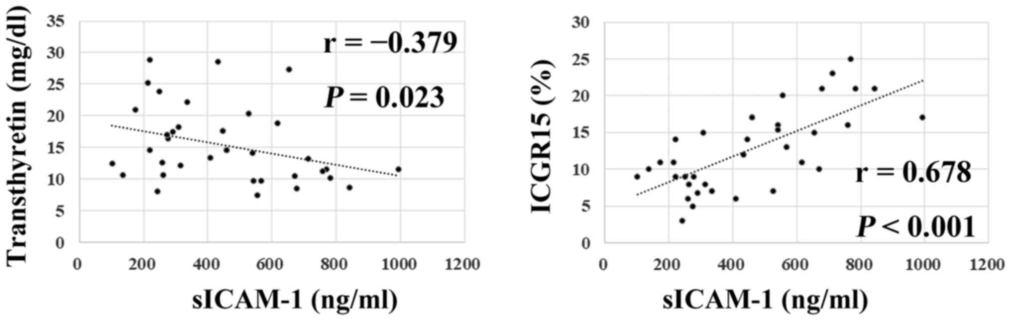
Association between sICAM-1 and other
parameters
Fig. 2 shows the
relationships between serum sICAM-1 levels and other parameters.
The serum sICAM-1 levels exhibited statistically significant
inverse correlations with TTR (r=−0.379, P=0.023), and showed
statistically significant correlations with ICGR15 (r=0.678,
P<0.001). However, the serum sICAM-1 showed no correlations with
BMI.
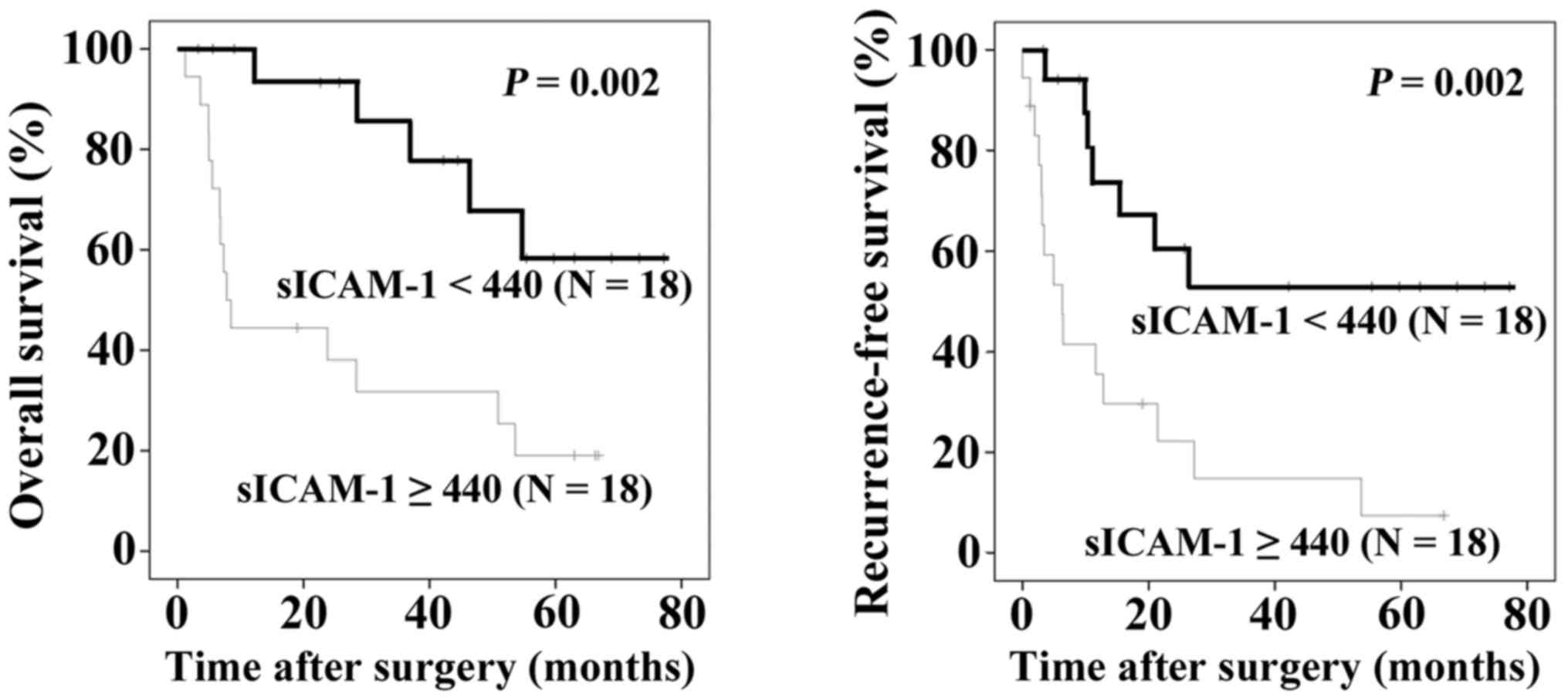
Prognostic impact of sICAM-1
The evaluation of the prognostic factors was
performed by dividing the patients into two groups for each
parameter: Age (<75 years vs. ≥75 years), gender (male vs.
female), serum sICAM-1 level (sICAM-1 <440 ng/ml vs. ≥440
ng/ml), T factor (T1 vs. T2), Child-Pugh classification (A vs. B),
intrahepatic metastasis (negative vs. positive), vascular invasion
(negative vs. positive), and biliary invasion (negative vs.
positive). As shown in Fig. 3, the
patients with sICAM-1 ≥440 ng/ml showed poorer OS and RFS than
those with sICAM-1 <440 ng/ml (P=0.002 and P=0.002,
respectively).
Table III summarizes
the analyses of a Cox proportional hazard model. With regard to OS,
sICAM-1 ≥440 ng/ml, T2, intrahepatic metastasis positive, vascular
invasion positive, and biliary invasion positive showed statistical
significance in the univariate analysis. sICAM-1 ≥440 ng/ml (hazard
ratio: 3.623, 95% confidence interval: 1.145–11.458, P=0.028) and
Child B (hazard ratio: 1.514, 95% confidence interval: 1.066–2.150,
P=0.021) were independent prognostic factors for OS in the
multivariate analysis. With regard to RFS, sICAM-1 ≥440 ng/ml, T2,
intrahepatic metastasis positive, vascular invasion positive, and
biliary invasion positive showed statistical significance in the
univariate analysis. In the multivariate analysis, sICAM-1 ≥440
ng/ml was an independent prognostic factor for the RFS of HCC
patients (hazard ratio: 3.625, 95% confidence interval:
1.233–10.659, P=0.019).
| Table III.Cox proportional hazards model. |
Table III.
Cox proportional hazards model.
| A, Overall
survival |
|---|
|
|---|
|
| Univariate
analysis | Multivariate
analysis |
|---|
|
|
|
|
|---|
| Variable | HR | 95% CI | P-value | HR | 95% CI | P-value |
|---|
| Age <75 vs.
≥75 | 1.092 | 0.383–3.116 | 0.869 |
|
|
|
| Sex male vs.
female | 0.622 | 0.144–2.696 | 0.526 |
|
|
|
| sICAM-1 <440
ng/ml vs. ≥440 ng/ml | 4.368 | 1.562–12.216 | 0.005a | 3.623 | 1.145–11.458 | 0.028a |
| T T1 vs. T2 | 4.011 | 1.576–10.210 | 0.004a | 0.782 | 0.148–4.116 | 0.771 |
| Child-Pugh A vs.
B | 1.521 | 1.155–2.003 | 0.003a | 1.514 | 1.066–2.150 | 0.021a |
| IM negative vs.
positive | 3.847 | 1.079–13.719 | 0.038a | 0.743 | 0.119–4.643 | 0.750 |
| V negative vs.
positive | 4.063 | 1.397–11.816 | 0.010a | 4.441 | 0.902–21.864 | 0.067 |
| B negative vs.
positive | 2.113 | 1.087–4.108 | 0.027a | 2.594 | 0.779–8.642 | 0.121 |
|
| B,
Recurrence-free survival |
|
| Univariate
analysis | Multivariate
analysis |
|
|
Variable | HR | 95% CI | P-value | HR | 95% CI | P-value |
|
| Age <75 vs.
≥75 | 1.031 | 0.416–2.553 | 0.947 |
|
|
|
| Sex male vs.
female | 0.952 | 0.321–2.823 | 0.930 |
|
|
|
| sICAM-1 <440
ng/ml vs. ≥440 ng/ml | 3.776 | 1.528–9.331 | 0.004a | 3.625 | 1.233–10.659 | 0.019a |
| T T1 vs. T2 | 6.119 | 2.389–16.085 |
<0.001a | 2.434 | 0.488–12.135 | 0.278 |
| Child-Pugh A vs.
B | 1.371 | 1.087–1.728 | 0.003a | 1.177 | 0.850–1.629 | 0.328 |
| IM negative vs.
positive | 3.553 | 1.013–12.467 | 0.048a | 1.088 | 0.166–7.120 | 0.930 |
| V negative vs.
positive | 2.736 | 1.024–7.309 | 0.045a | 1.142 | 0.234–5.575 | 0.870 |
| B negative vs.
positive | 3.002 | 1.539–5.858 | 0.001a | 2.808 | 0.906–8.706 | 0.930 |
Discussion
Immunohistochemically, ICAM-1 is expressed on
hepatocytes in cancerous areas but not on hepatocytes in
noncancerous areas (18). It has
recently been reported that ICAM-1 was a marker of HCC stem cells,
and increased numbers of CD45−ICAM+ tumor
cells in blood samples of HCC patients correlated with worse
clinical outcomes (19). On the other
hand, circulating sICAM-1 has been reported to be elevated in the
serum of patients with various malignancies (20–28). With
regard to HCC, Shimizu et al reported that sICAM-1 ≥1,000
ng/ml was associated with poor prognosis in HCC patients who had
been treated by transcatheter arterial chemoembolization (20), and Zhu et al reported that
sICAM-1 >684 ng/ml was an independent prognostic factor for OS
and RFS in HCC patients who had undergone surgical treatment
(21). Our results on the usefulness
of sICAM-1 for predicting the survival of HCC patients confirmed
their findings; however, our sICAM-1 cutoff threshold of 440 ng/ml
was lower than those of the other two studies. With regard to the
meanings of higher sICAM-1, it has been reported that sICAM-1
inhibits ICAM-1/LFA-1-mediated cell-to-cell interaction, resulting
in tumor cells escaping from cell-mediated immune surveillance
(27,29). This escape theory seems possible,
considering that a high amount of circulating sICAM-1 was an
independent prognostic factor for the RFS in patients with HCC in
the present study. Since the source of increased circulating level
of the serum sICAM-1 has yet to be elucidated, further
investigation will be needed.
We revealed the relationships of serum sICAM-1
levels with the TTR levels and ICGR15. TTR, also known as
prealbumin, has a relatively short half-life (approximately two
days) and is the earliest laboratory indicator of malnutrition
status, as it contains a high percentage of essential amino acids
(30). Systemic chronic inflammation
has been reported to induce angiogenesis and malnutrition. Thus,
higher sICAM-1 might be one of the causes of lower serum TTR
levels. The meaning of the correlation between sICAM-1 and ICGR15
remains unclear; however, angiogenesis in tumors may prolong the
retention of indocyanine green.
There are some limitations to the current study.
First is its small sample size. In addition, it is costly and
troublesome to examine sICAM-1 in every HCC patient. However,
further investigations are warranted whether higher serum sICAM-1
is due to HCC stem cells or circulating tumor cells expressing
ICAM-1.
In conclusion, our analysis using a ROC curve
revealed that the cutoff value of sICAM-1 for predicting the
prognosis of the HCC patients was 440 ng/ml. The serum sICAM-1
levels in the current study exhibited statistically significant
inverse correlations with TTR, and showed statistically significant
correlations with ICGR15. The patients with sICAM-1 ≥440 ng/ml
showed poorer OS and RFS than those with sICAM-1 <440 ng/ml.
Furthermore, sICAM-1 ≥440 ng/ml and Child B were independent
prognostic factors for OS, and sICAM-1 ≥440 ng/ml was an
independent prognostic factor for RFS in HCC patients.
Acknowledgements
Not applicable.
Funding
No funding was received.
Availability of data and materials
The analyzed data sets generated during the study
are available from the corresponding author upon reasonable
request.
Authors' contributions
TS and MS contributed to concept, design, and
integrity of this study. YF, RO, TI, TK, AK and NS performed data
acquisition, analysis, or data interpretation. TS and MS drafted
the manuscript and critically revised it for important intellectual
content.
Ethics approval and consent to
participate
This retrospective study was carried out in
accordance with the ethical standards of the institutional research
committee and with the 1964 Helsinki declaration and its later
amendments or ethical standards. Written informed consent was
obtained from all enrolled patients. All patient data were treated
in accordance with the local privacy regulations.
Patient consent for publication
Not applicable.
Competing interests
The authors declare that they have no competing
interests.
References
|
1
|
El-Serag HB: Hepatocellular carcinoma. N
Engl J Med. 365:1118–1127. 2011. View Article : Google Scholar : PubMed/NCBI
|
|
2
|
Torre LA, Bray F, Siegel RL, Ferlay J,
Lortet-Tieulent J and Jemal A: Global cancer statistics, 2012. CA
Cancer J Clin. 65:87–108. 2015. View Article : Google Scholar : PubMed/NCBI
|
|
3
|
Kohli A, Shaffer A, Sherman A and Kottilil
S: Treatment of hepatitis C: A systematic review. JAMA.
312:631–640. 2014. View Article : Google Scholar : PubMed/NCBI
|
|
4
|
Lim KC, Chow PK, Allen JC, Siddiqui FJ,
Chan ES and Tan SB: Systematic review of outcomes of liver
resection for early hepatocellular carcinoma within the Milan
criteria. Br J Surg. 99:1622–1629. 2012. View Article : Google Scholar : PubMed/NCBI
|
|
5
|
Thelen A, Benckert C, Tautenhahn HM, Hau
HM, Bartels M, Linnemann J, Bertolini J, Moche M, Wittekind C and
Jonas S: Liver resection for hepatocellular carcinoma in patients
without cirrhosis. Br J Surg. 100:130–137. 2013. View Article : Google Scholar : PubMed/NCBI
|
|
6
|
Springer TA: Adhesion receptors of the
immune system. Nature. 346:425–434. 1990. View Article : Google Scholar : PubMed/NCBI
|
|
7
|
Smith ME and Thomas JA: Cellular
expression of lymphocyte function associated antigens and the
intercellular adhesion molecule-1 in normal tissue. J Clin Pathol.
43:893–900. 1990. View Article : Google Scholar : PubMed/NCBI
|
|
8
|
Maio M, Pinto A, Carbone A, Zagonel V,
Gloghini A, Marotta G, Cirillo D, Colombatti A, Ferrara F, Del
Vecchio L, et al: Differential expression of CD54/intercellular
adhesion molecule-1 in myeloid leukemias and in lymphoproliferative
disorders. Blood. 76:783–790. 1990.PubMed/NCBI
|
|
9
|
Natali P, Nicotra MR, Cavaliere R, Bigotti
A, Romano G, Temponi M and Ferrone S: Differential expression of
intercellular adhesion molecule 1 in primary and metastatic
melanoma lesions. Cancer Res. 50:1271–1278. 1990.PubMed/NCBI
|
|
10
|
Vánky F, Wang P, Patarroyo M and Klein E:
Expression of the adhesion molecule ICAM-1 and major
histocompatibility complex class I antigens on human tumor cells is
required for their interaction with autologous lymphocytes in
vitro. Cancer Immunol Immunother. 31:19–27. 1990. View Article : Google Scholar : PubMed/NCBI
|
|
11
|
Diamond MS, Staunton DE, de Fougerolles
AR, Stacker SA, Garcia-Aguilar J, Hibbs ML and Springer TA: ICAM-1
(CD54): A counter-receptor for Mac-1 (CD11b/CD18). J Cell Biol.
111:3129–3139. 1990. View Article : Google Scholar : PubMed/NCBI
|
|
12
|
Carlos TM and Harlan JM:
Leukocyte-endothelial adhesion molecules. Blood. 84:2068–2101.
1994.PubMed/NCBI
|
|
13
|
Sallusto F and Lanzavecchia A: Efficient
presentation of soluble antigen by cultured human dendritic cells
is maintained by granulocyte/macrophage colony-stimulating factor
plus interleukin 4 and downregulated by tumor necrosis factor
alpha. J Exp Med. 179:1109–1118. 1994. View Article : Google Scholar : PubMed/NCBI
|
|
14
|
Shen J, Devery JM and King NJ: Adherence
status regulates the primary cellular activation responses to the
flavivirus West Nile. Immunology. 84:254–264. 1995.PubMed/NCBI
|
|
15
|
Gho YS, Kleinman HK and Sosne G:
Angiogenic activity of human soluble intercellular adhesion
molecule-1. Cancer Res. 59:5128–5132. 1999.PubMed/NCBI
|
|
16
|
Wittekind C: Hepatobiliary sectionTNM
Classification of Malignant Tumours. 8th. Brierley JD,
Gospodarowicz MK and Wittekind C: Wiley; West Sussex: pp. 80–82.
2017
|
|
17
|
Bedossa P and Poynard T: An algorithm for
the grading of activity in chronic hepatitis C. The METAVIR
Cooperative Study Group. Hepatology. 24:289–293. 1996. View Article : Google Scholar : PubMed/NCBI
|
|
18
|
Momosaki S, Yano H, Ogasawara S, Higaki K,
Hisaka T and Kojiro M: Expression of intercellular adhesion
molecule 1 in human hepatocellular carcinoma. Hepatology.
22:1708–1713. 1995. View Article : Google Scholar : PubMed/NCBI
|
|
19
|
Liu S, Li N, Yu X, Xiao X, Cheng K, Hu J,
Wang J, Zhang D, Cheng S and Liu S: Expression of intercellular
adhesion molecule 1 by hepatocellular carcinoma stem cells and
circulating tumor cells. Gastroenterology. 144:1031–1041. 2013.
View Article : Google Scholar : PubMed/NCBI
|
|
20
|
Shimizu Y, Minemura M, Tsukishiro T,
Kashii Y, Miyamoto M, Nishimori H, Higuchi K and Watanabe A: Serum
concentration of intercellular adhesion molecule-1 in patients with
hepatocellular carcinoma is a marker of the disease progression and
prognosis. Hepatology. 22:525–531. 1995. View Article : Google Scholar : PubMed/NCBI
|
|
21
|
Zhu PP, Yuan SG, Liao Y, Qin LL and Liao
WJ: High level of intercellular adhesion molecule-1 affects
prognosis of patients with hepatocellular carcinoma. World J
Gastroenterol. 21:7254–7263. 2015. View Article : Google Scholar : PubMed/NCBI
|
|
22
|
Harning R, Mainolfi E, Bystryn JC, Henn M,
Merluzzi VJ and Rothlein R: Serum levels of circulating
intercellular adhesion molecule 1 in human malignant melanoma.
Cancer Res. 51:5003–5005. 1991.PubMed/NCBI
|
|
23
|
Grothey A, Heistermann P, Philippou S and
Voigtmann R: Serum levels of soluble intercellular adhesion
molecule-1 (ICAM-1, CD54) in patients with non-small-cell lung
cancer: Correlation with histological expression of ICAM-1 and
tumour stage. Br J Cancer. 77:801–807. 1998. View Article : Google Scholar : PubMed/NCBI
|
|
24
|
Zhang GJ and Adachi I: Serum levels of
soluble intercellular adhesion molecule-1 and E-selectin in
metastatic breast carcinoma: Correlations with clinicopathological
features and prognosis. Int J Oncol. 14:71–77. 1999.PubMed/NCBI
|
|
25
|
Kitagawa T, Matsumoto K and Iriyama K:
Serum cell adhesion molecules in patients with colorectal cancer.
Surg Today. 28:262–267. 1998. View Article : Google Scholar : PubMed/NCBI
|
|
26
|
Benekli M, Güllü IH, Tekuzman G, Savaş MC,
Hayran M, Hasçelik G and Firat D: Circulating intercellular
adhesion molecule-1 and E-selectin levels in gastric cancer. Br J
Cancer. 78:267–271. 1998. View Article : Google Scholar : PubMed/NCBI
|
|
27
|
Becker JC, Termeer C, Schmidt RE and
Bröcker EB: Soluble intercellular adhesion molecule-1 inhibits
MHC-restricted specific T cell/tumor interaction. J Immunol.
151:7224–7232. 1993.PubMed/NCBI
|
|
28
|
Shimura T, Shibata M, Gonda K, Kofunato Y,
Okada R, Ishigame T, Kimura T, Kenjo A, Marubashi S, Kono K and
Takenoshita S: Clinical significance of soluble intercellular
adhesion molecule-1 and interleukin-6 in patients with extrahepatic
cholangiocarcinoma. J Invest Surg. Sep 19–2017.(Epub ahead of
print). View Article : Google Scholar : PubMed/NCBI
|
|
29
|
Altomonte M, Colizzi F, Esposito G and
Maio M: Circulating intercellular adhesion molecule 1 as a marker
of disease progression in cutaneous melanoma. N Engl J Med.
327:9591992. View Article : Google Scholar : PubMed/NCBI
|
|
30
|
Spiekerman AM: Nutritional assessment
(protein nutriture). Anal Chem. 67:429R–436R. 1995. View Article : Google Scholar : PubMed/NCBI
|