Introduction
Lung cancer is the leading cause of
cancer-associated mortalities in males and the second leading cause
of cancer-associated mortalities in females, with ~1.6 million new
lung cancer cases (1). Lung cancer
caused ~1,590,000 mortalities in 2012 globally and currently is the
leading cause of cancer-associated mortality (2). For developed and developing countries,
lung cancer remains the leading cause of cancer-associated
mortalities and a global challenge to human health, particularly to
those who are heavy smokers (3). In
China, due to pollution from the environment, indoor air,
occupations and cooking fumes, females have an increased lung
cancer incidence rate (21.3 cases/100,000 females) in 2000,
compared with females in European countries, even though they have
a reduced prevalence of smoking (4).
Among all lung cancer types in 2002, non-small cell lung cancer
(NSCLC) accounts for ~80% of cases, the majority of which are at an
advanced stage and unresectable when diagnosed (5). In 2008, the 1-, 2-, 3-, 4- and 5-year
survival rate of stage IV NSCLC in China was 44, 22, 13, 9 and 6%,
respectively (6). Surgery,
radiotherapy, combined chemoradiotherapy and adjuvant chemotherapy
are the most common treatments for NSCLC. Previously, targeted
therapies, including epidermal growth factor receptor- and
anaplastic lymphoma kinase-targeted therapies, are promising
effective methods to personalize the treatment of lung cancer;
however, resistance to these treatments and the side effects of the
radiotherapy or chemotherapy are challenging problems (7). According to the increased Hoechst 33342
dye efflux activity, NSCLC-initiating cells have been isolated from
human lung cancer cell lines (8).
Cancer side-population (SP) represents a sub-population of
stem-like cancer cells that have an important role in drug
resistance (9). The Hoechst dye low
SP cells are enriched for tumor-initiating activity, compared with
non-SP (NSP) cells, and express elevated ATP binding cassette (ABC)
subfamily G member 2 and other multi-drug resistance transporters
that may mediate therapeutic resistance (10).
Alternative complementary and alternative medicines
(CAM) display an attractive efficacy in the treatment of cancer.
The effect of Traditional Chinese Medicine, a type of CAM, in
treating lung cancer has attracted the attention of a number of
medical organizations (11,12). Curcumin is a key polyphenolic
curcuminoid extracted from the root of turmeric rhizome Curcuma
longa Linn, which is a frequently used Chinese herb for the
treatment of cancer, including colon cancer, central nervous system
diseases and respiratory diseases (13–15).
Currently, a number of studies have demonstrated the therapeutic
effect of curcumin on cancer, particularly lung cancer (16,17). For
instance, it has been indicated that curcumin exerted a strong
inhibition effect on lung cancer cells, characterized by the
regulation of the mitochondrial apoptosis pathway (18). Liu et al (19) reported that curcumin-induced autophagy
has anticancer effects on human lung adenocarcinoma cell line A549;
however, studies are yet to be conducted to assess the effect of
curcumin on lung cancer in vitro and its different role on
A549 cell subsets SP and NSP cells. In this present study, the
anticancer effects of curcumin on nude mice bearing lung cancer
A549 cell subsets SP and NSP cells were assessed; therefore, the
present study was designed to observe the effects of curcumin on
BALB/c mice subcutaneously injected with the tumor cells of A549 SP
or NSP subsets. To accomplish the stated objectives, a series of
indexes were performed including: The tumor weight and size; Notch
and hypoxia inducible factor 1 (HIF-1) mRNA expression; and
vascular endothelial growth factor (VEGF) and nuclear factor-κB
(NF-κB) expression.
Materials and methods
Animal grouping and treatment
The present experiment was approved by the Animal
Care Committee of Tianjin University of Traditional Chinese
Medicine (Tianjin, China) and in accordance with the UK Animals
(Scientific Procedures) Act of 1986 (20). A total of 40 male nude BALB/c mice
aged 4–6 weeks, weighing 18±2 g (purchased from Beijing Vital River
Laboratory Animal Technology Co., Ltd, Beijing, China) were kept in
standard cages at 25±1°C under a 12/12 h light/dark cycle and fed a
rodent standard diet with free access to water. The mice were
randomly divided into four groups, with each group containing 10
mice. For the group SP, mice were subcutaneously injected with the
tumor cells of A549 SP subsets consisting of 1×10⁹/l
cells (0.2 ml in total). According to a previous study (21), high dosage (500 mg/kg/day) or low
dosage (100 mg/kg/day) of curcumin had no clear difference from
that of the control group, in terms of eight hematological indexes,
general dissection and pathology. Additionally, curcumin was safe
if taken 80 days continuously under the dosage 100 mg/kg/day;
therefore, the low dose (100 mg/kg) was selected in the present
experiment. For the group SP+curcumin, mice were subcutaneously
injected with the tumor cells of A549 SP subsets consisting of
1×10⁹/l cells (0.2 ml in total), combined with 100 mg/kg
curcumin. For the group NSP, mice were subcutaneously injected with
the tumor cells of A549 NSP subsets consisting of
1×10⁹/l cells (0.2 ml in total). For the group
NSP+curcumin, mice were subcutaneously injected with the tumor
cells of A549 NSP subsets consisting of 1×10⁹/l cells
(0.2 ml in total), combined with curcumin. After 16 days of
inoculation with A549, the mice were intraperitoneally injected
with curcumin (100 mg/kg, 0.2 ml) once every other day, eight times
in total. For the mice without curcumin treatment, saline (25
ml/kg, 0.2 ml) was used as the control.
A549 cell culture
Lung cancer cell line A549 were purchased from the
Institute of Basic Medical Sciences of the China Science Academy
(Beijing, China) and cultured in Dulbecco's modified Eagle's medium
containing 10% fetal bovine serum and 1% penicillin and
streptomycin (Sigma-Aldrich; Merck KGaA, Darmstadt, Germany) in a
humidified 5% CO2 atmosphere at 37°C. The A549 cells
were passaged at 90% confluence, with 2–3 passages/week.
SP and NSP cells separation and soft
agar colony formation assay
The immunomagnetic-bead sorting method (22) was used to separate SP
(CD133+) cells and NSP cells (CD133−) from
the A549 cell line. Briefly, A549 cells were incubated with CD133
immunomagnetic beads (cat. no. 130-100-857; Miltenyi Biotec, Inc.,
Cambridge, MA, USA) for 30 min at 4°C. For magnetic separation,
separation column was used to retain the positive cells associated
with the beads. The CD133+ cells obtained from the
column were centrifuged at 300 × g at room temperature for 5 min
and resuspended in PBS. CD133+ cells were incubated with
0.1% Triton X-100 for 10 min at room temperature and incubated with
3% H2O2 at room temperature for 30 min. CD133
primary antibody (1:100; cat. no. sc-30219; Santa Cruz
Biotechnology, Inc., Dallas, TX, USA) was added and incubated at
4°C overnight, followed by horseradish peroxidase-conjugated goat
anti-rabbit secondary antibody (1:500; cat. no. ab6721; Abcam,
Cambridge, UK) incubation for 40 min at 37°C. Add DAB for 2 min.
The sorted SP cells and NSP cells at logarithmic growth phase were
adjusted at a cell density of 400 cells/ml and were cultured with
2X RPMI-1640 culture medium (Thermo Fisher Scientific, Inc.,
Waltham, MA, USA) in a 6-well plate coated with 0.5% agarose and 2X
RPMI-1640 medium (1:1). Pipette 2 ml the cell-agar mixture onto the
solidified bottom layer of agar in 6-well plate and allow it to
solidify for 30 min at room temperature. When the upper layer was
coagulated, the cells were cultured for 3 weeks in a humidified 5%
CO2 atmosphere at 37°C. The cell colony rate was tested by MTT
staining. Briefly, 5 mg/ml MTT were added into each well (1
ml/well) and incubated for 1 h at 37°C. The number of colonies with
>50 cells was counted under a light microscope (×40; ECLIPSETs2;
Nikon Corporation, Tokyo, Japan), and the colony clone rate was
calculated with the formula: Cell colony rate=(average cell colony
number/total cells) ×100%.
Measurement of tumor size and
weight
For the tumor-bearing mice, tumor size and weight
were used for evaluation of the effect of curcumin on tumor growth.
On the last day of the experiment, the tumor size was calculated
according to the formula: (LxS2) ×0.5, where L
represents the greatest diameter of the tumor and S represents the
shortest diameter of the tumor (23).
Additionally, 200 mg/kg pentobarbital was used to anesthetize all
mice. Subsequently, the mice were anaesthetized with aether and
used immediately for determination of tumor weight, which was
determined by weighing the wet tumor.
Notch-1 mRNA quantification by reverse
transcription-quantitative polymerase chain reaction (RT-qPCR)
mRNA was detected using RT-qPCR and the
2−ΔΔCq method (24). Total
RNA of tumor tissue was isolated and analyzed using a Reverse
Transcriptase kit (Suzhou GenePharma Co., Ltd., Suzhou, China.
http://www.genepharma.cn/), according to the
manufacturer's protocols. A total of 2 µl cDNA synthesized by the
RevertAid First Strand cDNA Synthesis kit (Thermo Fisher
Scientific, Inc.) was used for PCR. The level of mRNA was detected
using the Maxima SYBR® Green qPCR Master mix (Fermentas;
Thermo Fisher Scientific, Inc.). qPCR was conducted at 95°C for 10
min followed by 40 cycles of 95°C for 15 sec and 60°C for 60 sec.
The following primer sequences were used: Notch-1, forward,
5′-TCCGCGGCTCCATCGTCTACC-3′, and reverse,
5′-CTGCACGGCCTGGATCTTGTA-3′; HIF-1, forward,
5′-TCGGACAGCCTCACCAGACAG-3′ and reverse,
5′-TTCCATTTTTCGCTTCCTCTGA-3′; and β-actin, forward,
5′-TGCTGTCCCTGTATGCCTCT-3′ and reverse, 5′-TTTGATGTCACGCACGATTT-3′.
RT-qPCR was performed in duplicate and relative expression levels
of Notch-1 and HIF-1 were normalized to β-actin expression.
VEGF and NF-κB expression by
immunohistochemistry
All of the mice were sacrificed and the lung cancer
tissue was removed for VEGF and NF-κB expression detection.
Briefly, tumor tissues were fixed with 4% paraformaldehyde
overnight at room temperature, and then paraffin embedded tissue
was cut into 4-µm sections and were deparaffinized in histoclear
(National Diagnostics, Atlanta, GA, USA) for 10 min, rehydrated
with a graded ethanol series (100, 95, 90, 80, 60 and 30%) and
washed with 0.01 M PBS at room temperature. Endogenous peroxidase
was inactivated with 3% hydrogen peroxide at room temperature for
30 min. 10% goat serum (Wuhan Boster Biological Technology, Ltd.,
Wuhan, China) was used to block non-specific antibodies for 30 min
at room temperature, then the sections were incubated with
anti-VEGF (antigen recognition site is H-70; cat. no. sc-13083) or
anti-NF-κB (antigen recognition site is C-20; cat. no. sc-372)
(1:500; Santa Cruz Biotechnology, Inc.) at 4°C overnight. Following
washing with 0.01 M PBS, the sections were incubated with the
horseradish peroxidase-conjugated goat anti-rabbit secondary
antibody (cat. no. 31460; Thermo Fisher Scientific, Inc.) at 37°C
for 1 h, followed by counterstaining with hematoxylin for 5 min at
room temperature. Subsequently, the slices were coverslipped and
observed by a light microscope (×40; cat. no. LSI3-FV1000-Inverted;
Olympus Corporation, Tokyo, Japan) and Image-Pro Plus 6.0 software
(Media Cybernetics, Inc., Rockville, MD, USA).
Statistical analysis
The results are presented as the mean ± standard
deviation of the mean. SPSS 16.0 (SPSS, Inc., Chicago, IL, USA) and
Excel (Microsoft Corporation, Redmond, WA, USA) software were used
for further data and statistical analysis. One-way analysis of
variance combined with post-hoc test of Newman-Keuls were used to
determine statistically significant differences among the groups.
P<0.05 was considered to indicate a statistically significant
difference.
Results
Isolation and identification of A549
lung adenocarcinoma stem cells
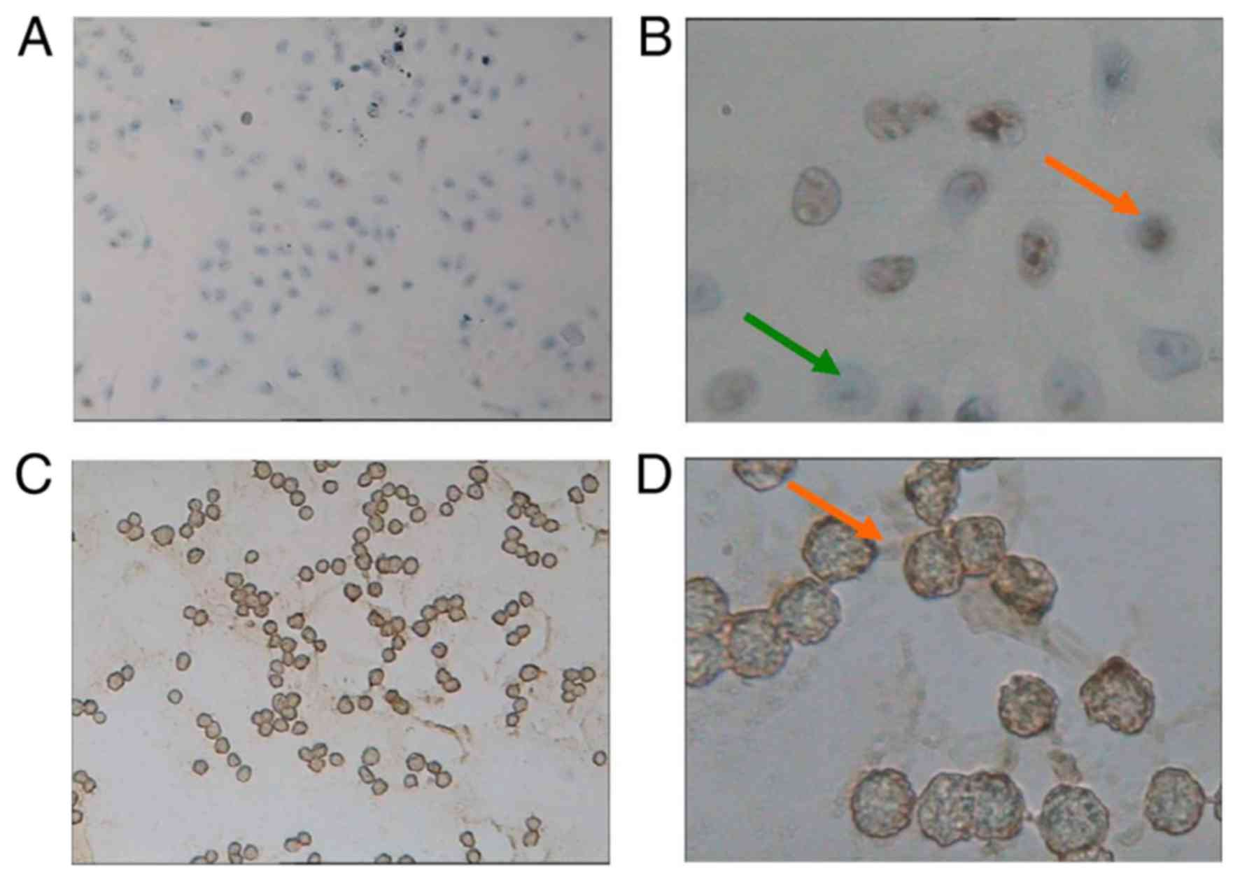
Among the normal cultured A549 cells (Fig. 1A), NSP cells were negatively stained
by CD133 (Fig. 1B); however, lung
cancer stem cells, SP cells, were positively stained by CD133,
characterized by brown staining (Fig. 1C
and D). The soft agar colony formation assay demonstrated that
SP and NSP cells had colony formation ability in vitro after
21 days of culture. The NSP colony formation rate was 13.33±1.76%,
which was significantly reduced compared with SP cells
(30.67±2.57%; P<0.05).
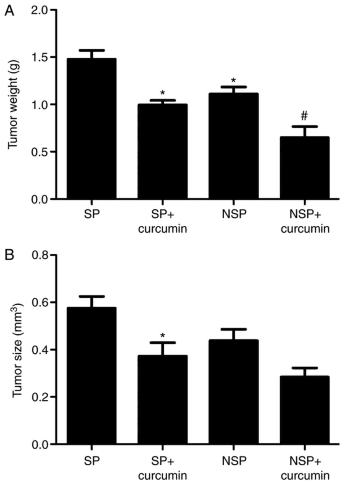
Effect of curcumin on tumor size and
weight
To measure tumor growth inhibition caused by
curcumin treatment, tumor tissues were measured and weighed.
Fig. 2A indicated that the tumor
weight (1.48±0.16 g) of group NSP was notably reduced, compared
with group SP (1.11±0.13 g), indicating that the tumor growth of SP
cells was increased, compared with NSP cells. Following the
treatment of curcumin, compared with the group SP and group NSP,
the tumor weight of group SP+curcumin was decreased by 32.7%, which
was less than the decrease in the group NSP+curcumin (45.1%). As
for the tumor volume (Fig. 2B), the
tumor size of group SP (0.58±0.09 mm3) had no
significant difference, compared with group NSP (0.44±0.08
mm3). Following treatment with curcumin, compared with
the group SP, the tumor size of group SP+curcumin significantly
decreased by 35.2% (P<0.05); however, compared with the group
NSP, treatment with curcumin, the tumor volume of group
NSP+curcumin was not significantly reduced (P>0.05).
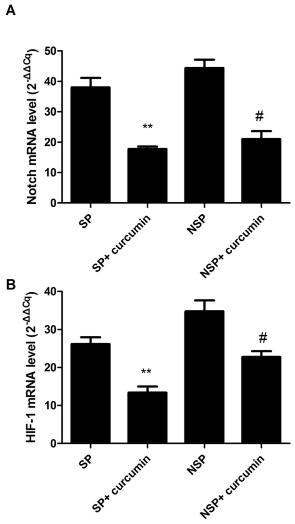
Effect of curcumin on Notch-1 and
HIF-1 mRNA expression
Notch-1 and HIF-1 mRNA expression was analyzed by
the relative ratio of Notch-1 and HIF-1, individually, against
β-actin. It was determined that Notch-1 and HIF-1 mRNA expression
levels in groups SP and NSP had no significant difference
(P>0.05; Fig. 3A and B). Curcumin
significantly suppressed the mRNA expression of Notch-1 in the
group SP+curcumin and group NSP+curcumin by 53.2% (P<0.01) and
52.7% (P<0.05), respectively. Additionally, HIF-1 mRNA
expression was also inhibited by curcumin in the group SP+curcumin
and group NSP+curcumin by 48.8% (P<0.01) and 34.4% (P<0.05),
respectively.
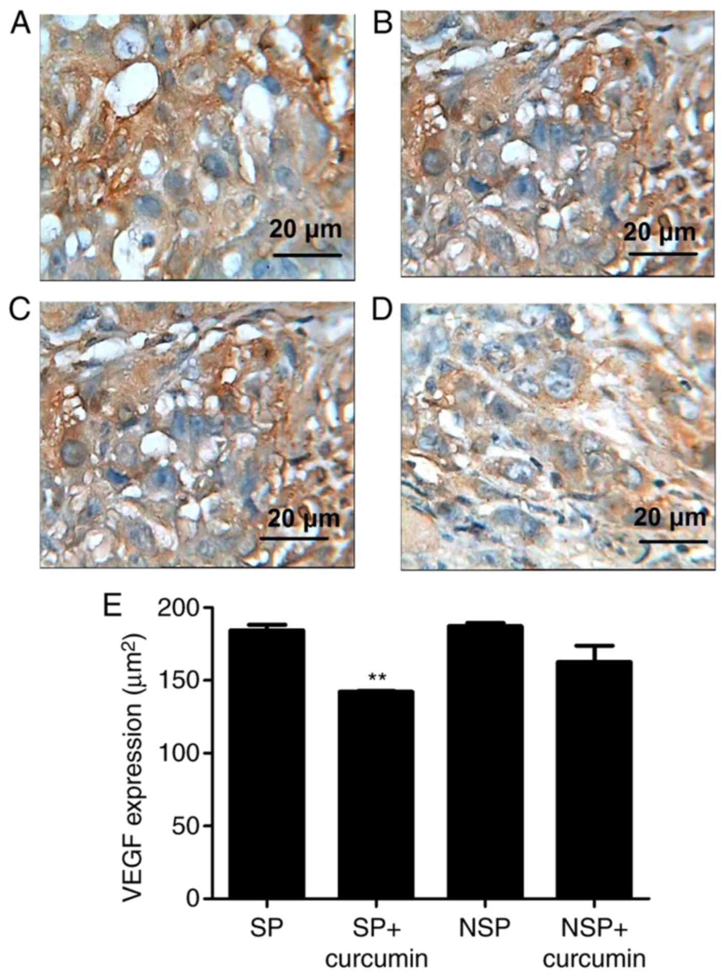
Effect of curcumin on VEGF and NF-κB
expression
To assess the in vivo protective effect of
curcumin against tumor angiogenesis, VEGF and NF-κB expression was
evaluated via immunohistochemistry. The result of
immunohistochemistry demonstrated that the VEGF (Fig. 4A and B) expressed in groups SP and
NSP, and were primarily located in the cytoplasm and characterized
by dark brown staining; however, following eight treatments of
curcumin, VEGF (Fig. 4C and D)
immunoreactivity was not prominent, indicating a notable
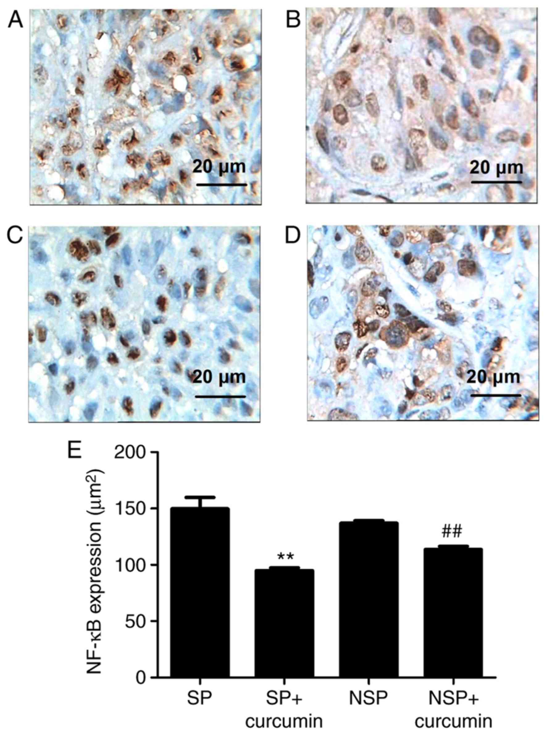
suppression of VEGF in the tumor tissue. NF-κB expression (Fig. 5A and B) was significantly increased,
while suppressed by curcumin (Fig. 5C and
D). Compared with group SP, the expression of VEGF in the group
SP+curcumin was notably decreased; however, curcumin treatment did
not inhibit the expression of VEGF for the mice subcutaneously
injected with the NSP cells. For the NF-κB expression, compared
with group SP or NSP, curcumin treatment did not significantly
suppress the expression in mice subcutaneously injected with SP or
NSP cells (P<0.01).
Discussion
Lung cancer is the leading cause of
cancer-associated mortalities in 2012 globally (2). Almost the same number of people from USA
succumbed from lung cancer as succumbed from prostate, breast and
colon cancer combined in 2011 (25).
Previously, palliative chemotherapy in the metastatic NSCLC setting
resulted in modest survival prolongation and preservation of
quality of life (26). The ability to
exclude Hoechst dye, as defined by SP fraction, was initially
indicated in normal hematopoietic cells (27), but was subsequently determined to be
present in hematopoietic malignancy and solid tumor types (28). SP cells comprise <0.1% of the whole
bone marrow cells and are enriched in drug-resistant hematopoietic
stem cells (29). Curcumin is an
important bioactive component extracted from turmeric rhizome
Curcuma longa Linn, which has been frequently studied for its
potential anticancer activity in vivo and in vitro.
The mechanisms of anticancer include: Inhibition of invasion and
metastasis; inhibition of the protein kinases activity; and
anti-angiogenesis.
In the present results, the SP colony formation rate
(30.67±2.57%) was notably increased, compared with NSP cells
(13.33±1.76%), which indicated that the SP fraction cells were
enriched in lung cancer cells capable of self-renewal and
differentiation with reconstitution of the original cell
population. As a result, the tumor weight of group SP was notably
increase, compared with group NSP. The tumor volume of group SP was
also increased, compared with group NSP, but there was no
significant difference. If the observation time was prolonged, a
significant difference may be observed between the two groups.
Curcumin may significantly reduce the tumor weight and size in the
group SP+curcumin and group NSP+curcumin. Furthermore, the
inhibition degree of group NSP+curcumin was notable increased,
compared with group NSP. According to a previous report, SP cells
that had the ability to exclude Hoechst dye were associated with
increased expression of drug transporters, primarily the ABC
transporters family, including ABC subfamily B member 1 and ABCG2,
which are capable of extrusion of the dye from the cell (30). The expression of ABC transporters is
associated with drug resistance, which is a characteristic of stem
cells from normal and malignant tissues (31). The aforementioned characteristic of SP
cells explained why NSP cells had increased sensitivity to curcumin
treatment.
Overexpression of VEGF has been determined in NSCLC,
and is associated with increased tumor recurrence, metastasis and
associated mortality (32). The VEGF
pathway activation results in endothelial cell survival,
mitogenesis, migration, differentiation and mobilization of
endothelial progenitor cells from the bone marrow into the
peripheral circulation (33).
Additionally, recognition of the VEGF pathway is considered an
important mediator of angiogenesis, which has resulted in the
clinical study of a number of VEGF-targeted therapies for lung
cancer. The coexpression of Notch-1/VEGF has a notable impact on
the survival of lung cancer cells, indicating that Notch-regulated
angiogenesis is involved in the metastasis and determines the
prognosis of NSCLC (34). The
involvement of Notch in lung cancer was experimentally demonstrated
in a transgenic mouse model by the alveolar epithelium specific
expression of activated Notch (35).
Notch is associated with the expression of numerous other
cancer-associated proteins, including HIF-1, as a marker of normal
lung physiology. Under the condition of hypoxia, Notch 1 could be
activated by HIF-1α in lung adenocarcinoma cells, including A549
cells (36). In the present study,
RT-qPCR was used to analyze the levels of Notch and HIF-1 in the
A549 cells. Statistical analysis demonstrated that curcumin
suppressed the expression of Notch and HIF-1. The significant
differences of VEGF expression were observed between the groups
treated with and without curcumin. These data are consistent with
curcumin-inhibited cancer cell growth associated with the
inhibition of angiogenesis.
As a transcription factor, NF-κB could induce
>200 genes expression, which are involved in diverse biological
processes, including cell survival, cell adhesion, inflammation,
differentiation and growth. Tumor tissues from patients with lung
cancer expressed high levels of NF-κB activation, and was
significantly associated with disease advancement, regarding
Tumor-Node-Metastasis stage, and poor prognosis in patients with
lung cancer (37). NF-κB in
inflammatory cells activates the secretion of a variety of
angiogenesis factors, including VEGF. Immunohistochemistry
demonstrated that curcumin inhibited NF-κB expression in the group
of SP+curcumin and group NSP+curcumin. Combined with the result of
VEGF and its downstream factors Notch and HIF-1, curcumin
suppressed VEGF expression via the inhibition of NF-κB.
To conclude, the present in vivo experiment
demonstrated that treatment with curcumin through intraperitoneal
injection inhibited SP and NSP cell-induced lung cancer, which may
be associated with the inhibition of angiogenesis. The present
study indicated curcumin as a potential alternative for the
prevention of tumor growth in lung cancer.
Acknowledgements
Not applicable.
Funding
This project was supported by Key projects of
Tianjin applied basic and frontier technology research program
grant no. 14JCZDJC36900).
Availability of data and materials
All data generated or analyzed during this study are
included in this published article.
Authors' contributions
XL and SM performed the majority of the experiments,
and analyzed and interpreted the data. PY and BS contributed to the
cell culture and RT-qPCR. YZ and YS were responsiible for
anaesthetizing the animals, removing the tumor tissue, analyzing
the animal tumor weight and calculating the tumor size according to
the formula. MH and RM performed the immunohistochemistry. YJ
designed the present study, analyzed the data and wrote the
manuscript. All authors read and approved the final manuscript.
Ethics approval and consent to
participate
The present experiment was approved by the Animal
Care Committee of Tianjin University of Traditional Chinese
Medicine (Tianjin, China) and in accordance with the UK Animals
(Scientific Procedures) Act of 1986 (18).
Patient consent for publication
Not applicable.
Competing interests
The authors declare that they have no competing
interests.
References
|
1
|
Ferlay J, Shin HR, Bray F, Forman D,
Mathers C and Parkin DM: Estimates of worldwide burden of cancer in
2008: GLOBOCAN 2008. Int J Cancer. 127:2893–2917. 2010. View Article : Google Scholar : PubMed/NCBI
|
|
2
|
Islami F, Torre LA and Jemal A: Global
trends of lung cancer mortality and smoking prevalence. Transl Lung
Cancer Res. 44:327–338. 2015.
|
|
3
|
Torre LA, Bray F, Siegel RL, Ferlay J,
Lortet-Tieulent J and Jemal A: Global cancer statistics, 2012. CA
Cancer J Clin. 65:87–108. 2015. View Article : Google Scholar : PubMed/NCBI
|
|
4
|
Lam WK, White NW and Chan-Yeung MM: Lung
cancer epidemiology and risk factors in Asia and Africa. Int J
Tuberc Lung Dis. 8:1045–1057. 2004.PubMed/NCBI
|
|
5
|
Parkin DM, Bray F, Ferlay J and Pisani P:
Global cancer statistics, 2002. CA Cancer J Clin. 55:74–108. 2005.
View Article : Google Scholar : PubMed/NCBI
|
|
6
|
Peng H, Ma M and Han B: Survival analysis
of 1,742 patients with stage IV non-small cell lung cancer.
Zhongguo Fei Ai Za Zhi. 14:362–636. 2011.(In Chinese). PubMed/NCBI
|
|
7
|
Ming X, Feng Y, Yang C, Wang W, Wang P and
Deng J: Radiation-induced heart disease in lung cancer
radiotherapy: A dosimetric update. Medicine (Baltimore).
95:e50512016. View Article : Google Scholar : PubMed/NCBI
|
|
8
|
Ho MM, Ng AV, Lam S and Hung JY: Side
population in human lung cancer cell lines and tumors is enriched
with stem-like cancer cells. Cancer Res. 67:4827–4833. 2007.
View Article : Google Scholar : PubMed/NCBI
|
|
9
|
Hou GX, Liu PP, Zhang S, Yang M, Liao J,
Yang J, Hu Y, Jiang WQ, Wen S and Huang P: Elimination of stem-like
cancer cell side-population by auranofin through modulation of ROS
and glycolysis. Cell Death Dis. 9:892018. View Article : Google Scholar : PubMed/NCBI
|
|
10
|
Akunuru S, Palumbo J, Zhai QJ and Zheng Y:
Rac1 targeting suppresses human non-small cell lung adenocarcinoma
cancer stem cell activity. PLoS One. 6:e169512011. View Article : Google Scholar : PubMed/NCBI
|
|
11
|
Qiu C, Zhang T, Zhang W, Zhou L, Yu B,
Wang W, Yang Z, Liu Z, Zou P and Liang G: Licochalcone A Inhibits
the proliferation of human lung cancer cell lines A549 and H460 by
Inducing G2/M cell cycle arrest and ER stress. Int J Mol Sci.
18:pii: E17612017. View Article : Google Scholar
|
|
12
|
Feng H, Lu JJ, Wang Y, Pei L and Chen X:
Osthole inhibited TGF β-induced epithelial-mesenchymal transition
(EMT) by suppressing NF-κB mediated Snail activation in lung cancer
A549 cells. Cell Adh Migr. 11:464–475. 2017. View Article : Google Scholar : PubMed/NCBI
|
|
13
|
Li B, Shi C, Li B, Zhao JM and Wang L: The
effects of curcumin on HCT-116 cells proliferation and apoptosis
via the miR-491/PEG10 pathway. J Cell Biochem. 119:3091–3098. 2018.
View Article : Google Scholar : PubMed/NCBI
|
|
14
|
Wang YL, Ju B, Zhang YZ, Yin HL, Liu YJ,
Wang SS, Zeng ZL, Yang XP, Wang HT and Li JF: Protective effect of
curcumin against oxidative stress-induced injury in rats with
Parkinson's disease through the Wnt/β-catenin signaling pathway.
Cell Physiol Biochem. 43:2226–2241. 2017. View Article : Google Scholar : PubMed/NCBI
|
|
15
|
Zhang M, Tang J, Li Y, Xie Y, Shan H, Chen
M, Zhang J and Yang X, Zhang Q and Yang X: Curcumin attenuates
skeletal muscle mitochondrial impairment in COPD rats: PGC-1α/SIRT3
pathway involved. Chem Biol Interact. 277:168–175. 2017. View Article : Google Scholar : PubMed/NCBI
|
|
16
|
Zhao Z, Yang Y, Liu W and Li Z: T59, a new
compound reconstructed from curcumin, induces cell Apoptosis
through reactive oxygen species activation in human lung cancer
cells. Molecules. 23:pii: E12512018. View Article : Google Scholar
|
|
17
|
Shimada K, Ushijima K, Suzuki C, Horiguchi
M, Ando H, Akita T, Shimamura M, Fujii J, Yamashita C and Fujimura
A: Pulmonary administration of curcumin inhibits B16F10 melanoma
lung metastasis and invasion in mice. Cancer Chemother Pharmacol.
82:265–273. 2018. View Article : Google Scholar : PubMed/NCBI
|
|
18
|
Li Y, Zhang S, Geng JX and Hu XY: Curcumin
inhibits human non-small cell lung cancer A549 cell proliferation
through regulation of Bcl-2/Bax and cytochrome C. Asian Pac J
Cancer Prev. 14:4599–602. 2013. View Article : Google Scholar : PubMed/NCBI
|
|
19
|
Liu F, Gao S, Yang Y, Zhao X, Fan Y, Ma W,
Yang D, Yang A and Yu Y: Curcumin induced autophagy anticancer
effects on human lung adenocarcinoma cell line A549. Oncol Lett.
14:2775–2782. 2017. View Article : Google Scholar : PubMed/NCBI
|
|
20
|
Hollands C: The Animals (scientific
procedures) Act 1986. Lancet. 8497:32–33. 1986. View Article : Google Scholar
|
|
21
|
Wo XD, H XQ and G CX: Long term toxicity
test of curcumin. J Zhejiang College TCM. 24:61–65. 2000.(In
Chinese).
|
|
22
|
Zhuang HW, Mo TT, Hou WJ, Xiong GX, Zhu
XL, Fu QL and Wen WP: Biological characteristics of CD133(+) cells
in nasopharyngeal carcinoma. Oncol Rep. 30:57–63. 2013. View Article : Google Scholar : PubMed/NCBI
|
|
23
|
Yu L, Garg HG, Li B, Linhardt RJ and Hales
CA: Antitumor effect of butanoylated heparin with low anticoagulant
activity on lung cancer growth in mice and rats. Curr Cancer Drug
Targets. 10:229–241. 2010. View Article : Google Scholar : PubMed/NCBI
|
|
24
|
Livak KJ and Schmittgen TD: Analysis of
relative gene expression data using real-time quantitative PCR and
the 2(-Delta Delta C(T)) method. Methods. 25:402–408. 2001.
View Article : Google Scholar : PubMed/NCBI
|
|
25
|
Dela Cruz CS, Tanoue LT and Matthay RA:
Lung cancer: Epidemiology, etiology, and prevention. Clin Chest
Med. 32:605–644. 2011. View Article : Google Scholar : PubMed/NCBI
|
|
26
|
Hopwood P and Stephens RJ: Symptoms at
presentation for treatment in patients with lung cancer:
Implications for the evaluation of palliative treatment. The
Medical Research Council (MRC) Lung Cancer Working Party. Br J
Cancer. 71:633–636. 1995. View Article : Google Scholar : PubMed/NCBI
|
|
27
|
Goodell MA, Brose K, Paradis G, Conner AS
and Mulligan RC: Isolation and functional properties of murine
hematopoietic stem cells that are replicating in vivo. J Exp Med.
183:1797–1806. 1996. View Article : Google Scholar : PubMed/NCBI
|
|
28
|
Hadnagy A, Gaboury L, Beaulieu R and
Balicki D: SP analysis may be used to identify cancer stem cell
populations. Exp Cell Res. 312:3701–3710. 2006. View Article : Google Scholar : PubMed/NCBI
|
|
29
|
Sales KM, Winslet MC and Seifalian AM:
Stem cells and cancer: An overview. Stem Cell Rev. 3:249–255. 2007.
View Article : Google Scholar : PubMed/NCBI
|
|
30
|
Kim M, Turnquist H, Jackson J, Sgagias M,
Yan Y, Gong M, Dean M, Sharp JG and Cowan K: The multidrug
resistance transporter ABCG2 (breast cancer resistance protein 1)
effluxes Hoechst 33342 and is overexpressed in hematopoietic stem
cells. Clin Cancer Res. 8:22–28. 2002.PubMed/NCBI
|
|
31
|
Szakács G, Paterson JK, Ludwig JA,
Booth-Genthe C and Gottesman MM: Targeting multidrug resistance in
cancer. Nat Rev Drug Discov. 5:219–234. 2006. View Article : Google Scholar : PubMed/NCBI
|
|
32
|
Ferrara N: The role of vascular
endothelial growth factor in pathological angiogenesis. Breast
Cancer Res Treat. 36:127–137. 1995. View Article : Google Scholar : PubMed/NCBI
|
|
33
|
Hicklin DJ and Ellis LM: Role of the
vascular endothelial growth factor pathway in tumor growth and
angiogenesis. J Clin Oncol. 23:1011–1027. 2005. View Article : Google Scholar : PubMed/NCBI
|
|
34
|
Yuan X, Wu H, Han N, Xu H, Chu Q, Yu S,
Chen Y and Wu K: Notch signaling and EMT in non-small cell lung
cancer: Biological significance and therapeutic application. J
Hematol Oncol. 7:872014. View Article : Google Scholar : PubMed/NCBI
|
|
35
|
Allen TD, Rodriguez EM, Jones KD and
Bishop JM: Activated Notch1 induces lung adenomas in mice and
cooperates with Myc in the generation of lung adenocarcinoma.
Cancer Res. 71:6010–6018. 2011. View Article : Google Scholar : PubMed/NCBI
|
|
36
|
Guo L, Zhang T, Xiong Y and Yang Y: Roles
of NOTCH1 as a therapeutic target and a biomarker for lung cancer:
Controversies and perspectives. Dis Markers. 2015:5205902015.
View Article : Google Scholar : PubMed/NCBI
|
|
37
|
Karin M and Greten FR: NF-kappaB: Linking
inflammation and immunity to cancer development and progression.
Nat Rev Immunol. 5:749–759. 2005. View
Article : Google Scholar : PubMed/NCBI
|