Introduction
Cancer is a substantial health and economic burden
to humans. Globally, there is an annual increase in the incidence
of cancer and its associated mortality rate (1). In 2017, it was estimated that there
would be ~1,688,780 new cancer cases and 600,920 cancer-related
mortalities in the United States alone (2). Biomarkers that may assist with the
clinical diagnosis of various tumors and the assessment of
prognosis are being investigated. To date, microRNAs (miRNAs/miRs)
have received considerable attention in research and are viewed as
potential biomarkers for the early detection and diagnosis of
cancer.
miRNAs are a class of small endogenous non-coding
regulatory RNAs that are 18–25 nucleotides in length; they serve
important roles in post-transcriptional gene regulation and are
strongly linked to biological processes, including cell
proliferation and metastasis (3–5).
Accumulating studies have demonstrated that miRNAs can function as
tumor suppressors or as oncogenes by targeting genes involved in
tumor cell differentiation, proliferation, apoptosis and metastasis
(6). Growing evidence also suggests
that miRNAs may act as novel and non-invasive biomarkers for
diagnosing a range of cancer types at an early stage (7–11).
miR-195 is located on chromosome 17p13.1, a region
frequently deleted in human cancer (12). Studies have suggested that abnormal
expression of miR-195 serves a critical role in tumorigenesis in
bladder, stomach, thyroid, colorectal, prostate and cervical cancer
types, as well as in hepatocellular carcinoma, glioblastoma, tongue
squamous cell carcinoma, esophageal squamous cell carcinoma and
osteosarcoma (13–23). More importantly, miR-195 has been
consistently detected in bodily fluids, including blood and saliva,
in cancer patients, suggesting that it may be valuable as a
non-invasive biomarker for cancer diagnosis and detection (12,24–29).
However, the diagnostic value of miR-195 remains uncertain. No
meta-analyses of the association between miR-195 expression and
cancer diagnosis and detection have been published. Therefore, a
quantitative meta-analysis to clarify the diagnostic value of
miR-195 expression in human cancer was performed in the present
study.
Materials and methods
Search strategy and study selection
criteria
A meta-analysis was performed based on published
guidelines for diagnostic meta-analyses (30). Searches were performed in the PubMed
(https://www.ncbi.nlm.nih.gov/pubmed),
Cochrane Library (https://www.cochranelibrary.com/), Wanfang (http://www.wanfangdata.com.cn/index.html) and China
National Knowledge Infrastructure databases (http://cnki.net/), for eligible articles published up
to December 10, 2017. The following search terms were used:
‘Cancer’ or ‘tumor’ or ‘carcinoma’ or ‘neoplasm’ or ‘malignancy’,
and ‘miR-195’, and ‘sensitivity’ or ‘specificity’ or ‘ROC curve’
(ROC, receiver operator characteristic). The titles and abstracts
of the studies were checked, and the full texts were scanned for
relevance by two investigators independently based on their titles
and abstracts, following which full texts were perused for
potential eligibility. The following article inclusion criteria
were used: i) Test indices for diagnosis [sensitivity, specificity
and area under the curve (AUC)] were provided or could be
calculated from the available data; ii) population and control
group(s) were explicitly defined; iii) the diagnostic value of
miR-195 for detecting cancer was assessed; and iv) the study was
published in English or Chinese.
Data extraction and quality
assessment
The following information from the included studies
was recorded by two researchers independently: Name of the first
author, year of publication, country, ethnicity of cohort, sample
size, specimen and cancer type, detection method, cut-off value,
and true-positive, false-positive, true-negative and false-negative
numbers. The quality of the diagnostic test studies was evaluated
using the Quality Assessment of Diagnostic Accuracy Studies 2
(QUADAS-2) tool (http://www.bristol.ac.uk/population-health-sciences/projects/quadas/).
Specifically, 14 items from the QUADAS checklist were applied to
each article, and an answer of ‘Yes’, ‘No’ or ‘Unclear’ was
determined. Only ‘Yes’ resulted in a score.
Statistical analysis
Statistical analyses were performed using the Stata
14.0 software (StataCorp, College Station, TX, USA). The bivariate
meta-analysis model was used to calculate the relevant measures,
including pooled sensitivity, specificity, diagnostic odds ratio
(DOR), positive likelihood ratio (PLR) and negative likelihood
ratio (NLR). Summary receiver operator characteristic (SROC) curve
analysis was performed and the AUC was calculated to evaluate the
overall diagnostic value of miR-195 in cancer detection and
diagnosis. The data were confirmed using a hierarchical summary
receiver operating characteristics (HSROC) model. Spearman's
correlation [testing that −1≤r≤1, with a permutation test, which
compares the sensitivity and (1-specificity)] and ROC plane
analyses were conducted to assess the heterogeneity of threshold
effects. The heterogeneity of non-threshold effects was assessed by
the Cochran's Q and inconsistency index (I2) tests
(31). P<0.10 for the Cochran's Q
test or I2>50% indicated obvious heterogeneity
between the studies (32). Fagan's
nomogram was used to certify associations between the pre-test
probability, the likelihood ratio and the post-test probability.
The publication bias was tested using Deeks' funnel plots (33).
Results
Study selection and characteristics of
included studies
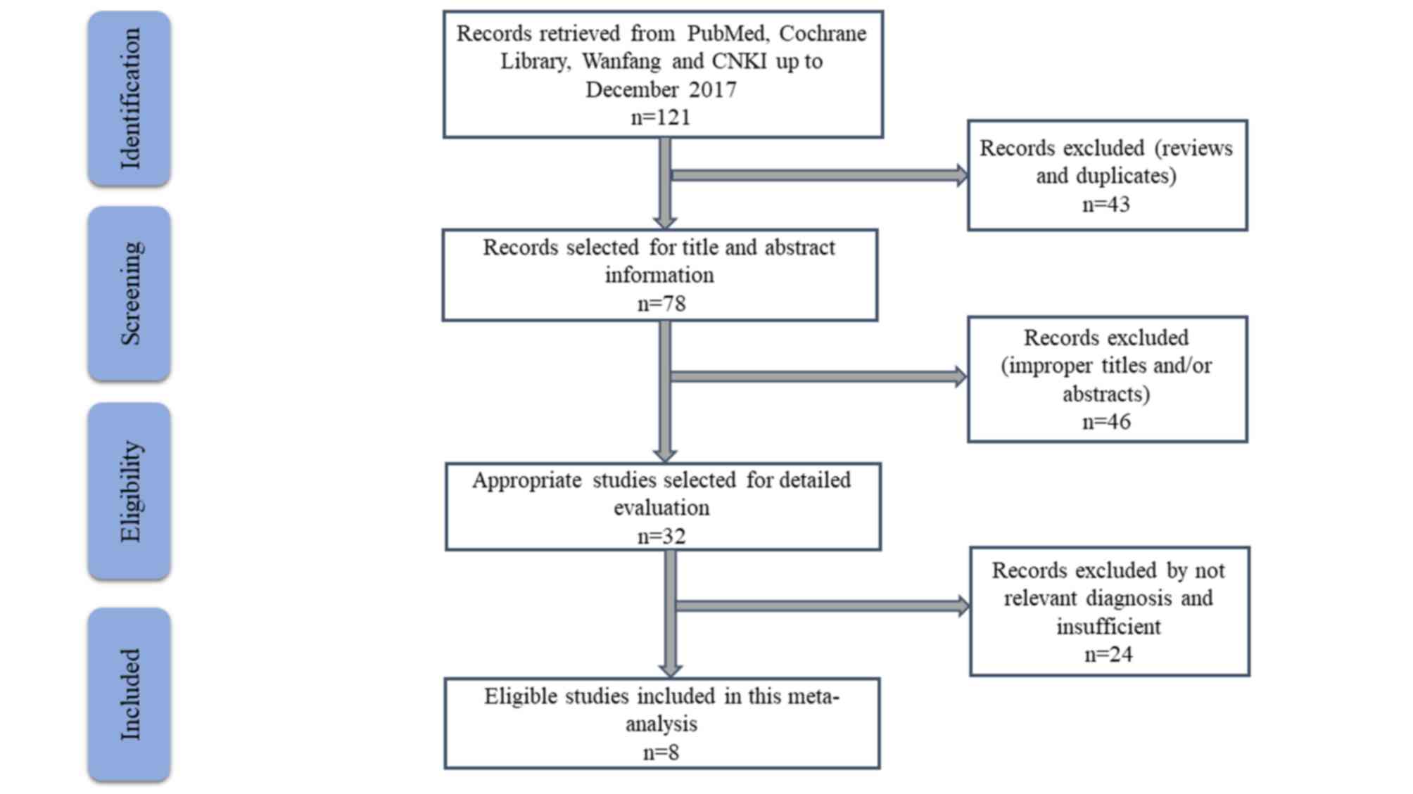
Of 121 published articles that were determined using
the stated search terms in the aforementioned databases, 8 studies
(12,24–29,34) met
the inclusion criteria for meta-analysis (Fig. 1). Details about the included subjects
(546 cases and 547 controls) are displayed in Table I. In total, 4 studies focused on
breast cancer, and 1 each on hepatic carcinoma, colorectal cancer,
non-small cell lung cancer and acute myeloid leukemia. The miR-195
expression was assessed using reverse transcription-quantitative
polymerase chain reaction methods in serum (n=6), plasma (n=1) or
tissue (n=1).
 | Table I.Summary of information of the included
studies. |
Table I.
Summary of information of the included
studies.
| First author | Publication
year | Country | Ethnicity | Cancer type | Sample type | Test method | Cut-off | Cases/controls | TP | FP | FN | TN | QUADAS-2 score | (Refs.) |
|---|
| Heneghan et
al | 2010 | Ireland | Caucasian | BRC | Serum | RT-qPCR | NA | 83/63 | 73 | 6 | 10 | 57 | 6 | (25) |
| Zhu et
al | 2013 | China | Asian | BRC | Serum | RT-qPCR | NA | 20/10 | 17 | 3 | 3 | 7 | 4 | (24) |
| Zhao et
al | 2014 | China | Asian | BRC | Serum | RT-qPCR | 0.505 | 210/102 | 145 | 11 | 65 | 91 | 5 | (29) |
| Motawi et
al | 2015 | Egypt | Caucasian | HCC | Serum | RT-qPCR | 0.77 | 112/42 | 75 | 10 | 37 | 32 | 6 | (26) |
| Su et
al | 2016 | China | Asian | NSCLC | Plasma | RT-qPCR | NA | 100/100 | 78 | 14 | 22 | 86 | 5 | (12) |
| Yang et
al | 2016 | China | Asian | CRC | Tissue | RT-qPCR | 0.93 | 50/54 | 48 | 40 | 2 | 14 | 4 | (27) |
| Zhang et
al | 2016 | China | Asian | BRC | Serum | RT-qPCR | NA | 54/70 | 38 | 5 | 16 | 65 | 5 | (28) |
| Hong et
al | 2018 | China | Asian | AML | Serum | RT-qPCR | 2.09 | 106/106 | 73 | 4 | 33 | 102 | 6 | (34) |
Quality assessment
The methodological quality of diagnosis in the
present study was assessed using the QUADAS-2 tool. All the studies
were of moderate to high quality, with QUADAS-2 scores between 4
and 7 (Table I), indicating a
reliable foundation for the present meta-analysis.
Data analysis
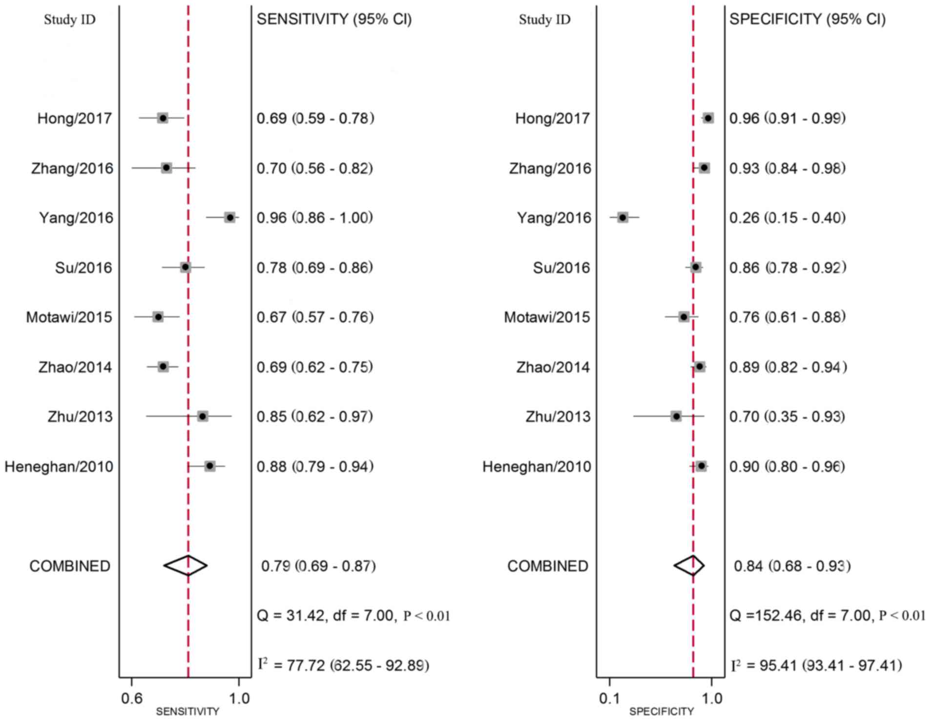
Forest plots of data from the 8 studies on the
sensitivity and specificity of miR-195 in diagnosing various cancer
types are shown in Fig. 2. The
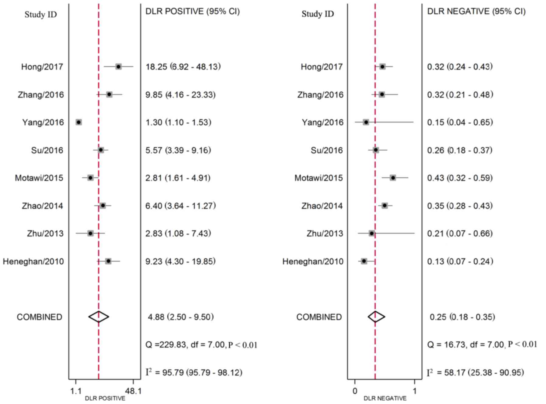
calculated metrics from all included studies are as follows:
Sensitivity 0.79 [95% confidence interval (CI), 0.69–0.87],
specificity 0.84 (95% CI, 0.68–0.93), PLR 4.9 (95% CI, 2.50–9.50),
NLR 0.25 (95% CI, 0.18–0.35; Fig. 3)
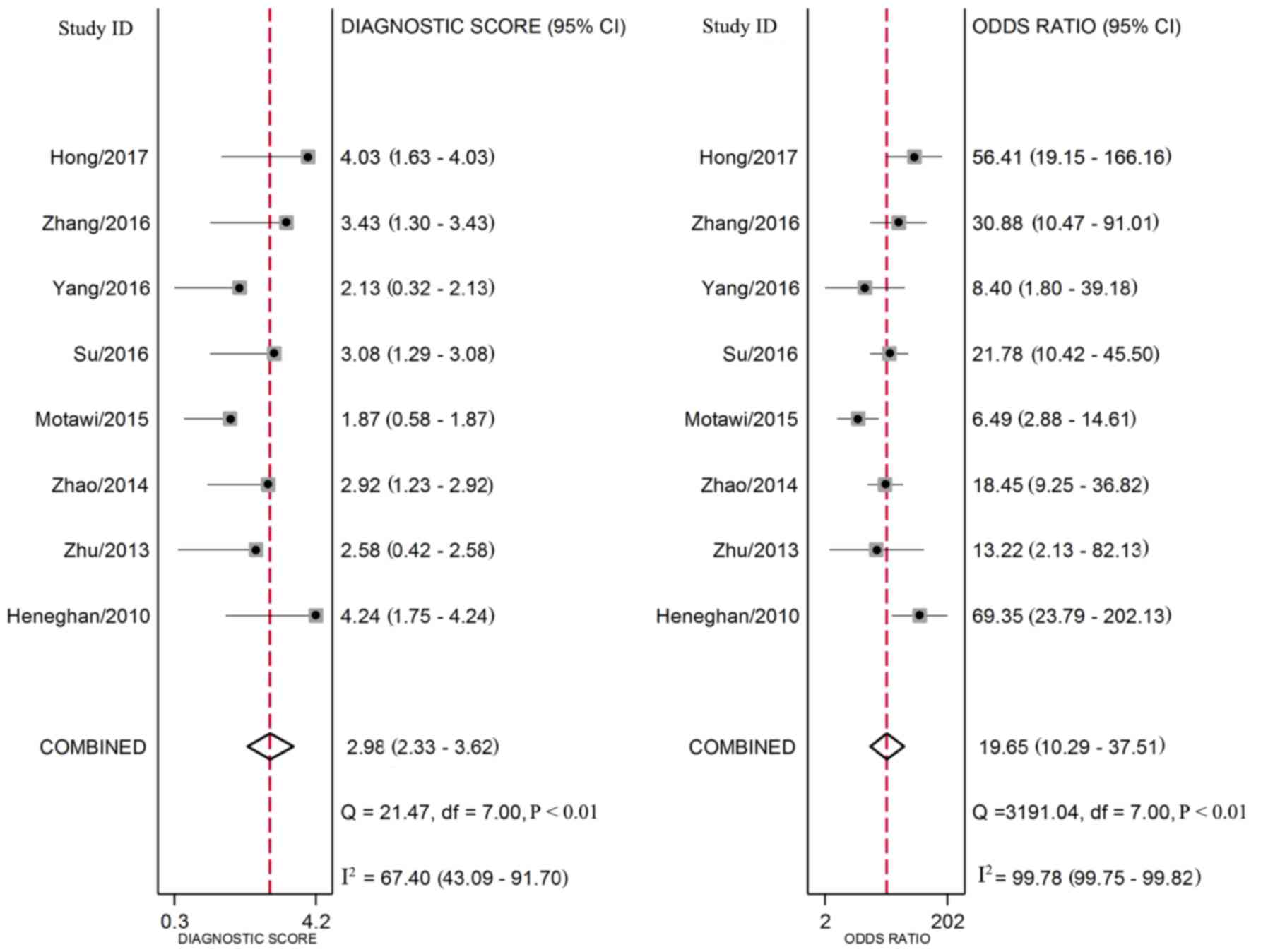
and DOR 20 (95% CI, 10.00–38.00; Fig.
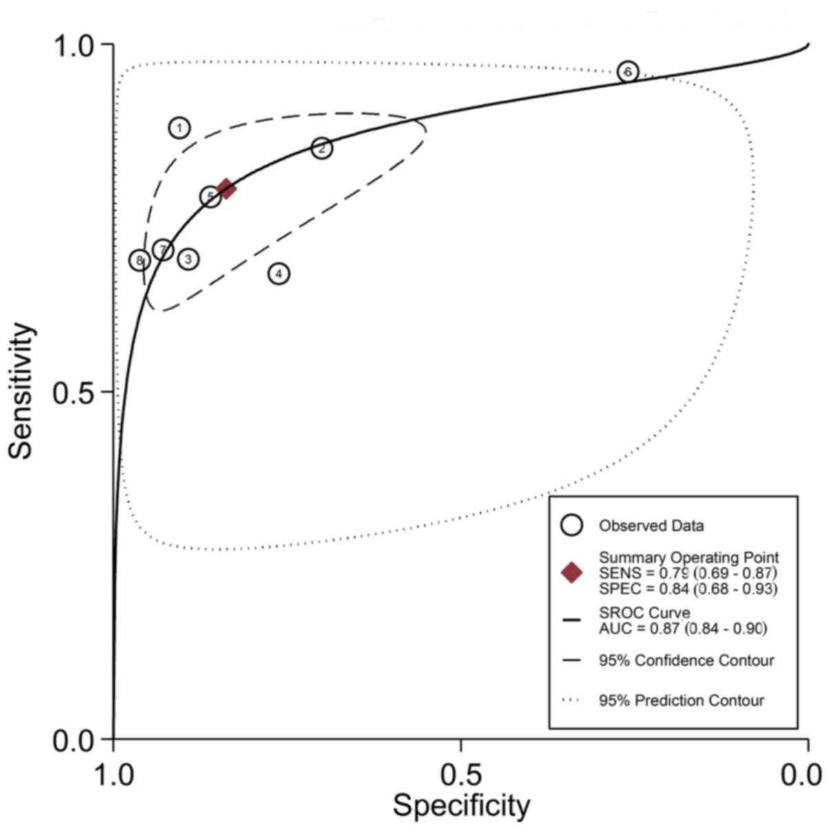
4). The overall SROC curve for the 8 included studies is shown
in Fig. 5. The AUC of miR-195 was
0.87 (95% CI, 0.84–0.90). These results indicate that miR-195
differentiates cancer patients from controls with high accuracy.
The HSROC curve of the included studies was in line with the
results from the bivariate model (Fig.
6). The value of β (a scale of parameter, which indicated the
asymmetry of the curve) was 0.579 (95% CI, −0.128–1.286) and the
P-value was 0.108, which indicated that the HSROC was symmetrical.
The value of γ (the statistical value representing the accuracy of
the diagnostic tests) was 3.015 (95% CI, 2.490–3.539), which
indicated that miR-195 had high accuracy in differentiating
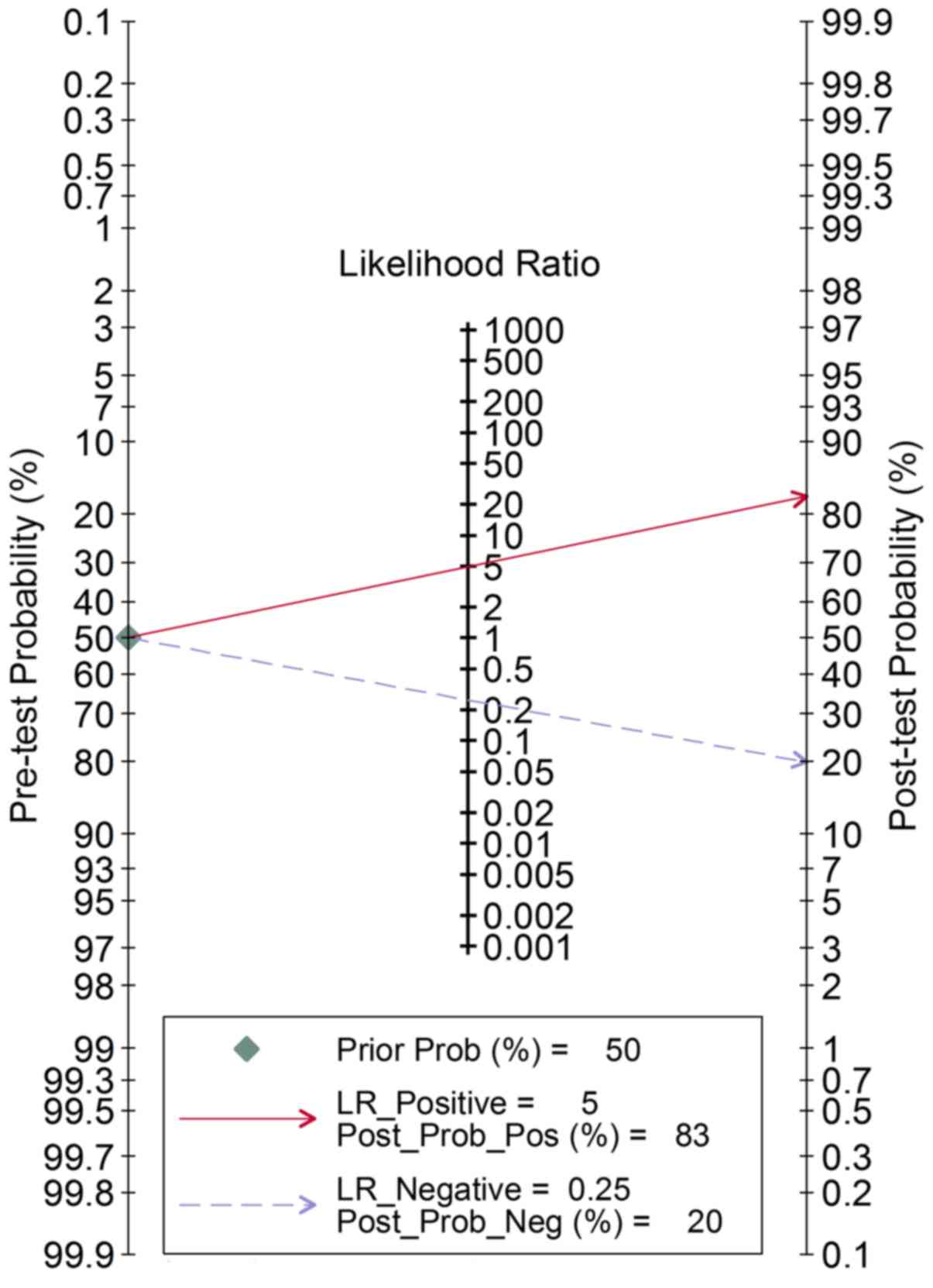
patients with cancer from control patients. In order to assess the
clinical utility of the index test, Fagan's nomogram was used to
predict the increasing inerrability of a positive diagnosis by
estimating post-test probabilities. As shown in Fig. 7, when miR-195 was tested in all
individuals with a pre-test probability of cancer of 50%, a
positive result improved the post-test probability of having cancer
to 83%, while a negative result dropped the post-test probability
to 20%. Combined, the results indicated that miR-195 displays a
moderate accuracy for distinguishing patients with cancer from all
individuals.
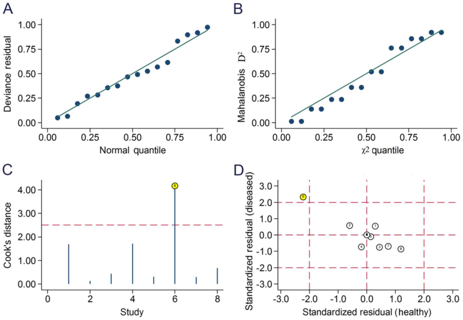
Influence analysis and robustness
tests
Goodness-of-fit (Fig.
8A) and bivariate normality (Fig.
8B) analyses demonstrated that the bivariate model was
moderately robust. Influence analysis (Fig. 8C) and outlier detection (Fig. 8D) only identified 1 outlier. Exclusion
of the outlier gave rise to small changes to the present analysis
results, with the overall metrics varying as follows: Sensitivity
decreased from 0.79 to 0.75, specificity increased from 0.84 to
0.89, PLR increased from 4.9 to 6.7, NLR increased from 0.25 to
0.29, DOR increased from 20 to 24 and AUC increased from 0.87 to
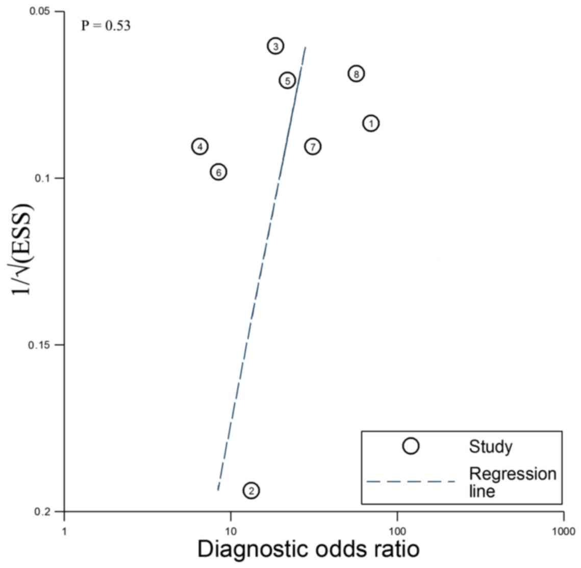
0.88. The Deeks' funnel plot asymmetry test suggested no
significant publication bias (P=0.53; Fig. 9). The tests confirm the robustness of
the present results in the pooled analysis.
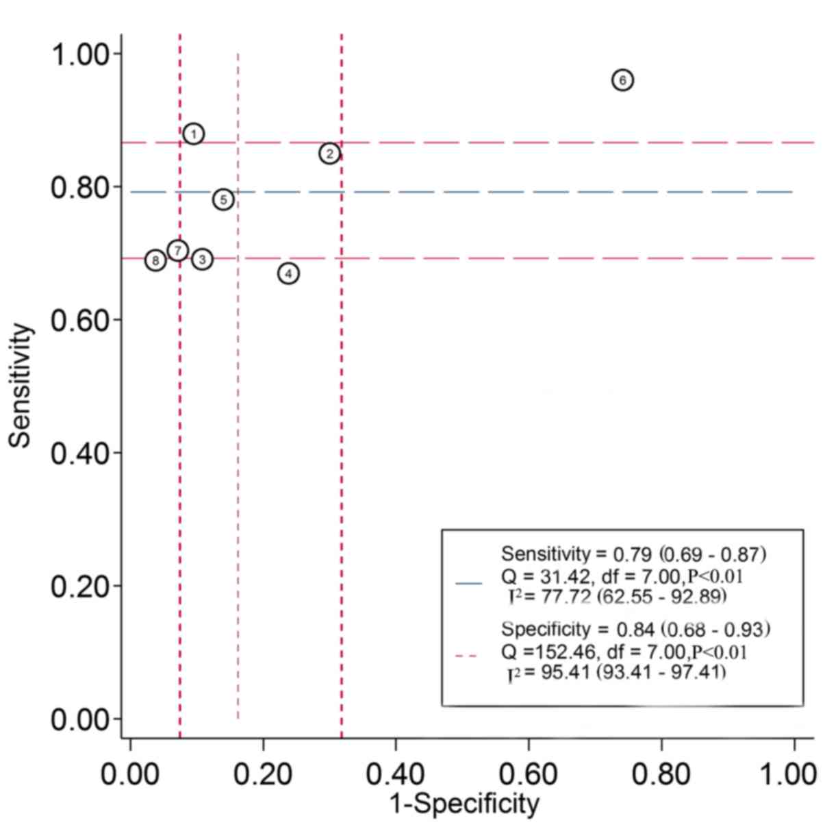
Threshold effect and
heterogeneity
In the present analysis, the ROC plane revealed a
non-typical shoulder arm appearance suggesting that there was no
threshold effect (Fig. 10). The
calculated Spearman's correlation coefficient was −0.81 (P=0.66),
also indicating no threshold effect. An I2 value of 98%
indicated high heterogeneity. Due to only 8 studies being included
in the present meta-analysis, subgroup and meta-regression analyses
could not be performed to investigate the sources of
heterogeneity.
Discussion
The initial diagnosis of malignant tumors currently
involves use of screening endoscopy, random tissue biopsies and
exfoliative cytological examination. The first two methods are
invasive and uncomfortable, and exhibit a low sensitivity, which
limits their application in the early diagnosis of cancer due to
issues with inter-observer reproducibility (35). Therefore, there is an urgent
requirement for reliable biomarkers that require minimally invasive
sampling to detect the presence of malignant tumor tissues.
The present study describes the first meta-analysis
to be performed for the evaluation of the diagnostic value of
miR-195 in cancer detection. An AUC of 0.87 (95% CI, 0.84–0.90),
with a sensitivity of 0.79 (95% CI, 0.69–0.87) and a specificity of
0.84 (95% CI, 0.68–0.93), demonstrated that miR-195 could be used
as a novel biomarker for the detection of cancer in patients. In
the present analysis, the pooled DOR of 20 (95% CI, 10.00–38.00)
suggested that use of miR-195 for cancer diagnosis is credible.
The PLRs and NLRs also reflected the diagnostic
accuracy of miR-195. In the present analysis, the pooled PLR was
4.9 (95% CI, 2.50–9.50) and the NLR was 0.25 (95% CI, 0.18–0.35),
reflecting the fact that patients with cancer had a 4.9-fold higher
probability of being miR-195-positive compared with control
patients, and 25% of all individuals were negative. Fagan's
nomogram revealed that when a pre-test probability of 50% was
specified, the positive post-test probability would increase to 83%
with a PLR of 5, and the negative post-test probability would
decrease to 20% with a NLR of 0.25. The results suggest that
miR-195 is reliable in cancer detection and diagnosis.
There was heterogeneity among the studies included
in the present meta-analysis due to the existence of other
confounding factors. In the present analysis, the Spearman's rank
correlation test was used to analyze the threshold effect and a
correlation coefficient of −0.81 (P=0.66) indicated that the
threshold effect was not a major source of heterogeneity. However,
meta-regression analysis and subgroup analysis could not be
performed due to the small number of eligible studies. Therefore,
factors such as ethnicity and the test method were not investigated
as potential sources of variance.
The role of long non-coding RNAs, including
miRNA-210, plasmacytoma variant translocation 1 gene and
metastasis-associated lung adenocarcinoma transcript-1, as
molecular diagnostic and prognostic markers for cancer, has been
investigated by previous meta-analyses (36–38). To
search for a suitable diagnostic marker, the diagnostic value of
miR-195 expression in human cancer was addressed. The present
meta-analysis was the first to investigate miR-195 expression and
cancer detection using published data.
The present study was restricted by several
limitations: First, the numbers of studies available and
participants included were small; second, a high proportion of data
was from Chinese populations, which may cause ethnicity bias;
third, the overall cut-off value for the miR-195 test could not be
determined as different cut-off values were adopted in each study;
fourth, the present analysis was retrospective, which may limit its
possible conclusions due to selection bias; fifth, only studies
published in English or Chinese were included, creating the
possibility that studies published in other languages were
neglected; and finally, it is difficult to detect cancer early
using just a single marker, and as such, a combination of several
markers may improve the accuracy.
In conclusion, the results of the present
meta-analysis indicate that miR-195 has a moderate diagnostic value
in distinguishing cancer patients from healthy controls. The data
suggest that miR-195 may supplement and improve the accuracy of
existing diagnostic methods. Future studies should concentrate on
the combined use of miR-195 with other miRNAs to improve the
diagnostic accuracy of human cancer detection.
Acknowledgements
Not applicable.
Funding
The present study was supported by the fund of
‘San-ming’ Project of Medicine in Shenzhen (grant no.
SZSM201612010), the Shenzhen Foundation of Science and Technology
International Cooperative Project (grant no.
GJHZ20160301164637011), and the Shenzhen Health and Family Planning
Commission Scientific Research Project (grant nos. 201601023 and
201601025).
Availability of data and materials
All data generated or analyzed during this study are
included in this published article.
Authors' contributions
BL and YL conducted the conception and design,
acquisition of data, analysis of data and drafting the manuscript.
XL and YP performed the acquisition of data, and the drafting and
revising of the manuscript. LY and FL assisted with acquisition of
data. RG assisted with the statistical analysis. WC and JH
participated in the design and coordination of the study and
assisted to revise the manuscript.
Ethics approval and consent to
participate
Not applicable.
Patient consent for publication
Not applicable.
Competing interests
All authors declare that they have no competing
interests.
References
|
1
|
Bray F, Ren JS, Masuyer E and Ferlay J:
Global estimates of cancer prevalence for 27 sites in the adult
population in 2008. Int J Cancer. 132:1133–1145. 2013. View Article : Google Scholar : PubMed/NCBI
|
|
2
|
Siegel RL, Miller KD and Jemal A: Cancer
statistics, 2017. CA Cancer J Clin. 67:7–30. 2017. View Article : Google Scholar : PubMed/NCBI
|
|
3
|
Shin VY and Chu KM: MiRNA as potential
biomarkers and therapeutic targets for gastric cancer. World J
Gastroenterol. 20:10432–10439. 2014. View Article : Google Scholar : PubMed/NCBI
|
|
4
|
Bushati N and Cohen SM: microRNA
functions. Annu Rev Cell Dev Biol. 23:175–205. 2007. View Article : Google Scholar : PubMed/NCBI
|
|
5
|
Hainaut P and Plymoth A: Targeting the
hallmarks of cancer: Towards a rational approach to next-generation
cancer therapy. Curr Opin Oncol. 25:50–51. 2013. View Article : Google Scholar : PubMed/NCBI
|
|
6
|
Macfarlane LA and Murphy PR: MicroRNA:
Biogenesis, function and role in cancer. Curr Genomics. 11:537–561.
2010. View Article : Google Scholar : PubMed/NCBI
|
|
7
|
Bartels CL and Tsongalis GJ: MicroRNAs:
Novel biomarkers for human cancer. Clin Chem. 55:623–631. 2009.
View Article : Google Scholar : PubMed/NCBI
|
|
8
|
Fabbri M: miRNAs as molecular biomarkers
of cancer. Expert Rev Mol Diagn. 10:435–444. 2010. View Article : Google Scholar : PubMed/NCBI
|
|
9
|
Madhavan D, Cuk K, Burwinkel B and Yang R:
Cancer diagnosis and prognosis decoded by blood-based circulating
microRNA signatures. Front Genet. 4:1162013. View Article : Google Scholar : PubMed/NCBI
|
|
10
|
Weiland M, Gao XH, Zhou L and Mi QS: Small
RNAs have a large impact: Circulating microRNAs as biomarkers for
human diseases. RNA Biol. 9:850–859. 2012. View Article : Google Scholar : PubMed/NCBI
|
|
11
|
Zen K and Zhang CY: Circulating microRNAs:
A novel class of biomarkers to diagnose and monitor human cancers.
Med Res Rev. 32:326–348. 2012. View Article : Google Scholar : PubMed/NCBI
|
|
12
|
Su K, Zhang T, Wang Y and Hao G:
Diagnostic and prognostic value of plasma microRNA-195 in patients
with non-small cell lung cancer. World J Surg Oncol. 14:2242016.
View Article : Google Scholar : PubMed/NCBI
|
|
13
|
Itesako T, Seki N, Yoshino H, Chiyomaru T,
Yamasaki T, Hidaka H, Yonezawa T, Nohata N, Kinoshita T, Nakagawa M
and Enokida H: The microRNA expression signature of bladder cancer
by deep sequencing: The functional significance of the miR-195/497
cluster. PLoS One. 9:e843112014. View Article : Google Scholar : PubMed/NCBI
|
|
14
|
Deng H, Guo Y, Song H, Xiao B, Sun W, Liu
Z, Yu X, Xia T, Cui L and Guo J: MicroRNA-195 and microRNA-378
mediate tumor growth suppression by epigenetical regulation in
gastric cancer. Gene. 518:351–359. 2013. View Article : Google Scholar : PubMed/NCBI
|
|
15
|
Wang F, Jiang C, Sun Q, Yan F, Wang L, Fu
Z, Liu T and Hu F: miR-195 is a key regulator of Raf1 in thyroid
cancer. Onco Targets Ther. 8:3021–3028. 2015. View Article : Google Scholar : PubMed/NCBI
|
|
16
|
Yang B, Tan Z and Song Y: Study on the
molecular regulatory mechanism of MicroRNA-195 in the invasion and
metastasis of colorectal carcinoma. Int J Clin Exp Med.
8:3793–3800. 2015.PubMed/NCBI
|
|
17
|
Cai C, Chen QB, Han ZD, Zhang YQ, He HC,
Chen JH, Chen YR, Yang SB, Wu YD, Zeng YR, et al: miR-195 inhibits
tumor progression by targeting RPS6KB1 in human prostate cancer.
Clin Cancer Res. 21:4922–4934. 2015. View Article : Google Scholar : PubMed/NCBI
|
|
18
|
Li Z, Wang H, Wang Z and Cai H: MiR-195
inhibits the proliferation of human cervical cancer cells by
directly targeting cyclin D1. Tumour Biol. 37:6457–6463. 2016.
View Article : Google Scholar : PubMed/NCBI
|
|
19
|
Wang M, Zhang J, Tong L, Ma X and Qiu X:
MiR-195 is a key negative regulator of hepatocellular carcinoma
metastasis by targeting FGF2 and VEGFA. Int J Clin Exp Pathol.
8:14110–14120. 2015.PubMed/NCBI
|
|
20
|
Susluer Yilaz S, Avci Biray C, Dodurga Y,
Ozlem Dogan Sigva Z, Oktar N and Gunduz C: Downregulation of
miR-195 via cyclosporin A in human glioblastoma cells. J BUON.
20:1337–1340. 2015.PubMed/NCBI
|
|
21
|
Jia LF, Wei SB, Gong K, Gan YH and Yu GY:
Prognostic implications of micoRNA miR-195 expression in human
tongue squamous cell carcinoma. PLoS One. 8:e566342013. View Article : Google Scholar : PubMed/NCBI
|
|
22
|
Fu MG, Li S, Yu TT, Qian LJ, Cao RS, Zhu
H, Xiao B, Jiao CH, Tang NN, Ma JJ, et al: Differential expression
of miR-195 in esophageal squamous cell carcinoma and miR-195
expression inhibits tumor cell proliferation and invasion by
targeting of Cdc42. FEBS Lett. 587:3471–3479. 2013. View Article : Google Scholar : PubMed/NCBI
|
|
23
|
Han K, Chen X, Bian N, Ma B, Yang T, Cai
C, Fan Q, Zhou Y and Zhao TB: MicroRNA profiling identifies MiR-195
suppresses osteosarcoma cell metastasis by targeting CCND1.
Oncotarget. 6:8875–8889. 2015. View Article : Google Scholar : PubMed/NCBI
|
|
24
|
Zhu Y, Wang EL, Li H, Zhong CC, Liu JN and
Duan GK: Clinical significance of serum miR-195 in the diagnosis of
breast cancer. J Tropical Med. 13:833–836. 2013.
|
|
25
|
Heneghan HM, Miller N, Kelly R, Newell J
and Kerin MJ: Systemic miRNA-195 differentiates breast cancer from
other malignancies and is a potential biomarker for detecting
noninvasive and early stage disease. Oncologist. 15:673–682. 2010.
View Article : Google Scholar : PubMed/NCBI
|
|
26
|
Motawi TK, Shaker OG, El-Maraghy SA and
Senousy MA: Serum MicroRNAs as potential biomarkers for early
diagnosis of hepatitis C virus-related hepatocellular carcinoma in
Egyptian patients. PLoS One. 10:e01377062015. View Article : Google Scholar : PubMed/NCBI
|
|
27
|
Yang IP, Tsai HL, Miao ZF, Huang CW, Kuo
CH, Wu JY, Wang WM, Juo SH and Wang JY: Development of a
deregulating microRNA panel for the detection of early relapse in
postoperative colorectal cancer patients. J Transl Med. 14:1082016.
View Article : Google Scholar : PubMed/NCBI
|
|
28
|
Zhang WZ, Li Y and Tao CH: Value of
combined detection of multiple miRNA in diagnosis of early breast
carcinoma. J Hainan Med Univ. 22:1591–1593. 2016.
|
|
29
|
Zhao FL, Dou YC, Wang XF, Han DC, Lv ZG,
Ge SL and Zhang YK: Serum microRNA-195 is down-regulated in breast
cancer: A potential marker for the diagnosis of breast cancer. Mol
Biol Rep. 41:5913–5922. 2014. View Article : Google Scholar : PubMed/NCBI
|
|
30
|
Leeflang MM: Systematic reviews and
meta-analyses of diagnostic test accuracy. Clin Microbiol Infect.
20:105–113. 2014. View Article : Google Scholar : PubMed/NCBI
|
|
31
|
Vamvakas EC: Meta-analyses of studies of
the diagnostic accuracy of laboratory tests: A review of the
concepts and methods. Arch Pathol Lab Med. 122:675–686.
1998.PubMed/NCBI
|
|
32
|
Rutter CM and Gatsonis CA: A hierarchical
regression approach to meta-analysis of diagnostic test accuracy
evaluations. Stat Med. 20:2865–2884. 2001. View Article : Google Scholar : PubMed/NCBI
|
|
33
|
Deeks JJ, Macaskill P and Irwig L: The
performance of tests of publication bias and other sample size
effects in systematic reviews of diagnostic test accuracy was
assessed. J Clin Epidemiol. 58:882–893. 2005. View Article : Google Scholar : PubMed/NCBI
|
|
34
|
Hong Z, Zhang R and Qi H: Diagnostic and
prognostic relevance of serum miR-195 in pediatric acute myeloid
leukemia. Cancer Biomark. 21:269–275. 2018. View Article : Google Scholar : PubMed/NCBI
|
|
35
|
Paranjape T, Slack FJ and Weidhaas JB:
MicroRNAs: Tools for cancer diagnostics. Gut. 58:1546–1554. 2009.
View Article : Google Scholar : PubMed/NCBI
|
|
36
|
Lu J, Xie F, Geng L, Shen W, Sui C and
Yang J: Potential role of MicroRNA-210 as biomarker in human
cancers detection: A Meta-analysis. Biomed Res Int.
2015:3039872015. View Article : Google Scholar : PubMed/NCBI
|
|
37
|
Lu D, Luo P, Wang Q, Ye Y and Wang B:
lncRNA PVT1 in cancer: A review and meta-analysis. Clin Chim Acta.
474:1–7. 2017. View Article : Google Scholar : PubMed/NCBI
|
|
38
|
Shuai P, Zhou Y, Gong B, Jiang Z, Yang C,
Yang H, Zhang D and Zhu S: Long noncoding RNA MALAT1 can serve as a
valuable biomarker for prognosis and lymph node metastasis in
various cancers: A meta-analysis. Springerplus. 5:17212016.
View Article : Google Scholar : PubMed/NCBI
|