Introduction
Epithelial ovarian cancer (EOC) is a malignant tumor
with the highest incidence rate in ovarian cancer (OC). According
to statistics, EOC patients account for more than 80% of OC
patients, and approximately 120,000 EOC patients die each year
around the world, showing an increasing trend year by year
(1). The occurrence and pathogenesis
of EOC have remained unclear through the study on EOC for years
(2). EOC has high malignancy, rapid
development, and no good biological or molecular markers in the
early diagnosis. Many patients are in the middle and late stages
when attending the hospital (3).
Besides, its prognosis is not ideal, and statistics reveal that the
5-year survival rate of patients with advanced EOC is not >30%.
Therefore, a practical and effective treatment method is needed in
clinical practice (4).
A micro ribonucleic acid (miRNA) is a small
non-coding RNA molecule with a length of approximately 21–25
nucleotides (5). miRNAs are involved
in such important cell physiological processes as cell regulation,
energy metabolism, and apoptosis, which can degrade mRNA or inhibit
protein translation by binding to the 3′ untranslated region (UTR)
of downstream target genes. At present, many studies on miRNAs have
revealed that miRNAs become more closely related to mental illness,
cardiovascular disease and tumors (6,7). In
particular, in the research of tumors, miRNAs participate in the
occurrence, development, diagnosis, treatment and prognosis of
tumors, and different miRNAs are differentially expressed in
different tumors (8). Moreover,
literature has manifested that expression of both miR-124 and
miR-152 are low in EOC, which can significantly inhibit the
proliferation of SKOV3 cells (9,10).
Therefore, a nude mouse model of subcutaneous
xenografts was established in this experiment to observe the
inhibitory effects of miR-124 mimics and miR-152
mimics on tumors, thus inserting new ideas and laying a foundation
for clinical treatment of EOC.
Materials and methods
Main reagents and materials
Human OC SKOV3 cells (cat. no. FDCC-HLC207) were
purchased from Shanghai Ruilu Biotechnology Co., Ltd. (Shanghai,
China). A total of 10% bovine fetal serum (FBS), Roswell Park
Memorial Institute (RPMI)-1640 medium, Lipofectamine®
2000, and TRIzol reagents (cat. nos. 10099141, 61870044, 11668019
and 15596026, respectively) were purchased from Invitrogen; Thermo
Fisher Scientific, Inc. (Waltham, MA, USA). Reverse
transcription-quantitative polymerase chain reaction (RT-qPCR) kit
was purchased from Sigma-Aldrich (QR0100; St. Louis, Missouri, USA)
and complementary deoxyribonucleic acid (cDNA) synthesis kit were
purchased from Roche Diagnostics (cat. no. 11483188001; Basel,
Switzerland). Primary antibodies (rabbit anti-mouse caspase-3,
Ki-67 and β-actin polyclonal antibodies) and a secondary antibody
[horseradish peroxidase (HRP) goat anti-rabbit antibody] (cat. nos.
AC033, AF1738, A0208 and AF0003, respectively) were purchased from
Shanghai Beyotime Institute of Biotechnology (Shanghai, China).
miRNA-124 and miR-152 mimics, controls of mimics, and
normal controls (NCs) of mimics were designed and synthesized by
Shanghai GenePharma Co., Ltd. (Shanghai, China). A 7900
fluorescence real-time qPCR instrument was purchased from Applied
Biosystems (Thermo Fisher Scientific, Inc.). Uv spectrophotometer
Alpha-1860SPlus was purchased from Shanghai Lab-Spectrum
Instruments Co. Ltd. (Shanghai, China,).
Animal sources
A total of 28 female, specific-pathogen-free
(SPF)-grade BALB/c nude mice, aged 5 weeks and weighing 18–20 g,
were purchased from Beijing Weitong Lihua Experimental Animal
Technology Co., Ltd. (Beijing, China). The mice were fed by
designated persons in the SPF-grade animal laboratories under
uniform temperature (20±2°C) and humidity (80±5%), with chow and
water ad libitum. The mice were kept in separate cages with
12 h day and 12 h night.
The study was approved by the Ethics Committee of
Yidu Central Hospital of Weifang (Weifang, China).
Methods
Cell culture and transfection
SKOV3 cells were cultured in RPMI-1640 medium
(containing 10% FBS), and further cultured in an incubator with 5%
CO2 at a constant temperature (37°C). Then the cells were observed,
and 0.25% trypsin was used for digestion and passage after they
were adherent to the wall. The SKOV3 cells were inoculated on a
6-well plate to observe cell growth. When the degree of cell fusion
reached approximately 80%, Lipofectamine® 2000 kit was
applied for plasmid transfection. In this study, mice were divided
into 7 groups: the observation group, the experimental group 1
(miR-124 mimics), the experimental group 2 (miR-152
mimics), the control group 1 (NCs of miR-124 mimics), the
control group 2 (NCs of miR-152 mimics), the blank group 1
(untreated cells), and the blank group 2 (untreated cells).
miR-124 mimics or NCs of miR-124 mimics (20 µl) were
added to 100 µl serum-free medium, and then 3 µl
Lipofectamine® 2000 was added. After 5 min of standing,
the reagents were evenly mixed, followed by standing for 20 min.
Then the prepared mixed solution was added to the 6-well plate
containing SKOV3 cells, and the volume was adjusted to 1 ml using
RPMI-1640 medium (without penicillin-streptomycin double antibody),
and the medium was changed 6 h later. After transfection, the cells
were collected for standby application after further culture for 48
h. The above experiments were repeated for the transcription of
miR-152.
Detection of the expression levels of
miR-124 and miR-152 in cells and cancer tissues via RT-qPCR
Cells or tissues were lysed by using TRIzol reagent,
and the total RNA was extracted in strict accordance with the
TRIzol reagent manufacturer's protocol. Cell or tissue grinding was
conducted on ice, followed by the addition of TRIzol reagent for
extraction. After that, RNA concentration, purity and integrity
were measured via an ultraviolet spectrophotometer and 1% denatured
agarose gel electrophoresis. The optical density (OD) value of the
total RNA solution: A260/A280 should be within the range of
1.8–2.1, and if it did not meet the standard, the extraction would
be conducted again. Then, it was reverse transcribed to cDNA with
reverse transcription kit (Beyotime, Shanghai, China). The
expression levels of miR-124 and miR-152 in cells and cancer
tissues after transfection were detected via RT-qPCR.
miR-124 and miR-152 primer sequences are shown in
Table I. PCR reaction system: 2′ SYBR
Green Taq ReadyMix for RT-qPCR (10 µl), upstream and downstream
primers each 0.5 µl, cDNA 1 µl, MgCl2 0.6 µl and finally added to
20 µl with DEPC. PCR amplification conditions: at 95°C for 10 min,
95°C for 45 sec, 60°C for 45 sec, 72°C for 45 sec, and a total of
40 cycles. U6 was used as an internal reference gene, and
the experiments were repeated 3 times. The results were expressed
by using 2−ΔΔCq (11).
 | Table I.Gene sequences. |
Table I.
Gene sequences.
| Gene name | Forward
primers | Reverse
primers |
|---|
|
miRNA-124 |
5′-GATACTCATAAGGCACGCGG-3′ |
5′-GTGCAGGGTCCGAGGT-3′ |
|
miRNA-152 |
5′-CCAGCTCAGTGCATGACAGA-3′ |
5′-GTGCAGGGTCCGAGGTATTC-3′ |
| U6 |
5′-CGCTTCGGCAGCACATATAC-3′ |
5′-CAGGGGCCATGCTAATCTT-3′ |
Animal model establishment
A total of 28 fed nude mice (BALB/c) were divided
into 7 groups [the observation group, miR-124 groups
(experimental group 1, control group 1 and blank group 1), and
miR-152 groups (experiment group 2, control group 2 and
blank group 2)] with 4 mice each according to the principle of
similarity in body weight. After cells were transfected for 24 h
(the density of cells was adjusted to 5×10⁶ cells/well
by resuspension in serum-free medium), they were subcutaneously
injected in the right axillary fossa of nude mice. After 1 week,
tumorigenesis was observed, and the tumor size >3 mm3
indicated successful modeling. Then the tumors in nude mice
received injection. Tumors in the observation group were injected
with the mixed solution of miR-124 mimics and miR-152
mimics [5 µl miR-124 mimics + 5 µl miR-152 mimics +
100 µl phosphate-buffered saline (PBS)] and 3 µl liposomes; those
in the experimental group 1 were injected with the mixed solution
of miR-124 mimics (5 µl miR-124 mimics + 100 µl PBS)
and 3 µl liposomes; those in the control group 1 received injection
of the mixed solution of NCs of miR-124 mimics (5 µl NCs of
miR-124 mimics + 100 µl PBS) and 3 µl liposomes; those in
the blank group 1 underwent injection of 100 µl PBS + 3 µl
liposomes, and the above experiments were repeated in the
miR-152 groups. The mice received the injection once a week
for a total of 6 weeks, and the tumor size was observed. At 36 days
after injection, the nude mice were sacrificed, and their tumor
tissues were preserved for subsequent experiments.
Detection of the expression of Ki-67
and caspase-3 proteins in each group via western blotting
The total protein was extracted from tumor tissues
of the nude mice, and lysed by the addition of the cell lysate (on
ice) for 30 min. Then the cells were crushed and centrifuged at 4°C
and 11,500 × g for 10 min by using an ultracentrifuge. The
supernatant after centrifugation was collected and stored in a
refrigerator at −80°C. Afterwards, the protein concentration was
determined by using bicinchoninic acid (BCA); sodium dodecyl
sulfate-polyacrylamide gel electrophoresis (SDS-PAGE) buffer was
added for 5 min of boiling, and the protein was transferred onto a
polyvinylidene fluoride (PVDF) membrane after electrophoresis. A
total of 5% skim milk and tris-buffered saline with Tween-20 (TBST)
solution were adopted for sealing in the dark for 2 h, followed by
washing with TBST solution. Primary antibodies (rabbit anti-mouse
Ki-67 and caspase-3 polyclonal antibodies) (1:1,000) were added for
incubation at 4°C overnight, followed by washing with PBS. After
that, horseradish perxiodase labeled goat anti-rat secondary
antibody (1:5,000) was added and it was incubated at 37°C for 1 h,
TBST was used to rinse three times, each time for 5 min, followed
by color development and grayscale measurement. β-actin was used as
an experimental internal reference. Quantity One software was used
to analyze the gray value. The relative expression level of protein
= the gray value of target protein bands/the gray value of β-actin
protein bands.
Statistical analysis
In this study, Statistical Product and Service
Solutions (SPSS) 20.0 software package (Beijing Sichuang Weida
Information Technology Co., Ltd., Beijing, China) was employed for
statistical analysis of all the collected results. Count data were
expressed as percentage (%). The Chi-square test was applied for
the comparison between the two groups. Measurement data were
expressed as mean ± standard deviation, and independent sample t
test was used for comparison between two groups. Single-factor
ANOVA analysis was used for comparison between multiple groups, and
LSD-t test was used in pairwise comparison. P<0.05 was
considered to indicate a statistically significant difference.
Results
Expression of miR-124 mimics and
miR-152 mimics in SKOV3 cells after transfection
The expression of miRNA-124 and
miRNA-152 mimics in SKOV3 cells after transfection was
detected by RT-qPCR. The results revealed that both
miRNA-124 mimics and miRNA-152 mimics in the
experimental groups were obviously highly expressed in SKOV3 cells,
and were remarkably higher than those in the control and blank
groups, displaying statistically significant differences
(P<0.01) (Table II).
| Table II.The expression of miRNA-124
and miRNA-152 mimics in SKOV3 cells after transfection. |
Table II.
The expression of miRNA-124
and miRNA-152 mimics in SKOV3 cells after transfection.
| Groups |
miRNA-124 |
miRNA-152 |
|---|
| Experimental |
7540.50±1153.50 |
8210.00±1284.00 |
| Control |
0.67±0.05a |
0.74±0.08a |
| Blank |
0.64±0.07a,b |
0.71±0.08a,b |
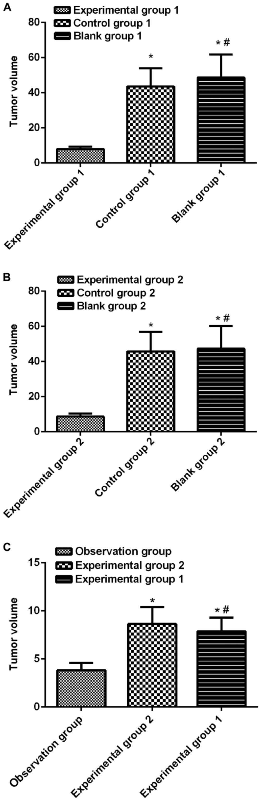
Subcutaneous tumor growth in nude
mice
The transfected SKOV3 cells were inoculated into
tumors, and it was manifested in the observation of tumor condition
1 week later that the tumor size in the 7 groups of nude mice met
the requirements of modeling, so the modeling was successful. At 36
days after the injection, the nude mice were sacrificed, and the
measurement of their tumors demonstrated that the tumor volumes in
the miRNA-124 groups were 7.88±2.84 mm3
(experimental group 1), 43.57±20.64 mm3 (control group
1) and 48.62±26.28 mm3 (blank group 1), respectively,
and those in the miRNA-152 groups were 8.64±3.52
mm3 (experimental group 2), 45.74±22.31 mm3
(control group 2) and 47.27±25.88 mm3 (blank group 2),
respectively, manifesting significant differences (P<0.01).
However, it was revealed through injection that in the observation
group, the volume was 3.83±1.54 mm3, which was
significantly different from that in experimental group 1 and 2
(P<0.01) (Fig. 1).
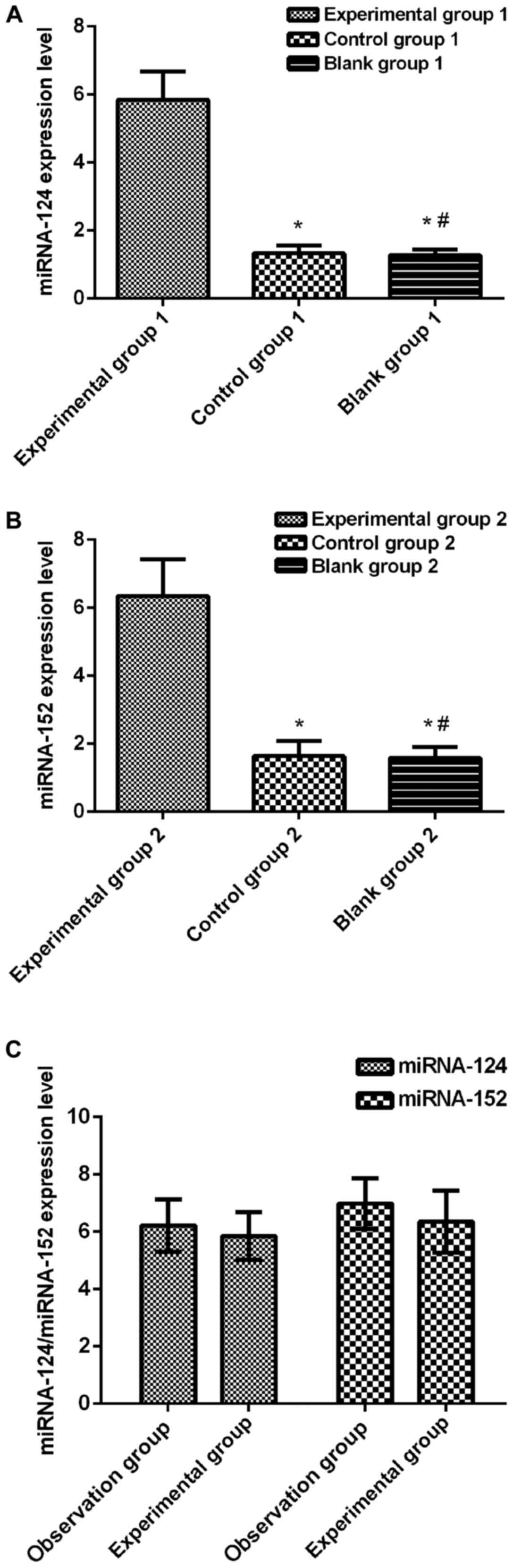
Detection of the relative expression level of miRNA
in animal model tissues in each group via RT-qPCR. According to the
detection of the relative expression levels of miRNA-124 and
miRNA-154 in the nude mouse tumor model in each group, the
relative expression level of miR-124 in the experimental
group 1 (5.84±1.67) was significantly higher than that in the
control group 1 (1.33±0.47) and blank group 1 (1.28±0.32) among the
miRNA-124 groups, with statistical differences (P<0.01 in
all comparisons). Besides, among the miRNA-152 groups, the
relative expression level of miR-152 in the experimental
group 2 (6.34±2.17) was markedly higher than that in the control
group 2 (1.64±0.87) and the blank group 2 (1.58±0.64), displaying
statistical differences (P<0.01 in all comparisons). However,
the relative expression levels of miRNA-124 and
miRNA-152 in the observation group were 6.21±1.84 and
6.98±1.77, respectively, manifesting no differences in comparisons
with those in the experimental group 1 and 2 (P>0.05) (Fig. 2).
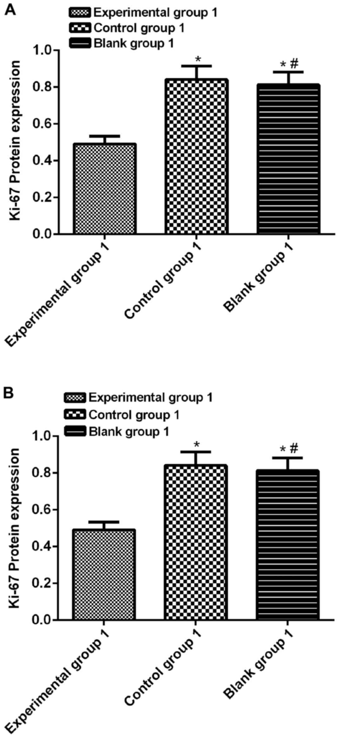
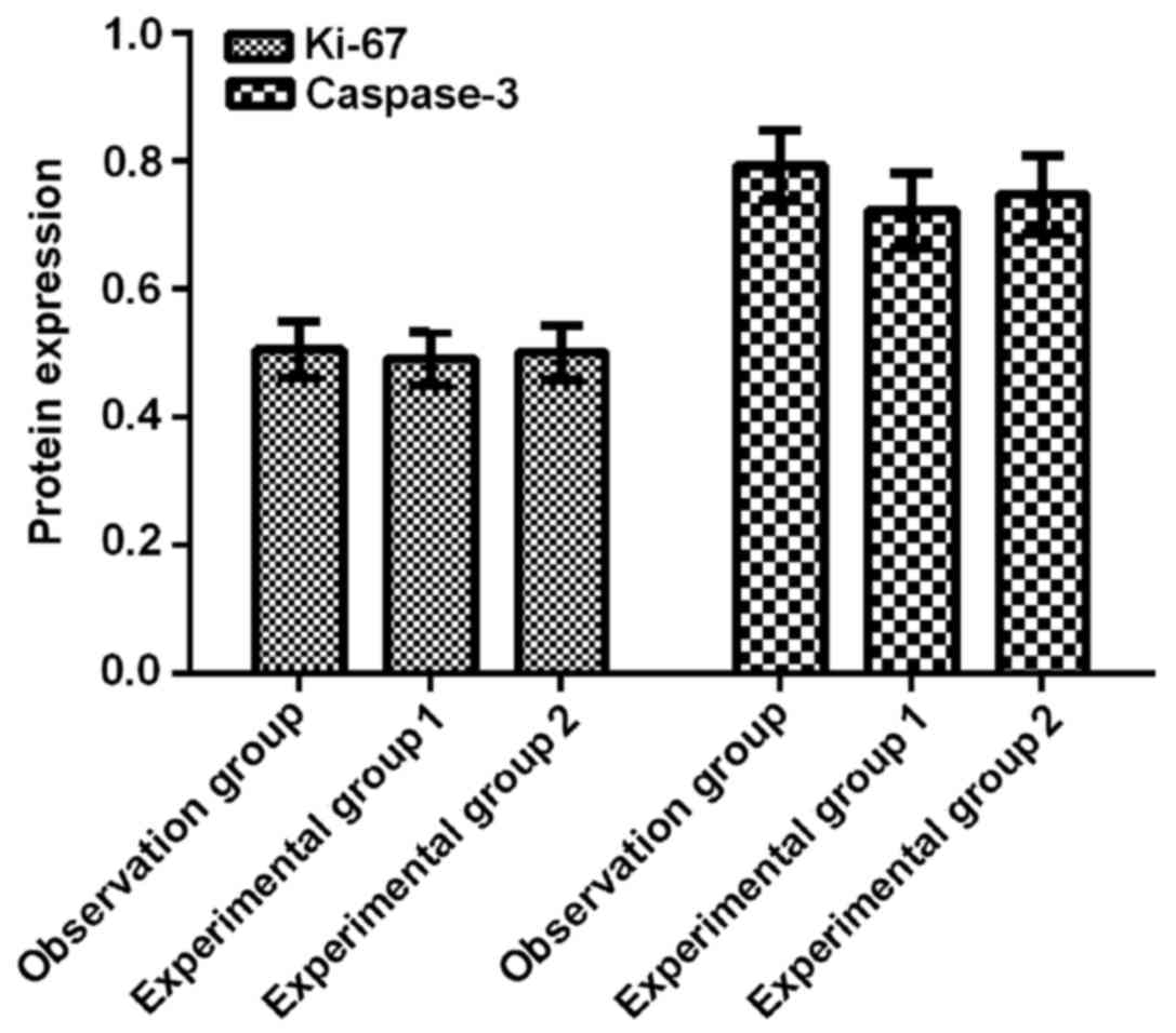
Protein expression level in each
group
The relative expression levels of Ki-67 and
caspase-3 proteins were detected by means of western blotting. The
results illustrated that among the Ki-67 groups, the expression
level of Ki-67 in the experimental group 1 (0.492±0.084) was
obviously lower than those in the control group 1 (0.841±0.148) and
blank group 1 (0.813±0.137), manifesting statistical differences
(P<0.01 in all comparisons). The expression level of Ki-67 in
the experimental group 2 (0.501±0.084) was decreased markedly
compared with that in the control group 2 (0.862±0.152) and blank
group 2 (0.823±0.147), and the differences were statistically
significant (P<0.01 in all comparisons). In addition, among the
caspase-3 groups, the expression level of caspase-3 in the
experimental group 1 (0.724±0.117) was significantly higher than
that in the control group 1 (0.392±0.067) and blank group 1
(0.411±0.077), and the differences were statistically significant
(P<0.01 in all comparisons). The expression level of caspase-3
in the experimental group 2 (0.748±0.122) was elevated notably
compared with that in the control group 2 (0.387±0.058) and blank
group 2 (0.406±0.062), showing statistical differences (P<0.01).
Nevertheless, the expression levels of Ki-67 and caspase-3 in the
observation group were 0.506±0.088 and 0.794±0.110, respectively,
and there were no differences in comparison with those in the
experimental group 1 and 2 (P>0.05) (Figs. 3–5).
Discussion
OC is one of the malignant cancers with a fairly
high mortality rate in women, and it ranks sixth among cancer in
women worldwide. EOC, occupying the highest proportion of cancers
with the highest malignancy, is hard to be detected, and patients
are in the advanced stage when diagnosed, so they lost the best
time for treatment. Nowadays, early diagnosis and treatment of EOC
are the major problems faced with the society (12). Cancerous adhesions triggered by late
lesions of EOC cannot be completely removed during surgery, and
distal metastasis may occur, which cannot be solved via surgery.
These problems, in turn, are also the main causes of poor prognosis
of EOC patients (13). At present,
the best treatment option is surgery combined with chemotherapy as
well as cisplatin and paclitaxel combined chemotherapy, but the
drug resistance resulted from the long-term application makes
clinical treatment efficacy worse and worse, so there is a need to
find a more effective treatment method for EOC, thus alleviating
the burden on patients (14).
In recent years, miRNA has become a hot topic. As a
non-coding RNA molecule, miRNAs can inhibit the degradation of
target genes by binding to them, causing differential expression of
post-transcriptional regulatory genes. miRNAs are involved in most
of the complex biological processes in the organism, especially the
occurrence and development of tumors, which have attracted
increasing attention from scholars (15). A study has illustrated that (16) miR-124 is expressed in a variety
of cancers such as liver cancer, gastric cancer, pancreatic cancer,
and OC. A study by Silber et al (17) demonstrated that the high expression of
miR-124 can reduce the proliferative capacity of neuroblasts
and decrease the growth rate of tumorigenic cells in in vivo
experiments. However, miR-152 has been confirmed to play an
important role in the occurrence and development of many tumors, as
well as cell proliferation and apoptosis (18).
In this study, the inhibitory effects of
miR-124 and miR-152 in EOC were investigated. An EOC
animal model was successfully established, and it was revealed
through the regular injection of miR-124 mimics and
miR-152 mimics that overexpressed miR-124 mimics and
miR-152 mimics markedly inhibited the growth of tumors.
Moreover, the combined injection of miR-124 and
miR-152 indicated that the tumor size detected by the
combined injection was significantly larger than that detected via
the injection of miRNA-124 mimics or miRNA-152 mimics
alone. Furthermore, subsequent RT-qPCR detection results revealed
that the expression of miR-124 and miR-152 in both
experimental groups were obviously increased compared with those in
other groups, manifesting significant differences, and there were
no differences in comparison with the observation group. A study by
Zhang et al (19) demonstrated
that miR-124 expression is low in OC, and this low
expression is more obvious in highly metastatic OC. In addition,
Shu et al (20) found from
in vitro experiments that highly expressed miRNA can inhibit
the proliferation, migration and invasion, and induce apoptosis of
OC cells. According to the study of Zhou et al (21), the overexpression of miR-152
can significantly suppress the proliferation of OC cells. The study
results are similar to those of our experiment. Ki-67 protein is
closely related to cell proliferation, differentiation, invasion
and apoptosis and participates in the pathological processes of all
malignant tumors (22). However,
caspase-3 protein, as a member of the caspase family, is involved
in apoptosis and is the initiator of apoptosis (23). The expression levels of Ki-67 and
caspase-3 proteins in tissues were detected via western blotting,
which manifested that the expression of Ki-67 protein in the two
experimental groups was significantly lower than that in other
groups, with significant differences, but there was no difference
compared with that in the control groups. However, caspase-3
protein was highly expressed in the two experimental groups
compared with that in the other groups, but this expression was not
different from that in the observation group. The underlying cause
may be the binding of miRNA-124 and miRNA-152 to
downstream target genes (24,25) [GLI family zinc finger 3 (Gli3) and
phosphoinositide-3-kinase regulatory subunit 3 (PIK3R3)].
However, there are still some defects in this
experiment. First of all, this experiment was an animal model
experiment and not conducted in clinical practice. Whether the
observed results are biased is not clear due to the few
experimental samples. Besides, the modeling time is short, and
patients were in the middle and advanced stage when they were
clinically diagnosed, so we expect to carry out research through
clinical trials in the future, so as to improve the treatment and
prognosis of EOC.
In conclusion, miR-124 and miR-152
exert inhibitory effects on the growth of EOC xenografts in nude
mice and are expected to serve as new targets for EOC
treatment.
Acknowledgements
Not applicable.
Funding
No funding was received.
Availability of data and materials
The datasets used and/or analyzed during the present
study are available from the corresponding author on reasonable
request.
Authors' contributions
WL and LZ wrote the manuscript and were responsible
for cell culture and transfection. JW performed PCR. XW constructed
the animal model. HS contributed to performing western blotting.
All authors read and approved the final manuscript.
Ethics approval and consent to
participate
The study was approved by the Ethics Committee of
Yidu Central Hospital of Weifang (Weifang, China).
Patient consent for publication
Not applicable.
Competing interests
The authors declare that they have no competing
interests.
References
|
1
|
Bean L, Sulzmaier FJ, Anderson KM,
Tancioni I, Kolev V, Plaxe SC, McHale MT, Schlaepfer DD and Pachter
J: Focal adhesion kinase (FAK) inhibition overcomes
cisplatin-resistance in epithelial ovarian cancer. Gynecol Oncol.
145:97–98. 2017. View Article : Google Scholar
|
|
2
|
Phelan CM, Kuchenbaecker KB, Tyrer JP, Kar
SP, Lawrenson K, Winham SJ, Dennis J, Pirie A, Riggan MJ, Chornokur
G, et al; AOCS study group; EMBRACE Study; GEMO Study
Collaborators; HEBON Study; KConFab Investigators; OPAL study
group, . Identification of 12 new susceptibility loci for different
histotypes of epithelial ovarian cancer. Nat Genet. 49:680–691.
2017. View
Article : Google Scholar : PubMed/NCBI
|
|
3
|
Kar SP, Adler E, Tyrer J, Hazelett D,
Anton-Culver H, Bandera EV, Beckmann MW, Berchuck A, Bogdanova N,
Brinton L, et al: Enrichment of putative PAX8 target genes at
serous epithelial ovarian cancer susceptibility loci. Br J Cancer.
116:524–535. 2017. View Article : Google Scholar : PubMed/NCBI
|
|
4
|
Yang WL, Gentry-Maharaj A, Simmons A, Ryan
A, Fourkala EO, Lu Z, Baggerly KA, Zhao Y, Lu KH, Bowtell D, et al;
AOCS Study Group, . Elevation of TP53 autoantibody before CA125 in
preclinical invasive epithelial ovarian cancer. Clin Cancer Res.
23:5912–5922. 2017. View Article : Google Scholar : PubMed/NCBI
|
|
5
|
Vitsios DM, Davis MP, van Dongen S and
Enright AJ: Large-scale analysis of microRNA expression,
epi-transcriptomic features and biogenesis. Nucleic Acids Res.
45:1079–1090. 2017. View Article : Google Scholar : PubMed/NCBI
|
|
6
|
Suzuki HI, Young RA and Sharp PA:
Super-enhancer-mediated RNA processing revealed by integrative
microRNA network analysis. Cell. 168:1000–1014.e15. 2017.
View Article : Google Scholar : PubMed/NCBI
|
|
7
|
Mazurek SR, Calway T, Harmon C, Farrell P
and Kim GH: MicroRNA-130a regulation of desmocollin 2 in a novel
model of arrhythmogenic cardiomyopathy. Microrna. 6:143–150. 2017.
View Article : Google Scholar : PubMed/NCBI
|
|
8
|
Bradshaw NJ, Ukkola-Vuoti L, Pankakoski M,
Zheutlin AB, Ortega-Alonso A, Torniainen-Holm M, Sinha V, Therman
S, Paunio T, Suvisaari J, et al: The NDE1 genomic locus can
affect treatment of psychiatric illness through gene expression
changes related to microRNA-484. Open Biol. 7:72017. View Article : Google Scholar
|
|
9
|
Zhang L, Volinia S, Bonome T, Calin GA,
Greshock J, Yang N, Liu CG, Giannakakis A, Alexiou P, Hasegawa K,
et al: Genomic and epigenetic alterations deregulate microRNA
expression in human epithelial ovarian cancer. Proc Natl Acad Sci
USA. 105:7004–7009. 2008. View Article : Google Scholar : PubMed/NCBI
|
|
10
|
Taylor DD and Gercel-Taylor C: MicroRNA
signatures of tumor-derived exosomes as diagnostic biomarkers of
ovarian cancer. Gynecol Oncol. 110:13–21. 2008. View Article : Google Scholar : PubMed/NCBI
|
|
11
|
Livak K J..Schmittgen T D.: Analysis of
relative gene expression data using real-time quantitative PCR and
the 2−ΔΔCT method. Methods. 25:402–408. 2001. View Article : Google Scholar : PubMed/NCBI
|
|
12
|
Baldwin LA, Chen Q, Tucker TC, White CG,
Ore RN and Huang B: Ovarian cancer incidence corrected for
oophorectomy. Diagnostics (Basel). 7:72017.
|
|
13
|
Darelius A, Lycke M, Kindblom JM,
Kristjansdottir B, Sundfeldt K and Strandell A: Efficacy of
salpingectomy at hysterectomy to reduce the risk of epithelial
ovarian cancer: A systematic review. BJOG. 124:880–889. 2017.
View Article : Google Scholar : PubMed/NCBI
|
|
14
|
Oronsky B, Ray CM, Spira AI, Trepel JB,
Carter CA and Cottrill HM: A brief review of the management of
platinum-resistant-platinum-refractory ovarian cancer. Med Oncol.
34:1032017. View Article : Google Scholar : PubMed/NCBI
|
|
15
|
Agarwal V, Bell GW, Nam JW and Bartel DP:
Predicting effective microRNA target sites in mammalian mRNAs.
eLife. 4:42015. View Article : Google Scholar
|
|
16
|
Lin S and Gregory RI: MicroRNA biogenesis
pathways in cancer. Nat Rev Cancer. 15:321–333. 2015. View Article : Google Scholar : PubMed/NCBI
|
|
17
|
Silber J, Hashizume R, Felix T, Hariono S,
Yu M, Berger MS, Huse JT, VandenBerg SR, James CD, Hodgson JG, et
al: Expression of miR-124 inhibits growth of medulloblastoma cells.
Neuro-oncol. 15:83–90. 2013. View Article : Google Scholar : PubMed/NCBI
|
|
18
|
Dang YW, Zeng J, He RQ, Rong MH, Luo DZ
and Chen G: Effects of miR-152 on cell growth inhibition, motility
suppression and apoptosis induction in hepatocellular carcinoma
cells. Asian Pac J Cancer Prev. 15:4969–4976. 2014. View Article : Google Scholar : PubMed/NCBI
|
|
19
|
Zhang H, Wang Q, Zhao Q and Di W: miR-124
inhibits the migration and invasion of ovarian cancer cells by
targeting SphK1. J Ovarian Res. 6:842013. View Article : Google Scholar : PubMed/NCBI
|
|
20
|
Shu J, Yuan L, Liu XM, Li SL and Zhou Q:
The expression of miR-124 in ovarian cancer tissues and its effect
on biological functions of ovarian cancer cells. Tumor. 34:430–436.
2014.
|
|
21
|
Zhou X, Zhao F, Wang ZN, Song YX, Chang H,
Chiang Y and Xu HM: Altered expression of miR-152 and miR-148a in
ovarian cancer is related to cell proliferation. Oncol Rep.
27:447–454. 2012.PubMed/NCBI
|
|
22
|
Brown DC and Gatter KC: Ki67 protein: The
immaculate deception? Histopathology. 40:2–11. 2002. View Article : Google Scholar : PubMed/NCBI
|
|
23
|
Dick SA, Chang NC, Dumont NA, Bell RA,
Putinski C, Kawabe Y, Litchfield DW, Rudnicki MA and Megeney LA:
Caspase 3 cleavage of Pax7 inhibits self-renewal of satellite
cells. Proc Natl Acad Sci USA. 112:E5246–E5252. 2015. View Article : Google Scholar : PubMed/NCBI
|
|
24
|
Wen SY, Lin Y, Yu YQ, Cao SJ, Zhang R,
Yang XM, Li J, Zhang YL, Wang YH, Ma MZ, et al: miR-506 acts as a
tumor suppressor by directly targeting the hedgehog pathway
transcription factor Gli3 in human cervical cancer. Oncogene.
34:717–725. 2015. View Article : Google Scholar : PubMed/NCBI
|
|
25
|
Li B, Xie Z and Li B: miR-152 functions as
a tumor suppressor in colorectal cancer by targeting PIK3R3. Tumour
Biol. 37:10075–10084. 2016. View Article : Google Scholar : PubMed/NCBI
|