Introduction
Oral squamous cell carcinoma (OSCC) is the most
frequently found oral neoplasm by a considerable margin, accounting
for ~90% all types oral cancer (1).
Due to a variety of factors, including a high degree of local
invasiveness and metastasis to cervical lymph nodes (2), the survival rate of traditionally
treated OSCC patients has improved slightly in the past three
decades (3,4). Regional or distant metastasis is one of
the most crucial factors for the survival of patients. Therefore,
searching for biomarkers associated with tumor progression has
become a significant challenge in the diagnosis of OSCC (5,6).
Proliferation marker protein Ki-67 (Ki-67), a
nuclear and nucleolar protein, is expressed in proliferating cells
from the G1 to the M phase of the cell cycle, with the
exception of the resting phase G0 (7). A sharp decrease in Ki-67 levels occurs
in later phases of mitosis (8).
Additionally, Ki-67 has been shown to serve an important role in
tumorigenesis due to its positive association with tumor
proliferation and invasion (9),
providing a marker of tumor aggressiveness.
Tumor cell proliferation is an important biological
parameter for tumor diagnosis. High expression of Ki-67 in breast
tumors shows higher proliferative and invasive activity (10). Kim et al found Ki-67 expression
to be inversely associated with age and young age/low
Ki-67 patients (young age <40 years, low Ki67 level <10%) had
significantly poorer recurrence-free survival (RFS) compared with
older age/high patients (old age ≥40 years, high Ki67
level ≥10%) with breast cancer (10).
Another study showed that high Ki-67 expression was associated with
good clinical outcomes and could act as a good independent
prognostic marker in colorectal cancer (11). It was also reported that Ki-67, a
proliferative marker, but not neuroendocrine expression, was an
independent factor in predicting the prognosis of patients with
prostate cancer (12). Furthermore,
Ki-67 combined with other proteins can also be of diagnostic value.
For example, certain studies found that a combination of B-cell
lymphoma 2 protein and Ki-67 improved the detection of gastric
cancer and identified metastatic castrate-resistant prostate cancer
more accurately by assessing vimentin and Ki-67 expression
(13,14). Ki-67 was also reported to correlate
with tumor progression in pancreatic neuroendocrine neoplasms
(15).
Although Ki-67 has been reported to provide a
diagnostic marker for neck metastasis in head and neck carcinomas
(16), its role in OSCC has not been
fully clarified. The present study aimed to evaluate the
association between Ki-67 expression and the clinicopathological
features of OSCC patients to further assess its diagnostic
value.
Patients and methods
Patients and tissue specimens
Paraffin-embedded surgical tissues were randomly
collected from 298 OSCC patients, 62 patients with oral leukoplakia
exhibiting various histological grades of oral epithelial dysplasia
and a control group (36 patients) with normal oral tissues. The
specimens were collected from resection surgery at Nanjing
Stomatological Hospital, Medical School of Nanjing University
(Nanjing, China) between March 2007 and December 2014. Diagnosis
was confirmed by postoperative pathology, and no patients received
radiotherapy or chemotherapy prior to surgery. Pregnant patients
and those diagnosed with other diseases were excluded from the
present study. The approval of the Ethics Committee of the
Stomatological Hospital Affiliated Medical School, Nanjing
University was obtained, as was informed consent from the patients
or their families. All patients were followed up bimonthly until
July 31, 2015.
Immunohistochemistry
Tissue specimens were fixed in 4% paraformaldehyde
solution at 4°C for 24 h, paraffin-embedded, and then cut into 2-µm
sections and placed on microscope slides for immunohistochemical
analysis. In brief, the sections were successively incubated in
xylene, 100% ethanol and 95% ethanol, blocked with 3%
H2O2 for 10 min at room temperature and
washed. A rabbit monoclonal antibody for Ki-67 (cat. no. ab15580;
1:200 dilution; Abcam, Cambridge, MA, USA) was incubated with all
slides at 4°C overnight followed by use of the secondary antibody
from the Dako Real™ Envision™ Detection System (cat. no. K500711;
Dako; Agilent Technologies, Inc., Santa Clara, CA, USA), incubated
at room temperature for 2 h. DAB (5 mg/ml) chromogen detection
(EnVision Detection System; Agilent Technologies, Inc., Sana Clara,
CA, USA) for 10 min was followed by nuclear staining using 1 mg/ml
of hematoxylin for 2 min at room temperature. Samples were washed
with tap water for 10 min, dehydrated, transparent, and detected
with an inverted microscope (magnification, ×200 and ×400; Olympus
CKX41; Olympus Corporation, Tokyo, Japan).
Quantification of
immunohistochemistry
Analysis and evaluation of immunostaining results
was independently determined by two pathologists. Differences of
opinion were reassessed together to reach consensus. Cells with
brown staining under the microscope were considered as positive
expression. The proportion of the number of Ki-67-positive cells to
the number of total cells was recorded, and three different fields
were examined within each micro-localization for the tumor specimen
of each patient (9). The median
proportion was used as a cutoff for further analysis of the Ki-67
high group (n=149) and the Ki-67 low group (n=149).
Statistical analysis
SPSS version 16.0 (SPSS Inc., Chicago, IL, USA) and
the Prism statistical software package version 6.0 (GraphPad
Software Inc., San Diego, CA, USA) were used for statistical
analyses. The χ2 test was used to compare the Ki67 high
group and the Ki67 low group. Overall survival (OS), disease-free
survival (DFS), recurrence-free survival (RFS) and metastasis-free
survival (MFS) were estimated comparing high expression groups and
low expression groups using Kaplan-Meier survival curves and the
long-rank test. Survival time was defined as the interval between
the date of surgery and the last date the patient was known to be
disease-free or alive (censored). The Cox regression model was used
to examine interactions between different prognostic factors in a
multivariate analysis. Differences were considered statistically
significant at P<0.05.
Results
Ki-67 expression is associated with
tumor progression
A total of 396 individuals (298 with OSCC, 62 with
dysplasia and 36 with normal epithelia) were examined in the
present study (Table I). Therefore,
differences in Ki-67 expression by immunohistochemistry were
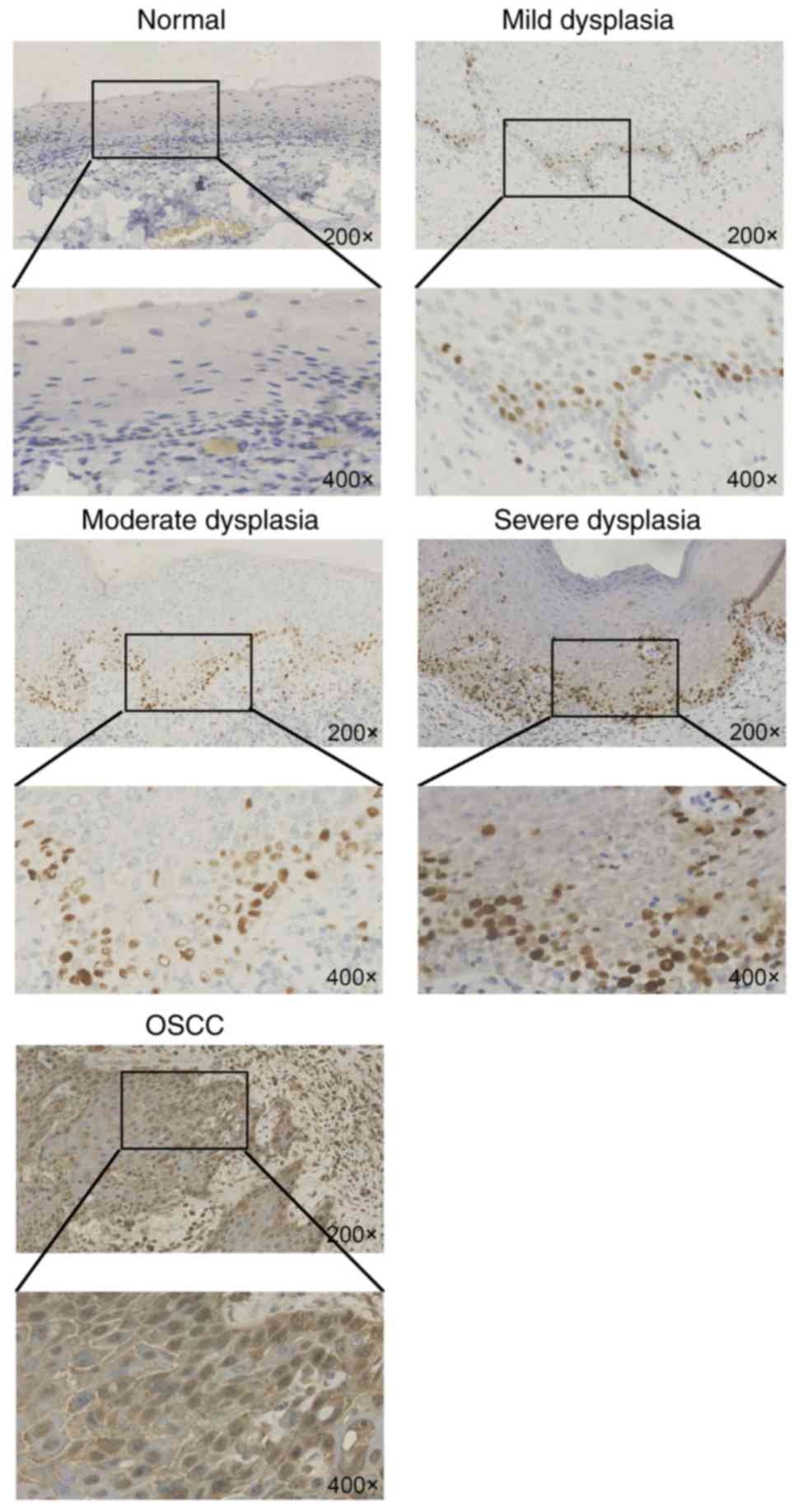
analyzed from normal tissue to dysplasia and tumor tissues. It was
found that Ki-67 expression was rare in normal oral mucosa and that
its expression was mainly located in the basal cells, where it
exhibited higher expression than that in normal mucosa (Fig. 1). Ki-67 was highly expressed in the
tumor tissues and was mainly expressed in the nuclei of tumor
cells. The immunohistochemistry indicated that the Ki-67 high group
with oral dysplasia of the epithelial mucosa accounted for 16.7%
(n=33) of the total number of high Ki-67 expression cases, while
the Ki-67 low group with oral dysplasia of the epithelial mucosa
accounted for 14.6% (n=29) of the total number of cases with low
Ki-67 expression (Table I).
 | Table I.Ki-67 expression in normal mucosa,
oral epithelial dysplasia and OSCC samples. |
Table I.
Ki-67 expression in normal mucosa,
oral epithelial dysplasia and OSCC samples.
|
| Ki-67 expression |
|
|
|
|---|
|
|
|
|
|
|
|---|
| Sample type | n (%) | Low, n (%) | High, n (%) | χ2 | P-value |
|---|
| Normal oral
mucosa | 36 (9.0) | 20 (10.1) | 16 (8.0) | 2.828 | 0.568 |
| Oral epithelial
dysplasia | 62 (15.7) | 29 (14.6) | 33 (16.7) |
|
|
| OSCC | 298 (75.3) | 149 (75.3) | 149 (75.3) |
|
|
As the dysplasia of oral epithelial mucosa included
mild, moderate and severe dysplasia, Ki-67 expression was analyzed
in these three types of tissues. The results showed that Ki-67
expression increased from mild dysplasia to moderate to severe, and
demonstrated that the expression of Ki-67 increased with the
decreased severity of oral leukoplakia: Mild dysplasia (n=9),
14.5%; moderate dysplasia (n=15), 24.2%; and severe dysplasia
(n=38), 61.3% (P=0.029; Table
II).
| Table II.Associations between Ki-67 expression
and various grades of oral epithelial dysplasia. |
Table II.
Associations between Ki-67 expression
and various grades of oral epithelial dysplasia.
|
|
| Ki-67 expression |
|
|
|---|
|
|
|
|
|
|
|---|
| Grade of oral
epithelial dysplasia | n (%) | Low, n (%) | High, n (%) | χ2 | P-value |
|---|
| Mild | 9 (14.5) | 3 (33.3) | 6 (66.7) | 7.685 | 0.029a |
| Moderate | 15 (24.2) | 4 (26.7) | 11 (73.3) |
|
|
| Severe | 38 (61.3) | 15 (39.5) | 23 (60.5) |
|
|
Associations between Ki-67 expression
and clinicopathological characteristics
The associations were investigated between Ki-67
expression and clinicopathological characteristics, including sex,
age, smoking habits, Tumor-Node-Metastasis (TNM) stage (17), tumor differentiation, lymph node
metastasis status, depth of tumor invasion (DOI) and worst pattern
of invasion (WPOI) type (18). The
results demonstrated that Ki-67 expression was significantly
associated with patient age (P=0.011; Table III), demonstrating that younger
people (<60 years) exhibited lower Ki-67 expression.
Additionally, Ki-67 was highly expressed in patients with
moderate-high differentiation (P=0.001; Table III) and patients with lymph node
metastasis (P=0.006; Table III). In
addition, Ki-67 expression was found to be higher in patients with
WPOI types 4–5 than in those with types 1–3 (P<0.0001; Table III), but there was no significant
difference between the DOI <5 and ≥5 mm groups (P=0.082;
Table III).
| Table III.Ki-67 expression ratio and
clinicopathological characteristics in patients with oral squamous
cell carcinoma. |
Table III.
Ki-67 expression ratio and
clinicopathological characteristics in patients with oral squamous
cell carcinoma.
|
|
|
| Ki-67 expression |
|
|
|---|
|
|
|
|
|
|
|
|---|
| Variable | Category | n (%) | Low, n (%) | High, n (%) | χ2 | P-value |
|---|
| Sex | Male | 152 (51) | 72 (48.3) | 80 (53.7) | 0.859 | 0.354 |
|
| Female | 146 (49) | 77 (51.7) | 69 (46.3) |
|
|
| Age | <60 years | 138 (46.3) | 80 (53.7) | 58 (38.9) | 6.532 | 0.011a |
|
| ≥60 years | 160 (53.7) | 69 (46.3) | 91 (61.1) |
|
|
| TNM | I–III | 295 (99) | 148 (99.3) | 147 (98.7) | 0.337 | 0.562 |
|
| IV–V | 3 (1) | 1 (0.7) | 2 (1.3) |
|
|
|
Differentiation | Low | 149 (50) | 89 (59.7) | 60 (40.3) | 11.289 | 0.001a |
|
| Moderate-high | 149 (50) | 60 (40.3) | 89 (59.7) |
|
|
| Lymph node
metastasis | Yes | 11 (3.7) | 1 (0.7) | 10 (6.7) | 7.646 | 0.006a |
|
| No | 287 (96.3) | 148 (99.3) | 139 (93.3) |
|
|
| Depth of
invasion | <5 mm | 143 (48) | 79 (53) | 64 (43) | 3.025 | 0.082 |
|
| ≥5 mm | 155 (52) | 70 (47) | 85 (57) |
|
|
| WPOI | I–III | 174 (58.4) | 103 (69.1) | 71 (47.7) | 14.143 |
<0.0001a |
|
| IV–V | 124 (41.6) | 46 (30.9) | 78 (52.3) |
|
|
Associations between Ki-67 expression
and survival time
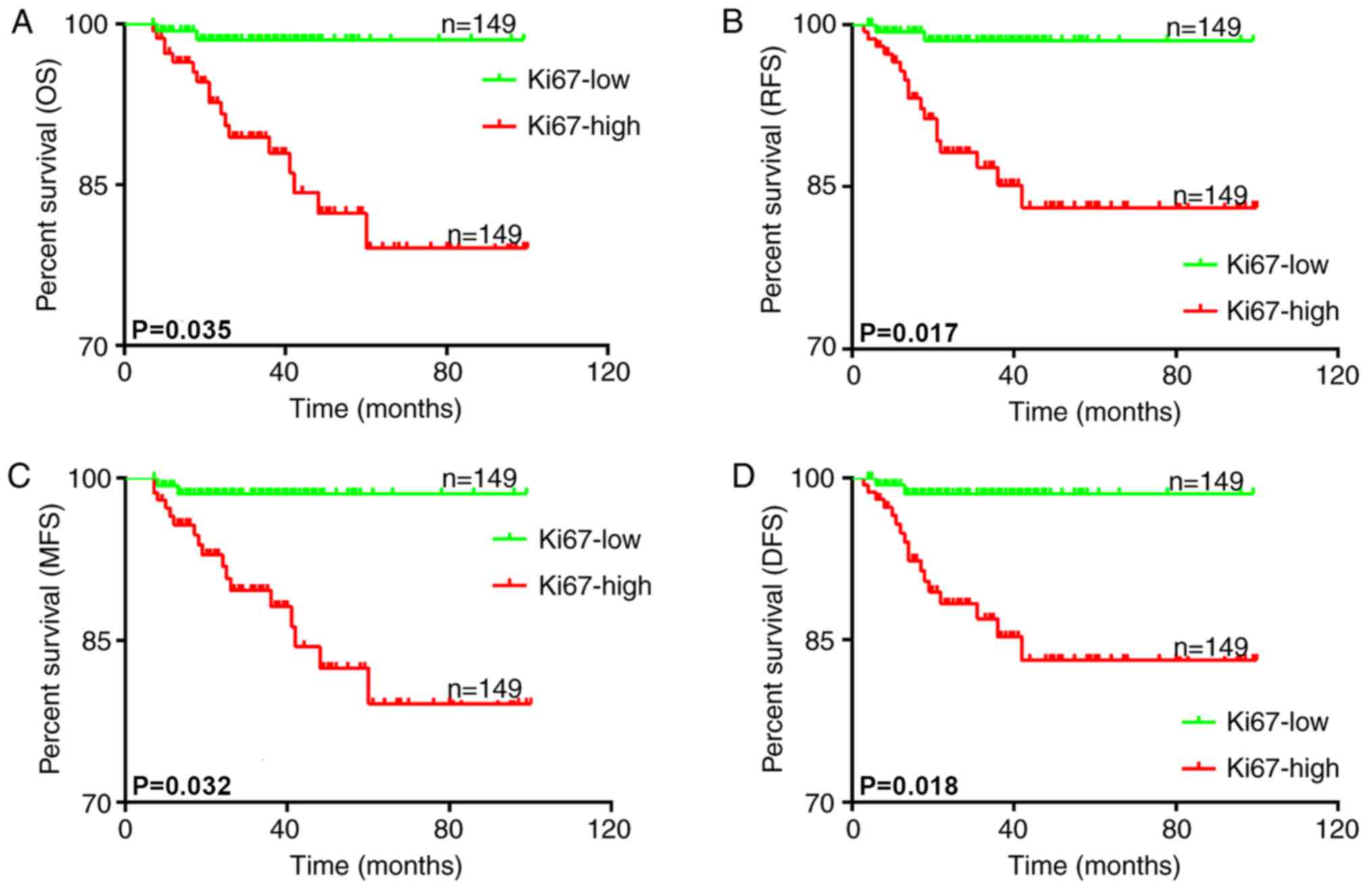
Follow-up data was obtained and the association
between Ki-67 expression and the clinical outcome of the patients
was analyzed. All patients were separated into two groups according
to Ki-67 expression using the median as the cutoff value. The
association between OS, DFS, RFS, metastasis-free survival (MFS)
and Ki-67 expression was then analyzed with Kaplan-Meier survival
curves. The results demonstrated that higher Ki-67 expression was
associated with poorer OS (P=0.035; Fig.
2A), RFS (P=0.017; Fig. 2B), MFS
(P=0.032; Fig. 2C) and DFS (P=0.018;
Fig. 2D) times. These results
suggested that higher Ki-67 expression in OSCC patients was
associated with an unsatisfactory clinical outcome.
Ki-67 expression is an independent
diagnostic marker for OSCC
Univariate analysis revealed that the following
parameters were associated with increased mortality: TNM [OS:
Hazard ratio (HR), 0.081; 95% confidence interval (CI),
0.025–0.262; P<0.0001; DFS: HR, 0.107; 95% CI, 0.033–0.345;
P<0.0001; RFS: HR, 0.106; 95% CI, 0.033–0.341; P<0.0001; MFS:
HR, 0.083; 95% CI, 0.025–0.267; P<0.0001], differentiation (OS:
HR, 0.662; 95% CI, 0.521–0.842; P=0.001; DFS: HR, 0.724; 95% CI,
0.57–0.919; P=0.008; RFS: HR, 0.735; 95% CI, 0.579–0.933; P=0.011;
MFS: HR, 0.652; 95% CI, 0.513–0.829; P<0.0001) and Ki-67
expression (OS: HR, 1.893; 95% CI, 1.136–3.573; P=0.035; DFS: HR,
2.283; 95% CI, 1.408–3.766; P=0.018; RFS: HR, 2.43; 95% CI,
1.437–3.428; P=0.017; MFS: HR, 2.048; 95% CI, 1.165–3.393;
P=0.032). In a further step of multivariate analysis, Ki-67
expression retained its negative impact on survival for OS, DFS,
RFS and MFS (Tables IV–VII).
| Table IV.Prognostic factors in the Cox
proportional hazards model for overall survival. |
Table IV.
Prognostic factors in the Cox
proportional hazards model for overall survival.
|
|
| Univariate | Multivariate |
|---|
|
|
|
|
|
|---|
| Variable | Category | HR | 95% CI | P-value | HR | 95% CI | P-value |
|---|
| Sex | Male vs.
female | 1.138 | 0.899–1.442 | 0.281 | 1.114 | 0.877–1.414 | 0.378 |
| Age | <60 vs. ≥60
years | 1.208 | 0.953–1.53 | 0.118 | 1.154 | 0.906–1.469 | 0.246 |
| TNM | I–III vs. IV–V | 0.081 | 0.025–0.262 |
<0.0001a | 0.08 | 0.024–0.264 |
<0.0001a |
|
Differentiation | Low vs.
moderate-high | 0.662 | 0.521–0.842 | 0.001a | 0.659 | 0.507–0.858 | 0.002a |
| Lymph node
metastasis | Yes vs. no | 1.003 | 0.372–2.704 | 0.996 | 1.159 | 0.425–3.162 | 0.773 |
| Depth of
invasion | <5 vs. ≥5
mm | 1.011 | 0.798–1.28 | 0.929 | 1.137 | 0.879–1.47 | 0.329 |
| WPOI | I–III vs. IV–V | 0.912 | 0.715–1.164 | 0.461 | 1.015 | 0.776–1.326 | 0.915 |
| Ki-67 | Low vs. high
expression | 1.893 | 1.136–3.573 | 0.035a | 1.261 | 1.051–3.287 | 0.044a |
| Table VII.Prognostic factors in the Cox
proportional hazards model for metastasis-free survival. |
Table VII.
Prognostic factors in the Cox
proportional hazards model for metastasis-free survival.
|
|
| Univariate | Multivariate |
|---|
|
|
|
|
|
|---|
| Variable | Category | HR | 95% CI | P-value | HR | 95% CI | P-value |
|---|
| Sex | Male vs.
female | 1.152 | 0.91–1.459 | 0.239 | 1.123 | 0.885–1.425 | 0.340 |
| Age | <60 vs. ≥60
years | 1.223 | 0.965–1.549 | 0.096 | 1.178 | 0.925–1.5 | 0.185 |
| TNM | I–III vs. IV–V | 0.083 | 0.025–0.267 |
<0.0001a | 0.078 | 0.024–0.258 |
<0.0001a |
|
Differentiation | Low vs.
moderate-high | 0.652 | 0.513–0.829 |
<0.0001a | 0.66 | 0.508–0.858 | 0.002a |
| Lymph node
metastasis | Yes vs. no | 0.498 | 0.182–1.363 | 0.175 | 0.506 | 0.183–1.398 | 0.189 |
| Depth of
invasion | <5 vs. ≥5
mm | 0.997 | 0.787–1.263 | 0.979 | 1.119 | 0.866–1.446 | 0.390 |
| WPOI | I–III vs. IV–V | 0.896 | 0.703–1.143 | 0.377 | 1.015 | 0.777–1.325 | 0.916 |
| Ki-67 | Low vs. high
expression | 2.048 | 1.165–3.393 | 0.032a | 3.301 | 1.077–10.116 | 0.037a |
Discussion
OSCC is among the most frequently diagnosed cancer
types worldwide. However, despite improved therapies, the 5-year
survival rate has not changed. Overall, >50% of patients with
OSCC demonstrate regional and distant metastases, which result in
treatment failures and occasionally mortality within a year due to
recurrent or metastatic disease (19,20). In
order to improve the survival rate for OSCC, identification of an
underlying molecular event differentiating patients at risk for
progression at the premalignant stage, is required (21). Factors, including tumor size, lymph
node metastasis, TNM type and differentiation, influence the
prognosis of OSCC (22).
Ki-67 is an indicator of cell proliferation and has
been shown to be upregulated in numerous tumors (23,24). Tumor
proliferative activity labeled by Ki-67 has been found to be
associated with tumor aggression, which is specified by tumor grade
and stage. Several studies have described these associations and
identified Ki-67 as a prognostic factor (14,25–27). The
present study found upregulation of Ki-67 expression with tumor
progression using normal epithelial mucosa, dysplasia and OSCC
samples. In addition, Ki-67 expression also increased from mild to
moderate to severe dysplasia. Another important finding was that
high expression of Ki-67 was associated with severe differentiation
(P=0.001), lymph node metastasis (P=0.006) and higher WPOI
(P<0.0001). Furthermore, high Ki-67 expression was significantly
associated with poor survival time with regards to OS (P=0.035),
RFS (P=0.017), MFS (P=0.032) and DFS (P=0.018). The biological
behavior of OSCC with highly expressed Ki-67 is verified by the
association between Ki-67 and affiliated potential
clinicopathological characteristics; for example, differentiation,
lymph node metastasis, TNM, DOI and WPOI. Unexpectedly, the
multivariate analysis revealed that Ki-67 could be an independent
predictor for OS, RFS, MFS and RFS.
In summary, the findings of the present study
strongly argue for the value of highly expressed Ki-67 as an
independent prognostic marker for OSCC. Considering the extensive
heterogeneity of tumors, further researches need to enrich
correlations of spatial expression of Ki67 with OSCC.
Acknowledgements
The authors would like to thank Dr K. D. Tanq
(School of Biomedical Sciences, Institute of Health and Biomedical
Innovation, Queensland University of Technology, Brisbane,
Queensland, Australia) for assisting in the drafting of the
original manuscript.
Funding
The present study was supported by the National
Natural Science Foundation of China (grant nos. 81772880 and
81702680), the Nanjing Medical Science and Technique Development
Foundation (grant nos. YKK16164 and QRX17083), the Jiangsu
Provincial Key Medical Discipline (since 2017), Nanjing Municipal
Key Medical Laboratory Constructional Project Funding (since 2016),
and the Center of Nanjing Clinical Medicine of Tumor project (since
2014).
Availability of data and materials
The datasets used and/or analyzed during the current
study are available from the corresponding author on reasonable
request.
Authors' contributions
YJ performed the histological examination of the
OSCC samples, analyzed the data and wrote the manuscript. QZ, HZ
and YZ analyzed the OSCC patient data. YS and XZ participated in
the statistical analyses. XH contributed to the interpretation of
the OSCC patient data. YY participated in the histological
examination of OSCC. YN designed the study and drafted the
manuscript. QH conceived and designed the study. All authors read
and approved the submitted manuscript.
Ethics approval and consent to
participate
The study was approved by the Ethics Committee of
the Stomatological Hospital Affiliated Medical School, Nanjing
University. Written informed consent was obtained from the
patients.
Patient consent for publication
Written informed consent was obtained from the
patients for the publication of any associated data and
accompanying images.
Competing interests
The authors declare that they have no competing
interests.
References
|
1
|
Bagan J, Sarrion G and Jimenez Y: Oral
cancer: Cral precancer and cancer and preventive measures. Clinical
Oral investigations. 5:207–213. 2001. View Article : Google Scholar : PubMed/NCBI
|
|
2
|
Reichart PA: Identification of risk groups
for oral precancer and cancer and preventive measures. Clin Oral
Investig. 5:207–213. 2001. View Article : Google Scholar : PubMed/NCBI
|
|
3
|
Perez-Sayans M, Somoza-Martin JM,
Barros-Angueira F, Reboiras-Lopez MD, Gandara Rey JM and
Garcia-Garcia A: Genetic and molecular alterations associated with
oral squamous cell cancer (Review). Oncol Rep. 22:1277–1282. 2009.
View Article : Google Scholar : PubMed/NCBI
|
|
4
|
Sasahira T, Kirita T and Kuniyasu H:
Update of molecular pathobiology in oral cancer: A review. Int J
Clin Oncol. 19:431–436. 2014. View Article : Google Scholar : PubMed/NCBI
|
|
5
|
Ost P, Bossi A, Decaestecker K, De
Meerleer G, Giannarini G, Karnes RJ, Roach M III and Briganti A:
Metastasis-directed therapy of regional and distant recurrences
after curative treatment of prostate cancer: A systematic review of
the literature. Eur Urol. 67:852–863. 2015. View Article : Google Scholar : PubMed/NCBI
|
|
6
|
Sannam Khan R, Khurshid Z, Akhbar S and
Faraz Moin S: Advances of Salivary Proteomics in Oral Squamous Cell
Carcinoma (OSCC) Detection: An Update. Proteomes. 4(piii): E412016.
View Article : Google Scholar : PubMed/NCBI
|
|
7
|
Gerdes J, Lemke H, Baisch H, Wacker HH,
Schwab U and Stein H: Cell cycle analysis of a cell
proliferation-associated human nuclear antigen defined by the
monoclonal antibody Ki-67. J Immunol. 133:1710–1715.
1984.PubMed/NCBI
|
|
8
|
Li LT, Jiang G, Chen Q and Zheng JN: Ki67
is a promising molecular target in the diagnosis of cancer
(review). Mol Med Rep. 11:1566–1572. 2015. View Article : Google Scholar : PubMed/NCBI
|
|
9
|
Antonarakis ES, Keizman D, Zhang Z, Gurel
B, Lotan TL, Hicks JL, Fedor HL, Carducci MA, De Marzo AM and
Eisenberger MA: An immunohistochemical signature comprising PTEN,
MYC, and Ki67 predicts progression in prostate cancer patients
receiving adjuvant docetaxel after prostatectomy. Cancer.
118:6063–6071. 2012. View Article : Google Scholar : PubMed/NCBI
|
|
10
|
Kim J, Han W, Jung SY, Park YH, Moon HG,
Ahn SK, Lee JW, Kim MK, Kim JJ, Lee ES, et al: The Value of Ki67 in
Very Young Women with hormone receptor-positive breast cancer:
Retrospective Analysis of 9,321 Korean Women. Ann Surg Oncol.
22:3481–3488. 2015. View Article : Google Scholar : PubMed/NCBI
|
|
11
|
Melling N, Kowitz CM, Simon R, Bokemeyer
C, Terracciano L, Sauter G, Izbicki JR and Marx AH: High Ki67
expression is an independent good prognostic marker in colorectal
cancer. J Clin Pathol. 69:209–214. 2016. View Article : Google Scholar : PubMed/NCBI
|
|
12
|
Pascale M, Aversa C, Barbazza R, Marongiu
B, Siracusano S, Stoffel F, Sulfaro S, Roggero E, Bonin S and
Stanta G: The proliferation marker Ki67, but not neuroendocrine
expression, is an independent factor in the prediction of prognosis
of primary prostate cancer patients. Radiol Oncol. 50:313–320.
2016. View Article : Google Scholar : PubMed/NCBI
|
|
13
|
Zhou Y, Li Y, Zheng J, Liu K and Zhang H:
Detecting of gastric cancer by Bcl-2 and Ki67. Int J Clin Exp
Pathol. 8:7287–7290. 2015.PubMed/NCBI
|
|
14
|
Lindsay CR, Le Moulec S, Billiot F, Loriot
Y, Ngo-Camus M, Vielh P, Fizazi K, Massard C and Farace F: Vimentin
and Ki67 expression in circulating tumour cells derived from
castrate-resistant prostate cancer. BMC Cancer. 16:1682016.
View Article : Google Scholar : PubMed/NCBI
|
|
15
|
Panzuto F, Cicchese N, Partelli S,
Rinzivillo M, Capurso G, Merola E, Manzoni M, Pucci E, Iannicelli
E, Pilozzi E, et al: Impact of Ki67 re-assessment at time of
disease progression in patients with pancreatic neuroendocrine
neoplasms. PLoS One. 12:e01794452017. View Article : Google Scholar : PubMed/NCBI
|
|
16
|
Liu M, Lawson G, Delos M, Jamart J, Ide C,
Coche E, Weynand B, Desuter G, Hamoir M, Remacle M and Marbaix E:
Predictive value of the fraction of cancer cells immunolabeled for
proliferating cell nuclear antigen or Ki67 in biopsies of head and
neck carcinomas to identify lymph node metastasis: Comparison with
clinical and radiologic examinations. Head Neck. 25:280–288. 2003.
View Article : Google Scholar : PubMed/NCBI
|
|
17
|
Keohane SG, Proby CM, Newlands C, Motley
RJ, Nasr I, Mohd Mustapa MF and Slater DN: The new 8th edition of
TNM staging and its implications for skin cancer: a review by the
British Association of Dermatologists and the Royal College of
Pathologists, United Kingdom. Br J Dermatol. 2018.(Epub ahead of
print). View Article : Google Scholar
|
|
18
|
Heerema MG, Melchers LJ, Roodenburg JL,
Schuuring E, de Bock GH and van der Vegt B: Reproducibility and
prognostic value of pattern of invasion scoring in low-stage oral
squamous cell carcinoma. Histopathology. 68:388–397. 2016.
View Article : Google Scholar : PubMed/NCBI
|
|
19
|
Kuperman DI, Auethavekiat V, Adkins DR,
Nussenbaum B, Collins S, Boonchalermvichian C, Trinkaus K, Chen L
and Morgensztern D: Squamous cell cancer of the head and neck with
distant metastasis at presentation. Head Neck. 33:714–718. 2011.
View Article : Google Scholar : PubMed/NCBI
|
|
20
|
Chen W, Zheng R, Baade PD, Zhang S, Zeng
H, Bray F, Jemal A, Yu XQ and He J: Cancer statistics in China,
2015. CA Cancer J Clin. 66:115–132. 2016. View Article : Google Scholar : PubMed/NCBI
|
|
21
|
Towle R, Truong D and Garnis C: Epigenetic
mediated silencing of EYA4 contributes to tumorigenesis in oral
dysplastic cells. Genes Chromosomes Cancer. 55:568–576. 2016.
View Article : Google Scholar : PubMed/NCBI
|
|
22
|
Noguti J, De Moura CF, De Jesus GP, Da
Silva VH, Hossaka TA, Oshima CT and Ribeiro DA: Metastasis from
oral cancer: An overview. Cancer Genomics Proteomics. 9:329–335.
2012.PubMed/NCBI
|
|
23
|
Yurakh AO, Ramos D, Calabuig-Farinas S,
López-Guerrero JA, Rubio J, Solsona E, Romanenko AM, Vozianov AF,
Pellin A and Llombart-Bosch A: Molecular and immunohistochemical
analysis of the prognostic value of cell-cycle regulators in
urothelial neoplasms of the bladder. Eur Urol. 50:506–515. 2006.
View Article : Google Scholar : PubMed/NCBI
|
|
24
|
Brown DC and Gatter KC: Ki67 protein: The
immaculate deception? Histopathology. 40:2–11. 2002. View Article : Google Scholar : PubMed/NCBI
|
|
25
|
Wu TT, Chen JH, Lee YH and Huang JK: The
role of bcl-2, p53, and ki-67 index in predicting tumor recurrence
for low grade superficial transitional cell bladder carcinoma. J
Urol. 163:758–760. 2000. View Article : Google Scholar : PubMed/NCBI
|
|
26
|
Li H, Han X, Liu Y, Liu G and Dong G: Ki67
as a predictor of poor prognosis in patients with triple-negative
breast cancer. Oncol Lett. 9:149–152. 2015. View Article : Google Scholar : PubMed/NCBI
|
|
27
|
Li W, Zhang G, Wang HL and Wang L:
Analysis of expression of cyclin E, p27kip1 and Ki67 protein in
colorectal cancer tissues and its value for diagnosis, treatment
and prognosis of disease. Eur Rev Med Pharmacol Sci. 20:4874–4879.
2016.PubMed/NCBI
|