Introduction
Laryngocarcinoma is a common head and neck malignant
tumor that mostly presents as squamous cell carcinoma (1). In addition, the morbidity rate in
northern China is increased compared with that in southern China
(2). The morbidity rate has increased
over the past 40 years, and laryngocarcinoma results in a serious
effect on the quality of life and human health. At present, surgery
remains the primary therapeutic method for laryngocarcinoma.
However, lymphatic metastasis is an essential factor that affects
the prognosis of laryngocarcinoma (3). Supraglottic and glottic cancer account
for the vast majority of cases of laryngocarcinoma (3), and supraglottic laryngocarcinoma is more
prone to metastasize than glottic laryngocarcinoma (1). Generally, the lower the differentiation
degree of the tumor, the earlier the occurrence of neck lymph node
metastasis will be. The course of lymphatic metastasis is
consistent with the flow of laryngopharyngeal lymph (4). The lymphatic vessels at in the
supraglottic region are abundant, and the differentiation degree of
supraglottic laryngocarcinoma is low; thus, the incidence rate of
cervical nodal metastases is high (1).
Cell cycle regulation is closely associated with
tumor development. The process of cell cycle regulation is
complicated (5), and this regulation
is involved in almost all biological effects of oncogenes and
cancer suppressor genes (5). Cell
cycle cytokines may reflect the progression of tumors from the
aspect of cell cycle kinetics (6).
Numerous oncogenes and cancer suppressor genes are directly
involved in cell cycle regulation, and are main components of the
cell cycle regulation checkpoints. Cell cycle regulation occurs
mainly via two key limiting points, G1/S and G2/M (7). The main regulation factors at the G2/M
phase include cyclin B, cyclin-dependent kinase 1, also termed cell
division cycle (Cdc) 2, cyclin-dependent kinase 2 and Cdc25C
(8).
Matrix metalloproteinases (MMPs) are a type of
proteolytic enzyme closely associated with tumor invasion and
metastasis (9). MMP-2 and MMP-9 are
important members of the MMP family (9). They can effectively degrade collagen IV,
the main component of the extracellular matrix and basilar
membrane, thereby destroying the integrity of the basilar membrane
(10). This process is closely
associated with tumor infiltration and metastasis (11). Previous studies have shown that MMP-2
and MMP-9 are overexpressed in human malignancies, and promote the
infiltration and metastasis of tumor cells; this is associated with
the prognosis of the patient (9,11). A
previous study also demonstrated that MMP-2 and MMP-9 are
overexpressed in laryngocarcinoma tissue, and they are associated
with lymphatic metastasis and poor prognosis (12).
Mitogen-activated protein kinase (MAPK) is an
important signaling pathway in cells, mainly involving c-Jun
NH2-terminal kinase/stress-activated protein kinase, extracellular
signal regulated protein kinase and p38-MAPK (13). This pathway has a close association
with cell proliferation, differentiation and apoptosis. The
p38-MAPK signaling pathway has complex effects on tumors (14). For example, transient activation of
p38-MAPK can promote tumor cell proliferation (15). Maintaining a long-term activated state
of phosphorylated p38-MAPK induces tumor cells apoptosis via the
tumor necrosis factor-α pathway (15).
Ophiopogon japonicus is a herb used as a
Traditional Chinese Medicine, and is considered a yin-nourishing
drug (16). With the deepening of
investigation into O. japonicus, the pharmacological actions
of Ophiopogonin D, the major active component in O.
japonicus, have been identified (17). Ophiopogonin D has significant
pharmacological effects and has anti-aging properties, improves
learning deficit and dysmnesia, increases resistance to
cardiovascular and cerebrovascular diseases, and has anti-tumor,
anti-inflammatory and immunoregulatory effects (16,18,19). The
aim of the present study was to investigate whether the anti-cancer
effect of Ophiopogonin D inhibits cell proliferation and induced
apoptosis of human laryngocarcinoma, and may therefore be a
potential therapy for the treatment of laryngocarcinoma.
Materials and methods
Cell culture
The human laryngocarcinoma AMC-HN-8 cell line was
purchased from the Shanghai Cell Bank of Chinese Academy of
Sciences (Shanghai, China) and cultured in complete RPMI-1640 with
10% fetal bovine serum (HyClone; GE Healthcare Life Sciences,
Logan, UT, USA), 100 U/ml penicillin, 100 mg/ml streptomycin and 1
mM sodium pyruvate (Invitrogen; Thermo Fisher Scientific, Inc.,
Waltham, MA, USA) at 37°C and in a 5% CO2 atmosphere.
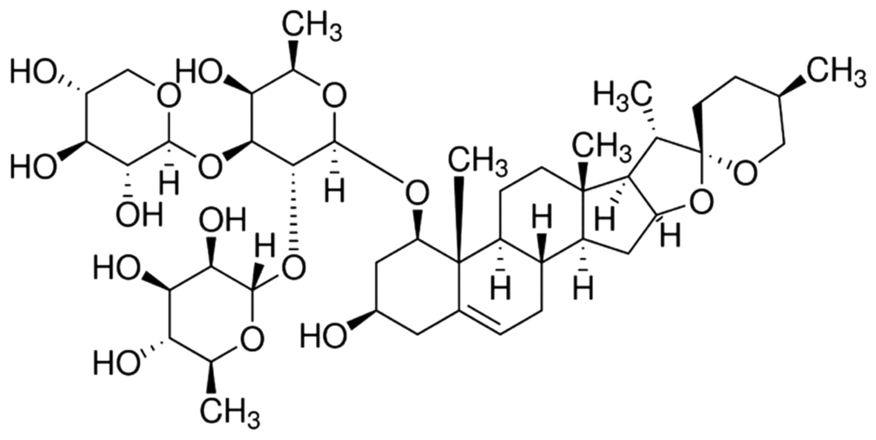
Ophiopogonin D (≥98%; Fig. 1) was
purchased from Sigma-Aldrich (Merck KGaA, Darmstadt, Germany).
MTT assay and lactate dehydrogenase
(LDH) assay
AMC-HN-8 cells (5×10⁶ cells/well) were
plated into a 96-well plate for 24 h at 37°C. The cultured cells
were treated with various concentrations of Ophiopogonin D (0,
12.5, 25 and 50 µmol/l) for 12 or 24 h. Subsequently, 20 µl MTT (5
mg/ml) was added daily to each well and incubated for an additional
4 h at 37°C. Dimethyl sulfoxide (200 µl) was added to each well and
dissolved for 10 min, and the absorbance at 490 nm was measured
using a plate reader (Synergy-HT; BioTek Instruments, Inc.,
Winooski, VT, USA).
LDH assay
AMC-HN-8 cells (5×10⁶ cells/well) were
plated onto a 96-well plate for 24 h culture, and were then treated
with various concentrations of Ophiopogonin D (0, 12.5, 25 and 50
µmol/l) for 12 or 24 h. Subsequently, 60 µl LDH (Beyotime Institute
of Biotechnology, Haimen, China) was added daily to each well and
incubated for an additional for 30 min at room temperature, and the
absorbance at 490 nm was measured using a plate reader (Synergy-HT;
BioTek Instruments, Inc.).
Apoptosis assay
AMC-HN-8 cells (5×10⁶ cells/well) were
plated onto a 6-well plate for 24 h and were treated with various
concentrations of Ophiopogonin D (0–50 µmol/l) for 24 h. AMC-HN-8
cells was stained with Annexin V (BD Biosciences, Franklin Lakes,
NJ, USA) and propidium iodide (PI) (BD Biosciences) in darkness for
30 min. Apoptosis was quantitated with the Guava EasyCyte™ flow
cytometer (Becton Dickinson, Bedford, MA).
DAPI assay
AMC-HN-8 cells (5×10⁶ cells/well) were
plated into a 6-well plate for 24 h and were treated with various
concentrations of Ophiopogonin D (0, 12.5, 25 and 50 µmol/l) for 24
h at 37°C. AMC-HN-8 cells was stained with DAPI assay (Beyotime
Institute of Biotechnology) for 1 h. Images were captured using a
DP70 fluorescence microscope (magnification, ×20; Olympus, Tokyo,
Japan).
Caspase-9/3 activity analysis
AMC-HN-8 cells (5×10⁶ cells/well) were
plated into a 6-well plate for 24 h and cultured were treated with
different concentrations of Ophiopogonin D (0–50 µmol/l) for 24 h.
Cell pellets were collected and lysed with radioimmunoprecipitation
assay (RIPA) lysis buffer (Beyotime Institute of Biotechnology)
containing 0.5 mM phenylmethylsulfonyl fluoride (PMSF) and protease
inhibitors cocktail. The supernatant was collected after
centrifuged at 13,000 × g for 10 min at 4°C and the protein
concentration measured using a bicinchoninic acid (BCA) kit
(Beyotime Institute of Biotechnology), according to the
manufacturer's protocol. Equal concentrations of protein were
incubated with Ac-DEVD-pNA (caspase-3) and Ac-LEHD-pNA (caspase-9)
at 37°C for 2 h in the dark, and the absorbance at 405 nm was
measured using a plate reader (Synergy-HY; BioTek Instruments,
Inc.).
Western blot analysis
AMC-HN-8 cells (5×10⁶ cells/ well) were
plated into a 6-well plate for 24 h and cultured were treated with
various concentrations of Ophiopogonin D (0, 12.5, 25 and 50
µmol/l) for 24 h at 37°C. Cell pellets were collected and lysed
with RIPA lysis buffer (Beyotime Institute of Biotechnology)
containing 0.5 mM PMSF and protease inhibitor cocktail. The
supernatant was collected after centrifugation at 13,000 × g for 10
min at 4°C and the protein concentration was measured using a BCA
kit (Beyotime Institute of Biotechnology), according to the
manufacturer's protocol. Equal protein amounts from each sample (50
µg) were electrophoresed on 8–15% SDS-PAGE gels and
electrotransferred onto a nitrocellulose membrane (0.22 mm; Bio-Rad
Laboratories, Inc., Hercules, CA, USA). Nitrocellulose membrane was
incubated with antibodies against cyclin B1 (sc-594), MMP-9
(sc-10737), p-p38 MAPK (sc-101759) and GAPDH (sc-25778), purchased
from Santa Cruz Biotechnology, Inc. (Dallas, TX, USA), at a
dilution of 1:500 overnight at 4°C. Membranes were blocked with 5%
skim milk powder, and incubated with horseradish
peroxidase-conjugated secondary antibody (sc-2004, 1:5,000; Santa
Cruz Biotechnology, Inc., Dallas, TX, USA) at 37°C for 1 h. The
relative protein expression was observed with chemiluminescence kit
(GE Healthcare Life Sciences) and analyzed using an Odyssey
Two-Color Infrared Imaging System (LI-COR Biosciences, Lincoln, NE,
USA).
Statistical analysis
Statistical data are expressed as the mean ±
standard error. Comparisons were made using one/two-way analysis of
variance followed by the Tukey post hoc test for multiple
comparisons. P<0.05 was used to indicate a statistically
significant difference.
Results
Ophiopogonin D inhibits cell
proliferation of human laryngocarcinoma AMC-HN-8 cells
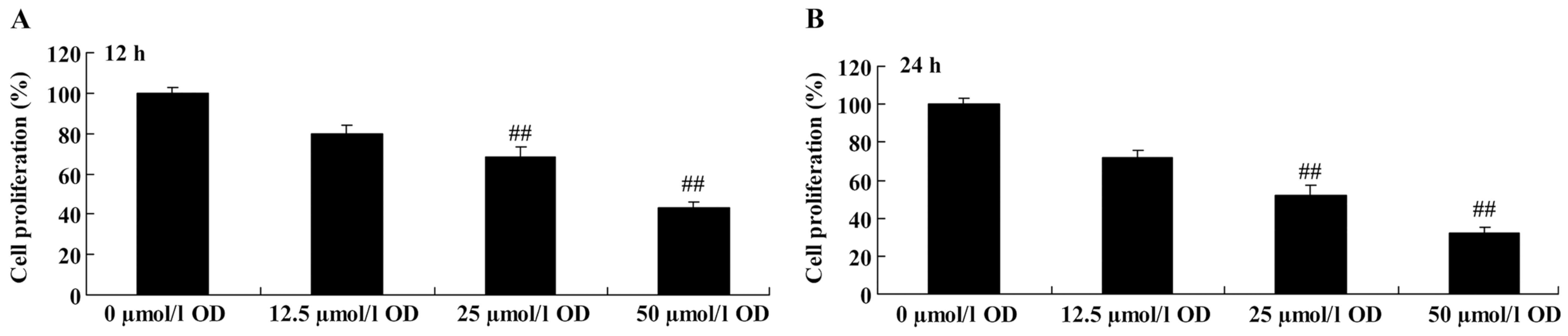
To investigate the effect of Ophiopogonin D on
laryngocarcinoma cell proliferation, AMC-HN-8 cells were treated
with different concentrations of Ophiopogonin D (0–50 µmol/l) for
12 or 24 h. After treatment with Ophiopogonin D, the inhibition of
cell proliferation in human laryngocarcinoma AMC-HN-8 cells was
found to be time and dose-dependent (Fig.
2). In particular, Ophiopogonin D (25 and 50 µmol/l)
significantly suppressed the cell proliferation in human
laryngocarcinoma AMC-HN-8 cells at 12 and 24 h, compared with the
control (0 µmol/l Ophiopogonin D; Fig.
2).
Ophiopogonin D is cytotoxic in human
laryngocarcinoma AMC-HN-8 cells
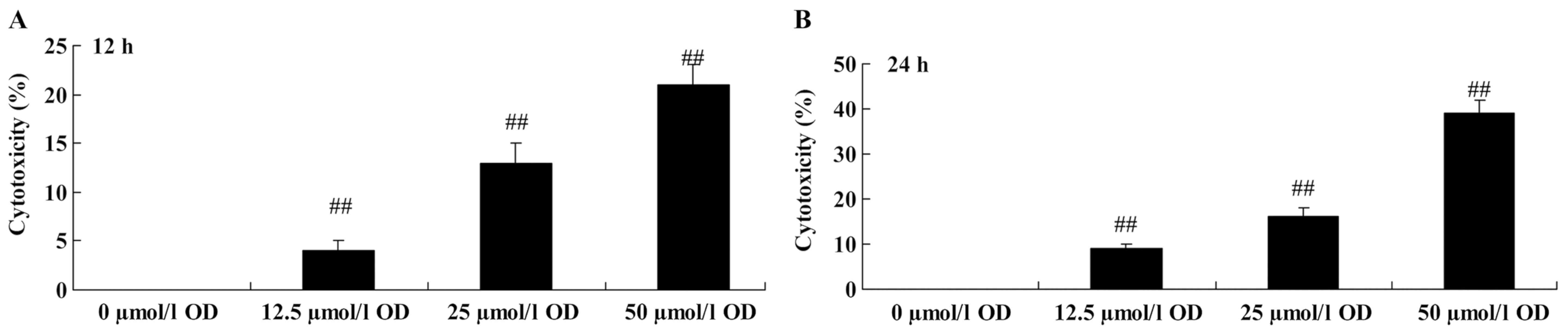
To analyze the anti-cancer effect of Ophiopogonin D
on laryngocarcinoma cells, the LDH assay was performed. Following
treatment with Ophiopogonin D (50 µmol/l) for 12 h, the
cytotoxicity rate of human laryngocarcinoma AMC-HN-8 cells was
effectively promoted, compared with the control (0 µmol/l
Ophiopogonin D; Fig. 3). Subsequent
to treatment with 25 and 50 µmol/l Ophiopogonin D for 24 h, the
cytotoxicity rate of human laryngocarcinoma AMC-HN-8 cells was also
increased, compared with the control (0 µmol/l Ophiopogonin D;
Fig. 3).
Ophiopogonin D induces apoptosis of
human laryngocarcinoma AMC-HN-8 cells
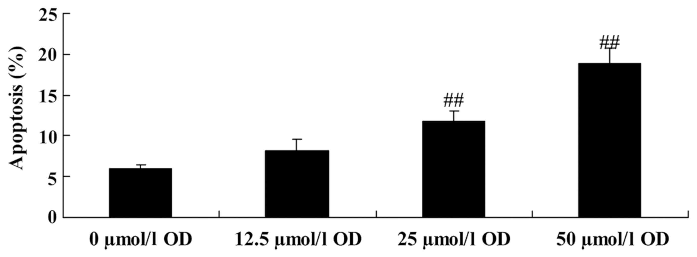
Annexin V/PI staining was performed to qualify the
efficacy of Ophiopogonin D on AMC-HN-8 cells apoptosis. As shown in
Fig. 4, 25 or 50 µmol/l Ophiopogonin
D treatment significantly induces apoptosis of human
laryngocarcinoma AMC-HN-8 cells, compared with the control (0
µmol/l Ophiopogonin D).
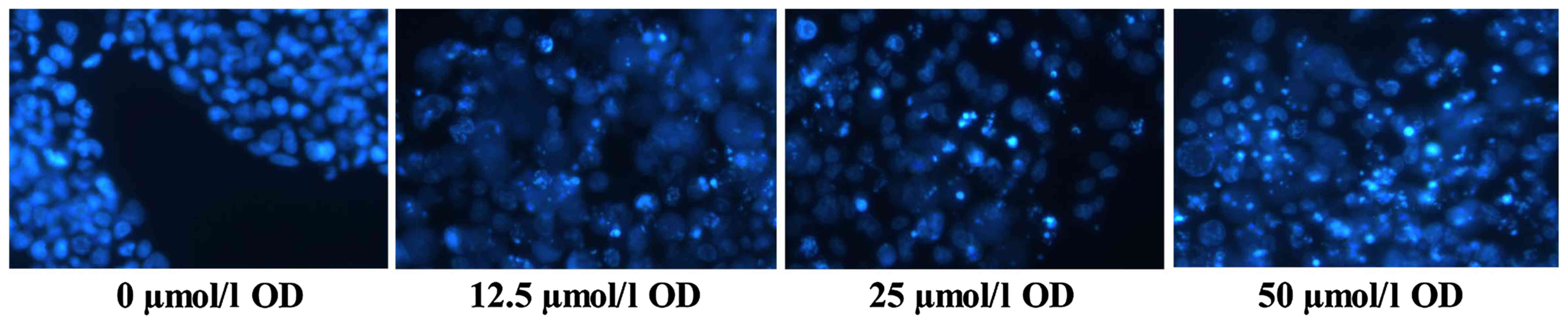
Ophiopogonin D induces degradation of
the nucleolus in human laryngocarcinoma AMC-HN-8 cells
The anti-cancer effect of Ophiopogonin D on the
nucleoli of AMC-HN-8 cells was detected using DAPI assay. Compared
with the control 0 µmol/l Ophiopogonin D, cell apoptosis was
observed in the nucleoli of AMC-HN-8 cells in the 5 or 50 µmol/l
Ophiopogonin D treated group (Fig.
5).
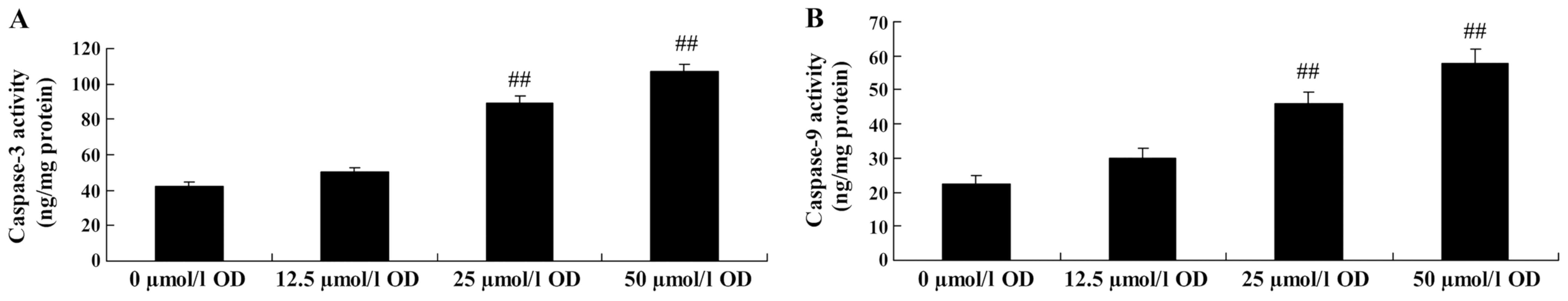
Ophiopogonin D induces caspase-3/9
activity in human laryngocarcinoma AMC-HN-8 cells
Commercially available PEDF ELISA kits were used to
analyze the effect of Ophiopogonin D on caspase-3/9 activity in of
AMC-HN-8 cells. Treatment with Ophiopogonin D (25 or 50 µmol/l)
significantly induced caspase-3/9 activity in AMC-HN-8 cells,
compared with the control (0 µmol/l Ophiopogonin D; Fig. 6).
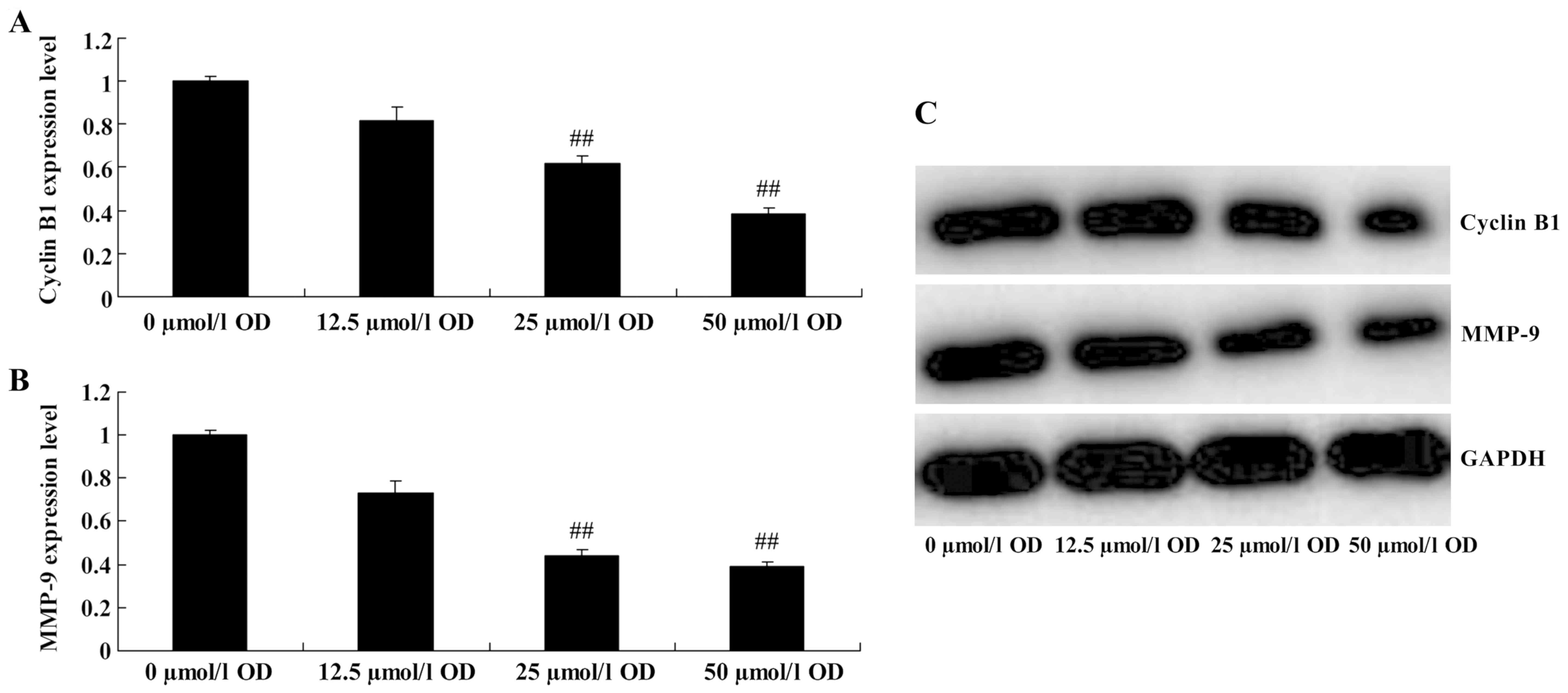
Ophiopogonin D downregulates cyclin B1
and MMP-9 protein expression in human laryngocarcinoma AMC-HN-8
cells
Furthermore, in order to investigate whether the
molecular mechanism of Ophiopogonin D on human laryngocarcinoma
AMC-HN-8 cells, cyclin B1 protein expression was measured using
western blot analysis. Ophiopogonin D (25 or 50 µmol/l)
significantly suppressed the protein of cyclin B1 and MMP-9
expression level in AMC-HN-8 cells, compared with control 0 µmol/l
of Ophiopogonin D (Fig. 7).
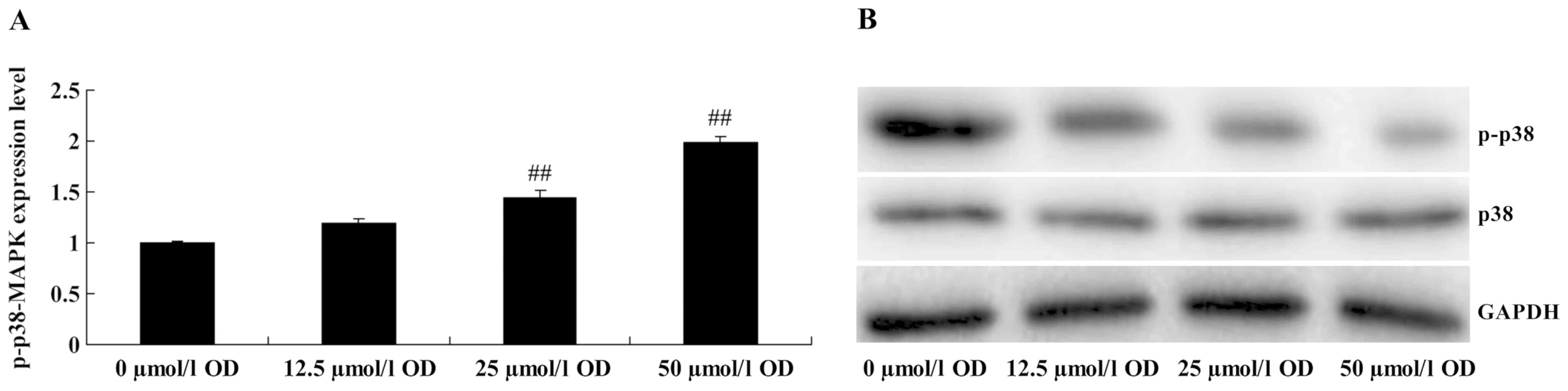
Ophiopogonin D upregulates p38-MAPK
protein expression in human laryngocarcinoma AMC-HN-8 cells
To analyze whether Ophiopogonin D upregulates
p38-MAPK protein expression of human laryngocarcinoma AMC-HN-8
cells, the present study also examined p-p38-MAPK protein
expression of AMC-HN-8 cells. As shown in Fig. 8, 25 or 50 µmol/l of Ophiopogonin D
treatment significantly activated p-p38-MAPK protein expression of
AMC-HN-8 cells, compared with the control 0 µmol/l of Ophiopogonin
D.
Discussion
Laryngocarcinoma is a common malignant tumor of the
ear, nose and throat, head and neck. It is most commonly diagnosed
as squamous cell carcinoma (3).
Laryngocarcinoma is divided into supraglottic, glottic, subglottic
and across glottis types. Supraglottic laryngocarcinoma is most
frequently observed in certain regions of China (Eastern, Southern
and Northern) (20). Studies on the
genesis and development mechanism of laryngocarcinoma, particularly
fundamental studies on laryngocarcinoma, are of great significance
for clinical effective prevention and control (1,20). The
present data indicate that Ophiopogonin D inhibits cell
proliferation, promotes cytotoxicity, induces apoptosis and
increases the caspase-3/9 activity of human laryngocarcinoma cells.
Furthermore, the present study provided evidence that Ophiopogonin
D may be a potential antitumor compound in human laryngocarcinoma
cells.
Cyclin B1 protein, as a regulative subunit of the
kinase Cdc2, forms active compounds, including mitosis-promoting
factor (also termed M phase-promoting factor), with Cdc2 (21). These compounds mainly participate in
transformation regulation at the G2/M phase and regulate the
activation and distribution of multiple microtubule
germinal-associated proteins, such as CENP-E and Eg5 (22). The compound also participates in
spindle formation, chromosome segregation and further produces a
series of actions. Cyclin then degrades and inactivates via
dependent protease hydrolysis (23).
The present results have shown that Ophiopogonin D significantly
suppressed the level of cyclin B1 protein expression in AMC-HN-8
cells. Zang et al (18)
suggested that Ophiopogonin D inhibits MCF-7 cell growth through
downregulation of cyclin B1 at the G2/M phase. Therefore, the
effect of Ophiopogonin D on AMC-HN-8 cells may be associated with
cyclin B1 expression.
MMPs belong to an endopeptidase family dependent on
zinc. This family is named due to its activity on ECM endopeptidase
(24). MMP-2 and MMP-9 are important
members of the family. MMP-2 and MMP-9 can be expressed on a
variety of cells, mainly consisting of inflammatory cells,
mesenchymal cells in tumor tissues and tumor cells (25). Numerous studies have shown that MMP-2
and MMP-9 are overexpressed in the majority of human malignancies,
which is associated with the infiltration and metastasis of tumor
cells and prognosis of the patient (24,25).
Previous studies have shown that MMP-2 and MMP-9 are also
overexpressed in laryngocarcinoma tissue, and they are associated
with lymphatic metastasis and poor prognosis (10). The present results demonstrated that
Ophiopogonin D significantly downregulates MMP-9 protein expression
in AMC-HN-8 cells. Zhang et al (26) suggested that Ophiopogonin-D suppresses
MDA-MB-435 cell adhesion and invasion through inhibition of
phosphorylation of the p38 and MMP-9 pathway, not the MMP-2
pathway. Therefore, MMP-9 expression may have a crucial role in
Ophiopogonin-D-induced apoptosis within laryngocarcinoma AMC-HN-8
cells.
Studies show that the occurrence of laryngocarcinoma
is associated with MAPK signal transduction. p38-MAPK is an
important member of the MAPK family (27). It participates in cell proliferation,
apoptosis and differentiation, and has an important role on cell
apoptosis process (15). The genesis
and development, rapid proliferation and indeterminate expression
of p38-MAPK in laryngocarcinoma are closely associated with
metastasis (28). p38-MAPK
participates in the process of laryngocarcinoma and carcinogenic
process of certain organics. These results suggest that
Ophiopogonin D treatment significantly activated p-p38-MAPK protein
expression in AMC-HN-8 cells. Zhang et al (26) suggested that Ophiopogonin D suppresses
MDA-MB-435 cell adhesion and invasion through inhibition of
phosphorylation of p38 and MMP-9 pathway. P38-MAPK pathways may
also be associated with the antitumor activity of Ophiopogonin D on
laryngocarcinoma. Additional studies are required to examine the
detailed pharmacological mechanisms that underlie the action of
Ophiopogonin D in vivo.
To conclude, the present study demonstrated that
Ophiopogonin D could inhibit cell proliferation, promoted
cytotoxicity, induced apoptosis and increased the activity of
caspase-3/9 in human laryngocarcinoma cells. The potential
mechanism underlying the antitumor effects of Ophiopogonin D may
result from a downregulatory effect upon cyclin B1/MMP-9 expression
and suppression of p38-MAPK, which ultimately induces cellular
apoptosis in AMC-HN-8 cells.
References
|
1
|
Dong QL and Wang GN: Effect of general
anaesthesia with combination of acupuncture and enflurane applied
in radical operation of laryngocarcinoma. Chin J Integr Med.
12:306–309. 2006. View Article : Google Scholar : PubMed/NCBI
|
|
2
|
Chen LW, Wang JL, Zhang LY, Yang SM, Li
CS, Yu N, Zhao W JD, Zhao LD, Li K, Liu MB and Zhai SQ:
Establishment of an animal model of spontaneous cervical lymph node
metastasis of laryngeal squamous cell carcinoma and obtaining
laryngocarcinoma cells with high metastatic potential. Neoplasma.
60:504–510. 2013. View Article : Google Scholar : PubMed/NCBI
|
|
3
|
Jiang LY, Lian M, Wang H, Fang JG and Wang
Q: Inhibitory effects of 5-Aza-2′-deoxycytidine and trichostatin A
in combination with p53-expressing adenovirus on human
laryngocarcinoma cells. Chin J Cancer Res. 24:232–237. 2012.
View Article : Google Scholar : PubMed/NCBI
|
|
4
|
Wang XX, Yao XB, Qiang ZS and Zhu HL:
Attenuation of EGFL7 inhibits human laryngocarcinoma cells growth
and invasion. Int J Clin Exp Med. 8:3141–3155. 2015.PubMed/NCBI
|
|
5
|
Fodor T, Szántó M, Abdul-Rahman O, Nagy L,
Dér Á, Kiss B and Bai P: Combined treatment of MCF-7 cells with
AICAR and methotrexate, arrests cell cycle and reverses warburg
metabolism through AMP-activated protein kinase (AMPK) and FOXO1.
PLoS One. 11:e01502322016. View Article : Google Scholar : PubMed/NCBI
|
|
6
|
Makarević J, Tsaur I, Juengel E, Borgmann
H, Nelson K, Thomas C, Bartsch G, Haferkamp A and Blaheta RA:
Amygdalin delays cell cycle progression and blocks growth of
prostate cancer cells in vitro. Life Sci. 147:137–142. 2016.
View Article : Google Scholar : PubMed/NCBI
|
|
7
|
Liu YF, Qu GQ, Lu YM, Kong WM, Liu Y, Chen
WX and Liao XH: Silencing of MAP4K4 by short hairpin RNA suppresses
proliferation, induces G1 cell cycle arrest and induces apoptosis
in gastric cancer cells. Mol Med Rep. 13:41–48. 2016. View Article : Google Scholar : PubMed/NCBI
|
|
8
|
Liu G, Ren X, Gao C and Zhang W:
Acylglycerol kinase promotes the proliferation and cell cycle
progression of oral squamous cell carcinoma. Mol Med Rep.
12:2225–2230. 2015. View Article : Google Scholar : PubMed/NCBI
|
|
9
|
Latocha M, Płonka J, Kuśmierz D, Jurzak M,
Polaniak R and Nowosad A: Transcripional activity of genes encoding
MMPs and TIMPs in breast cancer cells treated by genistein and in
normal cancer-associated fibroblasts-in vitro studies. Acta Pol
Pharm. 71:1095–1102. 2014.PubMed/NCBI
|
|
10
|
Gurgel DC, Valença-Junior JT, Dornelas CA,
Vieira RB, Maia-Filho JT, Lima-Junior RC, Ribeiro RA and Almeida
PR: Immunoexpression of metalloproteinases 2 and 14 and TIMP-2
inhibitor in main types of primary gastric carcinomas and lymph
node metastasis. Pathol Oncol Res. 21:73–81. 2015. View Article : Google Scholar : PubMed/NCBI
|
|
11
|
Navarini NF, Araújo VC, Brown AL,
Passador-Santos F, Souza IF, Napimoga MH, Araújo NS and Martinez
EF: The EGF signaling pathway influences cell migration and the
secretion of metalloproteinases by myoepithelial cells in
pleomorphic adenoma. Tumour Biol. 36:205–211. 2015. View Article : Google Scholar : PubMed/NCBI
|
|
12
|
Kang N, Hu G, Wu W, Jiang J and Yang X:
The impact of PDTC on circulating tumor cells of laryngocarcinoma
in nude mice model. Lin Chung Er Bi Yan Hou Tou Jing Wai Ke Za Zhi.
25:419–422. 2011.(In Chinese). PubMed/NCBI
|
|
13
|
Gaundar SS and Bendall LJ: The potential
and limitations of p38MAPK as a drug target for the treatment of
hematological malignancies. Curr Drug Targets. 11:823–833. 2010.
View Article : Google Scholar : PubMed/NCBI
|
|
14
|
Noel JK, Crean S, Claflin JE, Ranganathan
G, Linz H and Lahn M: Systematic review to establish the safety
profiles for direct and indirect inhibitors of p38
Mitogen-activated protein kinases for treatment of cancer. A
systematic review of the literature. Med Oncol. 25:323–330. 2008.
View Article : Google Scholar : PubMed/NCBI
|
|
15
|
Zhang ZD, Li Y, Fan Q, Zhao B, Tan B and
Zhao XF: Annexin A2 is implicated in multi-drug-resistance in
gastric cancer through p38MAPK and AKT pathway. Neoplasma.
61:627–637. 2014. View Article : Google Scholar : PubMed/NCBI
|
|
16
|
Park SH, Lee HJ, Ryu J, Son KH, Kwon SY,
Lee SK, Kim YS, Hong JH, Seok JH and Lee CJ: Effects of
ophiopogonin D and spicatoside A derived from Liriope Tuber on
secretion and production of mucin from airway epithelial cells.
Phytomedicine. 21:172–176. 2014. View Article : Google Scholar : PubMed/NCBI
|
|
17
|
Ishibashi H, Mochidome T, Okai J, Ichiki
H, Shimada H and Takahama K: Activation of potassium conductance by
ophiopogonin-D in acutely dissociated rat paratracheal neurones. Br
J Pharmacol. 132:461–466. 2001. View Article : Google Scholar : PubMed/NCBI
|
|
18
|
Zang QQ, Zhang L, Gao N and Huang C:
Ophiopogonin D inhibits cell proliferation, causes cell cycle
arrest at G2/M, and induces apoptosis in human breast carcinoma
MCF-7 cells. J Integr Med. 14:51–59. 2016. View Article : Google Scholar : PubMed/NCBI
|
|
19
|
Qian J, Jiang F, Wang B, Yu Y, Zhang X and
Yin Z: Ophiopogonin D prevents H2O2-induced injury in primary human
umbilical vein endothelial cells. J Ethnopharmacol. 128:438–445.
2010. View Article : Google Scholar : PubMed/NCBI
|
|
20
|
Li X, Shen Y, Di B, Li J, Geng J, Lu X and
He Z: Biological and clinical significance of p75NTR expression in
laryngeal squamous epithelia and laryngocarcinoma. Acta
Otolaryngol. 132:314–324. 2012. View Article : Google Scholar : PubMed/NCBI
|
|
21
|
Lohberger B, Leithner A, Stuendl N,
Kaltenegger H, Kullich W and Steinecker-Frohnwieser B: Diacerein
retards cell growth of chondrosarcoma cells at the G2/M cell cycle
checkpoint via cyclin B1/CDK1 and CDK2 downregulation. BMC Cancer.
15:8912015. View Article : Google Scholar : PubMed/NCBI
|
|
22
|
Song Y, Zhao C, Dong L, Fu M, Xue L, Huang
Z, Tong T, Zhou Z, Chen A, Yang Z, et al: Overexpression of cyclin
B1 in human esophageal squamous cell carcinoma cells induces tumor
cell invasive growth and metastasis. Carcinogenesis. 29:307–315.
2008. View Article : Google Scholar : PubMed/NCBI
|
|
23
|
Dao T, Korontsvit T, Zakhaleva V, Haro K,
Packin J and Scheinberg DA: Identification of a human cyclin
D1-derived peptide that induces human cytotoxic CD4 T cells. PLoS
One. 4:e67302009. View Article : Google Scholar : PubMed/NCBI
|
|
24
|
Luukkaa H, Klemi P, Leivo I, Mäkitie AA,
Irish J, Gilbert R, Perez-Ordonez B, Hirsimäki P, Vahlberg T,
Kivisaari A, et al: Expression of matrix metalloproteinase-1, −7,
−9, −13, Ki-67, and HER-2 in epithelial-myoepithelial salivary
gland cancer. Head Neck. 32:1019–1027. 2010. View Article : Google Scholar : PubMed/NCBI
|
|
25
|
Munaut C, Noël A, Hougrand O, Foidart JM,
Boniver J and Deprez M: Vascular endothelial growth factor
expression correlates with matrix metalloproteinases MT1-MMP, MMP-2
and MMP-9 in human glioblastomas. Int J Cancer. 106:848–855. 2003.
View Article : Google Scholar : PubMed/NCBI
|
|
26
|
Zhang Y, Han Y, Zhai K, Sun M, Liu J, Yu B
and Kou J: Ophiopogonin-D suppresses MDA-MB-435 cell adhesion and
invasion by inhibiting matrix metalloproteinase-9. Mol Med Rep.
12:1493–1498. 2015. View Article : Google Scholar : PubMed/NCBI
|
|
27
|
Singh P, Sarkar S, Umar S, Rengifo-Cam W,
Singh AP and Wood TG: Insulin-like growth factors are more
effective than progastrin in reversing proapoptotic effects of
curcumin: Critical role of p38MAPK. Am J Physiol Gastrointest Liver
Physiol. 298:G551–G562. 2010. View Article : Google Scholar : PubMed/NCBI
|
|
28
|
Estrella VC, Eder AM, Liu S, Pustilnik TB,
Tabassam FH, Claret FX, Gallick GE, Mills GB and Wiener JR:
Lysophosphatidic acid induction of urokinase plasminogen activator
secretion requires activation of the p38MAPK pathway. Int J Oncol.
31:441–449. 2007.PubMed/NCBI
|