Introduction
Colon cancer is a common malignant tumor in the
digestive system. In recent years, the incidence rate of colon
cancer has gradually increased and the 5-year survival rate is
still not high. Patients with colon cancer often die of tumor
recurrence and metastasis (1). At
present, the mechanisms of occurrence and development of colon
cancerare not completely understood. Hepatocyte growth factor (HGF)
is a factor in vivo with multiple biological functions,
which strongly promotes cell division, inducing epithelial cell
migration, invasion and angiogenesis in vivo (2,3). Setia
et al (4) found that the HGF
expression is significantly increased in patients with colon
cancer, and it is even higher in patients complicated with
metastasis, so it is believed that HGF is involved in growth and
metastasis processes of colon cancer. HGF can bind to c-methionine
(c-Met) receptor and activates its activity, thus resulting in
tyrosine phosphorylation of various substrate proteins, including
phospholipase C-γ (PLC-γ), phosphatidylinositol
3-kinase-serine/threonine kinase/protein kinase B (PI3K-AKT/PKB),
mitogen-activated protein kinase (MAPK) and Grb2-associated
binder-1 (Gab1) (5,6). MAPK participates in the physiological
functions of various cells in vivo, including in
proliferation, apoptosis and differentiation (7). In this study, methyl ethyl ketone (MEK)
inhibitor U0126 was used to inhibit the extracellular
signal-regulated kinase (ERK)/MAPK signal transduction pathway, so
as to explore the roles of ERK/MAPK signaling pathway in the
effects of HGF on promoting proliferation, and regulating cycle and
apoptosis of human colon cancer cells to find new therapeutic
approaches of colon cancer.
Materials and methods
Materials
Human colon cancer SW620 cells were purchased from
Beijing Beina Chuanglian Biotechnology Research Institute (cat. no.
BNCC337664, Beijing, China). Cells were cultured using Roswell Park
Memorial Institute (RPMI)-1640 medium and 15% fetal bovine serum
(FBS) in an incubator with 5% CO2 at 37°C. Cells were
cryopreserved using basal medium, 5% dimethyl sulfoxide (DMSO) and
15% FBS, and those in logarithmic growth phase were used for
subsequent experiments.
Reagents: Roswell Park Memorial Institute
(RPMI)-1640 (Hyclone; GE Healthcare Life Sciences; Logan, UT, USA),
Dimethyl sulfoxide (DMSO) (Beyotime, Shanghai, China), FBS, 0.25%
trypsin (both from Thermo Fisher Scientific, Inc., Waltham, MA,
USA), Cell Counting Kit 8 (CCK8; Shanghai Yubo Biological
Technology Co., Ltd., Shanghai, China), Annexin V-fluorescein
isothiocyanate (FITC) apoptosis detection kits, polyclonal
antibodies (all from BD Pharmingen; BD Biosciences, Franklin Lakes,
NJ, USA), and MAPK MEK1/2 efficient selective inhibitor U0126
(Shanghai Yeasen Biological Technology Co., Ltd., Shanghai,
China).
The study was approved by the Ethics Committee of
Chinese PLA General Hospital (Beijing, China).
Methods
Detection of effect of U0126 on SW620
cell proliferation via CCK8
In this experiment, cells were divided into seven
groups, including five experimental groups (U0126: 0.5, 1, 2, 4 and
8 µmol/l, respectively + HGF), the control group (+ HGF) and the
DMSO group. Each group had five repeated wells, and HGF was added
into each group after cell culture for 30 min and after culture for
another 48 h, 10 µl CCK8 reagent was dropwise added into each well,
followed by incubation in the dark for 2 h. The optical density
(OD) value of each well was detected using a Sunrise microplate
reader (Tecan Group, Ltd., Mannedorf, Switzerland) at a wavelength
of 570 nm. Inhibition rate = (ODcontrol group -
ODU0126 group)/ODcontrol group ×100%.
Detection of cell cycle and apoptosis
via flow cytometry
Cells were cultured, collected, and fixed at 4°C for
at least 24 h. The fixing solution was removed before use, and
cells were washed with phosphate-buffered saline (PBS). The cell
density was adjusted to 1.0×10⁶/ml. A total of 0.1 ml
cell suspension was taken, added with 1 ml propidium iodide dye
liquor for staining in the dark at 4°C for half an hour and
filtered. The cell cycle and apoptosis were detected on the flow
cytometer (Thermo Fisher Scientific, Inc., Waltham, MA, USA).
Multicycle AV software (De Novo Software, Glendale, CA, USA) was
used to analyze the DNA cell cycle, and the distribution percentage
of each time phase in DNA histogram was calculated. The cell
proliferative activity was presented as proliferation index (PI):
PI = (S + G2/M)/(G0/G1 + S + G2/M) ×100%.
Detection of cell migration via wound
healing assay
Cells were inoculated and cultured at a density of
2×10⁶/well. After 8 h, the culture plate was scratched
vertically using a spearhead, 6 scratches/well. After the plate was
washed with PBS, complete medium was added into control group, 0.1%
DMSO was added into DMSO group, and 4 and 8 µmol/l U0126 was added
into experimental group. After half an hour, 20 ng/l HGF was added,
and cell growth (0 h) was observed under a light microscope (×100)
(Nikon Instrument Inc., NY, USA). The width of 3 scratches was
measured using Image-Pro Plus (Media Cybernetics, Inc., Rockville,
MD, USA), and cells continued to be cultured. After 24 h, the
complete medium was replaced, each group was added with the
above-mentioned corresponding reagents, and cell migration distance
was observed and measured under the light microscope: Migration
distance (d) = (scratch width at 0 h - scratch width at 24 h)/2.
Cells continued to be cultured, the complete medium was replaced
after 48 h, and cell migration distance was observed and measured
under the light microscope: Migration distance (d) = (scratch width
at 0 h - scratch width at 48 h)/2. Cells continued to be cultured
until 72 h. Inhibition rate of migration distance = (dcontrol
group - dU0126 group)/dcontrol group
×100%.
Statistical analysis
Statistical Product and Service Solutions (SPSS)
20.0 software (SPSS, Inc., Chicago, IL, USA) was used for
statistical analysis. Measurement data were presented as mean ±
standard deviation (SD), one-way analysis of variance (ANOVA) was
used for quantitative data, and Student-Newman-Keuls (SNK)-q test
was used for multiple comparisons as a post hoc test. α=0.05
indicated the inspection level.
Results
Detection of colon cancer cell
proliferation via CCK8
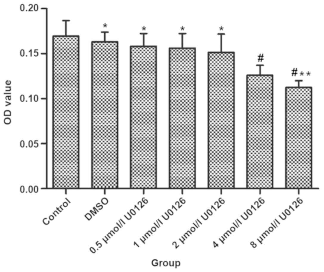
After drug treatment for 48 h, there were
statistically significant differences in 4 and 8 µmol/l U0126
experimental group compared with control group (P<0.05). The
inhibition rate had no significant difference between 4 and 8
µmol/l U0126 in experimental group (P>0.05) (Table I and Fig.
1).
 | Table I.Inhibition rate of U0126 on colon
cancer cell proliferation. |
Table I.
Inhibition rate of U0126 on colon
cancer cell proliferation.
| Variables | OD | Inhibition rate
(%) |
|---|
| Control | 0.16121±0.021294 | – |
| DMSO |
0.15374±0.013859a | – |
| 0.5 µmol/l U0126 |
0.15194±0.014857a | – |
| 1 µmol/l U0126 |
0.14982±0.020958a | – |
| 2 µmol/l U0126 |
0.13957±0.031857a | – |
| 4 µmol/l U0126 |
0.12194±0.015392b | 24.7 |
| 8 µmol/l U0126 |
0.11482±0.005928b,c | 28.9 |
Detection of cell cycle and apoptosis
via flow cytometry
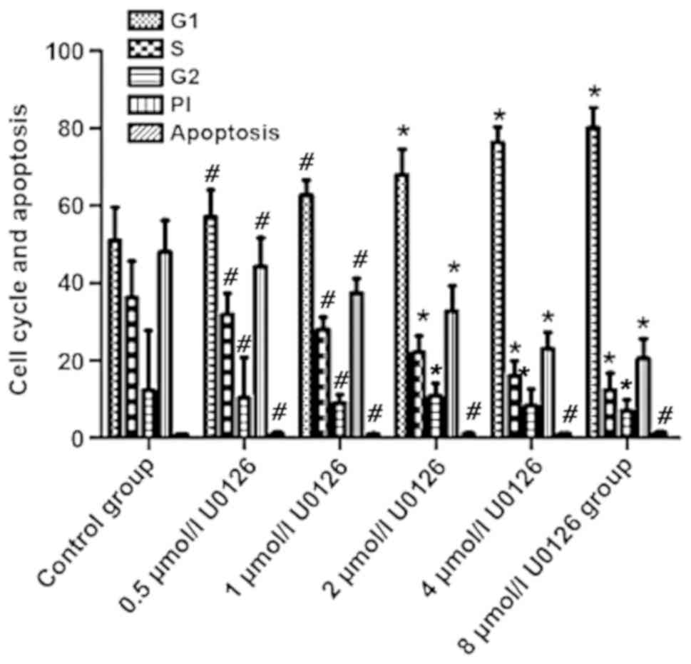
Compared with those in control group, G1 phase, S
phase, G2 phase and PI in 2, 4 and 8 µmol/l U0126 group had
statistically significant differences (P<0.05). G1 phase, S
phase, G2 phase and PI in control and 8 µmol/l U0126 group were
51.05±8.59 vs. 80.14±5.27%, 36.28±9.42 vs. 12.44±4.26%, 12.39±15.37
vs. 7.15±2.74%, and 48.03±8.21 vs. 20.57±5.07%, respectively, and
differences were statistically significant (P<0.05) (Table II, Fig.
2).
| Table II.Cell cycle and apoptosis (mean
±SD). |
Table II.
Cell cycle and apoptosis (mean
±SD).
| Variables | G1 | S | G2 | PI | Apoptosis |
|---|
| Control | 51.05±8.59 | 36.28±9.42 | 12.39±15.37 | 48.03±8.21 | 0.94±0.13 |
| 0.5 µmol/l U0126 |
57.13±7.02b |
31.98±5.38b |
10.35±10.48b |
44.32±7.41b |
1.17±0.36b |
| 1 µmol/l U0126 |
62.77±3.84b |
28.13±3.06b |
9.04±2.14b |
37.46±3.77b |
1.03±0.11b |
| 2 µmol/l U0126 |
68.02±6.62a |
22.17±4.18a |
10.95±3.19a |
32.81±6.52a |
1.15±0.19b |
| 4 µmol/l U0126 |
76.35±4.06a |
16.02±3.86a |
8.49±4.13a |
23.05±4.22a |
1.09±0.13b |
| 8 µmol/l U0126 |
80.14±5.27a |
12.44±4.26a |
7.15±2.74a |
20.57±5.07a |
1.37±0.35b |
Detection of cell migration via wound
healing assay
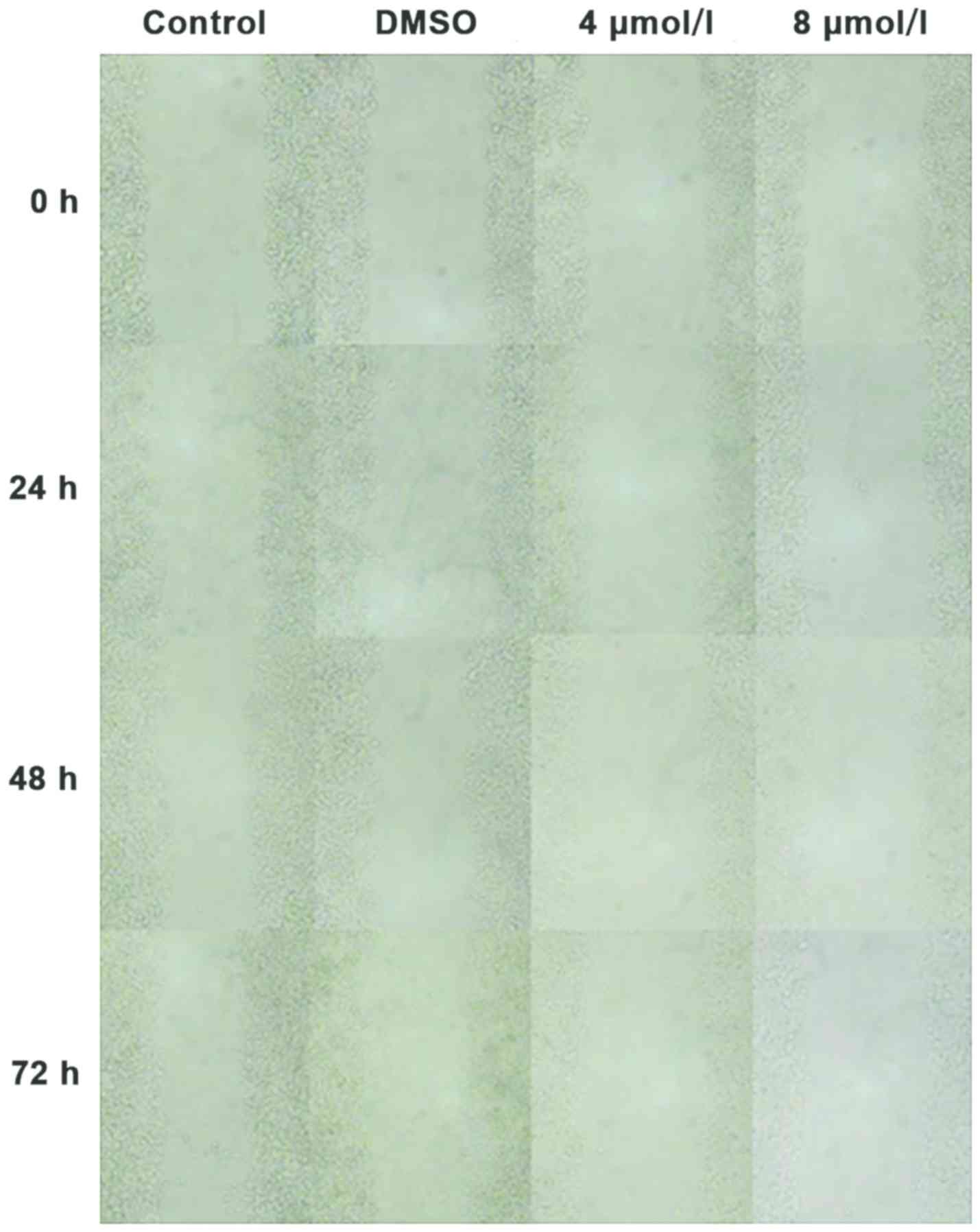
After drug treatment for 24 h, compared with that in
control group, the cell migration distance in 8 µmol/l U0126 group
had a statistically significant difference (P<0.05), but it had
no significant difference between DMSO and 4 µmol/l U0126 group
(P>0.05). After drug treatment for 48 and 72 h, the cell
migration distance in 4 and 8 µmol/l U0126 group was significantly
reduced, and the differences were statistically significant
compared with those in control group (P<0.05). However, the cell
migration distance had no statistically significant difference
between 4 and 8 µmol/l U0126 group (P>0.05) (Fig. 3, Table
III).
| Table III.Effect of drug on inhibition rate of
cell migration (mean ±SD). |
Table III.
Effect of drug on inhibition rate of
cell migration (mean ±SD).
|
| 24 h | 48 h | 72 h |
|---|
|
|
|
|
|
|---|
| Variables | d (µm) | Migration inhibition
rate (%) | d (µm) | Migration inhibition
rate (%) | d (µm) | Migration inhibition
rate (%) |
|---|
| Control | 34.9±14.3 | – | 47.2±9.4 | – | 50.2±14.4 | – |
| DMSO |
31.5±11.7b | – |
40.8±9.9b | – |
52.6±18.2b | – |
| 4 µmol/l U0126 |
19.9±7.2b | – |
25.3±15.4a | 46.2 |
28.6±15.1a | 44.7 |
| 8 µmol/l U0126 |
13.8±6.2a | 61.1 |
18.4±6.1a,c | 61.4 |
23.4±10.7a,c | 54.1 |
Discussion
The incidence rate of colon cancer is increasing
year by year, seriously threatening human health. The widely-used
treatment means is surgery, and the postoperative 5-year survival
rate of patients is also different due to different staging of
colon cancer. The 5-year survival rate of patients in Dukes A stage
is >90%, but that of patients in Dukes C stage is only 50%
(8). Although the 5-year survival
rate of patients can be increased to some extent through vaious
comprehensive treatments, the prognosis is still unsatisfactory.
Tumor recurrence and metastasis are still primary causes of
patients' death (9), and these
factors are closely related to the proliferation and invasion
capacities of tumor cells.
In a variety of tissues in the human body, there is
an extracellular signal factor, namely HGF, and it is essentially a
polypeptide growth factor (10). HGF
can be expressed and secreted in normal human and tumor cells. Some
scholars found via experiments that HGF can effectively promote the
proliferation and invasion processes of SW620 cells in vitro
(11). ERK/MAPK signaling pathway is
involved in a variety of physiological cell functions, such as
proliferation, differentiation and apoptosis. These functions of
tumor cells are also associated with the ERK/MAPK signal
transduction pathway (12).
Radziwon-Balicka et al (13)
found that MEK phosphorylation level is overexpressed in villous
adenoma tissues, and its expression is significantly increased
compared with that in para-carcinoma tissues and normal tissues.
Lee et al (14) also found
similar results in tubular adenomas. HGF binds to c-Met receptor
in vivo and activates its kinase activity and multiple
downstream signaling pathways, including ERK/MAPK (15), which provides a new idea for
inhibiting ERK/MAPK signaling pathway to block the effects of HGF
on promoting proliferation and invasion of colon cancer cells.
Enayat et al (16) showed that
inhibiting ERK/MAPK signal transduction pathway can produce more
significant inhibitory effects on proliferation and invasion of
tumor cells.
In this study, after drug treatment for 48 h, there
were statistically significant differences in 4 and 8 µmol/l U0126
experimental group compared with control group (P<0.05). The
inhibition rate had no significant difference between the
experimental groups of 4 and 8 µmol/l U0126 (P>0.05), and the
tumor cell proliferation was not inhibited in DMSO, 0.5, 1 and 2
µmol/l U0126 groups. The above results suggest that inhibiting
ERK/MAPK signaling pathway can effectively block the ability of HGF
to promote tumor cell proliferation, but there is no
concentration-dependent effect. This is consistent with the results
of Chen et al (17). It is
speculated that the possible reason is that there are other
downstream signaling pathways in HGF, such as PLC-γ and PI3K/AKT,
directly leading to no dose-dependence in inhibition effect.
Results of flow cytometry showed that U0126
inhibited the cell cycle from entering S phase, and U0126 had no
obvious effect on apoptosis of colon cancer cells. However, Bodur
et al (18) found that
inhibiting ERK/MAPK signaling pathway can promote apoptosis. It is
speculated that the application of U0126 cannot completely
antagonize the effect of HGF of inhibiting tumor cell apoptosis,
and ERK/MAPK signal transduction pathway does not play a major role
in regulating the apoptosis of colon cancer SW620 cells.
Wound healing assay showed that after drug treatment
for 24 h, compared with that in control group, the cell migration
distance in 8 µmol/l U0126 group had a statistically significant
difference (P<0.05), but it had no significant difference
between DMSO and 4 µmol/l U0126 group (P>0.05). After drug
treatment for 48 and 72 h, the cell migration distance in 4 and 8
µmol/l U0126 group was significantly reduced, and the differences
were statistically significant compared with those in control group
(P<0.05). However, the cell migration distance had no
statistically significant difference between 4 and 8 µmol/l U0126
group (P>0.05). These results indicate that inhibiting ERK/MAPK
signal transduction pathway can significantly inhibit SW620 cell
migration, during which the number of cell processes is reduced,
and the length is shortened. ERK/MAPK signaling pathway may exert
inhibition effect via inhibiting the cytoskeleton and cell
processes. Zhang et al (19)
also found a similar phenomenon. Najar et al (20) found that ERK signal transduction
regulates the expression of cell transcription factor, causing
cytoskeletal degeneration and enhancing invasion and metastasis
capacities of tumor cells.
In conclusion, ERK/MAPK signaling pathway is
involved in the effects of HGF on promoting proliferation and
regulating cell cycle and apoptosis of human colon cancer cells,
providing a new approach for the treatment of colon cancer.
Acknowledgements
Not applicable.
Funding
No funding was received.
Availability of data and materials
All data generated or analyzed during this study are
included in this published article.
Authors' contributions
GZ and JY were responsible for CCK-8 assay. GZ and
PS contributed to flow cytometry. All authors read and approved the
final manuscript.
Ethics approval and consent to
participate
The study was approved by the Ethics Committee of
Chinese PLA General Hospital (Beijing, China).
Patient consent for publication
Not applicable.
Competing interests
The authors declare that they have no competing
interests.
References
|
1
|
Chen Y, Xie H, Gao Q, Zhan H, Xiao H, Zou
Y, Zhang F, Liu Y and Li J: Colon cancer associated transcripts in
human cancers. Biomed Pharmacother. 94:531–540. 2017. View Article : Google Scholar : PubMed/NCBI
|
|
2
|
Tahir AA, Sani NF, Murad NA, Makpol S,
Ngah WZ and Yusof YA: Combined ginger extract & Gelam honey
modulate Ras/ERK and PI3K/AKT pathway genes in colon cancer HT29
cells. Nutr J. 14:312015. View Article : Google Scholar : PubMed/NCBI
|
|
3
|
Ye J, Talaiti A, Ma Y, Zhang Q, Ma L and
Zheng H: Allergies and risk of colorectal cancer: A systematic
review and meta-analysis of observational studies. Oncotarget.
8:14646–14654. 2017.PubMed/NCBI
|
|
4
|
Setia S, Nehru B and Sanyal SN:
Upregulation of MAPK/Erk and PI3K/Akt pathways in ulcerative
colitis-associated colon cancer. Biomed Pharmacother. 68:1023–1029.
2014. View Article : Google Scholar : PubMed/NCBI
|
|
5
|
Zhang Y, Yuan J, Zhang X, Yan F, Huang M,
Wang T, Zheng X and Zhang M: Angiomotin promotes the malignant
potential of colon cancer cells by activating the YAP-ERK/PI3K-AKT
signaling pathway. Oncol Rep. 36:3619–3626. 2016. View Article : Google Scholar : PubMed/NCBI
|
|
6
|
Urosevic J, Nebreda AR and Gomis RR: MAPK
signaling control of colon cancer metastasis. Cell Cycle.
13:2641–2642. 2014. View Article : Google Scholar : PubMed/NCBI
|
|
7
|
Dziegielewska B, Brautigan DL, Larner JM
and Dziegielewski J: T-type Ca2+ channel inhibition
induces p53-dependent cell growth arrest and apoptosis through
activation of p38-MAPK in colon cancer cells. Mol Cancer Res.
12:348–358. 2014. View Article : Google Scholar : PubMed/NCBI
|
|
8
|
Zuo L, Yang X, Lu M, Hu R, Zhu H, Zhang S,
Zhou Q, Chen F, Gui S and Wang Y: All-trans retinoic acid inhibits
human colorectal cancer cells RKO migration via downregulating
myosin light chain kinase expression through MAPK signaling
pathway. Nutr Cancer. 68:1225–1233. 2016. View Article : Google Scholar : PubMed/NCBI
|
|
9
|
Gröschl B, Bettstetter M, Giedl C,
Woenckhaus M, Edmonston T, Hofstädter F and Dietmaier W: Expression
of the MAP kinase phosphatase DUSP4 is associated with
microsatellite instability in colorectal cancer (CRC) and causes
increased cell proliferation. Int J Cancer. 132:1537–1546. 2013.
View Article : Google Scholar : PubMed/NCBI
|
|
10
|
Randhawa H, Kibble K, Zeng H, Moyer MP and
Reindl KM: Activation of ERK signaling and induction of colon
cancer cell death by piperlongumine. Toxicol In Vitro.
27:1626–1633. 2013. View Article : Google Scholar : PubMed/NCBI
|
|
11
|
Mao JD, Wu P, Huang JX, Wu J and Yang G:
Role of ERK-MAPK signaling pathway in pentagastrin-regulated growth
of large intestinal carcinoma. World J Gastroenterol.
20:12542–12550. 2014. View Article : Google Scholar : PubMed/NCBI
|
|
12
|
Zhao Y, Fan D, Zheng ZP, Li ET, Chen F,
Cheng KW and Wang M: 8-C-(E-phenylethenyl)quercetin from onion/beef
soup induces autophagic cell death in colon cancer cells through
ERK activation. Mol Nutr Food Res. 61:16004372017. View Article : Google Scholar
|
|
13
|
Radziwon-Balicka A, Santos-Martinez MJ,
Corbalan JJ, O'Sullivan S, Treumann A, Gilmer JF, Radomski MW and
Medina C: Mechanisms of platelet-stimulated colon cancer invasion:
Role of clusterin and thrombospondin 1 in regulation of the
P38MAPK-MMP-9 pathway. Carcinogenesis. 35:324–332. 2014. View Article : Google Scholar : PubMed/NCBI
|
|
14
|
Lee M, Young Kim S, Kim J, Kim HS, Kim SM
and Kim EJ: Mitogen-activated protein kinase phosphatase-1
inhibition and sustained extracellular signal-regulated kinase 1/2
activation in camptothecin-induced human colon cancer cell death.
Cancer Biol Ther. 14:1007–1015. 2013. View Article : Google Scholar : PubMed/NCBI
|
|
15
|
Slattery ML, Lundgreen A and Wolff RK:
Dietary influence on MAPK-signaling pathways and risk of colon and
rectal cancer. Nutr Cancer. 65:729–738. 2013. View Article : Google Scholar : PubMed/NCBI
|
|
16
|
Enayat S, Ceyhan MŞ, Başaran AA, Gürsel M
and Banerjee S: Anticarcinogenic effects of the ethanolic extract
of Salix aegyptiaca in colon cancer cells: Involvement of
Akt/PKB and MAPK pathways. Nutr Cancer. 65:1045–1058. 2013.
View Article : Google Scholar : PubMed/NCBI
|
|
17
|
Chen B, Zeng X, He Y, Wang X, Liang Z, Liu
J, Zhang P, Zhu H, Xu N and Liang S: STC2 promotes the
epithelial-mesenchymal transition of colorectal cancer cells
through AKT-ERK signaling pathways. Oncotarget. 7:71400–71416.
2016.PubMed/NCBI
|
|
18
|
Bodur C, Kutuk O, Karsli-Uzunbas G,
Isimjan TT, Harrison P and Basaga H: Pramanicin analog induces
apoptosis in human colon cancer cells: Critical roles for Bcl-2,
Bim, and p38 MAPK signaling. PLoS One. 8:e563692013. View Article : Google Scholar : PubMed/NCBI
|
|
19
|
Zhang P, Kawakami H, Liu W, Zeng X,
Strebhardt K, Tao K, Huang S and Sinicrope FA: Targeting CDK1 and
MEK/ERK overcomes apoptotic resistance in BRAF-mutant human
colorectal cancer. Mol Cancer Res. 16:378–389. 2018. View Article : Google Scholar : PubMed/NCBI
|
|
20
|
Najar AG, Pashaei-Asl R, Omidi Y, Farajnia
S and Nourazarian AR: EGFR antisense oligonucleotides encapsulated
with nanoparticles decrease EGFR, MAPK1 and STAT5 expression in a
human colon cancer cell line. Asian Pac J Cancer Prev. 14:495–498.
2013. View Article : Google Scholar : PubMed/NCBI
|