Introduction
Colon cancer is a common malignancy (1,2). In the
United States, morbidity and mortality of colon cancer ranks third
among all malignancies (3,4). Colon cancer is also the third most
common malignancy in developing countries. It has been reported
that >600,000 patients die of this cancer every year. Incidence
of colon cancer is higher in females than in males (5). Colon cancer affects >1,2 million new
cases (6). The average age of onset
of colon cancer is ~40 years, and the highest incidence is observed
in the population age group of 50–60 years. The onset age in
developing countries is 10 years earlier than that in developed
countries (7). Studies have confirmed
that the pathological process of tumors is related to the
expression of serum miRNAs. The expression of miRNAs plays an
important role in the development and progression of colon cancer,
as well as in the diagnosis and prognosis (8). miRNAs are a group of non-coding RNAs of
~19–25 nucleotides in length that regulate the expression of
multiple genes at the post-transcriptional level, thereby playing
an important role in cell proliferation, differentiation, apoptosis
and tumor development (9).
It has been reported that the increased expression
of miR-21 is closely related to various malignant tumors such as
breast, lung and prostate cancer, and it has been shown to promote
cell proliferation and inhibit cell apoptosis of various tumor cell
lines (10–13). miR-138 is ~23–24 nt in size, and its
gene is located on human chromosomes 3p21 and 16q13. miR-138 plays
a role as tumor suppressor gene in tumor regulation (14–16).
miR-138 is lowly expressed in various types of tumors and
negatively regulates the proliferation and metastasis of gastric
cancer cells and other tumor cells (17). Therefore, miR-21 and miR-138 are
closely associated with tumor development. Our study aimed to
analyze the expression of miR-21 and miR-138 in patients with colon
cancer and its effect on proliferation of SW480 cells and the
prognosis of patients, so as to investigate the role of miR-21 and
miR-138 in the development of colon cancer and provide references
for the diagnosis and prognosis of this disease.
Materials and methods
Specimen collection
Tumor tissues and adjacent healthy tissues within 50
mm around the tumors were collected from 128 patients who underwent
surgical resection from January 2010 to January 2013 in Nanfang
Hospital (Guangzhou, China). The patients included 68 males and 60
females, and their age ranged from 30–78 years. Tissues were stored
at −80°C before using. All cancer specimens were confirmed by
pathological examination, and no cancer cells and obvious
inflammatory cells were observed in cancer tissues. Colon cancer
tissues were diagnosed and staged according to the TNM staging
criteria of the American Joint Committee on Cancer (7th edition).
No history of a tumor, major organ dysfunction, such as liver or
kidney, and abnormal hemorrhage or abnormal coagulation function
were observed. All patients had complete medical record and
follow-up data. No preoperative radiotherapy, chemotherapy or other
anticancer treatments were performed. All patients and their
families were informed and signed a consent. The study was approved
by the Ethics Committee of Nanfang Hospital.
Reagents and instruments
Human colon cancer cell line SW480 was purchased
from Shanghai Cell Bank of the Chinese Academy of Sciences (cat.
no. TCHu172; Shanghai, China). Leibovitz's L-15 medium was
purchased from Changzhou Beiyuanxin Biotechnology Co., Ltd.
(Changzhou, China), and RNA extraction kit (TRIzol) was purchased
from Shanghai Pufei Biotechnology Co., Ltd. (Shanghai, China).
TaqMan microRNA assay kit was purchased from (ABI; Thermo Fisher
Scientific, Inc., Waltham, MA, USA). NanoDrop 2000 UV
spectrophotometer was purchased from Thermo Fisher Scientific, Inc.
Countess II FL Automated Cell Counter was purchased from ABI
(Thermo Fisher Scientific, Inc.). MTT test kit was purchased from
Shanghai Lianmai Biological Engineering Co., Ltd. (Shanghai,
China).
Methods
Total RNA extraction
TRIzol reagent was used to extract total RNA from
colon cancer and adjacent tissues in accordance with the
manufacturer's instructions. Concentration and purity of extracted
RNA were measured using NanoDrop 2000 UV spectrophotometer, and RNA
integrity was detected by agarose gel electrophoresis.
Reverse transcription
Reverse transcription was performed on the extracted
RNA. The primer sequences were designed and synthesized by Takara
Biotechnology Co., Ltd. (Beijing, China). U6 was used as an
endogenous control. Reverse transcription system (15 µl): 0.5 µl of
reverse transcript primer, 0.5 µl of reverse transcriptase, 2.0 µl
of buffer, 2 µl of RNA, and RNase Free dH2O was added to
make a final volume of 15 µl. Reaction conditions: 37°C for 10 min
and 95°C for 5 min. Synthesized cDNA was stored at 4°C.
RT-qPCR
Quantitative PCR reaction system: 1 µl of TaqMan
microRNA assay (20X), 1 µl of cDNA (1:15 dilution), 1.33 µl of
TaqMan 2X Universal PCR Master Mix II, and 10 µl of TaqMan 2X
Universal PCR Master Mix II, RNase Free dH2O were added
to make a final volume of 20 µl. Countess II FL Automated Cell
Counter quantitative PCR instrument was used for PCR amplification.
Reaction conditions were: 95°C for 5 min, followed by 45 cycles of
95°C for 20 sec and 60°C for 45 sec. U6 was used as the endogenous
control. Each reaction was repeated 3 times. Expression level of
miR-21 and miR-138 was analyzed by 2−ΔΔCq method. Primer
sequences are listed in Table I
(18).
 | Table I.Primer sequences of of miR-21, miR-138
and U6. |
Table I.
Primer sequences of of miR-21, miR-138
and U6.
| Items | Forward | Reverse |
|---|
| miR-21 |
5′-GCGGTAGCTTATCAGACTGA-3′ |
5′-TGCGTGTCGTGGAGTC-3′ |
| miR-138 |
5′-GGTGTCGTGGAGTCGGCAA-3′ |
5′-AACTTCACAACACCAGCTTA-3′ |
| U6 |
5′-CTCGCTTCGGCAGCACA-3′ |
5′-AACGCTTCACGAATTTGCGT-3′ |
Detection of human colon cancer cell
proliferation by MTT assay
Construction of miR-21 and miR-138 expression
vectors was performed by GenePharma Co., Ltd. (Shanghai, China).
miR-21 expression vector (positive group A) and blank vector
(negative group A), miR-138 expression vector (positive group B)
and blank vector (negative group B) and trypsin-digested SW480
cells were cultured in Leibovitz's L-15 medium at 37°C with pH
6.8–7.4 and 5% CO2, according to the instructions of
Lipofectamine 2000.
After transfection, the cells of human colon cancer
cell line SW480 were used to prepare single cell suspension and
were cultured in 96-well plates. Some cells were collected 6 h
later and 20 µl MTT (5 mg/ml) was added, followed by cell culture
for another 4 h at 37°C. Supernatant was removed and DMSO was
added. After shaking on a shaker for 15 min, OD values at a
wavelength of 570 nm were measured at 6, 24, 48, and 72 h using an
enzyme-linked immunosorbent assay detector and a growth curve was
generated. MTT test kit was purchased from Shanghai Lianmai
Biological Engineering Co., Ltd.
Follow-up
Patients were followed up by telephone and
outpatient visit. The relationship between the expression levels of
miR-21 and miR-138 and clinicopathological features of colon cancer
was observed. The endpoint of follow-up was the patient's death or
until April 2017, and associations between miR-21 and miR-138
expression and survival of patients were analyzed.
Statistical analysis
SPSS 21.0 statistical software package (Shanghai
Kabei Information Technology Co., Ltd., Shanghai, China) was used
for statistical analysis. Measurement data are expressed as mean ±
standard deviation. Measurement data with normal distribution were
analyzed by t-test. Kaplan-Meier was used for survival analysis.
Log-rank test was used to compare survival rates. Cox proportional
hazards model was used to analyze factors related to prognosis of
colon cancer patients. P<0.05 was considered to indicate a
statistically significant difference.
Results
Expression of miR-21 and miR-138 in
colon cancer and adjacent tissues
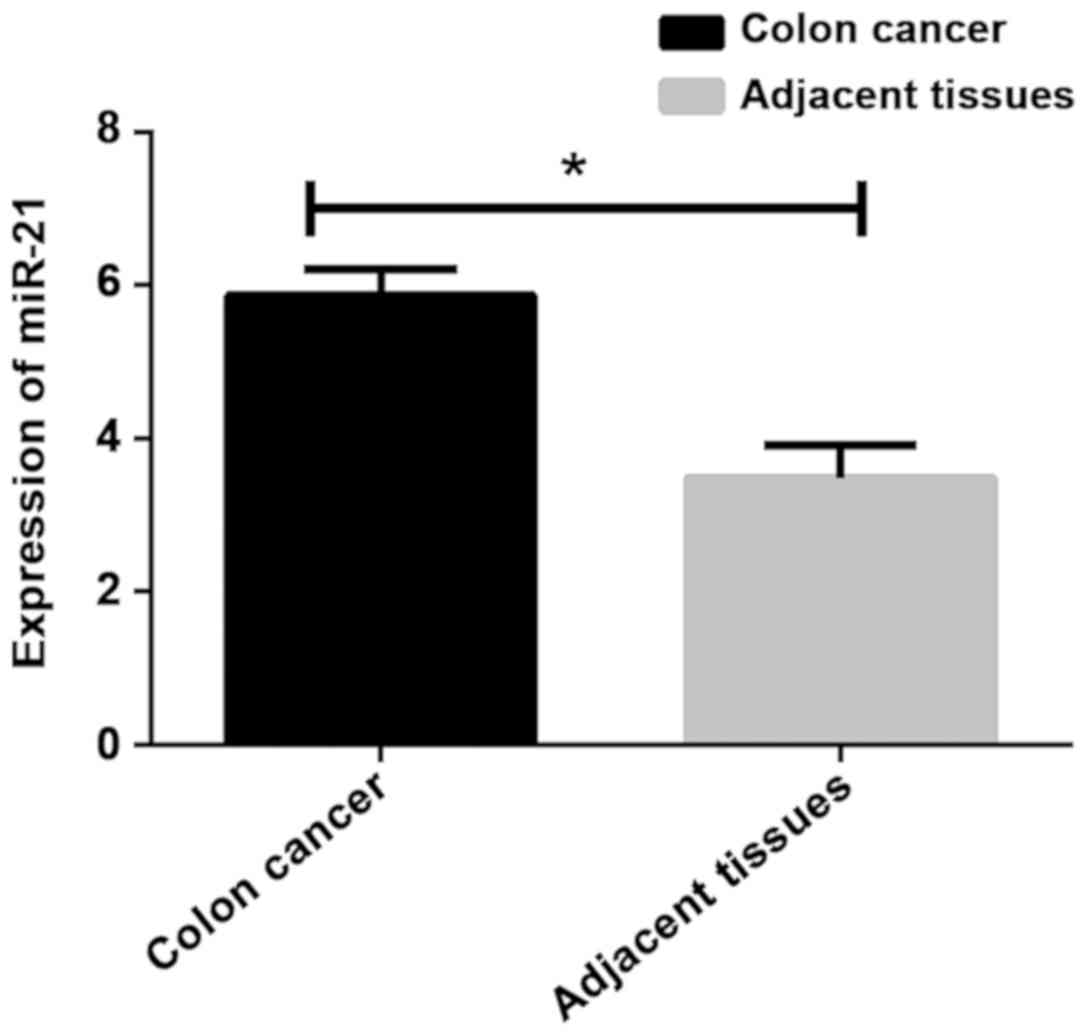
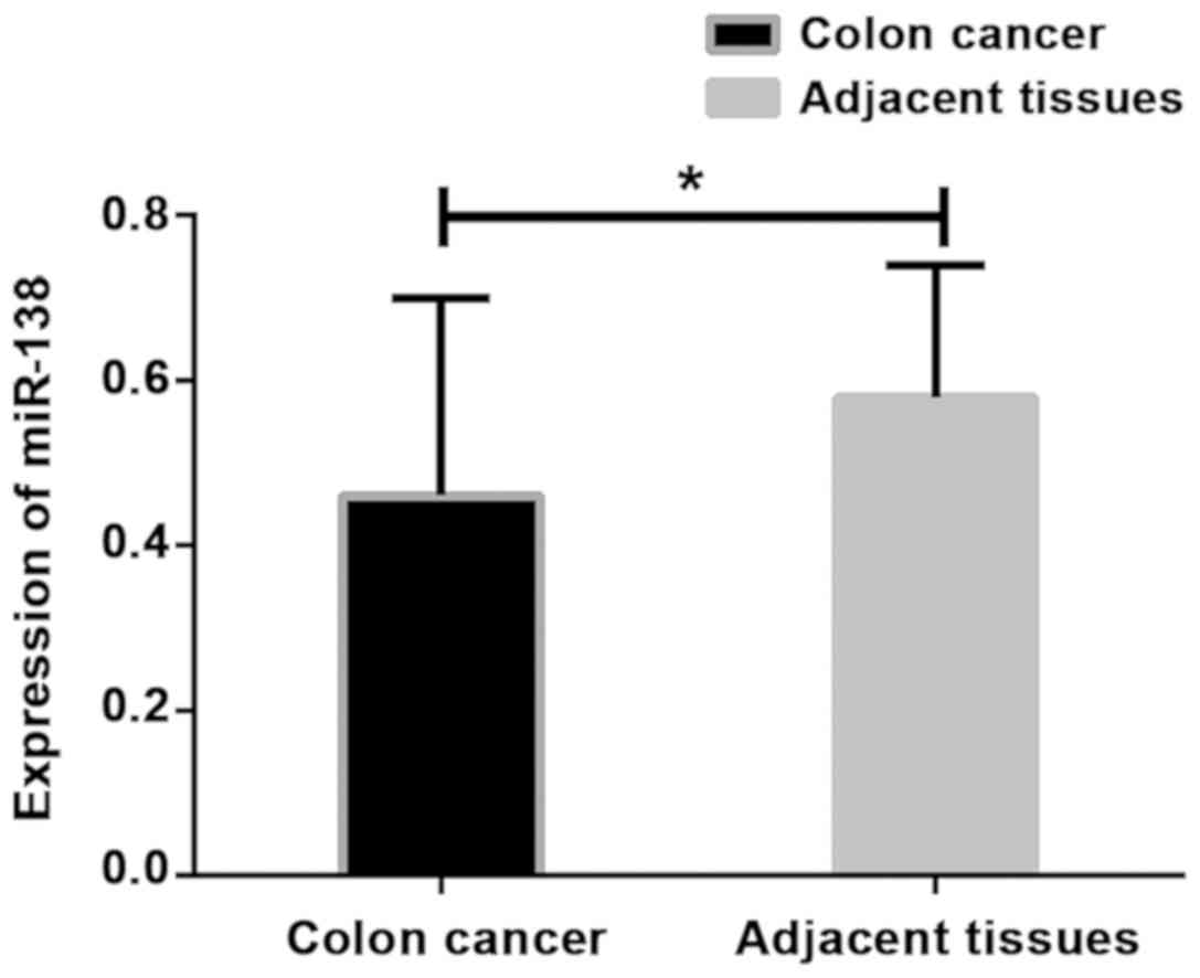
The expression levels of miR-21 and miR-138 were
detected by RT-qPCR. Results showed that the expression level of
miR-21 in colon cancer tissues was significantly higher than that
in adjacent healthy tissues, while the expression level of miR-138
was lower in colon cancer tissues than in adjacent healthy tissues
(P<0.001) (Table II; Figs. 1 and 2).
| Table II.Expression of miR-21 and miR-138 in
colon cancer and adjacent tissues. |
Table II.
Expression of miR-21 and miR-138 in
colon cancer and adjacent tissues.
| Groups | n | miR-21 | t value | P-value | miR-138 | t value | P-value |
|---|
| Colon cancer
tissues | 128 | 5.86±0.34 | 49.120 | <0.001 | 0.46±0.24 | 4.707 | <0.001 |
| Adjacent healthy
tissues | 128 | 3.48±0.43 |
|
| 0.58±0.16 |
|
|
Association between the expression of
miR-21 and miR-138 and clinicopathological features
There was no association between the expression
levels of miR-21 and miR-138 and age and sex (P>0.05). The
expression levels of miR-21 and miR-138 were associated with
differentiation, lymph node metastasis, distant metastasis, and TNM
staging (P<0.05) (Table
III).
| Table III.Association between the expressions of
miR-21 and miR-138 and clinicopathological features. |
Table III.
Association between the expressions of
miR-21 and miR-138 and clinicopathological features.
| Variables | n (%) | miR-21 | t value | P-value | miR-138 | t value | P-value |
|---|
| Age (years) |
|
| 0.438 | 0.662 |
| 0.622 | 0.535 |
| ≥45 | 76 (59.4) | 5.47±2.06 |
|
| 0.48±0.12 |
|
|
|
<45 | 52 (40.6) | 5.31±1.98 |
|
| 0.46±0.24 |
|
|
| Sex |
|
| 0.408 | 0.684 |
| 1.220 | 0.225 |
| Male | 68 (53.1) | 5.78±2.47 |
|
| 0.43±0.16 |
|
|
|
Female | 60 (46.9) | 5.64±1.05 |
|
| 0.47±0.21 |
|
|
|
Differentiation |
|
| 2.000 | 0.048 |
| 3.541 | 0.001 |
|
Low-moderate | 89 (69.5) | 5.08±2.13 |
|
| 0.48±0.15 |
|
|
|
High | 39 (30.5) | 5.89±2.06 |
|
| 0.38±0.14 |
|
|
| Lymph node
metastasis |
|
| 3.237 | 0.002 |
| 2.094 | 0.038 |
|
Yes | 62 (48.4) | 5.86±1.64 |
|
| 0.42±0.23 |
|
|
| No | 66 (51.6) | 5.07±1.08 |
|
| 0.49±0.14 |
|
|
| Distant
metastasis |
|
| 3.786 | <0.001 |
| 4.051 | <0.001 |
|
Yes | 48 (37.5) | 6.18±2.13 |
|
| 0.42±0.17 |
|
|
| No | 80 (62.5) | 5.04±1.28 |
|
| 0.57±0.22 |
|
|
| TNM stage |
|
| 3.681 | <0.001 |
| 3.818 | <0.001 |
|
I+II | 84 (65.6) | 5.01±1.47 |
|
| 0.52±0.14 |
|
|
|
III+IV | 44 (34.4) | 6.14±1.95 |
|
| 0.41±0.18 |
|
|
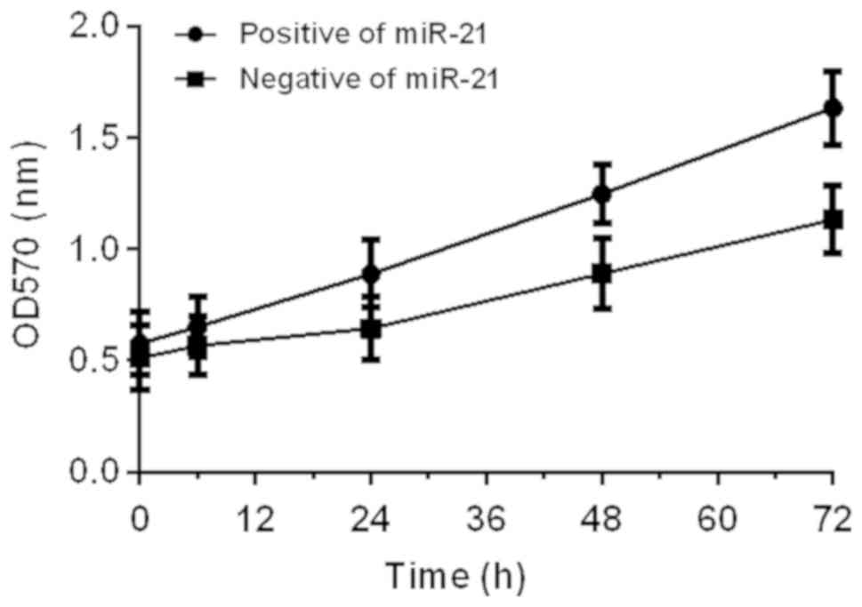
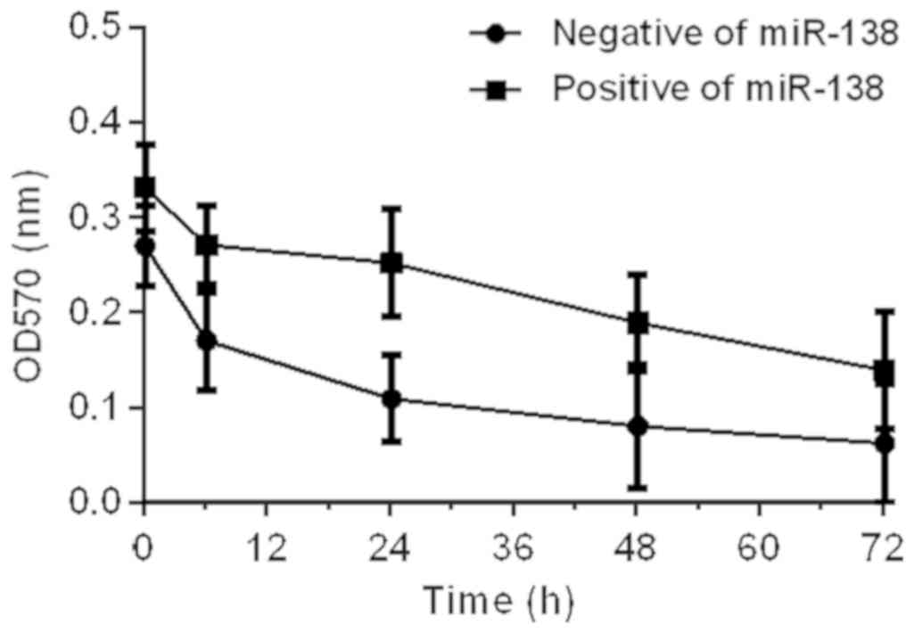
Effects of miR-21 and miR-138 on
proliferation of colon cancer cells
Effects of miR-21 and miR-138 on proliferation of
colon cancer SW480 cells were detected by MTT assay. Results showed
that miR-21 could promote cell proliferation. OD values at 6, 24,
48, and 72 h after miR-21 expression vector transfection (positive
group A) were higher than those of cells with blank vector
transfection (negative group B). miR-138 inhibited cell
proliferation, and OD values at 6, 24, 48, and 72 h after miR-1338
expression vector transfection (positive group A) were lower than
those of cells with blank vector transfection (negative group B)
(P<0.05) (Figs. 3 and 4).
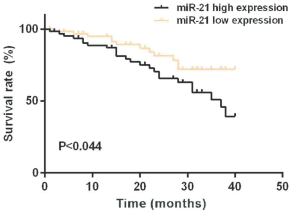
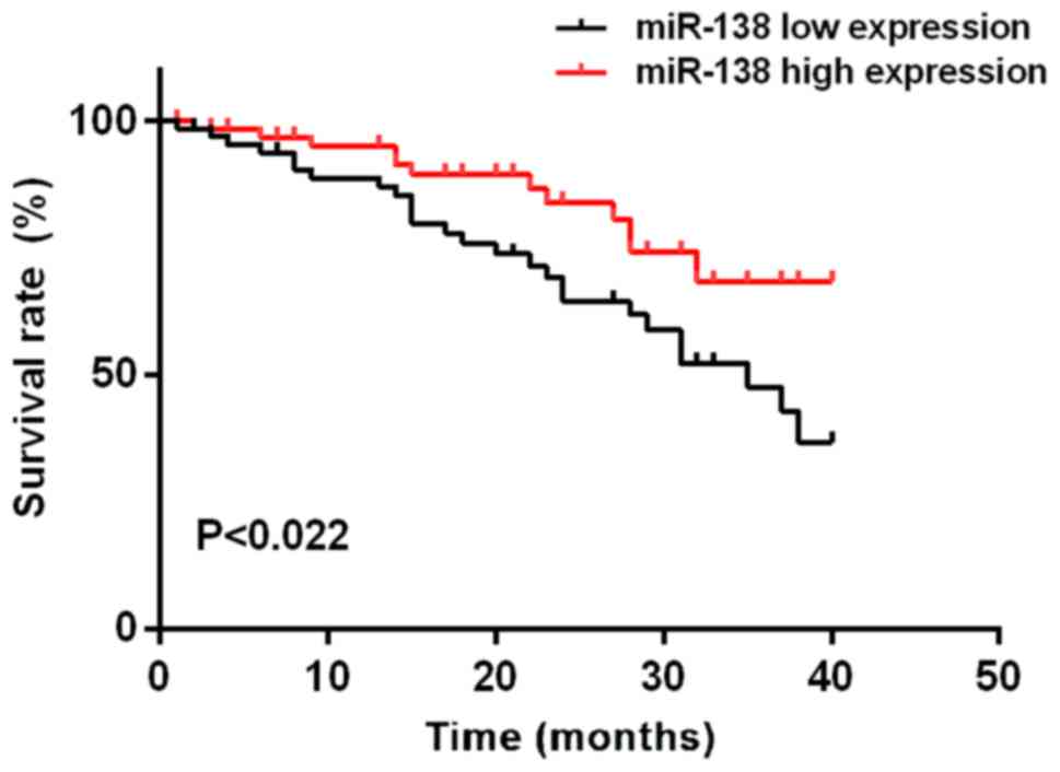
Survival analysis
The median expression level of miR-21 in colon
cancer tissues was 5.595. According to the median expression level
of miR-21, patients were divided into high expression group (n=64)
and low expression group (n=64). The median expression level of
miR-138 in colon cancer tissues was 0.483. According to the median
expression level of miR-138, patients were divided into high
expression group (n=64) and low expression group (n=64). Mean
survival time of the miR-21 low expression group was 35.46±1.08
months, and the mean survival time of the miR-21 high expression
group was 30.82±2.84 months. Mean survival time of the miR-138 low
expression group was 34.16±1.88 months, and the mean survival time
of the miR-138 high expression group was 36.86±2.02 months.
Survival analysis showed that the survival time of patients with
high expression of miR-21 was significantly shorter than that of
patients with low expression of miR-21. The survival time of
miR-138 high expression group was longer than that of miR-138 low
expression group (log-rank, P<0.05). Patient's survival was
negatively associated with the level of miR-21 expression.
Univariate prognostic analysis showed that the factors affecting
the prognosis of patients with colon cancer include miR-21 and
miR-138 expression, tissue differentiation, TNM staging, and lymph
node metastasis. Cox multivariate regression analysis showed that
miR-21, miR-138 expression, and TNM staging were independent risk
factors for poor prognosis of colon cancer (Figs. 5 and 6;
Tables IV and V).
| Table IV.Univariate analysis of factors
affecting colon cancer. |
Table IV.
Univariate analysis of factors
affecting colon cancer.
|
| miR-21 | miR-138 |
|---|
|
|
|
|
|---|
| Variables | P-value | HR (95% CI) | P-value | HR (95% CI) |
|---|
| Expression level
(low vs. high) | <0.001 | 38.842
(15.203–96.215) | <0.001 | 32.562
(13.524–87.347) |
| Sex (male vs.
female) | 0.559 | 1.012
(0.797–1.620) | 0.845 | 1.062
(0.523–1.946) |
| Age (<45 years
vs. ≥45 years) | 0.114 | 1.132
(1.028–1.715) | 0.173 | 0.986
(0.946–1.042) |
| TNM stage (I and II
vs. III and IV) | 0.013 | 2.821
(1.346–2.857) | 0.013 | 4.182
(2.152–7.864) |
| Lymph node
metastasis (yes vs. no) | 0.011 | 3.053
(1.282–7.323) | 0.004 | 2.452
(1.265–4.124) |
| Differentiation
(low vs. high) | 0.025 | 0.921
(0.831–1.525) | 0.032 | 0.042
(0.152–0.945) |
| Table V.Multivariate analysis of association
between miR-21 and miR-138 and colon cancer. |
Table V.
Multivariate analysis of association
between miR-21 and miR-138 and colon cancer.
|
| miR-21 | miR-138 |
|---|
|
|
|
|
|---|
| Variables | P-value | HR (95% CI) | P-value | HR (95% CI) |
|---|
| Expression level
(low vs. high) | 0.034 | 36.842
(15.203–96.215) | 0.016 | 33.542
(13.524–87.347) |
| TNM stage (I and II
vs. III and IV) | 0.028 | 2.867
(1.348–2.857) | 0.021 | 4.325
(2.248–7.856) |
Discussion
With the improvement of people's living standards
and changes in eating habits, the incidence of colon cancer has
shown an increasing trend (19).
Multiple factors, multiple stages, and multiple genetic mutations
lead to the development and progression of colon cancer, and
multiple oncogenes and tumor suppressor genes are involved.
Treatment of colon cancer is currently dominated by comprehensive
treatment, and the most effective one is surgical treatment
combined with targeted chemotherapy before and after surgery. With
the development of molecular biology and cell biology, it has been
found that the expression of oncogenes and tumor suppressor genes
can affect the proliferation and differentiation of tumor cells,
and has a correlation with the occurrence and development of tumors
and prognosis (20–22). At present, it has been confirmed that
at least 400 miRNAs in the human genome are closely related to
tumors (23).
The results of this study showed that the expression
level of miR-21 in colon cancer tissues was significantly higher
than that in adjacent healthy tissues, and the expression level of
miR-138 was lower in colon cancer tissues than in adjacent tissues
(P<0.001). The expression levels of miR-21 and miR-138 were
associated with the degree of differentiation, lymph node
metastasis, distant metastasis, and TNM stage (P<0.05). MTT
results showed that miR-21 could promote cell proliferation. OD
values at 6, 24, 48, and 72 h after miR-21 expression vector
transfection (positive group A) were higher than those of cells
with blank vector transfection (negative group B). miR-138
inhibited cell proliferation, and OD values at 6, 24, 48, and 72 h
after miR-1338 expression vector transfection (positive group A)
were lower than those of cells with blank vector transfection
(negative group B) (P<0.05). Studies on different tumor tissues
and cell lines have shown that miR-21 can promote cell
proliferation (24,25). Capraro et al (26) have shown that miR-138 expression is
low in glioma cells and can significantly inhibit the proliferation
and migration of glioma cells, and thus functions as a tumor
suppressor gene, which is consistent with the findings of the
present study. Therefore, miR-21 may be a potential marker for the
diagnosis and prognosis of colon cancer. miR-138 can inhibit the
proliferation of cancer cells. Survival analysis showed that
survival time of patients with high expression of miR-21 was
significantly shorter than that of patients with low expression of
miR-21. Survival time of miR-138 high expression group was longer
than that of miR-138 low expression group (log-rank, P<0.05).
Cox multivariate regression analysis showed that miR-21 and miR-138
expression, and TNM staging were independent risk factors for poor
prognosis of colon cancer (Table V).
Thus, miR-21 can predict the recurrence of colon cancer, and it is
also one of the risk factors for the progression of colon cancer.
Expression of miR-21 was positively associated with the degree of
malignancy and the risk of disease progression, and was negatively
associated with the survival of patients, while miR-138 is the
opposite. Expression of miR-21 and miR-138 in the pathological
specimens of patients with colon cancer is an index that can
determine the prognosis of colon cancer. Combination of routine
postoperative immunohistochemical pathological examination and
detection of miR-21 and miR-138 expression may improve the
diagnosis of colon cancer and is worthy of clinical
application.
Our study has also some limitations. The sample size
was small, and the patients were from a single hospital, which may
affect our conclusions. Our study only investigated the effects of
the two miRNAs on colon cancer cell proliferation, while the
involvement of other genes and interactions with other factors were
not studied. Further studies are needed to explore the mechanism
and the roles of those two miRNAs.
In conclusion, miR-21 is highly expressed in colon
cancer tissues, and is positively associated with the degree of
malignancy of patients and negatively associated with survival.
miR-138 expression is low in colon cancer tissues, and negatively
associates with the degree of malignancy of patients and positively
associated with survival. miR-21 and miR-138 may be involved in the
regulation of colon cancer cell proliferation. Our study provides
references for clinical diagnosis, treatment and prognosis of colon
cancer.
Acknowledgements
Not applicable.
Funding
This study was supported by the Science and
Technology Project of Guangdong Province (no. 2017B090901067) and
the Foundation of Social Development Project of the Science and
Technology Department of Jiangsu Province (BE2015719).
Availability of data and materials
The datasets used and/or analyzed during the present
study are available from the corresponding author on reasonable
request.
Authors' contributions
CY wrote the manuscript and analyzed the follow-up
data. QX and LJ extracted total RNA. QX and BS assisted with
reverse transcription. XJ performed PCR and XH was responsible for
MTT assay. All authors read and approved the final manuscript.
Ethics approval and consent to
participate
The study was approved by the Ethics Committee of
Nanfang Hospital (Guangzhou, China). Signed informed consents were
obtained from the patients or the guardians.
Patient consent for publication
Not applicable.
Competing interests
The authors declare that they have no competing
interests.
References
|
1
|
Ferlay J, Soerjomataram I, Dikshit R, Eser
S, Mathers C, Rebelo M, Parkin DM, Forman D and Bray F: Cancer
incidence and mortality worldwide: Sources, methods and major
patterns in GLOBOCAN 2012. Int J Cancer. 136:E359–E386. 2015.
View Article : Google Scholar : PubMed/NCBI
|
|
2
|
Sung JJ, Lau JY, Goh KL and Leung WK: Asia
Pacific Working Group on Colorectal Cancer: Increasing incidence of
colorectal cancer in Asia: Implications for screening. Lancet
Oncol. 6:871–876. 2005. View Article : Google Scholar : PubMed/NCBI
|
|
3
|
Bae JM, Cho NY, Kim TY and Kang GH:
Clinicopathologic and molecular characteristics of synchronous
colorectal cancers: Heterogeneity of clinical outcome depending on
microsatellite instability status of individual tumors. Dis Colon
Rectum. 55:181–190. 2012. View Article : Google Scholar : PubMed/NCBI
|
|
4
|
Hu H, Chang DT, Nikiforova MN, Kuan SF and
Pai RK: Clinicopathologic features of synchronous colorectal
carcinoma: A distinct subset arising from multiple sessile serrated
adenomas and associated with high levels of microsatellite
instability and favorable prognosis. Am J Surg Pathol.
37:1660–1670. 2013. View Article : Google Scholar : PubMed/NCBI
|
|
5
|
Gong C, Yao Y, Wang Y, Liu B, Wu W, Chen
J, Su F, Yao H and Song E: Up-regulation of miR-21 mediates
resistance to trastuzumab therapy for breast cancer. J Biol Chem.
286:19127–19137. 2011. View Article : Google Scholar : PubMed/NCBI
|
|
6
|
Ferlay J, Shin HR, Bray F, Forman D,
Mathers C and Parkin DM: Estimates of worldwide burden of cancer in
2008: GLOBOCAN 2008. Int J Cancer. 127:2893–2917. 2010. View Article : Google Scholar : PubMed/NCBI
|
|
7
|
Simoglou C, Gymnopoulou E, Simoglou L,
Gymnopoulou M, Nikolaou K and Gymnopoulos D: Surgery for colorectal
cancer in the small town of Komotini. J Multidiscip Healthc.
5:273–276. 2012. View Article : Google Scholar : PubMed/NCBI
|
|
8
|
Siegel R, Naishadham D and Jemal A: Cancer
statistics for Hispanics/Latinos, 2012. CA Cancer J Clin.
62:283–298. 2012. View Article : Google Scholar : PubMed/NCBI
|
|
9
|
Garalde DR, Snell EA, Jachimowicz D, Sipos
B, Lloyd JH, Bruce M, Pantic N, Admassu T, James P, Warland A, et
al: Highly parallel direct RNA sequencing on an array of nanopores.
Nat Methods. 15:201–206. 2018. View Article : Google Scholar : PubMed/NCBI
|
|
10
|
Hollis M, Nair K, Vyas A, Chaturvedi LS,
Gambhir S and Vyas D: MicroRNAs potential utility in colon cancer:
Early detection, prognosis, and chemosensitivity. World J
Gastroenterol. 21:8284–8292. 2015. View Article : Google Scholar : PubMed/NCBI
|
|
11
|
Ribas J and Lupold SE: The transcriptional
regulation of miR-21, its multiple transcripts, and their
implication in prostate cancer. Cell Cycle. 9:923–929. 2010.
View Article : Google Scholar : PubMed/NCBI
|
|
12
|
Zheng Y, Cui L, Sun W, Zhou H, Yuan X, Huo
M, Chen J, Lou Y and Guo J: MicroRNA-21 is a new marker of
circulating tumor cells in gastric cancer patients. Cancer Biomark.
10:71–77. 2011-2012. View Article : Google Scholar
|
|
13
|
Reis ST, Pontes-Junior J, Antunes AA,
Dall'Oglio MF, Dip N, Passerotti CC, Rossini GA, Morais DR,
Nesrallah AJ, Piantino C, et al: miR-21 may acts as an oncomir by
targeting RECK, a matrix metalloproteinase regulator, in prostate
cancer. BMC Urol. 12:142012. View Article : Google Scholar : PubMed/NCBI
|
|
14
|
Lagos-Quintana M, Rauhut R, Yalcin A,
Meyer J, Lendeckel W and Tuschl T: Identification of
tissue-specific microRNAs from mouse. Curr Biol. 12:735–739. 2002.
View Article : Google Scholar : PubMed/NCBI
|
|
15
|
Griffiths-Jones S: The microRNA registry.
Nucleic Acids Res. 32:D109–D111. 2004. View Article : Google Scholar : PubMed/NCBI
|
|
16
|
Na YJ, Sung JH, Lee SC, Lee YJ, Choi YJ,
Park WY, Shin HS and Kim JH: Comprehensive analysis of
microRNA-mRNA co-expression in circadian rhythm. Exp Mol Med.
41:638–647. 2009. View Article : Google Scholar : PubMed/NCBI
|
|
17
|
Yu ST, Chen L, Wang HJ, Tang XD, Fang DC
and Yang SM: hTERT promotes the invasion of telomerase-negative
tumor cells in vitro. Int J Oncol. 35:329–336.
2009.PubMed/NCBI
|
|
18
|
Livak KJ and Schmittgen TD: Analysis of
relative gene expression data using real-time quantitative PCR and
the 2(-Delta Delta C(T)) method. Methods. 25:402–408. 2001.
View Article : Google Scholar : PubMed/NCBI
|
|
19
|
Ji BC, Yu CC, Yang ST, Hsia TC, Yang JS,
Lai KC, Ko YC, Lin JJ, Lai TY and Chung JG: Induction of DNA damage
by deguelin is mediated through reducing DNA repair genes in human
non-small cell lung cancer NCI-H460 cells. Oncol Rep. 27:959–964.
2012. View Article : Google Scholar : PubMed/NCBI
|
|
20
|
Fish JE, Santoro MM, Morton SU, Yu S, Yeh
RF, Wythe JD, Ivey KN, Bruneau BG, Stainier DY and Srivastava D:
miR-126 regulates angiogenic signaling and vascular integrity. Dev
Cell. 15:272–284. 2008. View Article : Google Scholar : PubMed/NCBI
|
|
21
|
Wang S, Aurora AB, Johnson BA, Qi X,
McAnally J, Hill JA, Richardson JA, Bassel-Duby R and Olson EN: The
endothelial-specific microRNA miR-126 governs vascular integrity
and angiogenesis. Dev Cell. 15:261–271. 2008. View Article : Google Scholar : PubMed/NCBI
|
|
22
|
Png KJ, Halberg N, Yoshida M and Tavazoie
SF: A microRNA regulon that mediates endothelial recruitment and
metastasis by cancer cells. Nature. 481:190–194. 2011. View Article : Google Scholar : PubMed/NCBI
|
|
23
|
Pillai RS: MicroRNA function: Multiple
mechanisms for a tiny RNA? RNA. 11:1753–1761. 2005. View Article : Google Scholar : PubMed/NCBI
|
|
24
|
Iorio MV, Ferracin M, Liu CG, Veronese A,
Spizzo R, Sabbioni S, Magri E, Pedriali M, Fabbri M, Campiglio M,
et al: MicroRNA gene expression deregulation in human breast
cancer. Cancer Res. 65:7065–7070. 2005. View Article : Google Scholar : PubMed/NCBI
|
|
25
|
Buscaglia LE and Li Y: Apoptosis and the
target genes of microRNA-21. Chin J Cancer. 30:371–380. 2011.
View Article : Google Scholar : PubMed/NCBI
|
|
26
|
Capraro V, Zane L, Poncet D, Perol D,
Galia P, Preudhomme C, Bonnefoy-Berard N, Gilson E, Thomas X and
El-Hamri M: Telomere deregulations possess cytogenetic, phenotype,
and prognostic specificities in acute leukemias. Exp Hematol.
39:195–202.e2. 2011. View Article : Google Scholar : PubMed/NCBI
|