Introduction
Bladder cancer is the seventh most common type of
cancer in the male population worldwide, and eleventh including
females (1). There are 2 major types
of bladder cancer: Non-muscle invasive bladder cancer (NMIBC) and
muscle-invasive bladder cancer (MIBC). Bladder cancer exhibits a
high frequency of relapse and a poor clinical outcome once the
tumors progress to muscle-invasive disease (2). Patients with MIBC have a poor prognosis
due to the aggressive nature of the tumor and its resistance to
chemo- and radiotherapy (3).
Furthermore, for patients with MIBC, the risk of developing lymph
node metastases is increased and chemotherapy is less effective in
comparison with patients with NMIBC (4). Therapies and prognosis for this type of
cancer depend on the clinical information and individual tumor
pathology. However, in certain cases, even when tumors present with
similar histology, they respond differently to the same treatment,
resulting in different survival outcomes for the patients (5).
Protein-coding genes constitute only 2% of the total
genome sequence; the remaining sequences produce various classes of
functional non-coding RNAs (6).
There are two major classes of non-coding RNAs, including small and
long non-coding RNAs (lncRNAs). The difference between lncRNAs and
small non-coding RNAs is primarily their size, with the length of
lncRNAs defined as being >200 nucleotides. Similar to other
non-coding RNAs, lncRNA transcripts are associated with a wide
range of cancer types, including bladder cancer (7–10).
Alterations in the expression levels of lncRNAs have been
demonstrated to be associated with a number of important cellular
functions, and may promote the migration and invasion of cancer
cells (4,11). Non-coding RNAs have attracted
increasing attention in previous years regarding their role in
bladder cancer, with the number of studies on this topic increasing
considerably. The combination of traditional methods of treatment
with the use of non-coding RNAs may provide improved therapy for
patients. The accumulation of data in this field may assist in
elucidating the molecular profiles of patients with bladder cancer
and contribute to the use of non-coding RNAs as tools for precision
medicine to target critical genes in bladder cancer.
LINC00460 is a human lncRNA gene, transcribed from
chromosome 13, measuring 935 bp; its function is poorly understood.
Recent studies have demonstrated that LINC00460 is associated with
kidney cancer, nasopharyngeal carcinoma and head and neck squamous
cell carcinoma (12–14). A previous study demonstrated, using a
bioinformatics approach, that LINC00460 may serve an important role
in tumorigenesis and metastasis through the regulation of the cell
cycle and cell death (15). During
the analysis of the differential expression of lncRNAs in bladder
cancer tissues in the present study, it was identified that
LINC00460 was highly upregulated compared with the normal adjacent
tissue. Furthermore, the upregulation of LINC00460 was demonstrated
to be associated with the poor survival of patients. We
hypothesized that this lncRNA may serve an important role in the
regulation of biological processes in bladder urothelial carcinoma.
However, the precise function and the underlying molecular
mechanisms remain to be elucidated.
Bladder cancer predominantly presents in men
(16). Factors that are exclusive to
males are likely to serve critical roles in the development of
bladder cancer (17,18). An increasing amount of evidence has
suggested the involvement of androgen receptor (AR) signaling in
the development and progression of bladder cancer (19–21). The
present study investigated the role of the lncRNA LINC00460 in
bladder urothelial carcinoma using The Cancer Genome Atlas (TCGA)
data and cell experiments. The results indicated that increased
LINC00460 expression was a characteristic molecular change in
bladder urothelial carcinoma tissues, and in 5637 and T24 cell
lines. Therefore, the effects of aberrant LINC00460 expression on
the biological behavior of 5637 and T24 cells were additionally
investigated. The results provided novel insights into the function
and mechanisms of LINC00460 in bladder urothelial carcinoma
pathogenesis, and identified LINC0046 as a potential therapeutic
target for cancer intervention.
Materials and methods
TCGA database
Gene expression data obtained by RNA sequencing and
the corresponding clinical data for 412 patients (including 413
samples) with bladder urothelial carcinoma were downloaded from
TCGA (https://cancergenome.nih.gov/). All
RNA expression levels of the samples were normalized. The edgeR
Bioconductor package was used to analyze P-values and fold-change
(FC) using R (version 3.4.0) (22).
A gene was defined as differentially expressed between cancerous
and normal tissues when the false discovery rate-adjusted P<0.01
and the FC was ≥2-fold increased or decreased.
Cell culture and transfection
The expression of LINC00460 and the basic
characterization of bladder urothelial carcinoma cell lines were
initially investigated using the Expression Atlas database
(23) and the American Type Culture
Collection website (https://www.atcc.org/), respectively (Table I). Based on these results, the 5637
cell line was selected, as the expression of LINC00460 was the
highest in these cells compared with the others included in the
analysis. From the verification of LINC00460 expression using
reverse transcription-quantitative polymerase chain reaction
(RT-qPCR) in bladder cancer cell lines (T24, J82, TCCSUP and
UM-UC-3), it was identified that LINC00460 was also upregulated in
the T24 cell line compared with the normal bladder epithelial
SV-HUC-1 cell line (data not show).
 | Table I.Expression of LINC00460 and
characterization of bladder cancer cell lines from the Expression
Atlas database and the website of American Type Culture Collection
cell lines. |
Table I.
Expression of LINC00460 and
characterization of bladder cancer cell lines from the Expression
Atlas database and the website of American Type Culture Collection
cell lines.
| Cell line | Expression value,
transcripts per million | Histological
grade | Age | Sex |
|---|
| 5637 | 51 | Grade 2 | 68 | Male |
| U-BLC1 | 43 | Grade 3 | 84 | Female |
| HT-1197 | 11 | Grade 4 | 44 | Male |
| TCCSUP | 4 | Grade 4 | 67 | Female |
| J82 | 3 | Grade 3 | 58 | Male |
| RT-112 | 0.9 | Grade 2 | Not reported | Female |
| 253J | 0 | Grade 4 | 53 | Male |
| HT-1376 | 0 | Grade 3 | 58 | Female |
| RT4 | 0 | Grade 1 | 63 | Male |
| SW780 | 0 | Grade 1 | 80 | Female |
| T24 | 0 | Grade 3 | 81 | Female |
The 5637, T24, J82, TCCSUP, UM-UC-3 and SV-HUC-1
cells were purchased from The Cell Bank of Type Culture Collection
of Chinese Academy of Sciences (Shanghai, China). The 5637 and T24
cells were cultured in RPMI-1640 medium (Gibco; Thermo Fisher
Scientific, Inc., Waltham, MA, USA) and the SV-HUC-1 cell line was
cultured in F12 K medium (Gibco; Thermo Fisher Scientific, Inc.).
J82, TCCSUP and UM-UC-3 cells were cultured in Dulbecco's modified
Eagle's medium (Gibco; Thermo Fisher Scientific, Inc.). These media
were supplemented with 10% fetal bovine serum (Gibco, Thermo Fisher
Scientific, Inc.) and 1% penicillin and streptomycin, in a
humidified atmosphere with 5% CO2 at 37°C.
A total of three short hairpin RNAs (shRNAs)
targeting LINC00460 were purchased from Shanghai GenePharma Co.,
Ltd. (Shanghai, China) and transfected into 5637 and T24 cells with
Lipofectamine® 3000 (Invitrogen; Thermo Fisher
Scientific, Inc.) according to the manufacturer's protocol. The
three shRNA sequences specially targeting LINC00460 were designed
and cloned into a pGU6/green fluorescent protein (GFP)/Neo-shRNA
vector (Shanghai GenePharma Co., Ltd.). Cells (1×10⁵ T24
cells/well; 3×10⁵ 5,637 cells/well) were seeded in
six-well culture plates and transfected with the 2.5 µg
sh-LINC00460 in each well. The most effective shRNA sequence (sh-3)
in achieving knockdown of LINC00460 expression was selected for
subsequent experiments 48 h after the transfection. The
sh-LINC00460 sequences were as follows: sh-1,
5′-GCTAAGACCTAATAGCCAATA-3′; sh-2, 5′-GCCATCCACTTCAAAGTATTC-3′; and
sh-3, 5′-ACCTTGGTCAAACGTTTAACC-3′. A scrambled shRNA was used as
the negative control (sh-NC) in the experiments with the following
sequence: 5′-GTTCTCCGAACGTGTCACGT-3′.
RT-qPCR assays
Total RNA was extracted from 5637, T24, J82, TCCSUP,
UM-UC-3 and SV-HUC-1 cells using TRIzol® reagent (Thermo
Fisher Scientific, Inc.). Reverse transcription was conducted using
the PrimeScript™ RT reagent kit with gDNA Eraser (Takara
Biotechnology Co., Ltd., Dalian, China). qPCR was performed to
detect the expression of LINC00460 using SYBR® Premix Ex
Taq™ II kit (Takara Biotechnology Co., Ltd.). RT-qPCR assays were
performed using an Applied Biosystems QuantStudio 3 system (Applied
Biosystems; Thermo Fisher Scientific, Inc.), with β-actin as an
endogenous control. The primer sequences used were as follows:
LINC00460 forward, 5′-CGAGAAGGCCACCTATGAGC-3′ and reverse,
5′-TGAAGTGGATGGCTCAGGAA-3′; β-actin forward,
5′-CCGTGAAAAGATGACCCAGATC-3′ and reverse,
5′-CACAGCCTGGATGGCTACGT-3′. A two-step qPCR was performed as
follows: Initial denaturation in 95°C for 30 sec; 40 of cycles of
denaturation at 95°C for 5 sec; and annealing and elongation at
60°C for 34 sec. The 2−ΔΔCq method was used to quantify
LINC00460 (24).
Cell proliferation assay
Cells were seeded in 96-well plates at a density of
1.0×10⁴ and 1.5×10⁴ cells per well for T24
and 5637 cells, respectively. Following incubation at 37°C with 5%
CO2 for 0, 24, 48 and 72 h, the absorbance of each
sample was measured at 450 nm. Cell proliferation was evaluated
using a Cell Counting Kit-8 (CCK-8) assay (Dojindo Molecular
Technologies, Inc., Shanghai, China) according to the
manufacturer's protocol.
Cell migration assay
The 5637 and T24 cells were seeded (1×10⁶
cells/plate) in 6-well plates and incubated in serum-free RPMI-1640
medium. A wound was produced using a sterile 100 µl pipette tip
when a confluent cell monolayer had formed. The size of the wound
was measured using an inverted light microscope (magnification,
×200) and images were captured at 0 and 24 h.
Statistical analysis
SPSS 23.0 software (IBM Corp., Armonk, NY, USA) was
used to perform statistical analysis. The differential expression
levels of LINC00460 and AR between the cancerous and adjacent
tissues were analyzed with t-tests. The differences in LINC00460
expression between bladder cancer 5637 and T24 cell lines and the
normal bladder epithelial SV-HUC-1 cell line was assessed using a
one-way analysis of variance with Tamhane's post-hoc test. The
correlation between LINC00460 and AR was analyzed by Spearman's
rank correlation analysis following logarithmic (log10)
conversion of the original data. For the survival analysis, the
LINC00460 expression data matrix and clinical data files were
matched for each sample using the sample ID. The samples were
divided into the LINC00460-low and LINC00460-high groups based on
the cut-off of the median value, and according to sex. The
LINC00460-high group contained the samples with an exact median
value. A Kaplan-Meier plot was generated using the survival package
in R (version 3.4.0) with log-rank tests. P<0.05 was considered
to indicate a statistically significant difference.
Results
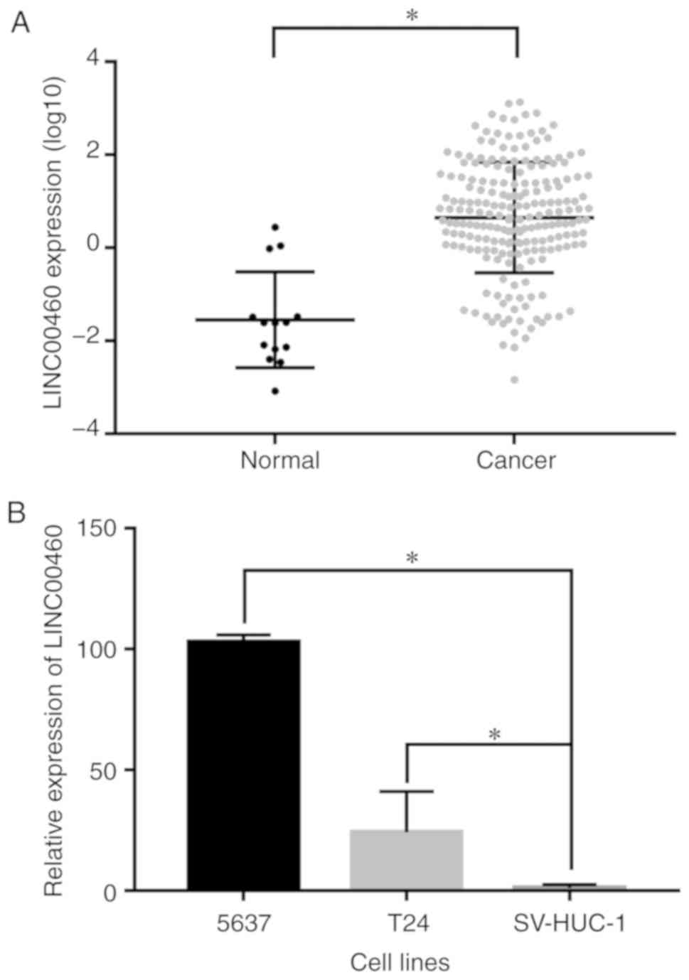
LINC00460 is upregulated in bladder
urothelial carcinoma tissues and cell lines
The differences in LINC00460 expression between the
bladder urothelial carcinoma tissues and the normal controls were
analyzed using TCGA data. LINC00460 was significantly upregulated
in bladder urothelial carcinoma tissues compared with the normal
controls (P<0.0001; Fig. 1A). The
expression level of LINC00460 was detected by RT-qPCR in bladder
urothelial carcinoma and normal bladder epithelial cell lines. An
increase in LINC00460 expression was observed in bladder cancer
5637 and T24 cell lines compared with the normal bladder epithelial
SV-HUC-1 cell line (P<0.05; Fig.
1B).
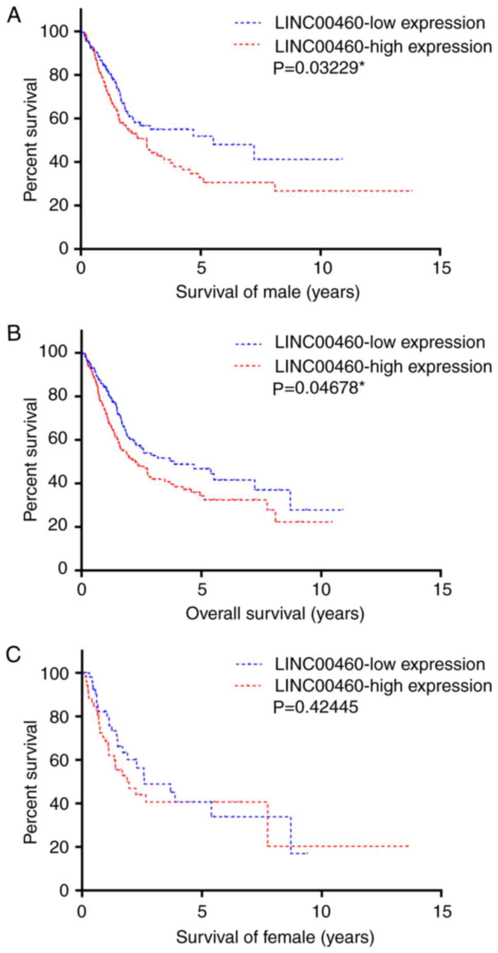
Upregulation of LINC00460 is
associated with poor survival
LINC00460 expression levels and the clinical data of
412 patients with bladder urothelial carcinoma were downloaded from
TCGA. All patients, male and female, were divided into
LINC00460-high and LINC00460-low groups using the mean expression
value as the cut-off. The survival time was plotted using a
Kaplan-Meier curve stratified by LINC00460-high and LINC00460-low
groups. High levels of LINC00460 were significantly associated with
a decreased survival time in male patients (P=0.03229; Fig. 2A). There was a similar association
observed in the overall group (P=0.04678; Fig. 2B), but not among the female patients
(P=0.42445; Fig. 2C).
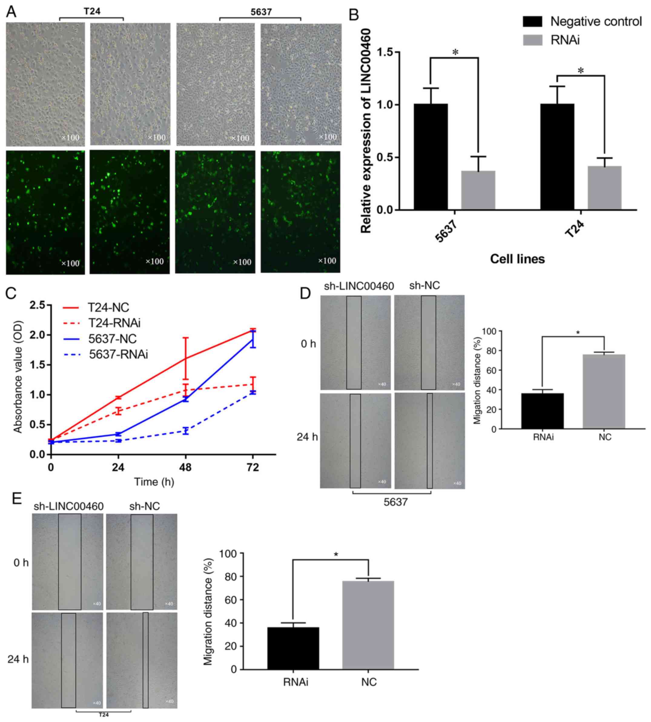
Downregulation of LINC00460 inhibits
the proliferation and migration of bladder urothelial carcinoma
cells
To explore the function of LINC00460, an shRNA
targeting LINC00460 was transfected into 5637 and T24 cells. GFP
visualization indicated that the shRNA was efficiently transfected
into cells (Fig. 3A). RT-qPCR
indicated that LINC00460 was significantly downregulated at 48 h
after transfection of shRNA in the 5637 and T24 cell lines compared
with the control group (P<0.05; Fig.
3B). A CCK-8 assay demonstrated that the downregulation of
LINC00460 inhibited the proliferation of 5637 and T24 cells in
vitro (P<0.05; Fig. 3C). In
addition, the effect of LINC00460 on the migration capacity of 5637
and T24 cells was observed via a wound-healing assay. The
wound-healing assay revealed that the knockdown of LINC00460
decreased the migration distance of cells (Fig. 3D and E). Overall, the results
demonstrated that the silencing of LINC00460 may inhibit the
proliferation and migration abilities of 5637 and T24 cells.
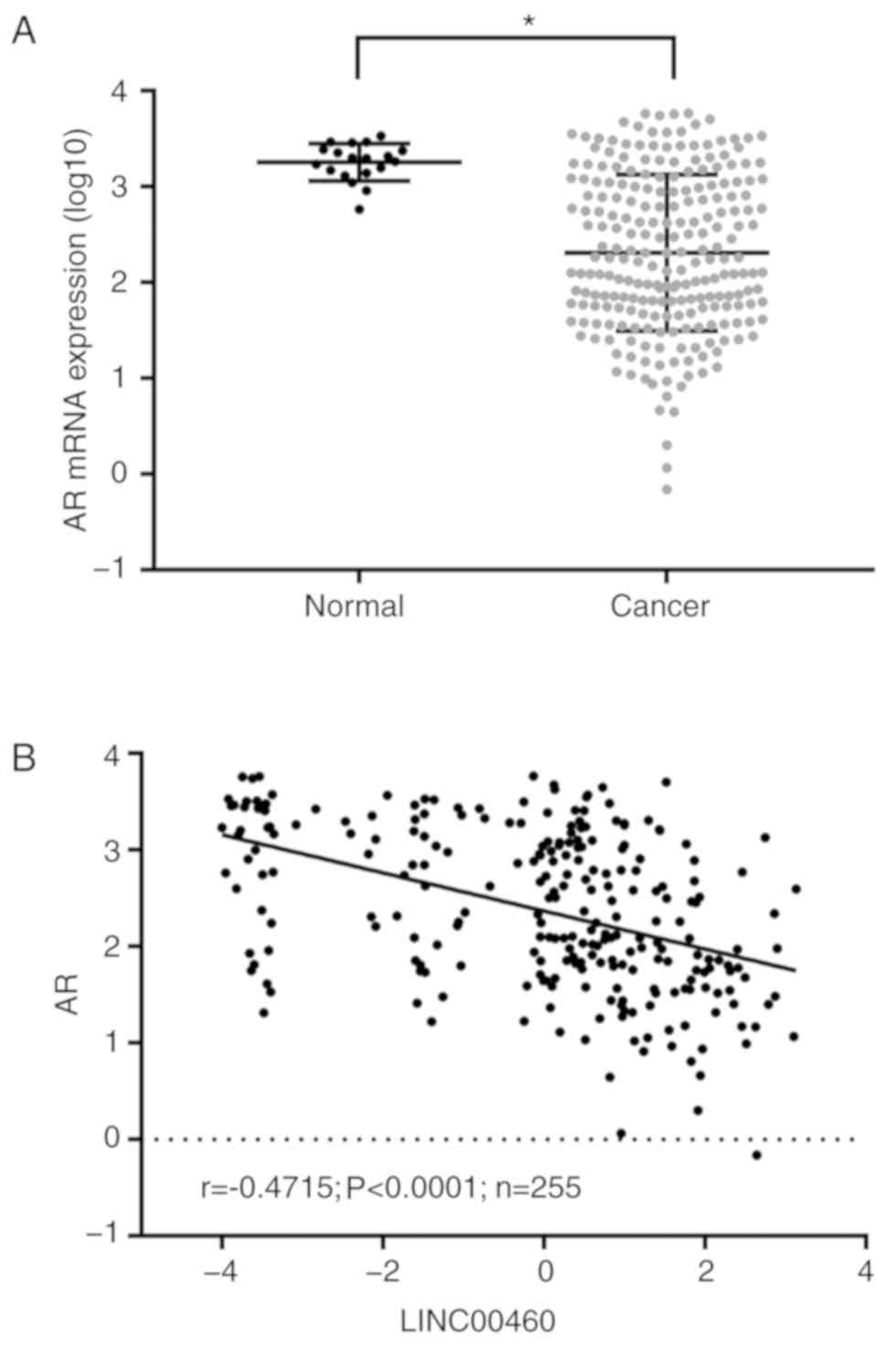
AR is downregulated in bladder
urothelial carcinoma tissues and is negatively correlated with
LINC00460 expression
AR expression was analyzed in bladder urothelial
carcinoma and adjacent tissues, and its correlation with LINC00460
expression, using TCGA data. AR was significantly downregulated in
bladder urothelial carcinoma tissues compared with the normal
tissues (P<0.05; Fig. 4A) and was
negatively correlated with the expression of LINC00460
(P<0.0001; r=−0.4715; Fig.
4B).
Discussion
The majority of cases of bladder cancer are
non-muscle invasive at the time of initial diagnosis, and the
initial treatment options generally focus on tumor resection, and
the prevention of recurrence or progression to muscle-invasive
disease (25). The 5-year
recurrence-free survival rate ranges from 62–89% for localized
muscle invasive bladder cancer following a radical cystectomy
(26). However, for patients with
metastatic disease, the 5-year survival is decreased, at ~5%
(27). The standard drugs used in
chemotherapy for the perioperative therapy for MIBC and metastatic
disease are cisplatin-based. The median overall survival time with
chemotherapy treatment is slightly >1 year, with an objective
response rate of 40–60% (28–30). In
May 2016, atezolizumab was approved as a second-line therapy for
patients with locally advanced or metastatic urothelial carcinoma
that had progressed during or following platinum-containing
chemotherapy; it became the first available immunotherapeutic
antibody to target programmed death-ligand 1. Although targeted
therapies have become standard for numerous other malignancies, the
number of approved targeted agents in bladder cancer is limited.
lncRNAs modulate the expression of genes that are pivotal in the
pathways associated with bladder cancer development and
progression; for example, the HOX transcript antisense RNA/zinc
finger E-box binding homeobox 1 (ZEB1) interaction affects
epithelial-mesenchymal transition in bladder cancer cell lines
(31), and metastasis associated
lung adenocarcinoma transcript 1/Epithelial cadherin (32) and urothelial cancer associated
1/ZEB1/zinc finger E-box binding homeobox 2 (33) interact to affect the invasion and
metastasis of bladder cancer cells. Data from the Cancer Research
Network (CRN) database (34)
indicated that LINC00460 is upregulated in 14 types of cancer,
suggesting that LINC00460 may be a regulator in cancer cell
development (35). The role of
LINC00460 in bladder cancer was selected for analysis in the
present study, as the expression fold change in bladder urothelial
carcinoma was the highest among all the types of cancer included in
the CRN database.
The present study explored the role of LINC00460 in
bladder urothelial carcinoma using TCGA data and cell experiments.
The results demonstrated that LINC00460 was upregulated in bladder
cancer tissues compared with the corresponding normal controls,
while the effect of LINC00460 on the prognosis for bladder
urothelial carcinoma was only observed in male patients.
Concomitantly, the results also indicated that AR was downregulated
in bladder urothelial carcinoma tissues, which was negatively
correlated with LINC00460 expression. Epidemiological and clinical
data have suggested that males are 3–4 times more likely to develop
bladder urothelial carcinoma compared with females (16,36).
Emerging preclinical evidence has indicated the involvement of AR
signaling in the development and progression of bladder urothelial
carcinoma; androgens, β-catenin, cluster of differentiation 24,
cyclins, epidermal growth factor receptor/receptor tyrosine-protein
kinase erbB-2, ETS domain-containing protein Elk-1, several AR
coregulators and orphan receptors have been demonstrated to
directly or indirectly modulate several molecules/pathways involved
in bladder urothelial carcinoma cell proliferation (19,20,37–40). As
a result, we hypothesized that LINC00460 may serve its oncogenic
role by regulating the expression of AR.
The effects of LINC00460 on the proliferation and
migration of 5637 and T24 cells were determined using gain- and
loss-of-function approaches. The data revealed that the
downregulation of LINC00460 inhibited the proliferation and
migration of 5637 and T24 cells. Zhang et al (15) performed a regulatory network analysis
of LINC00460, and the results indicated that LINC00460 was
associated with various biological processes, consistent with the
results from the present study.
However, the function of LINC00460 was only
investigated in bladder urothelial carcinoma cell lines. An in
vivo model is required to confirm the results. In addition, the
mechanisms underlying the effect of LINC00460 on 5637 and T24 cells
are not yet fully characterized. There was a recurring node,
double-strand-break repair protein rad21 homolog, between mRNAs and
transcription factors associated with LINC00460, as identified
through bioinformatics methods in a study conducted by Zhang et
al (15), which was previously
demonstrated to affect cell growth in breast cancer (35). The results from the present study
suggested that the expression level of AR mRNA was downregulated in
bladder urothelial carcinoma tissues and was negatively correlated
with LINC00460. LINC00460 functions as a competing endogenous RNA
to upregulate interleukin-6 through sponging miR-149-5p in the
cytoplasm of nasopharyngeal carcinoma (NPC) cells (13). LINC00460 was distributed in the
cytoplasm and nucleus in NPC cells (13). The data from the present study
implied that LINC00460 distributed in the nucleus may serve its
role by regulating the expression level of AR mRNA. However, the
underlying mechanisms require additional investigation.
In summary, the present study demonstrated that
LINC00460 has potential as a clinically promising biomarker for
bladder urothelial carcinoma. LINC00460 regulated the proliferation
and migration of 5637 and T24 cells, and these data may provide
novel insights into molecular cancer therapy.
Acknowledgements
Not applicable.
Funding
The present study was supported by the Program of
Translational Medicine Research on Bladder Cancer: Construction of
Translational Medicine Research Center and Collaborative Network in
the Area of Bladder Diseases of Liaoning Province (grant no.
2015225009).
Availability of data and materials
The datasets used and analyzed during the current
study are available from the corresponding author on reasonable
request.
Authors' contributions
PW, MH, MW conceived and supervised the study. LW
and XZ performed the experiments. JB and LH conducted the analysis
of data from The Cancer Genome Atlas. HH analyzed the experimental
data. LW wrote the manuscript. All authors read and approved the
final manuscript.
Ethics approval and consent to
participate
Not applicable.
Patient consent for publication
Not applicable.
Competing interests
The authors declare that they have no competing
interests.
References
|
1
|
Ferlay J, Soerjomataram I, Ervik M,
Dikshit R, Eser S, Mathers C, Rebelo M, Parkin DM, Forman D and
Bray F: Cancer incidence and mortality worldwide sources, methods
and major patterns in GLOBOCAN 2012. Int J Cancer. 136:E359–86.
2015. View Article : Google Scholar : PubMed/NCBI
|
|
2
|
van Rhijn BW, Burger M, Lotan Y, Solsona
E, Stief CG, Sylvester RJ, Witjes JA and Zlotta AR: Recurrence and
progression of disease in non-muscle-invasive bladder cancer: From
epidemiology to treatment strategy. Eur Urol. 56:430–442. 2009.
View Article : Google Scholar : PubMed/NCBI
|
|
3
|
Drayton RM and Catto JW: Molecular
mechanisms of cisplatin resistance in bladder cancer. Expert Rev
Anticancer Ther. 12:271–281. 2012. View Article : Google Scholar : PubMed/NCBI
|
|
4
|
Fang Y and Fullwood MJ: Roles, functions,
and mechanisms of long non-coding RNAs in cancer. Genomics
Proteomics Bioinformatics. 14:42–54. 2016. View Article : Google Scholar : PubMed/NCBI
|
|
5
|
Jacobs BL, Lee CT and Montie JE: Bladder
cancer in 2010: How far have we come? CA Cancer J Clin. 60:244–272.
2010. View Article : Google Scholar : PubMed/NCBI
|
|
6
|
Gibb EA, Brown CJ and Lam WL: The
functional role of long non-coding RNA in human carcinomas. Mol
Cancer. 10:382011. View Article : Google Scholar : PubMed/NCBI
|
|
7
|
Olivieri M, Ferro M, Terreri S, Durso M,
Romanelli A, Avitabile C, De Cobelli O, Messere A, Bruzzese D,
Vannini I, et al: Long non-coding RNA containing ultraconserved
genomic region 8 promotes bladder cancer tumorigenesis. Oncotarget.
7:20636–20654. 2016. View Article : Google Scholar : PubMed/NCBI
|
|
8
|
Zhao F, Lin T, He W, Han J, Zhu D, Hu K,
Li W, Zheng Z, Huang J and Xie W: Knockdown of a novel lincRNA
AATBC suppresses proliferation and induces apoptosis in bladder
cancer. Oncotarget. 6:1064–1078. 2015.PubMed/NCBI
|
|
9
|
Zhu H, Li X, Song Y, Zhang P, Xiao Y and
Xing Y: Long non-coding RNA ANRIL is up-regulated in bladder cancer
and regulates bladder cancer cell proliferation and apoptosis
through the intrinsic pathway. Biochem Biophys Res Commun.
467:223–228. 2015. View Article : Google Scholar : PubMed/NCBI
|
|
10
|
He A, Liu Y, Chen Z, Li J, Chen M, Liu L,
Liao X, Lv Z, Zhan Y, Zhuang C, et al: Over-expression of long
noncoding RNA BANCR inhibits malignant phenotypes of human bladder
cancer. J Exp Clin Cancer Res. 35:1252016. View Article : Google Scholar : PubMed/NCBI
|
|
11
|
Taft RJ, Pang KC, Mercer TR, Dinger M and
Mattick JS: Non-coding RNAs: Regulators of disease. J Pathol.
220:126–139. 2010. View Article : Google Scholar : PubMed/NCBI
|
|
12
|
Wang ZL, Li B, Piccolo SR, Zhang XQ, Li
JH, Zhou H, Yang JH and Qu LH: Integrative analysis reveals
clinical phenotypes and oncogenic potentials of long non-coding
RNAs across 15 cancer types. Oncotarget. 7:35044–35055.
2016.PubMed/NCBI
|
|
13
|
Kong YG, Cui M, Chen SM, Xu Y, Xu Y and
Tao ZZ: LncRNA-LINC00460 facilitates nasopharyngeal carcinoma
tumorigenesis through sponging miR-149-5p to up-regulate IL6. Gene.
639:77–84. 2018. View Article : Google Scholar : PubMed/NCBI
|
|
14
|
Cao W, Liu JN, Liu Z, Wang X, Han ZG, Ji
T, Chen WT and Zou X: A three-lncRNA signature derived from the
Atlas of ncRNA in cancer (TANRIC) database predicts the survival of
patients with head and neck squamous cell carcinoma. Oral Oncol.
65:94–101. 2017. View Article : Google Scholar : PubMed/NCBI
|
|
15
|
Zhang Y, Tao Y and Liao Q: Long noncoding
RNA: A crosslink in biological regulatory network. Brief Bioinform.
19:930–945. 2018. View Article : Google Scholar : PubMed/NCBI
|
|
16
|
Siegel RL, Miller KD and Jemal A: Cancer
statistics, 2016. CA Cancer J Clin. 65:7–30. 2016. View Article : Google Scholar
|
|
17
|
Hartge P, Harvey EB, Linehan WM, Silverman
DT, Sullivan JW, Hoover RN and Fraumeni JF Jr: Unexplained excess
risk of bladder cancer in men. J Natl Cancer Inst. 82:1636–1640.
1990. View Article : Google Scholar : PubMed/NCBI
|
|
18
|
Hemelt M, Yamamoto H, Cheng KK and Zeegers
MP: The effect of smoking on the male excess of bladder cancer: A
meta-analysis and geographicalanalyses. Int J Cancer. 124:412–419.
2009. View Article : Google Scholar : PubMed/NCBI
|
|
19
|
Overdevest JB, Knubel KH, Duex JE, Thomas
S, Nitz MD, Harding MA, Smith SC, Frierson HF, Conaway M and
Theodorescu D: CD24 expression is important in male urothelial
tumorigenesis and metastasis in mice and is androgen regulated.
Proc Natl Acad Sci USA. 109:E3588–E3596. 2012. View Article : Google Scholar : PubMed/NCBI
|
|
20
|
Kawahara T, Shareef HK, Aljarah AK, Ide H,
Li Y, Kashiwagi E, Netto GJ, Zheng Y and Miyamoto H: ELK1 is
up-regulated by androgen in bladder cancer cells and promotes tumor
progression. Oncotarget. 6:29860–29876. 2015. View Article : Google Scholar : PubMed/NCBI
|
|
21
|
Ding G, Yu S, Cheng S, Li G and Yu Y:
Androgen receptor (AR) promotes male bladder cancer cell
proliferation and migration via regulating CD24 and VEGF. Am J
Transl Res. 8:578–587. 2016.PubMed/NCBI
|
|
22
|
Robinson MD, McCarthy DJ and Smyth GK:
edgeR: A Bioconductor package for differential expression analysis
of digital gene expression data. Bioinformatics. 26:139–140. 2010.
View Article : Google Scholar : PubMed/NCBI
|
|
23
|
Papatheodorou I, Fonseca NA, Keays M, Tang
YA, Barrera E, Bazant W, Burke M, Füllgrabe A, Fuentes AM, George
N, et al: Expression Atlas: Gene and protein expression across
multiple studies and organisms. Nucleic Acids Res. 46(D1):
D246–D251. 2018. View Article : Google Scholar : PubMed/NCBI
|
|
24
|
Livak KJ and Schmittgen TD: Analysis of
relative gene expression data using real-time quantitative PCR and
the 2(-Delta C(T)) method. Methods. 25:402–408. 2001. View Article : Google Scholar : PubMed/NCBI
|
|
25
|
Pasin E, Josephson DY, Mitra AP, Cote RJ
and Stein JP: Superficial bladder cancer: An update on etiology,
molecular development, classification, and natural history. Rev
Urol. 10:31–43. 2008.PubMed/NCBI
|
|
26
|
Stein JP, Lieskovsky G, Cote R, Groshen S,
Feng AC, Boyd S, Skinner E, Bochner B, Thangathurai D, Mikhail M,
et al: Radical cystectomy in the treatment of invasive bladder
cancer: Long-term results in 1,054 patients. J Clin Oncol.
19:666–675. 2001. View Article : Google Scholar : PubMed/NCBI
|
|
27
|
Howlader N, Noone AM, Krapcho M, Garshell
J, Miller D, Altekruse SF, Kosary CL, Yu M, Ruhl J, Tatalovich Z,
et al: SEER cancer statistics review, 1975–2012. Natl Cancer Inst.
2015.
|
|
28
|
Sternberg CN, de Mulder P, Schornagel JH,
Theodore C, Fossa SD, van Oosterom AT, Witjes JA, Spina M, van
Groeningen CJ, Duclos B, et al: Seven year update of an EORTC phase
III trial of high-dose intensity M-VAC chemotherapy and G-CSF
versus classic M-VAC in advanced urothelial tract tumours. Eur J
Cancer. 42:50–54. 2006. View Article : Google Scholar : PubMed/NCBI
|
|
29
|
von der Maase H, Sengelov L, Roberts JT,
Ricci S, Dogliotti L, Oliver T, Moore MJ, Zimmermann A and Arning
M: Long-term survival results of a randomized trial comparing
gemcitabine plus cisplatin, with methotrexate, vinblastine,
doxorubicin, plus cisplatin in patients with bladder cancer. J Clin
Oncol. 23:4602–4608. 2005. View Article : Google Scholar : PubMed/NCBI
|
|
30
|
Saxman SB, Propert KJ, Einhorn LH,
Crawford ED, Tannock I, Raghavan D, Loehrer PJ Sr and Trump D:
Long-term follow-up of a phase III intergroup study of cisplatin
alone or in combination with methotrexate, vinblastine, and
doxorubicin in patients with metastatic urothelial carcinoma: A
cooperative group study. J Clin Oncol. 15:2564–2569. 1997.
View Article : Google Scholar : PubMed/NCBI
|
|
31
|
Berrondo C, Flax J, Kucherov V, Siebert A,
Osinski T, Rosenberg A, Fucile C, Richheimer S and Beckham CJ:
Expression of the long non-coding RNA HOTAIR Correlates with
disease progression in bladder cancer and is contained in bladder
cancer patient urinary exosomes. PLoS One. 11:e01472362016.
View Article : Google Scholar : PubMed/NCBI
|
|
32
|
Fan Y, Shen B, Tan M, Mu X, Qin Y, Zhang F
and Liu Y: TGF-β-induced upregulation of malat1 promotes bladder
cancer metastasis by associating with suz12. Clin Cancer Res.
20:1531–1541. 2014. View Article : Google Scholar : PubMed/NCBI
|
|
33
|
Xue M, Pang H, Li X, Li H, Pan J and Chen
W: Long non-coding RNA urothelial cancer-associated 1 promotes
bladder cancer cell migration and invasion by way of the
hsa-miR-145-ZEB1/2-FSCN1 pathway. Cancer Sci. 107:18–27. 2016.
View Article : Google Scholar : PubMed/NCBI
|
|
34
|
Li JR, Sun CH, Li W, Chao RF, Huang CC,
Zhou XJ and Liu CC: Cancer RNA-Seq Nexus: A database of
phenotype-specific transcriptome profiling in cancer cells. Nucleic
Acids Res. 44:D944–D951. 2016. View Article : Google Scholar : PubMed/NCBI
|
|
35
|
Atienza JM, Roth RB, Rosette C, Smylie KJ,
Kammerer S, Rehbock J, Ekblom J and Denissenko MF: Suppression of
RAD21 gene expression decreases cell growth and enhances
cytotoxicity of etoposide and bleomycin in human breast cancer
cells. Mol Cancer Ther. 4:361–368. 2005.PubMed/NCBI
|
|
36
|
Torre LA, Bray F, Siegel RL, Ferlay J,
Lortet-Tieulent J and Jemal A: Global cancer statistics, 2012. CA
Cancer J Clin. 65:87–108. 2015. View Article : Google Scholar : PubMed/NCBI
|
|
37
|
Li Y, Zheng Y, Izumi K, Ishiguro H, Ye B,
Li F and Miyamoto H: Androgen activates β-catenin signaling in
bladder cancer cells. Endocr Relat Cancer. 20:293–304. 2013.
View Article : Google Scholar : PubMed/NCBI
|
|
38
|
Shiota M, Takeuchi A, Yokomizo A,
Kashiwagi E, Tatsugami K, Kuroiwa K and Naito S: Androgen receptor
signaling regulates cell growth and vulnerability to doxorubicin in
bladder cancer. J Urol. 188:276–286. 2012. View Article : Google Scholar : PubMed/NCBI
|
|
39
|
Wu JT, Han BM, Yu SQ, Wang HP and Xia SJ:
Androgen receptor is a potential therapeutic target for bladder
cancer. Urology. 75:820–827. 2010. View Article : Google Scholar : PubMed/NCBI
|
|
40
|
Zheng Y, Izumi K, Yao JL and Miyamoto H:
Dihydrotestosterone upregulates the expression of epidermal growth
factor receptor and ERBB2 in androgen receptor-positive bladder
cancer cells. Endocr Relat Cancer. 18:451–464. 2011. View Article : Google Scholar : PubMed/NCBI
|