Introduction
Breast cancer (BC) is the most frequently diagnosed
cancer type and the second leading cause of tumor-associated
mortality in women worldwide (1). In
China, the top five commonly diagnosed types of cancer among women,
in descending order, are: BC, lung and bronchial cancer, stomach
cancer, colorectal cancer and esophageal cancer (2). Although significant progress regarding
treatment methods for BC has been made, the prognosis of BC remains
poor (3,4).
Extensive studies have revealed the critical role of
microRNAs (miRNAs/miRs) in almost all cancer types (5,6). miRNAs
are a class of non-coding RNAs that are widely involved in cell
proliferation, invasion and apoptotic processes (7,8). miRNAs
can be generally classified into two groups, namely oncogenic
miRNAs and tumor suppressive miRNAs (5,6,8). The role of oncogenic miRNAs is to
promote cancer development, while tumor suppressive miRNAs exert
the opposite effect (5,6).
Numerous aberrantly expressed miRNAs have been
identified in BC (5,6). Members of the miR-200 family have been
reported to function as tumor suppressors in BC and prevent the
epithelial-mesenchymal transition process (9,10).
miR-206 was found to be downregulated in BC and is correlated with
tumor progression through targeting cyclin D1 or notch3 (7,11,12).
miR-140-3p downregulation results in lung cancer cell
proliferation, invasion and migration stimulation by regulating
ATPase H+ transporting accessory protein 2 (13). miR-140-3p expression is also elevated
in chordoma and is correlated with worse recurrence-free survival
(14). However, the role of
miR-140-3p in BC remains unclear.
In the present study, miR-140-3p was revealed to be
downregulated in BC tissues and cell lines. In vitro
functional assays indicated that downregulation of miR-140-3p could
promote BC cell proliferation and migration. The present study
further revealed that tripartite motif 28 (TRIM28) was a direct
target of miR-140-3p, which could help in understanding the
regulatory mechanism of miR-140-3p in BC.
Materials and methods
Tissue samples and cell culture
BC and adjacent normal tissues were collected from
74 female patients (range, 43–68 years old; mean, 54.6 years old)
between March 2010 and November 2012 at First Affiliated Hospital
of Jiamusi University (Jiamusi, China). Tissues were immediately
frozen in liquid nitrogen and stored at −80°C until further use.
The study protocol was approved by the ethics committee of First
Affiliated Hospital of Jiamusi University (Jiamusi, China). Written
informed consent was obtained from all enrolled patients.
BC cell lines (MCF-7 and MDA-MB-453) and normal
breast epithelial cells (MCF-10A) were purchased from American Type
Culture Collection (Manassas, VA, USA). Cell lines were incubated
in Dulbecco's modified Eagle's medium supplemented (Thermo Fisher
Scientific, Inc., Waltham, MA, USA) with 10% fetal bovine serum
(FBS, Thermo Fisher Scientific, Inc.), 100 U/ml penicillin and 100
mg/ml streptomycin in a 37°C humidified atmosphere containing 5%
CO2.
Transfection of BC cell lines
miR-140-3p mimic (5′-UACCACAGGGUAGAACCACGG-3′),
inhibitor (5′-CCGUGGUUCUACCCUGUGGUA-3′), and their corresponding
negative controls (NC-mimic, 5′-GCAAGAGACAAGCGCUUAGCC-3′ and
NC-inhibitor, 5′-GGUCCUGAUUCGUGCUACUCG-3′) were synthesized by
Guangzhou Ribobio Co., Ltd. (Guangzhou, China). Small interfering
RNA targeting TRIM28 (si-TRIM28, 5′-GACCAAACCTGTGCTTATGTT-3′) and
NC (5′-GTTCTCCGAACGTGTCACGT-3′) was synthesized by Shanghai
GenePharma Co., Ltd. (Shanghai, China). A total of 2,000 cells
(MCF-7 and MDA-MB-453) were seeded into 6-well plate and incubated
until they reached 70–80% confluency. Transfection was conducted
using Lipofectamine® 2000 (Invitrogen; Thermo Fisher
Scientific, Inc., Waltham, MA, USA) with 50 nM miRNA or 50 nM siRNA
into MCF-7 and MDA-MB-453 cells, according to the manufacturer's
protocols. After 48 h of transfection, cells were collected for
following assays.
RNA isolation and reverse
transcription-quantitative polymerase chain reaction (RT-qPCR)
Total RNA was extracted from tissues and cell lines
with the RNeasy Mini kit (Qiagen GmbH, Hilden, Germany), according
to the manufacturer's protocol. RNA was reverse transcribed to cDNA
using PrimeScript™ RT reagent (Takara Biotechnology Co., Ltd.,
Dalian, China), following the manufacturer's protocols. miR-140-3p
levels were quantified using SYBR Premix Ex Taq kit (Takara
Biotechnology Co., Ltd.) on an ABI 7500 real-time PCR system
(Applied Biosystems; Thermo Fisher Scientific, Inc.), with the
following primers: miR-140-3p, forward
5′-ACACTCCAGCTGGGAGGCGGGGCGCCGCGGGA-3′, reverse
5′-CTCAACTGGTGTCGTGGA-3′; and U6, forward 5′-CTCGCTTCGGCAGCACA-3′
and reverse 5′-AACGCTTCACGAATTTGCGT-3′. The RT-qPCR condition was
95°C for 10 min followed by 40 cycles at 95°C for 15 sec, 60°C for
25 sec, and 72°C for 35 sec. Expression levels were measured using
the 2−ΔΔCq method (15),
with U6 small nuclear RNA used as an internal control.
Protein isolation and western blot
analysis
Total protein was extracted using
radioimmunoprecipitation assay buffer containing
phenylmethylsulfonyl fluoride (Beyotime Institute of Biotechnology,
Haimen, China). The concentration of extracted samples was analyzed
with Enhanced bicinchoninic acid protein assay kit (Beyotime
Institute of Biotechnology), according to the manufacturer's
protocols. A total of 50 µg extracted protein samples were
separated via SDS-PAGE on a 10% gel and transferred to a
polyvinylidene fluoride membrane. After blocking with 5% fat-free
milk at 4°C for 4 h, the membranes were incubated with primary
antibodies (TRIM28, cat. no. ab22553; dilution, 1:1,000, GAPDH,
cat. no. ab9484; dilution, 1:1,000; both Abcam, Cambridge, MA, USA)
at 4°C for overnight. Subsequently, the membranes were incubated
with horseradish peroxidase-conjugated goat anti-mouse secondary
antibodies (cat. no. ab205719; dilution, 1:5,000; Abcam) at room
temperature for 4 h. Band signals were detected using enhanced
chemiluminescence (Beyotime Institute of Biotechnology) and
analyzed with ImageJ v1.43 software (National Institutes of Health,
Bethesda, MD, USA).
Cell Counting Kit-8 (CCK-8) assay
Cells were seeded in 96-well plates at a density of
3,000 cells/well. Following transfection for 0, 24, 48 or 72 h, 10
µl CCK-8 reagent (Beyotime Institute of Biotechnology) was added to
each well and incubated for another 2 h at 37°C. Optical density
was measured at 450 nm using a microplate reader. All experiments
were repeated at least three times.
Cell migration assay
Cells were seeded in 96-well plates at a density of
3×10⁴ cells/well and cultured to ~90% confluence. A
wound was created at the cell surface and washed twice with PBS.
Images of the cells were captured after 0 and 24 h to record wound
width. Cell migration rate was calculated by subtracting the value
of the wound distance at 0 h from the value at 24 h after
scratching.
Transwell invasion assay
For invasion assays, 4×10⁴ cells in
serum-free DMEM were placed into the upper chamber of an insert (8
µm, Corning Inc., Corning, NY, USA) coated with Matrigel (BD
Biosciences, San Jose, CA, USA). DMEM containing 10% FBS were added
to the lower chamber. After 24 h of incubation, cells remaining on
the upper membrane were removed with cotton wool, whereas migratory
cells were stained with 10% Giemsa in methanol for 4 h at room
temperature, imaged and counted using an inverted light microscope
(magnification ×200; Canon, Inc., Tokyo, Japan).
Luciferase activity assay
Using the online prediction algorithm TargetScan
(http://www.targetscan.org), a total of
665 genes were identified to contain putative binding sites for
miR-140-3p. From these predicted genes, TRIM28 was selected for
further study as it was reported to be abnormally expressed in
human cancers (16–18). The 3′untranslated region (3′-UTR) of
TRIM28 obtained from genome was cloned into pMIRREPORT (Promega
Corporation, Madison, WI, USA) and designated wild-type (Wt)
3′-UTR. A site-directed mutagenesis kit (Takara Biotechnology Co.,
Ltd.) was used to generate mutant (Mt) 3′-UTR. MCF-7 and MDA-MB-453
cells (2,000 cells/well) were co-transfected with 50 nM miR-140-3p
mimic or NC, and 1 µg Wt or Mt 3′-UTR, using
Lipofectamine® 2000 (Invitrogen; Thermo Fisher
Scientific, Inc.) at 37°C. Following transfection for 48 h,
luciferase activities were measured with a dual-luciferase reporter
assay system (Promega Corporation) following normalized to
Renilla luciferase activity, according to the manufacturer's
protocol.
Statistical analysis
Data were expressed as the means ± standard
deviation and analyzed using GraphPad 5.0 software (GraphPad
Software, Inc., La Jolla, CA, USA). All experiments were repeated
in triplicates. Students' t-test was used to compare two groups,
and one-way analysis of variance and Tukey post-hoc test were used
to make group comparisons. Pearson correlation coefficients were
used for correlation analysis between miR-140-3p and TRIM28. The
Kaplan-Meier method and log-rank test were used to estimate the
effect of miR-140-3p on overall survival of patients with BC.
P<0.05 was considered to indicate a statistically significant
difference.
Results
Expression of miR-140-3p and TRIM28 in
BC tissues and cell lines
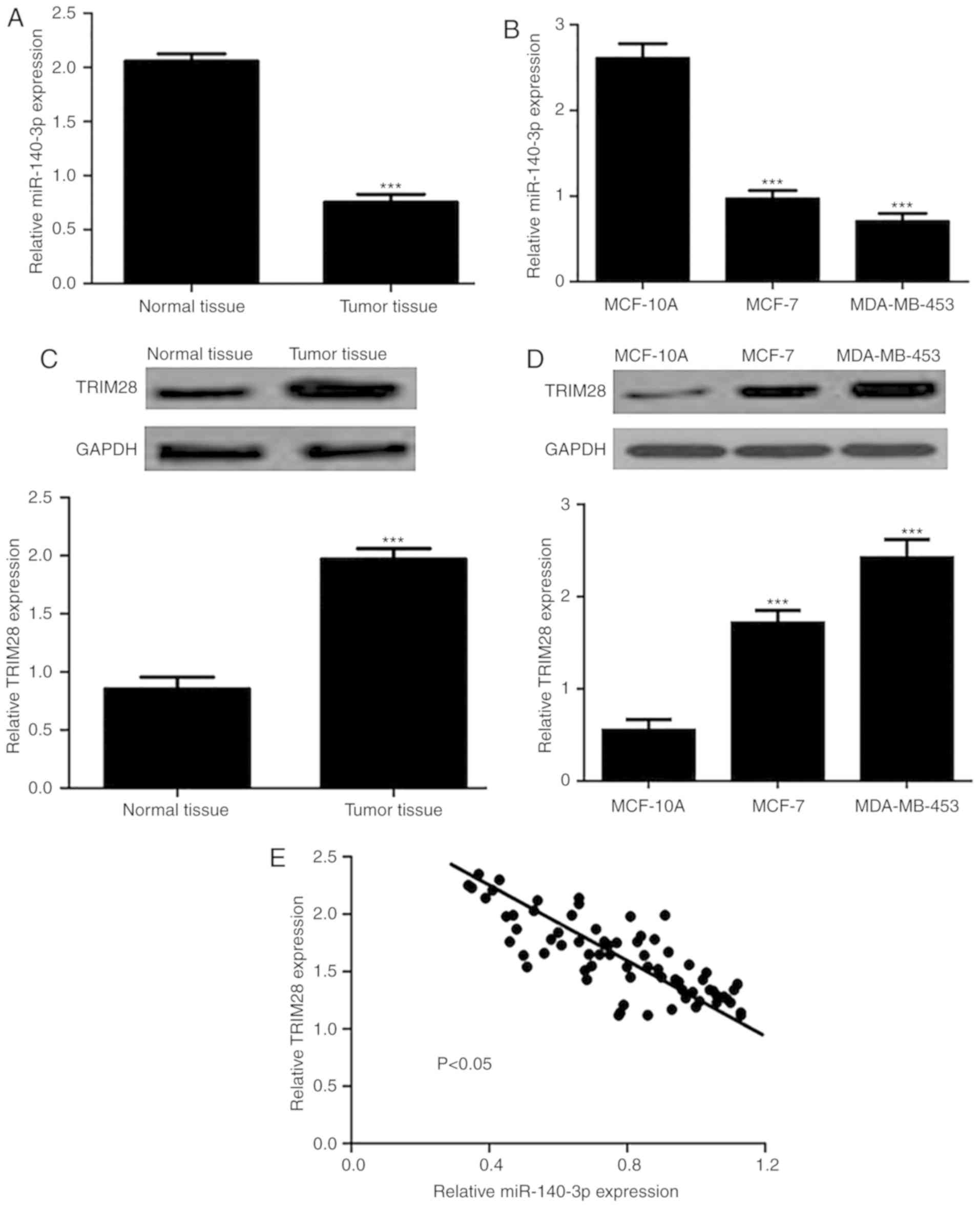
As shown in Fig. 1A and
B, the expression levels of miR-140-3p were significantly
reduced in BC tissues and cell lines compared with in normal
tissues and cells. Conversely, TRIM28 expression was markedly
elevated in BC tissues and cell lines (Fig. 1C and D). In addition, the correlation
between miR-140-3p and TRIM28 in BC tissues was examined and an
inverse correlation was identified (Fig.
1E). Collectively, these results demonstrated that in BC,
miR-140-3p expression was reduced and TRIM28 was overexpressed.
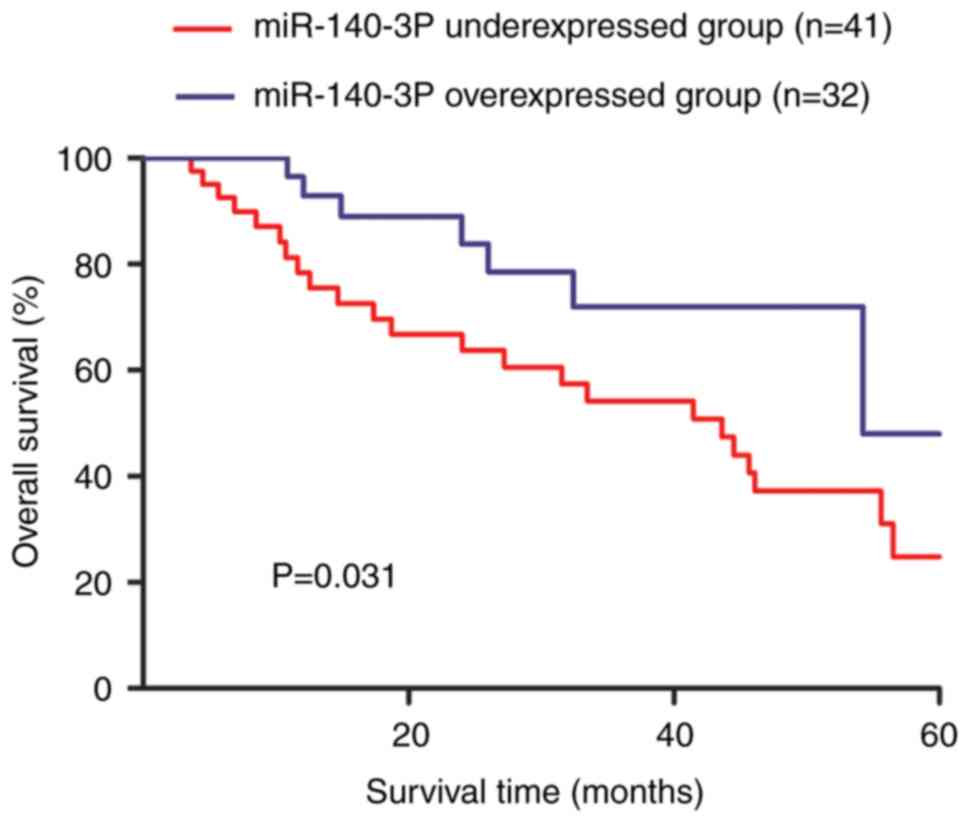
Clinical significance of miR-140-3p
expression in BC
The median value of miR-140-3p expression was used
to classify the enrolled patients with BC into high or low
miR-140-3p expression groups. Kaplan-Meier curve revealed that low
miR-140-3p expression predicted a worse 5-year overall survival for
patients with BC compared with high miR-140-3p expression (Fig. 2).
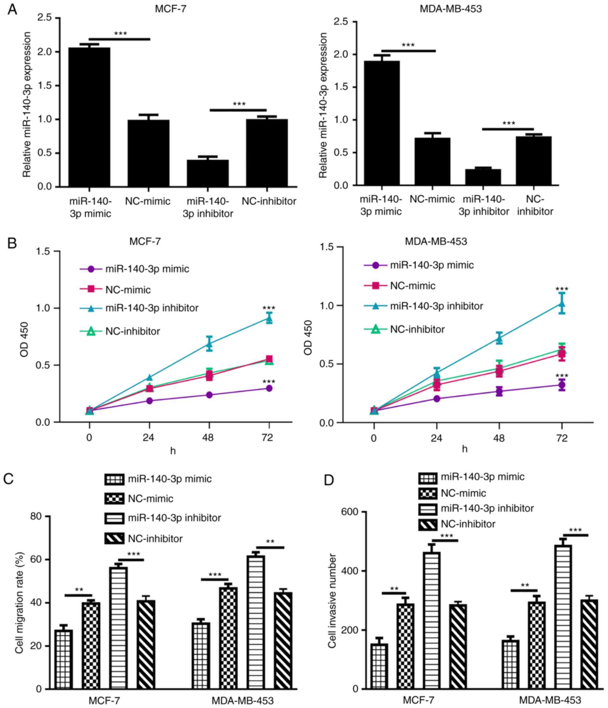
miR-140-3p inhibits cell
proliferation, migration and invasion of BC cell lines in
vitro
Proliferation and migration are indicators of the
malignancy of tumor cells (19).
RT-qPCR results demonstrated that the expression levels of
miR-140-3p were significantly elevated following miR-140-3p mimic
transfection, but were reduced by miR-140-3p inhibitor transfection
(Fig. 3A). Subsequently, a CCK-8
assay demonstrated that cell proliferation in the miR-140-3p mimic
group was significantly lower than in the NC-mimic group (Fig. 3B). Conversely, miR-140-3p inhibitor
significantly elevated cell proliferation compared with
NC-inhibitor (Fig. 3B). As shown in
Fig. 4C, cell migration in the
miR-140-3p mimic-transfected group was significantly decreased
compared with that in the NC-mimic group, whereas migration in the
miR-140-3p inhibitor group was markedly increased compared with
that in the NC-inhibitor group. Transwell invasion assays indicated
that cell invasion was enhanced by miR-140-3p inhibitor but
decreased by miR-140-3p mimic (Fig.
3D).
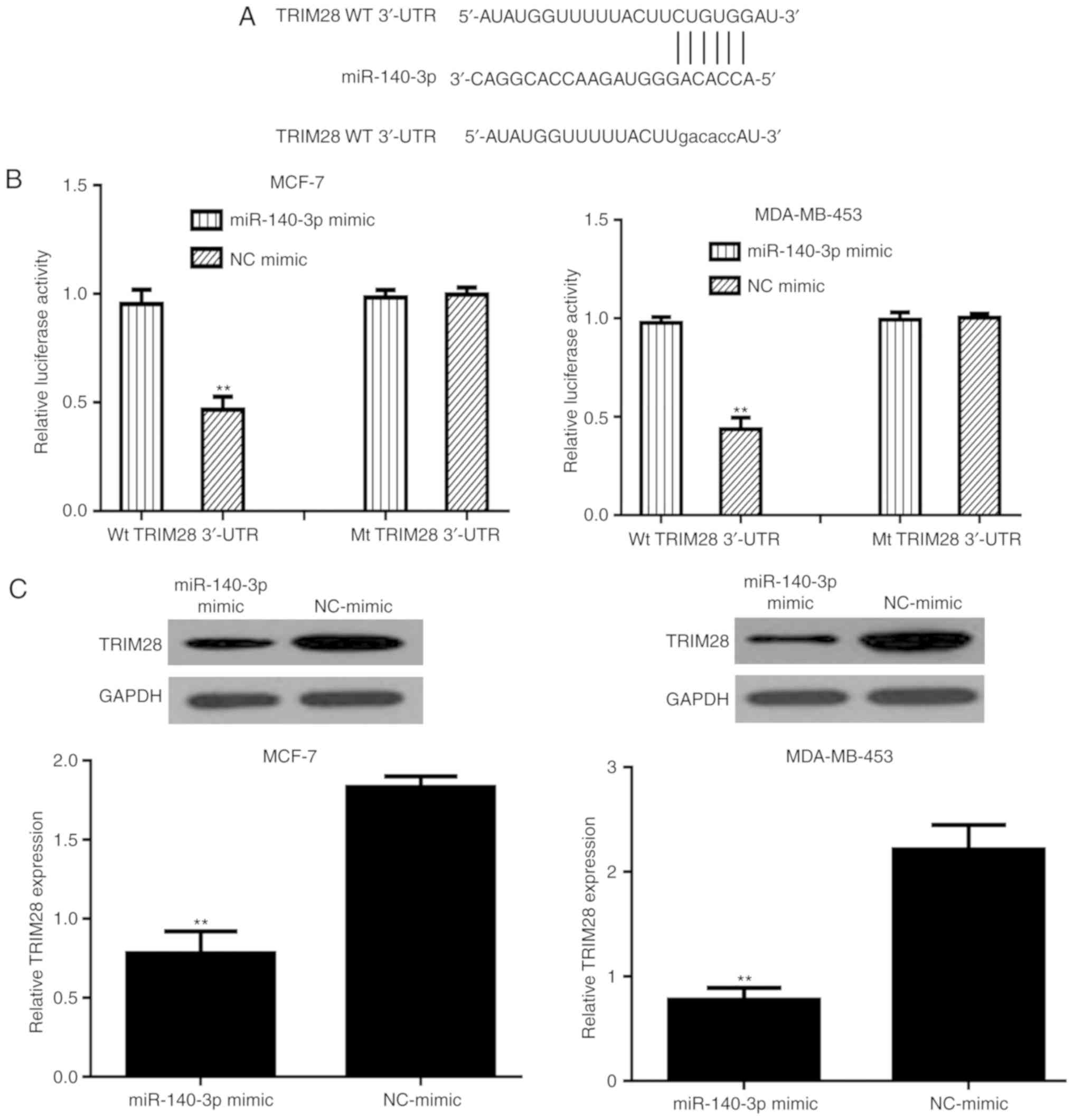
TRIM28 is a direct target of
miR-140-3p
The binding site between miR-140-3p and the 3′-UTR
of TRIM28 is presented in Fig. 4A.
The mutant sequence at the binding site between the 3′-UTR of
TRIM28 and miR-140-3p is also shown in Fig. 4A. To confirm this prediction, a
luciferase reporter system with TRIM28 Wt 3′-UTR or Mt 3′-UTR and
miR-140-3p mimic or NC-mimic co-transfection was constructed. As
shown in Fig. 4B, the luciferase
activity of cells transfected with Wt 3′-UTR was suppressed by the
miR-140-3p mimic. However, the miR-140-3p mimic did not alter the
luciferase activity in cells transfected with Mt 3′-UTR (Fig. 4B). To validate whether TRIM28 can be
regulated by miR-140-3p, the levels of TRIM28 in miR-140-3p mimic-
or NC-mimic-transfected cells were measured. As expected, the
levels of TRIM28 were downregulated by miR-140-3p mimic in BC cell
lines (Fig. 4C).
Effects of miR-140-3p on cell
proliferation, migration and invasion are partially reversed by
TRIM28
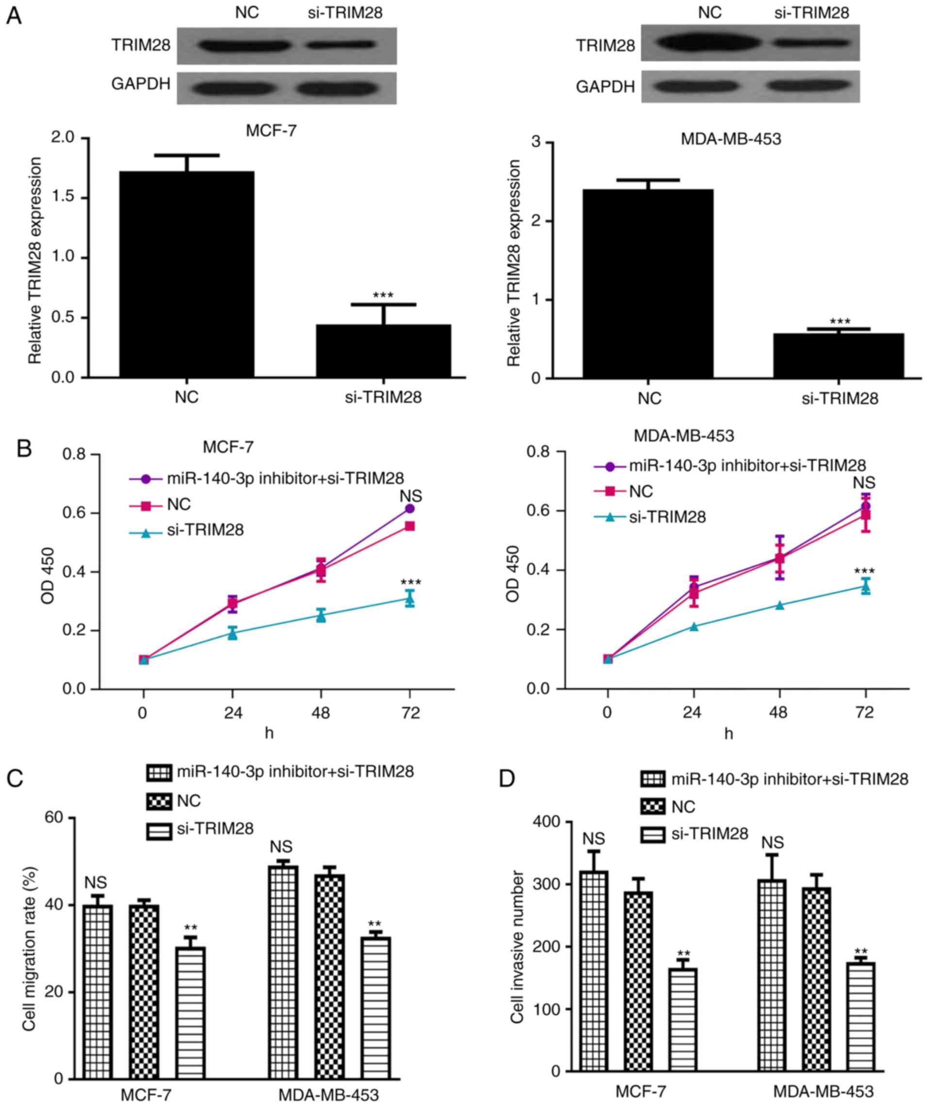
To determine whether TRIM28 can reverse the
inhibitory effects of miR-140-3p, si-TRIM28 and miR-140-3p
inhibitor were co-transfected into BC cells. Transfection with
si-TRIM28 could downregulate the expression of TRIM28 (Fig. 5A). In addition, the inhibitory
effects of si-TRIM28 on cell proliferation, migration and invasion
were reversed by miR-140-3p inhibitor, suggesting that TRIM28 may
be a functional target of miR-140-3p (Fig. 5B-D).
Discussion
It has been demonstrated that an improved
understanding of tumor progression mechanisms could greatly improve
the survival of cancer patients (5–8). In
recent decades, the importance of miRNAs in tumors has attracted
more attention (5–7). miR-140 is located at the intron of the
WW domain containing E3 ubiquitin protein ligase 2 gene and
produces two mature microRNAs: miR-140-5p and miR-140-3p (20). miR-140-5p has been reported to act as
a tumor suppressor in several cancer types, including gastric
cancer, BC and ovarian cancer (21–23).
miR-140-3p has also been revealed to be abnormally expressed in
several types of cancer, including lung cancer and spinal chordoma
(13,14). However, whether miR-140-3p serves a
role in BC remains largely unknown.
In the present study, miR-140-3p expression was
downregulated in BC tissues and cell lines compared with in
adjacent normal tissues and a normal cell line. Furthermore, low
miR-140-3p expression predicted a poor prognosis of patients with
BC. Functional assays revealed that miR-140-3p overexpression
inhibited BC cell proliferation, migration and invasion. These
results collectively suggested that miR-140-3p may function as a
tumor suppressor in BC. Using a miRNA target prediction algorithm,
it was suggested that TRIM28 may contain a putative binding site
for miR-140-3p in its 3′-UTR. Additionally, TRIM28 expression was
upregulated in BC tissues and cell lines.
TRIM28 belongs to the TRIM protein family and serves
a critical role in a wide range of biological activities (24,25). Hao
et al revealed that high TRIM28 expression is a predictor
for poor prognosis in patients with BC (16). Wei et al reported that TRIM28
enhances BC metastasis through direct interaction with
twist-related protein 1 (TWIST1), to protect TWIST1 from
degradation, suggesting that TRIM28 could be a target for BC
treatment (17). These studies
highlight the importance of TRIM28 in the progression of BC. A
previous study demonstrated that TRIM28 expression could be
regulated by miR-491 in glioma (18). Therefore, this study investigated
whether TRIM28 is also a downstream target of miR-140-3p. A
luciferase activity reporter assay and western blot analysis were
performed to validate TRIM28 as a direct target of miR-140-3p.
Additionally, an inverse correlation between miR-140-3p and TRIM28
expression was detected in BC tissues. Furthermore, BC cells were
co-transfected with si-TRIM28 and miR-140-3p inhibitor, revealing
that the inhibitory effects of si-TRIM28 on BC cell behaviors may
be reversed by miR-140-3p inhibitor. These results demonstrated
that TRIM28 was an effector for the role of miR-140-3p on BC cell
behaviors.
In conclusion, these findings clearly demonstrated
that miR-140-3p may act as a tumor suppressor and that its effects
were exerted by regulating TRIM28 in BC. Additionally, restoration
of miR-140-3p expression may be a novel strategy for BC
treatment.
Acknowledgements
Not applicable.
Funding
No funding was received.
Availability of data and materials
The datasets used and/or analyzed during the present
study are available from the corresponding author on reasonable
request.
Authors' contributions
YZ, BW and HW conceived and designed the study. YZ,
BW, YW, GC, QL and HW performed the experiments. YZ, BW, YW, GC, QL
and HW wrote the paper. YZ, BW, YW, GC, QL and HW reviewed and
edited the manuscript. All authors read and approved the
manuscript.
Ethics approval and consent to
participate
The collection and the use of all tissue samples
were approved by the Ethics Committee of First Affiliated Hospital
of Jiamusi University (Jiamusi, China). Written informed consent
was obtained from all participating patients.
Patient consent for publication
Not applicable.
Competing interests
The authors declare that they have no competing
interests.
References
|
1
|
DeSantis C, Ma J, Bryan L and Jemal A:
Breast cancer statistics, 2013. CA Cancer J Clin. 64:52–62. 2014.
View Article : Google Scholar : PubMed/NCBI
|
|
2
|
Chen W, Zheng R, Baade PD, Zhang S, Zeng
H, Bray F, Jemal A, Yu XQ and He J: Cancer statistics in China,
2015. CA Cancer J Clin. 66:115–132. 2016. View Article : Google Scholar : PubMed/NCBI
|
|
3
|
Tzanninis IG, Kotteas EA,
Ntanasis-Stathopoulos I, Kontogianni P and Fotopoulos G: Management
and outcomes in metaplastic breast cancer. Clin Breast Cancer.
16:437–443. 2016. View Article : Google Scholar : PubMed/NCBI
|
|
4
|
Sonnenblick A, Pondé N and Piccart M:
Metastatic breast cancer: The Odyssey of personalization. Mol
Oncol. 10:1147–1159. 2016. View Article : Google Scholar : PubMed/NCBI
|
|
5
|
Hemmatzadeh M, Mohammadi H, Jadidi-Niaragh
F, Asghari F and Yousefi M: The role of oncomirs in the
pathogenesis and treatment of breast cancer. Biomed Pharmacother.
78:129–139. 2016. View Article : Google Scholar : PubMed/NCBI
|
|
6
|
Asghari F, Haghnavaz N, Baradaran B,
Hemmatzadeh M and Kazemi T: Tumor suppressor microRNAs: Targeted
molecules and signaling pathways in breast cancer. Biomed
Pharmacother. 81:305–317. 2016. View Article : Google Scholar : PubMed/NCBI
|
|
7
|
Tavazoie SF, Alarcón C, Oskarsson T, Padua
D, Wang Q, Bos PD, Gerald WL and Massagué J: Endogenous human
microRNAs that suppress breast cancer metastasis. Nature.
451:147–152. 2008. View Article : Google Scholar : PubMed/NCBI
|
|
8
|
Ambros V: The functions of animal
microRNAs. Nature. 431:350–355. 2004. View Article : Google Scholar : PubMed/NCBI
|
|
9
|
Gravgaard KH, Lyng MB, Laenkholm AV,
Søkilde R, Nielsen BS, Litman T and Ditzel HJ: The miRNA-200 family
and miRNA-9 exhibit differential expression in primary versus
corresponding metastatic tissue in breast cancer. Breast Cancer Res
Treat. 134:207–217. 2012. View Article : Google Scholar : PubMed/NCBI
|
|
10
|
Korpal M, Lee ES, Hu G and Kang Y: The
miR-200 family inhibits epithelial-mesenchymal transition and
cancer cell migration by direct targeting of E-cadherin
transcriptional repressors ZEB1 and ZEB2. J Biol Chem.
283:14910–14914. 2008. View Article : Google Scholar : PubMed/NCBI
|
|
11
|
Elliman SJ, Howley BV, Mehta DS, Fearnhead
HO, Kemp DM and Barkley LR: Selective repression of the oncogene
cyclin D1 by the tumor suppressor miR-206 in cancers. Oncogenesis.
3:e1132014. View Article : Google Scholar : PubMed/NCBI
|
|
12
|
Song G, Zhang Y and Wang L: MicroRNA-206
targets notch3, activates apoptosis, and inhibits tumor cell
migration and focus formation. J Biol Chem. 284:31921–31927. 2009.
View Article : Google Scholar : PubMed/NCBI
|
|
13
|
Kong XM, Zhang GH, Huo YK, Zhao XH, Cao
DW, Guo SF, Li AM and Zhang XR: MicroRNA-140-3p inhibits
proliferation, migration and invasion of lung cancer cells by
targeting ATP6AP2. Int J Clin Exp Pathol. 8:12845–12852.
2015.PubMed/NCBI
|
|
14
|
Zou MX, Huang W, Wang XB, Lv GH, Li J and
Deng YW: Identification of miR-140-3p as a marker associated with
poor prognosis in spinal chordoma. Int J Clin Exp Pathol.
7:4877–4885. 2014.PubMed/NCBI
|
|
15
|
Livak KJ and Schmittgen TD: Analysis of
relative gene expression data using real-time quantitative PCR and
the 2(-Delta Delta C(T)) method. Methods. 25:402–408. 2001.
View Article : Google Scholar : PubMed/NCBI
|
|
16
|
Hao L, Leng J, Xiao R, Kingsley T, Li X,
Tu Z, Yang X, Deng X, Xiong M, Xiong J and Zhang Q: Bioinformatics
analysis of the prognostic value of Tripartite Motif 28 in breast
cancer. Oncol Lett. 13:2670–2678. 2017. View Article : Google Scholar : PubMed/NCBI
|
|
17
|
Wei C, Cheng J, Zhou B, Zhu L, Khan MA, He
T, Zhou S, He J, Lu X, Chen H, et al: Tripartite motif containing
28 (TRIM28) promotes breast cancer metastasis by stabilizing TWIST1
protein. Sci Rep. 6:298222016. View Article : Google Scholar : PubMed/NCBI
|
|
18
|
Qi Z, Cai S, Cai J, Chen L, Yao Y, Chen L
and Mao Y: miR-491 regulates glioma cells proliferation by
targeting TRIM28 in vitro. BMC Neurol. 16:2482016. View Article : Google Scholar : PubMed/NCBI
|
|
19
|
Hanahan D and Weinberg RA: Hallmarks of
cancer: The next generation. Cell. 144:646–674. 2011. View Article : Google Scholar : PubMed/NCBI
|
|
20
|
Rodriguez A, Griffiths-Jones S, Ashurst JL
and Bradley A: Identification of mammalian microRNA host genes and
transcription units. Genome Res. 14:1902–1910. 2004. View Article : Google Scholar : PubMed/NCBI
|
|
21
|
Fang Z, Yin S, Sun R, Zhang S, Fu M, Wu Y,
Zhang T, Khaliq J and Li Y: miR-140-5p suppresses the
proliferation, migration and invasion of gastric cancer by
regulating YES1. Mol Cancer. 16:1392017. View Article : Google Scholar : PubMed/NCBI
|
|
22
|
Lu Y, Qin T, Li J, Wang L, Zhang Q, Jiang
Z and Mao J: MicroRNA-140-5p inhibits invasion and angiogenesis
through targeting VEGF-A in breast cancer. Cancer Gene Ther.
24:386–392. 2017. View Article : Google Scholar : PubMed/NCBI
|
|
23
|
Lan H, Chen W, He G and Yang S: miR-140-5p
inhibits ovarian cancer growth partially by repression of PDGFRA.
Biomed Pharmacother. 75:117–122. 2015. View Article : Google Scholar : PubMed/NCBI
|
|
24
|
Hatakeyama S: TRIM proteins and cancer.
Nat Rev Cancer. 11:792–804. 2011. View
Article : Google Scholar : PubMed/NCBI
|
|
25
|
Chen L, Chen DT, Kurtyka C, Rawal B, Fulp
WJ, Haura EB and Cress WD: Tripartite motif containing 28 (Trim28)
can regulate cell proliferation by bridging HDAC1/E2F interactions.
J Biol Chem. 287:40106–40118. 2012. View Article : Google Scholar : PubMed/NCBI
|