Introduction
Giant cell tumor of bone (GCTB) is benign in
histology, but presents as a local invasive growth, and its
biological behavior is hard to predict (1). Although it is not a fatal tumor,
pulmonary metastasis easily occurs in young patients with recurrent
GCTB at Enneking stage 3 (2). GCTB
is characterized by expansive growth and usually occurs at the end
of a long bone, and in >50% patients, it occurs at the
knee-joint (3). In addition, the
tumor tissue often approaches the articular surface by damaging
bones covered by articular cartilage (4). Curettage can be used to remove the
tumor without damaging the joint function, but a high recurrence
rate following surgery remains a difficult problem to solve
(5). Although the wide excision of
tumors combined with bone tumor prosthesis can markedly decrease
the recurrence rate, the function of the reconstructed joint is
worse, when compared with the function of the joint following
curettage, due to joint defects caused by wide excision (6–8). The
range of osteotomy is correlated with tumor recurrence and joint
reconstruction (9,10). Therefore, investigating the
morphology of GCTB may assist in the accurate excision of tumors,
thus decreasing recurrence caused by residual tumor cells. A major
problem encountered with GCTB is that the range of excision is
restricted due to limitations on bone tumor prosthesis, which
increases the risk of failing to reconstruct the joint. The use of
personalized prosthesis for GCTB decreases unnecessary excision and
allows for the retention of more healthy bone. The morphological
changes of GCTB negatively affect the biomechanical property of
bones and increase the risk of pathological fractures, which may
influence the surrounding soft tissue and lead to tumor recurrence
following surgery (5,11,12).
Therefore, morphological characteristics, including the size of the
tumor, the distance of the tumor edge from the articular surface
and occurrence of pathological fractures, have an important impact
on the treatment of GCTB and on estimating prognosis.
The present study summarizes the morphological
features of GCTB in the knee and provides evidence to support the
use of these characteristics to improve the accuracy of tumor
excision, to decrease damage to the host bone and improve the
survival rate of prostheses. In addition, the present study
provides a theoretical basis for predicting the occurrence of
pathological fractures in GCTB and for designing a personalized
GCTB prosthesis for the knee.
Materials and methods
Inclusion and exclusion criteria
Inclusion criteria: i) Patients with tumors around
the knee confirmed as GCTB by pathology; ii) patients with complete
imaging data, including X-ray films covering the whole area of the
lesion, computed tomography (CT) scanning images in Digital Imaging
and Communications in Medicine (DICOM) format or multi-dimensional
magnetic resonance imaging (MRI); and iii) cases in which the
morphological measurement of GCTB was not influenced by surgery.
Exclusion criteria: i) Patients without a definite pathological
diagnosis; ii) patients without complete clinical and imaging data;
iii) patients with imaging data that was not in DICOM format; and
iv) cases in which the imaging data did not cover the whole area of
the lesion.
On the basis of the above criteria, a total of 255
patients who were diagnosed with GCTB by pathological surgery
between June 2000 and December 2016 in eight hospitals were
included in the present study. These hospitals are institutional
members of the Chinese Giant Cell Tumor Team of China
(GTOC)-associated group, including Tianjin Hospital (Tianjin,
China), Jinan Military Region General Hospital (Jinan, China),
Xijing Hospital (Xian, China), The Second Affiliated Hospital of
Zhejiang University School of Medicine (Hangzhou, China), Nanjing
Military Region General Hospital (Nanjing, China), West China
Hospital of Sichuan University (Chengdu, China), The Second
Hospital of Tianjin Medical University (Tianjin, China) and The
Third Hospital of Hebei Medical University (Shijiazhuang, China).
The clinical and imaging data of these 255 patients were
retrospectively analyzed. Of these patients, 5 had GCTB in the
proximal fibula (2.0%). As GCTB rarely occurs in the proximal
fibula, and the fibula bears no weight, these 5 cases were excluded
from the present study. The present study involving 250 patients
was conducted in accordance with The Declaration of Helsinki, and
approved by the Medical Ethics Committee of Tianjin Hospital.
Written informed consent was obtained from all participants and the
patients gave permission for publication of their images.
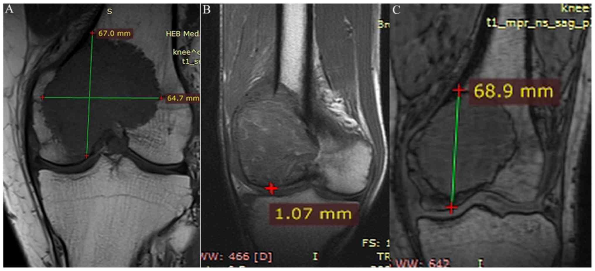
Morphological parameters and
measurement of the GCTB lesion
The morphological parameters of GCTB included the
following: i) Maximal longitudinal diameter (MLD) of the tumor
defined by the maximal diameter of the tumor parallel with the
femur and tibia; ii) maximal transverse diameter (MTD) of the tumor
defined by the maximal diameter of the tumor vertical or almost
vertical with the femur and tibia; iii) the shortest and longest
distance from the articular surface (SDAS and LDAS, respectively)
defined as the shortest or longest distance of the tumor edge from
the articular surface under the cartilage; and iv) the presence or
absence of pathological fractures. It should be noted that the
thickness of the articular cartilage did not require measurement.
CT images of the coronal/sagittal reconstruction or MRI images of
the coronal/sagittal plane with the maximal diameter were selected
to measure the MLD, MTD, SDAS and LDAS of the tumors.
All these parameters were measured by two observers
who had >10 years of experience in radiology, and were trained
in the standards prior to observation and measurement of the
current samples, including determining the optimal measuring plane
and point.
Statistical analysis
SPSS (version 19.0; IBM Corp., Armonk, NY, USA) was
used to analyze the data. The intra-group correlation coefficient
(ICC) was evaluated to compare the measurements of MLD, MTD, SDAS
and LDAS from the two observers. Age, morphological parameters, and
other parameters were expressed as the mean ± standard deviation,
and these data were compared using an independent sample t-test. A
χ2 test was used to compare the enumeration data between
two groups, such as the packet count data of SDAS and the incidence
of pathological fractures between these two groups. P<0.05 was
considered to indicate a statistically significant difference.
Results
Clinical parameters
A total of 250 patients with GCTB were enrolled in
the present study. The age range of the patients was 17.2–78.5
years, and the average age was 35.3±12.4 years. Among the patients,
133 (53.2%) were male and 117 (46.8%) were female. Additionally,
137 patients (54.8%) had GCTB in the distal femur, whereas 113
(45.2%) had GCTB in the proximal tibia. The sex distribution of
patients with GCTB in the distal femur was 73 male and 64 female,
and the sex distribution of patients with GCTB in the proximal
tibia was 62 male and 51 female. The difference in sex distribution
was not significant, using a χ2 test. The average age of
patients with GCTB in the distal femur was 35.6±12.7 years, and the
average age of patients with GCTB in the proximal tibia was
34.8±13.2 years. There was no significant difference in the age of
patients with GCTB between these two groups (t=0.639, P=0.523).
According to the imaging features, all patients presented with
typical manifestations of GCBT, including expansive growth,
osteolytic destruction and no calcification. Of the patients, 187
were treated with tumor curettage and 63 underwent wide excision of
tumors combined with bone tumor prosthesis (Fig. 1).
Intra-group consistency of the two
observers
The morphological parameters were measured by two
observers. For patients with GCTB in the distal femur, the ICC
r values of the MLD, MTD, SDAS and LDAS measurements were
0.9797, 0.9760, 0.9728 and 0.9650, respectively, whereas those in
patients with GCTB in the proximal tibia were 0.9971, 0.9720,
0.9605 and 0.9935, respectively. These results indicate that there
was high intra-group consistency in the measurements of the two
observers.
Measurement of the morphological
parameters
Morphological parameters of GCTB were measured from
the MRI/CT images (Fig. 2). The
results of the measured morphological parameters were as
follows.
MLD
Mean MLD in the group with GCTB in the distal femur
was 6.616±2.322 cm, and MLD in the group with GCTB in the proximal
tibia was 5.738±2.278 cm. The 95% confidence interval (CI) of MLD
in the distal femur group was 6.222–7.008 cm, and that in the
proximal tibia group was 5.313–6.163 cm. The measurements of MLD
were significantly different between the two groups (t=2.999,
P=0.003). The frequency distribution of MLD in the two groups
demonstrates that the MLD measurement was generally normally
distributed, with the exception of a number of tumors in the distal
femur that were >12 cm, and a number in the proximal tibia that
were >10 cm (Fig. 3). In
addition, 80% of the MLD measurements of tumors in the distal femur
were 4.4–8.9 cm, and 80% of the MLD values in the proximal tibia
were 4.1–7.1 cm.
MTD
The average MTD of tumors in patients with GCTB in
the distal femur was 4.865±1.525 cm, and that in patients with GCTB
in the proximal tibia was 4.313±1.309 cm. The 95% CI of MTD in
patients with GCTB in the distal femur was 4.607–5.122 cm, whereas
that in patients with GCTB in the proximal tibia was 4.069–4.557
cm. There was a significant difference in MTD measurements between
the two groups (t=3.232, P=0.003). In patients with GCTB in the
distal femur, 80% of MTD values were 3.1–6.4 cm, whereas 80% of the
MTD values in patients with GCTB in the proximal tibia were 2.7–6.0
cm.
LDAS
The average LDAS in patients with GCTB in the distal
femur was 6.924±2.135 cm, and that in patients with GCTB in the
proximal tibia was 5.878±1.825 cm. The 95% CI of LDAS in patients
with GCTB in the distal femur was 6.563–7.284 cm, and that in
patients with GCTB in the proximal tibia was 5.537–6.217 cm. There
was a significant difference in LDAS between the two groups
(t=4.116, P=0.001). The histogram of the frequency distribution of
LDAS in the two groups was similar to the histograms of MLD and
MTD, and was normally distributed (data not shown). In patients
with GCTB in the distal femur, 80% of the LDAS values were within
4.9–9.2 cm, whereas in patients with GCTB in the proximal tibia,
80% of the LDAS values were within 4.4–7.5 cm.
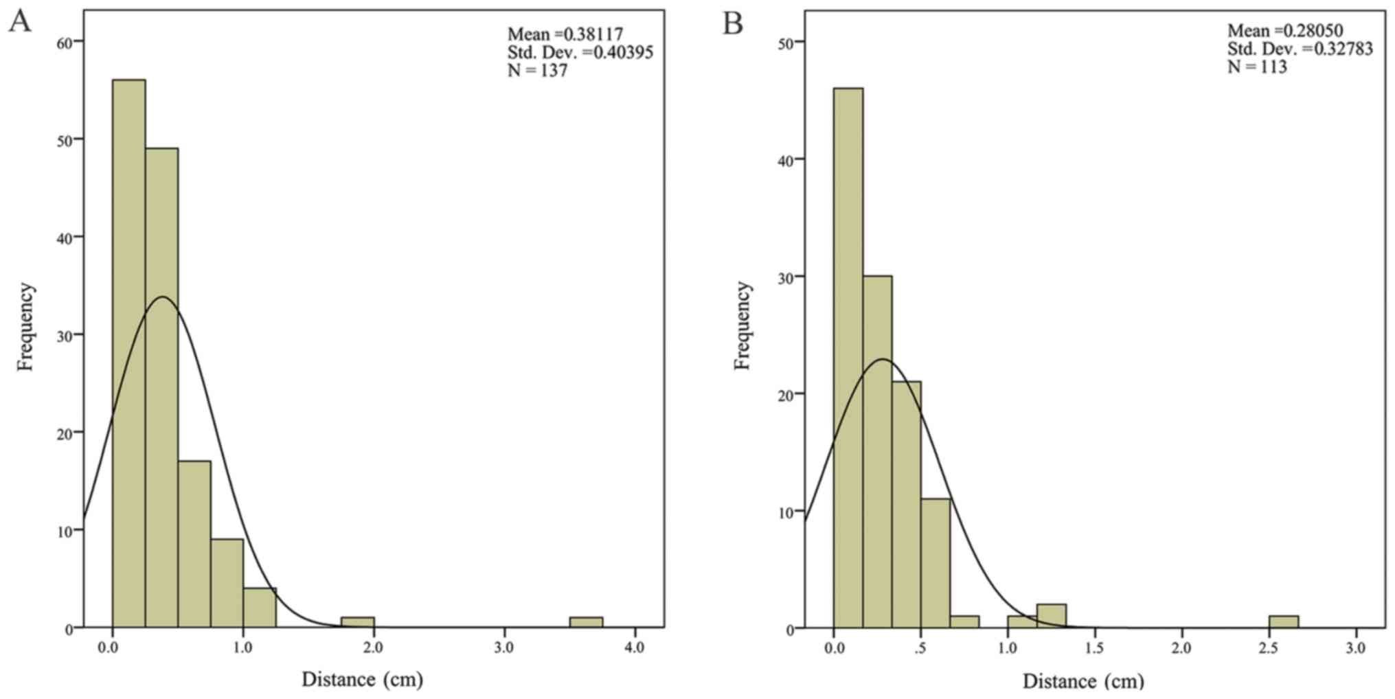
SDAS
The average SDAS in patients with GCTB in the distal
femur was 0.381±0.404 cm, and that in patients with GCTB in the
proximal tibia was 0.280±0.328 cm. The 95% CI of SDAS in patients
with GCTB in the distal femur was 0.313–0.449 cm, and that in
patients with GCTB in the proximal tibia was 0.219–0.342 cm. There
was a significant difference in SDAS between the two groups
(t=2.211, P=0.035). The histogram of the frequency distribution of
SDAS in the two groups demonstrates that lower SDAS measurements
were more frequent (Fig. 4). In
patients with GCTB in the distal femur, 80% of the SDAS values were
within 0.01–0.75 cm, whereas in patients with GCTB in the proximal
tibia, 80% of the SDAS values were within 0.01–0.51 cm.
Grouping statistics of SDAS
To further evaluate the association between the
distance from the tumor edge and the articular surface and the
location of the tumor, all subjects were divided into five ranges
on the basis of the SDAS values: 0–1, 1–3, 3–6, 6–10 and >10 mm
(Table I). These results indicate
that there was a significant difference between the two groups when
SDAS was 0–1 mm (χ2=9.837, P=0.043). The majority of
patients in the two groups fell into the range 0–3 mm (66 cases in
the distal femur and 69 cases in the proximal tibia). Therefore,
SDAS presented a markedly skewed distribution in the two groups.
The edge of the tumor in the two groups was evidently close to the
articular surface. The further the tumor edge was from the
articular surface, the lower the frequency.
 | Table I.Range of SDAS measurements of giant
cell tumors of the bone in the distal femur and in the proximal
tibia. |
Table I.
Range of SDAS measurements of giant
cell tumors of the bone in the distal femur and in the proximal
tibia.
|
| SDAS, mm |
|
|
|---|
|
|
|
|
|
|---|
| Location of
tumor | 0–1 | 1–3 | 3–6 | 6–10 | >10 | χ2 | P-value |
|---|
| Distal femur, n
(%) | 23 (16.9) | 43 (31.4) | 37 (27.0) | 18 (13.1) | 16 (11.6) | 9.837 | 0.043 |
| Proximal tibia, n
(%) | 35 (30.9) | 34 (30.0) | 29 (25.6) | 9 (8.1) | 6 (5.4) |
|
|
Association between the size of the
tumor and incidence of pathological fractures
A total of 64 patients (25.1% of 255) presented with
pathological fractures. Of those patients, 47 patients had GCTB in
the distal femur (34.4% of 137 patients with GCTB in the distal
femur), whereas 17 patients had GCTB in the proximal tibia (15.0%
of 113 patients with GCTB in the proximal tibia). There was a
significant difference in the incidence of pathological fractures
between the two groups (P=0.001). The MLD was divided into
different ranges, with intervals of 3 cm. With the exception of the
0–3 cm range, the incidence of pathological fractures in the distal
femur was higher compared with that in the proximal tibia (Table II). The MTD was divided into ranges
with 2 cm intervals. For all ranges, the incidence of pathological
fractures was higher in the distal femur compared with that in the
proximal tibia (Table III). The
trend of pathological fracture incidence in Tables II and III indicated that the frequency of
fracture increased with the increase in longitudinal and transverse
diameter in the two groups.
| Table II.Incidence of pathological fractures
and range of MLD of giant cell tumors of the bone in the distal
femur and in the proximal tibia. |
Table II.
Incidence of pathological fractures
and range of MLD of giant cell tumors of the bone in the distal
femur and in the proximal tibia.
|
| MLD, cm |
|
|
|---|
|
|
|
|
|
|---|
| Location of
tumor | 0–3 | 3–6 | 6–9 | >9 | Total | χ2 | P-value |
|---|
| Distal femur, n/total
(%) | 0/3 (0.0) | 23/71 (32.4) | 21/57 (36.8) | 3/6 (50.0) | 47/137 (34.3) | 12.063 | 0.001 |
| Proximal tibia,
n/total (%) | 1/5 (20.0) | 8/67 (12.0) | 6/35 (17.1) | 2/6 (33.0) | 17/113 (15.0) |
|
|
| Table III.Incidence of pathological fractures
and range of MTD of giant cell tumors of bone in the distal femur
and in the proximal tibia. |
Table III.
Incidence of pathological fractures
and range of MTD of giant cell tumors of bone in the distal femur
and in the proximal tibia.
|
| MTH, cm |
|
|
|---|
|
|
|
|
|
|---|
| Location of
tumor | 0–2 | 2–4 | 4–6 | 6–8 | >8 | Total | χ2 | P-value |
|---|
| Distal femur, n/total
(%) | 0/1 (0.0) | 10/37 (27.0) | 27/78 (34.6) | 8/18 (44.4) | 2/3 (66.7) | 47/137 (34.3) | 12.063 | 0.001 |
| Proximal tibia,
n/total (%) | 0/3 (0.0) | 6/44 (13.6) | 7/50 (14.0) | 4/15 (26.7) | 0/1 (0.0) | 17/113 (15.0) |
|
|
Discussion
The aim of treating GCTB is to improve knee function
as much as possible following surgery, while also ensuring that the
risk of tumor recurrence is decreased. A GCTB with a large diameter
can cause extensive bone cortical damage, and induce damage in a
large surrounding soft tissue mass and pathological fractures, or
extensive invasion to the articular surface. These types of tumor
should be treated with segmental resection and reconstruction, in
order to remove the tumor and maintain knee function (12). Therefore, accurate measurement of the
size and range of the tumor is essential to improve the success
rate of surgery (9). If the tumor
lesion is not sufficiently excised, there is a high risk of
recurrence (10). Alternatively, if
the tumor lesion is excessively excised, the normal host bone may
be damaged and limb function may be affected. Therefore, similar to
other bone tumors, the morphological features of GCTB, including
size, distance from the articular surface and pathological
fractures, are important factors that influence the surgical method
and reconstruction of the tumor cavity, and the prognosis.
The extent of surgical excision required is decided
on the basis of the length of the GCTB and the length of the
prosthesis in the reconstruction. Therefore, a definite length of
the longitudinal diameter of the GCTB would assist when devising a
surgical plan, and in determining the plane in which the bone
should be cut. Kivioja et al (13) measured the morphological parameters
of GCTB in a group of patients with tumors in long tubular bones,
and reported that the mean tumor length was 5 cm. However, that
cohort included tumors in upper and lower limbs, the sample size
was small, and a single method of measurement was used. The present
study had a large sample size and focused on GCTB in the knee, and
therefore avoided introducing variation due to differences in
growth characteristics and biological behavior of GCTB in different
locations. Hu (14) measured the
length of GCTB in 60 patients in the knee using MRI, and reported
that the mean length of the lesion was 6.7 cm, and the range was
4–12 cm. However, tumors in the femur and tibia were not
investigated separately. In the present study, the mean
longitudinal diameter of tumor in the distal femur was similar to
that identified in the study by Hu (14). However, for tumors in the proximal
tibia, the mean longitudinal diameter in the present study was
markedly smaller compared with that in the study by Hu (14). Furthermore, the mean longitudinal
diameter of tumors in the present study was higher compared with
that observed by Kivioja et al (13), indicating that the longitudinal
diameter of GCTB in the knee may be higher compared with that in
the humerus, radius and other locations. Therefore, the range of
osteotomy and the reconstructed length in the knee may be bigger
compared with that in other locations. Szendröi (15) identified that in ~1/3 of patients,
the size of the tumor was >50% of the diameter of the host bone
at the initial diagnosis. The present study revealed that the mean
MTD of GCTB in the distal femur was >50% of the diameter of the
pulp cavity of the distal femur. Thus, the mechanical strength of
the distal femur may be easily damaged, and pathological fractures
easily occur.
The clinical significance of the distance of the
tumor edge from the articular surface was recognized in previous
studies. Ward and Li (16) suggested
that the surgical excision plan should be influenced by the degree
by which the GCTB has invaded the articular surface. In a
retrospective study conducted by Blackley et al (17) on 59 cases with GCTB in the long bone,
an association between the distance of the tumor edge from the
articular surface and the incidence of postoperative articular
degeneration was identified. In the present study, the mean
shortest distance of GCBT in the knee from the articular surface
was <5 mm. Patients with GCTB in the femur at a distance of 0–3
mm from the articular surface accounted for 48.2% (66/137 cases),
and those with tumors in the tibia at that distance accounted for
61% (68/113 cases). This indicates that the majority of GCTB cases
in the knee may occur close to the articular surface.
Dreinhofer et al (18) reported 15 cases of GCTB with
pathological fractures. The distance between the tumor and
articular surface was observed by measuring the thickness of the
subchondral bone, and in 8 cases (53.3%) this measurement was 0 mm.
The present study identified a statistically significant difference
in the SDAS of GCTB in the distal femur and proximal tibia,
suggesting that GCTB in the proximal tibia could more easily invade
the articular surface under the soft bone, when compared with that
in the distal femur. Thus, collapse of the articular surface and
articular degeneration more easily occurred in the patients with
GCTB in the proximal tibia. Therefore, in treating GCTB of the
proximal tibia, more attention should be given to articular
integrity.
Pathological fractures increase the complexity of
the surgery required to completely remove tumors. Therefore,
understanding the association between the incidence of pathological
fractures and tumor morphology is important to determining
treatment of GCTB. In a retrospective study of 54 cases of distal
femur GCTB, Jeys et al (19)
identified that 22% of the 12 cases had pathological fractures. The
present study revealed that the incidence of pathological fractures
in GCTB in the distal femur (34.4%) was higher compared with that
reported by Jeys et al (19),
and compared with that in the proximal tibia (15.0%). The incidence
of fracture in the femur and tibia groups increased as the tumor
diameter increased. In addition, the present study revealed that
GCTB diameter in the distal femur was larger compared with that in
the proximal tibia, which may explain the increased incidence of
pathological fractures in the distal femur compared with that in
the proximal tibia.
Owing to the lack of studies on morphological
parameters of GCTB and the accurate measurement of the size of the
tumor, for early GCTB excision, the range of the tumor lesion is
determined by X-ray plus the addition of 3–5 cm to the border of
the osteotomy to avoid recurrence caused by residual tumor cells
(14). However, in increasing the
length of reconstruction, the risk of complications with the
prosthesis increases. If the size of the tumor is determined prior
to surgery, an accurate osteotomy can be conducted. Thus, the
healthy host bone can be preserved as much as possible, decreasing
the risk of complications with the prosthesis. This may assist in
increasing the service life of the prosthesis and developing a
personalized prosthesis in the future. Chen et al (20) performed a multicenter retrospective
study on 42 patients with distal femoral GCTB, who underwent
artificial prosthesis replacement. It was demonstrated that the
length of the osteotomy was significantly associated with loosening
of the prosthesis, and decreased flexion and extension function of
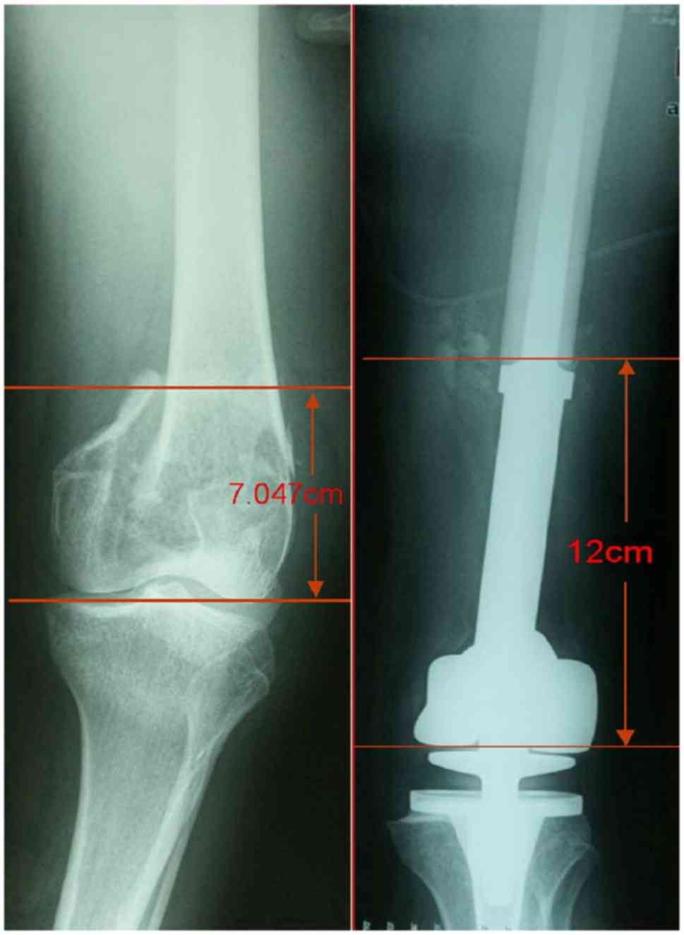
the knee, particularly when the osteotomy length was >12 cm. As
the number of patients included in the present and previous studies
is small, more patients are required to develop a database of
morphological parameters of GCTB in the knee and other sites, in
order to provide a theoretical basis for the diagnosis and
treatment of GCTB.
Acknowledgements
Not applicable.
Funding
No funding was received.
Availability of data and materials
The datasets used and/or analyzed during the current
study are available from the corresponding author on reasonable
request.
Authors' contributions
YCH and XNZ designed the study. YCH, LG and JM
analyzed and interpreted the patient data. PJ, MS, ZW and JM
collected the patient data, and LG, YCH and XNZ were major
contributors in writing the manuscript. All authors read and
approved the final manuscript.
Ethics approval and consent to
participate
This study was approved by the Medical Ethics
Committee of Tianjin Hospital (Tianjin, China).
Patient consent for publication
Written informed consent was obtained from all
participants, and the patients gave permission for publication of
their images.
Competing interests
The authors declare that they have no competing
interests.
References
|
1
|
Traub F, Singh J, Dickson BC, Leung S,
Mohankumar R, Blackstein ME, Razak AR, Griffin AM, Ferguson PC and
Wunder JS: Efficacy of denosumab in joint preservation for patients
with giant cell tumour of the bone. Eur J Cancer. 59:1–12. 2016.
View Article : Google Scholar : PubMed/NCBI
|
|
2
|
Chan CM, Adler Z, Reith JD and Gibbs CP
Jr: Risk factors for pulmonary metastases from giant cell tumor of
bone. J Bone Joint Surg Am. 97:420–428. 2015. View Article : Google Scholar : PubMed/NCBI
|
|
3
|
Sobti A, Agrawal P, Agarwala S and Agarwal
M: Giant cell tumor of bone-an overview. Arch Bone Jt Surg. 4:2–9.
2016.PubMed/NCBI
|
|
4
|
Fraquet N, Faizon G, Rosset P, Phillipeau
JM, Waast D and Gouin F: Long bones giant cells tumors: Treatment
by curretage and cavity filling cementation. Orthop Traumatol Surg
Res. 95:402–406. 2009. View Article : Google Scholar : PubMed/NCBI
|
|
5
|
Klenke FM, Wenger DE, Inwards CY, Rose PS
and Sim FH: Giant cell tumor of bone: Risk factors for recurrence.
Clin Orthop Relat Res. 469:591–599. 2011. View Article : Google Scholar : PubMed/NCBI
|
|
6
|
Zhang S, Zhang J and Wang X: Comparison of
tumor curettage and resection for treatment of giant cell tumor of
the bone around the knee joint. Pak J Med Sci. 32:662–666. 2016.
View Article : Google Scholar : PubMed/NCBI
|
|
7
|
Balke M, Schremper L, Gebert C, Ahrens H,
Streitbuerger A, Koehler G, Hardes J and Gosheger G: Giant cell
tumor of bone: Treatment and outcome of 214 cases. J Cancer Res
Clin Oncol. 134:969–978. 2008. View Article : Google Scholar : PubMed/NCBI
|
|
8
|
Boons HW, Keijser LC, Schreuder HW,
Pruszczynski M, Lemmens JA and Veth RP: Oncologic and functional
results after treatment of giant cell tumors of bone. Arch Orthop
Trauma Surg. 122:17–23. 2002. View Article : Google Scholar : PubMed/NCBI
|
|
9
|
Shin KH, Park HJ, Yoo JH and Hahn SB:
Reconstructive surgery in primary malignant and aggressive benign
bone tumor of the proximal humerus. Yonsei Med J. 41:304–311. 2000.
View Article : Google Scholar : PubMed/NCBI
|
|
10
|
Dürr HR, Maier M, Jansson V, Baur A and
Refior HJ: Phenol as an adjuvant for local control in the treatment
of giant cell tumour of the bone. Eur J Surg Oncol. 25:610–618.
1999. View Article : Google Scholar : PubMed/NCBI
|
|
11
|
Deheshi BM, Jaffer SN, Griffin AM,
Ferguson PC, Bell RS and Wunder JS: Joint salvage for pathologic
fracture of giant cell tumor of the lower extremity. Clin Orthop
Relat Res. 459:96–104. 2007. View Article : Google Scholar : PubMed/NCBI
|
|
12
|
van der Heijden L, Dijkstra PD, Campanacci
DA, Gibbons CL and van de Sande MA: Giant cell tumor with
pathologic fracture: Should we curette or resect? Clin Orthop Relat
Res. 471:820–829. 2013. View Article : Google Scholar
|
|
13
|
Kivioja AH, Blomqvist C, Hietaniemi K,
Trovik C, Walloe A, Bauer HC, Jorgensen PH, Bergh P and Follerås G:
Cement is recommended in intralesional surgery of giant cell
tumors: A scandinavian sarcoma group study of 294 patients followed
for a median time of 5 years. Acta Orthop. 79:86–93. 2008.
View Article : Google Scholar : PubMed/NCBI
|
|
14
|
Hu YC: Related prognostic factors in
surgical treatment of giant cell tumor of the knee around the knee.
Chin J Orthop. 32:1083–1090. 2012.(In Chinese).
|
|
15
|
Szendröi M: Giant-cell tumour of bone. J
Bone Joint Surg Br. 86:5–12. 2004. View Article : Google Scholar : PubMed/NCBI
|
|
16
|
Ward WG Sr and Li G III: Customized
treatment algorithm for giant cell tumor of bone: Report of a
series. Clin Orthop Relat Res. 259–270. 2002. View Article : Google Scholar : PubMed/NCBI
|
|
17
|
Blackley HR, Wunder JS, Davis AM, White
LM, Kandel R and Bell RS: Treatment of giant-cell tumors of long
bones with curettage and bone-grafting. J Bone Joint Surg Am.
81:811–820. 1999. View Article : Google Scholar : PubMed/NCBI
|
|
18
|
Dreinhöfer KE, Rydholm A, Bauer HC and
Kreicbergs A: Giant-cell tumours with fracture at diagnosis.
Curettage and acrylic cementing in ten cases. J Bone Joint Surg Br.
77:189–193. 1995. View Article : Google Scholar : PubMed/NCBI
|
|
19
|
Jeys LM, Suneja R, Chami G, Grimer RJ,
Carter SR and Tillman RM: Impending fractures in giant cell tumours
of the distal femur: Incidence and outcome. Int Orthop. 30:135–138.
2006. View Article : Google Scholar : PubMed/NCBI
|
|
20
|
Chen GJ, Wang Z, Wang L, Hu YC, Yu XC, Ye
ZM, Wu SJ, Zhang GC and Guo SB: A multicenter retrospective study
of artificial joint replacement on giant cell tumor in distal
femur. Chin J Orthop. 38:338–345. 2018.
|