Hepatocellular carcinoma (HCC) is the most common
primary malignant tumor of the liver and the second most common
cause of cancer-associated mortality worldwide (1). The associated risk factors for HCC have
been established and include viral hepatitis, alcohol consumption,
nonalcoholic steatohepatitis, genetic metabolic diseases and
environmental exposure (2,3). However, comparative studies and data
have identified that a marked feature of HCC is that males have a
higher incidence and worse prognosis compared with females in low-
and high-incidence areas (4). The
American Cancer Society estimated the numbers of new liver cancer
cases that occurred in the United States in 2017 to be 40,710
(29,200 males and 11,510 females), with 28,920 mortalities (19,610
males and 9,310 females) (5). In
China the most recent statistics indicate an incidence rate of
466,100 (343,700 males and 122,300 females), with 422,100
mortalities (310,600 males and 111,500 in females) (6). The sex disparity of HCC has
demonstrated that the ratio of estrogen and testosterone levels may
be associated with the initiation and progression of HCC,
suggesting that active estrogen- and androgen-mediated signaling
pathways may regulate the risk of HCC (7,8). In
recent years, increasing attention has been focused on the genetic
alterations of sex chromosomes, which may be responsible for the
sex disparity in HCC (9–11). Considerable efforts have been exerted
in exploring the molecular mechanisms involved in the sex disparity
in HCC (7–9). The current article reviewed the
molecular mechanisms underlying the involvement of the sex
hormones, including androgens and estrogens and their corresponding
receptors, as well as of the sex chromosomes in the pathogenesis of
HCC.
In contrast to the tumor-promoting activity of the
androgens, the preventive and inhibitory effects of estrogen have
been epidemiologically demonstrated by studies revealing an
increased incidence of HCC following the menopause (12–14).
This is consistent with animal studies in which treatment with
estrogen decreased the incidence and metastasis of HCC, and
ovariectomy increased susceptibility to HCC in female mice
(15). In past studies, chronic
inflammation was a major contributor to tumorigenesis and estrogen
modulated inflammatory tumor microenvironment via suppression of
pro-inflammatory cytokines (16–20). In
addition, the metabolism of 17β-estradiol (E2) is involved in the
sex disparity in HCC. Overexpression of liver-specific cytochrome
P450 1A2 (CYP1A2) markedly contributed to the inhibitory effect in
HCC cells by converting E2 to the cytotoxic 2-methoxyestradiol
(21,22).
However, in addition to the frequently reported
molecular mechanisms underlining the role of estrogen in the gender
disparity of HCC, recent studies have proposed that estrogen may
serve an inhibitory role in sex disparity of HCC via micro RNA, DNA
repair and obesity associated pathways (23–25).
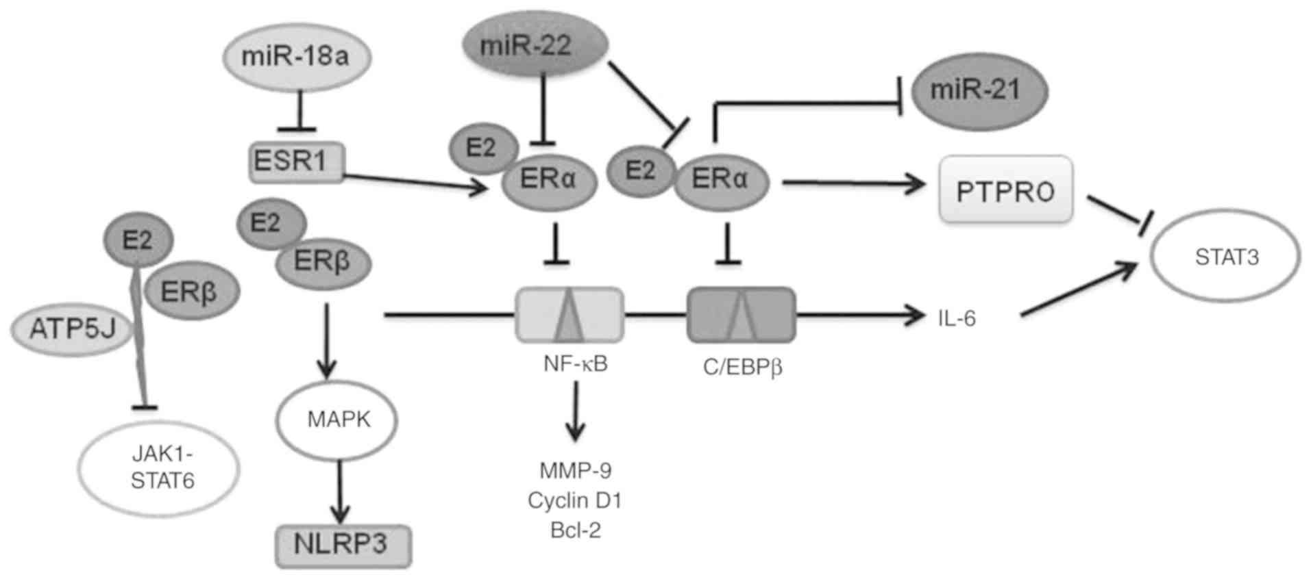
With different intracellular expression patterns in
the nucleus, cytoplasm or membrane, estrogen is involved in various
cellular processes including proliferation, survival, apoptosis and
differentiation through the estrogen receptors (ERs) (26). ERα and ERβ, two forms of ERs, share
significant structural homology and ligand binding properties, and
yet function very differently. As in breast cancer, aberrant
increases in ER gene expression have been reported in liver
tumors compared with normal or non-tumorous liver in patients with
HCC (27). ERα-mediated inhibition
of nuclear factor-κB binding activity is a pivotal event in the
process of inhibiting tumor formation (28). A previous study suggested that the
malignant behavior of HCC cells is markedly suppressed by treatment
with E2 through the E2/ERβ/mitogen-activated protein kinase (MAPK)
pathway-mediated increase of the nucleotide-binding domain,
leucine-rich-containing family, pyrin domain-containing-3
inflammasome (29). ERα transfection
effectively promotes the upregulation of estrogen to protein
tyrosine phosphatase receptor type O (PTPRO) in HCC cell lines and
it is positively correlated with the expression of ERα and PTPRO in
liver tissues (30). It has also
been identified that estrogen functions as a suppressor of
macrophage alternative activation and tumor progression by
preventing ERβ-adenosine triphosphate 5J interaction, thus
inhibiting the Janus kinase 1/signal transducer and activator of
transcription 6 signaling pathway (Fig.
1) (31). Other studies revealed
that ER inhibited the proliferation and invasion of human HCC cells
by decreasing the transcription of metastatic tumor antigen 1 and
peroxisome proliferator activated receptor γ (32,33).
miRNAs are small noncoding RNAs of ~20 nucleotides
that bind to conserved 3′-untranslated region sequences of their
target mRNAs and induce the inhibition of their translation
(34). Thereby miRNAs regulate gene
transcription and expression to modulate important physiological
functions (35,36). miRNAs serve a vital role in numerous
pathological events and in the cell response to various stresses
(35). In the hepatocarcinogenic
process, numerous miRNAs show abnormal expression in HCC tissues
compared with paired adjacent nontumorous tissues. Therefore,
miRNAs are recognized as a group of host genetic factors associated
with hepatocarcinogenesis (36–38). The
cross-linking of some miRNAs with ER is involved in the sex
difference in HCC. Zheng et al (22) concluded the correlation between some
miRNAs and sex disparity in HCC, including miR-23a, miR-545 and
miR-221. Other miRNAs associated with sex disparity in HCC will be
discussed in the current review (Fig.
1). miR-21 exhibits reduced mRNA binding and silencing activity
in healthy mouse liver, but its expression is significantly
elevated in HCC (39). Teng et
al (23) reported that
dehydroepiandrosterone, a precursor for adrenal androgen
biosynthesis, activates ERβ and androgen receptors and increases
miR-21 transcription. On the contrary, E2 inhibits miR-21
expression via ERα (23). The role
of circulating miR-22, as an independent prognostic marker of poor
clinical outcome, has been demonstrated by Cox regression analysis
(40). Jiang et al (41) demonstrated that overexpression of
miR-22 in male tumor-adjacent tissue was associated with
downregulated ERα expression by targeting its 3′-untranslated
region. miR-22 suppresses ER transcription and attenuates the
protective effect of estrogen, eventually increasing interleukin
(IL)-1α expression. The persistently high level of IL-1α may lead
to compensatory proliferation and tumorigenesis (41). In addition, by comparing the
expression pattern of miRNAs between male and female patients with
HCC, miR-18a was identified to be increased in female HCCs.
Furthermore, miR-18a targets the estrogen receptor 1 gene, which
encodes the ERα protein, and prevents translation of ER,
preferentially blocking the protective effects of estrogen and
promoting the development of HCC in women (42). In addition, elevated p53 promotes
miR-18a processing to decrease the expression level of ERα in
female patients with HCC, thereby suppressing the tumor-protective
function of the estrogen pathway (43). The production of estrogen is
associated with steroidogenesis pathways, including steroidogenesis
enzymes (44). However, to the best
of our knowledge, there have been no reports regarding the
interaction of miRNAs with steroidogenesis genes involved in sex
disparity in HCC.
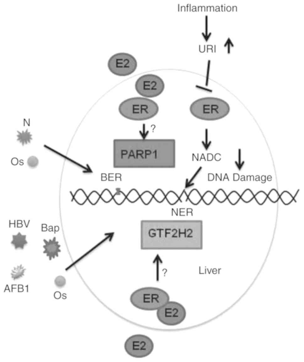
Genetic alterations and genomic instability,
possibly resulting from unrepaired DNA lesions, are increasingly
recognized as a common feature of human HCC (45,46). In
particular, next-generation sequencing technologies have revealed
numerous genetic alterations, including recurrently mutated genes
and dysregulated signaling pathways in HCC (45). Therefore, timely repair of DNA damage
is necessary. However, whether DNA damage repair is involved in the
sex disparity in HCC is largely unclear. The potential association
between estrogen and genomic instability is worth exploring.
Previous studies have reported the development of HCC from the
aspects of DNA damage repair-associated genes, including poly
(ADP-ribose) polymerase (PARP1), transcription factor IIH (TFIIH)
and nicotinamide adenine dinucleotide (NADC) (47–49),
which were associated with estrogen signaling pathways (49–51). The
present review explored the roles of these DNA repair associated
genes in the sex disparity of HCC (Fig.
2).
PARP1, a well-known DNA-binding enzyme, has a
potential role in DNA repair, especially in triggering the
base-excision repair process in the early stage of oxidative DNA
damage repair (52). PARP1 is also
involved in a variety of other biological processes, including
transcriptional regulation, apoptosis, mitosis and protein
degradation (53). The hepatitis B
virus (HBV) core promoter region binds to PARP1 and inhibits the
DNA repair capacity of PARP1, potentially disrupting host DNA
damage repair (54). PARP-1 is
downregulated in HBV-infected patients compared with uninfected
controls (55). It has been reported
that the physical interaction of hepatitis B virus X protein (HBX)
and PARP1 accelerated DNA damage by inhibiting recruitment of the
DNA repair complex to damaged DNA sites, which lead to
hepatocarcinogenesis (47). In
breast tissue, there is a positive association between PARP1 and ER
expression (50). However, there are
few studies on the association between ER and PARP1 in HCC, and
this merits further exploration.
Research implies that HBX impedes the DNA repair
process via its physical interactions with the helical components
of TFIIH, including excision repair cross-complementing rodent
repair deficiency, complementation groups 2 and 3 proteins
(56). TFIIH is a multiprotein
complex of 10 polypeptides and has clearly been shown to be an
integral component of the DNA repair pathway (57,58). Lee
et al (59) reported the
interaction of HBX with a probable cellular repair protein
UV-damaged DNA-binding protein, which acts as an essential factor
in HBV-associated hepatocarcinogenesis. General transcription
factor IIH subunit (GTF2H) is located on 5q13.2 and encodes the
44-kDa RNA polymerase II TFIIH protein subunit 2 that interacts
with other TFIIH subunits in the nucleotide excision repair
pathway. Zhao et al (48)
identified 30 (36.1%) of 83 HCC cases with loss of heterogeneity at
5q13.2, in which the tumor-associated gene GTF2H2 was
present. GTF2H2 is an estrogen signaling pathway gene in
breast cancer and is downregulated by luteolin (51). Therefore, the sex disparity in HCC
partly attributed to GTF2H2 is increasingly plausible.
In the early stage of many types of cancer,
including HCC, oncogene activation induces replication stress,
resulting in DNA damage and chromosomal instability and
acceleration of tumor development. Tummala et al (49) reported that increasing
NAD+ concentration is a critical mechanism in the
prevention of HCC. They described that unconventional prefoldin
RPB5 interactor (URI) inhibits the aryl hydrocarbon receptor (AhR)
and ER-mediated transcription of enzymes implicated in
NAD+ metabolism and synthesis, which causes DNA damage
in the early stages of tumorigenesis (49). Djouder (60) proposed boosting NAD+ as a
strategy to prevent and cure HCC and revealed that the activation
of AhR and ER was beneficial in HCC. Tummala et al (49) reported that AhR and ER could reverse
URI-induced transcription of L-tryptophan/kynurenine catabolism and
reduce the expression of tryptophan 2,3-dioxygenase through
establishing AhR and ER knockout mice and conducting experiments in
which AhR and ER were depleted in HepG2 cells.
Unhealthy lifestyles including smoking and alcohol
consumption are more prevalent among males compared with females,
and are also speculated to be susceptibility factors for sex
disparity of HCC (61). Obesity is a
significant risk factor for certain types of cancer, including HCC
(61,62). Park et al (63) described that both dietary and genetic
obesity enhance the inflammation-dependent increase in IL-6 and
tumor necrosis factor expression and promote liver inflammation and
tumorigenesis. Leptin, a 16 kD protein hormone secreted by white
adipose tissue, participates in the regulation of numerous
physiological functions including atherosclerosis and
carcinogenesis (64). Abnormal
regulation of leptin-signaling serves a crucial role in
obesity-associated liver cancer (64,65).
Shen and Shi (25) investigated the
function of E2 in opposing oncogenic actions of leptin in HepG2
cells, which are poor host cells for supporting the replication of
HBV or hepatitis C virus. The researchers used small
interfering-RNAs specific for ER-α, ER-β and G protein-coupled ER
(GPER) to verify that E2 decreased activation of the
leptin-signaling pathway through its receptors (25). E2 enhanced the activity of
extracellular signal-regulated kinase via activation of ER-α and
GPER and upregulated p38/MAPK via activation of ERβ. These
responses reversed leptin-induced alterations, eventually
inhibiting cell proliferation and stimulating cell apoptosis
(25).
Androgens are male hormones that have been
increasingly reported in male-predominant HCC (66,67).
They are mainly involved in various physiological and pathological
activities by combining with androgen receptors (ARs) (68,69). A
study by Wu et al (70)
identified that overexpression of ARs enhanced HCC cell growth and
invasion in vitro, and HCC initiation in vivo.
Previous studies have reported higher androgen levels and more
active androgen response elements (AREs) in liver tumor tissues,
compared with control tissues (8,71).
Further investigation revealed that when male mice with AR knockout
were induced by diethylnitrosamine (DEN), fewer tumors formed
compared with wild-type mice (72).
Androgen binding directly to AREs in the enhancer I of HBV genes
activated the androgen-signaling pathway and increased the rate of
HBV-induced hepatocarcinogenesis (8,73). AR
binding to ARE of the cell cycle related kinase promoter region
controls activation of the β-catenin/T-cell factor signaling
pathway, and has been identified as a major carcinogenic event and
described in animal models and up to 90% of HCC cases (74). Ligand-stimulated AR upregulated
miR-216a, resulting in tumorigenesis, and AR and miR-216a were
concordantly over-expressed in clinical specimens (38). Both activity and secretion of
aromatase, an enzyme which converts androgens to estrogens, was
markedly increased in human HCC tissues and HepG2 cells (75,76).
This contradicts the protective effect of estrogen and promoting
effect of androgen, and further studies are required to verify this
observation.
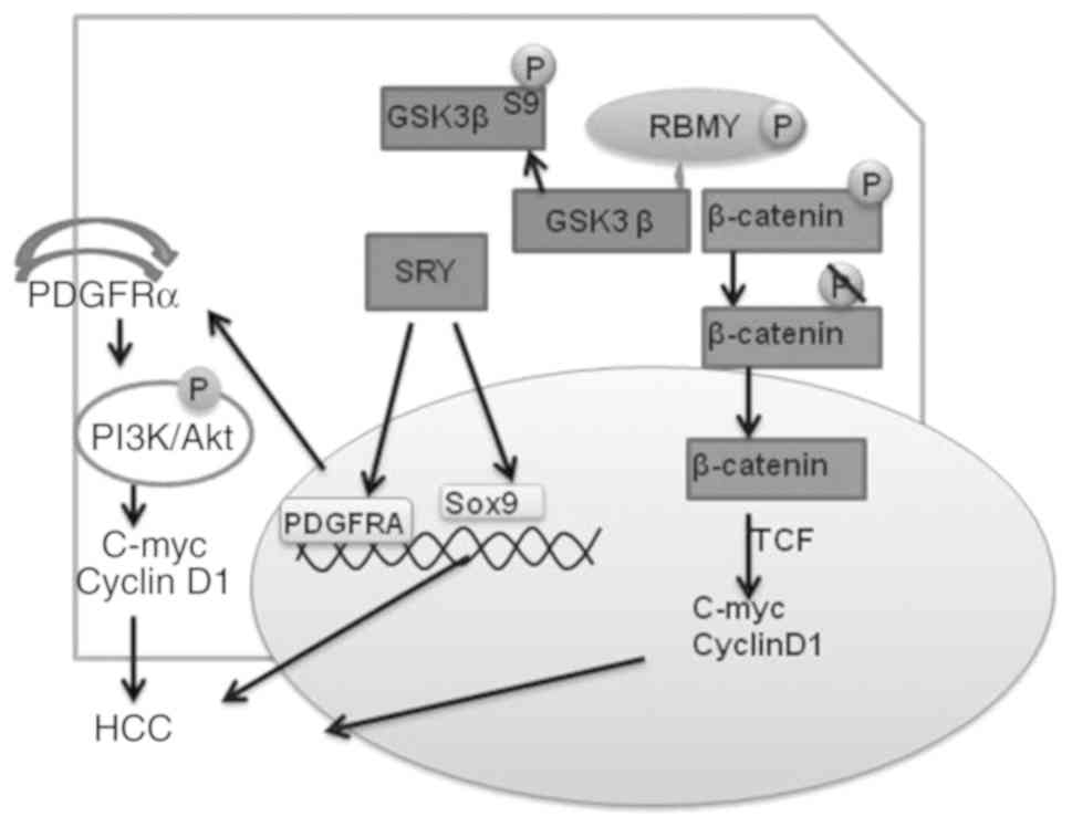
Previous studies have revealed that genetic
alterations of chromosomes X and Y are frequently observed in
patients with HCC, including chromosome-specific gene change,
oncogene and/or tumor suppressor gene expression and structural
rearrangements of chromosomes (9,11,77).
This indicates that genes located on sex chromosomes may be
responsible for HCC (78,79).
X-chromosome-coupled zinc finger protein is
abundantly expressed in HCC cells, and is associated with the
proliferation and survival of tumor cells (77). In addition, mRNA and protein levels
of dosage-sensitive sex reversal adrenal hypoplasia congenital
critical region on X chromosome, gene 1 (DAX-1), are downregulated
in HCC tissues and cell lines (10).
DAX-1 is known for its fundamental roles in sex steroid-dependent
neoplasms and interacts with β-catenin to attenuate its
transcriptional activity (80,81).
Jiang et al (10) first
reported the role of the DAX-1/β-catenin molecular network in
controlling HCC development. Furthermore, it was revealed that
DAX-1 is regulated by androgens (82).
SRY has been recognized as an oncogene and cancer
stem cell promoter in male HCC in in vitro studies (85,86). Liu
et al (9) reported
overexpression of SRY in ~84% of male patients with HCC. A
liver-specific transgenic murine model with overexpression of SRY
was susceptible to DEN-induced hepatocarcinogenesis compared with
age- and sex-matched wild-type mice (9). SRY activates its downstream target SOX9
and the platelet-derived growth factor receptor α/phosphoinositide
3-kinase/protein kinase B pathway, which stimulates the expression
of proliferation-associated genes MYC and cyclin D1
(CCND1), eventually accelerating tumorigenesis (9).
Oncogenic activation of RBMY is an important factor
in hepatocarcinogenesis, and a marked increase of cytoplasmic and
nuclear RBMY has been noted in HCC tissues (11,87). The
cytoplasmic expression of RBMY is associated with poor prognosis
and decreased survival rate in patients with HCC (11). Cytoplasmic RBMY competes with the
β-catenin destruction complex for binding GSK3b and enhancing the
phosphorylation of glycogen synthase kinase 3β Ser9 residue, which
eventually induces nuclear entry of β-catenin for transcription of
downstream oncogenes (11). The
tumorigenicity of RBMY has also been demonstrated though its
ability to induce cell transformation and tumor formation in nude
mice, and RBMY transgenic mice exhibited an increased DEN-induced
liver cancer incidence (83).
The genomic imbalances in HCC tissues have been
studied mostly by comparative genomic hybridization (CGH) (88,89). Y
chromosome loss and other genomic alterations in HCC cell lines
were analyzed by CGH and CGH array by Park et al (78). Park et al (78) detected the karyotypes of 21 male HCC
cell lines and identified 18 HCC cell lines with Y chromosome loss,
which may be responsible for the male preponderance in HCC. In
addition, increased copy number of several genes, CCND1 and
fibroblast growth factor 3/4 at 11q13, sarcoma amplified
sequence/cyclin-dependent kinase 4 at 12q13, telomerase RNA
component at 3q26, MET at 7q31, and MYC at 8q24, were
identified in 20 primary HCC tissues (90).
HCC is characterized by an apparent sex disparity
for which there lacks a clear mechanistic understanding. This
current review summarized the recent research exploring the role of
sex hormones and sex chromosomes in this process. Sex hormones and
their receptors constitute two tumor-promoting and inhibiting axes
through different channels. Genetic alterations in sex chromosomes
could also contribute to the underlying mechanism of the sex
disparity in HCC. In summary, the sex disparity in HCC is
attributed to multiple mechanisms, and the targeting of both sex
hormones and sex chromosomes is a novel and promising therapeutic
approach for patients with HCC.
The authors would like to thank Dr Cathel Kerr for
editing the English text of a draft of this manuscript.
The current study was supported by grants from the
Nature Science Foundation of China (grant no. 81650014) and Beijing
Natural Science Foundation (grant no. 7132058).
The datasets used during the present study are
available from the corresponding author upon reasonable
request.
JH designed the study and revised it. YL, AX and SJ
drafted the manuscript and revised the manuscript. All authors read
and approved the manuscript and agree to be accountable for all
aspects of the research in ensuring that the accuracy or integrity
of any part of the work are appropriately investigated and
resolved.
This review does not contain any studies with human
participants or animals performed by any of the authors.
Not applicable.
The authors declare that they have no competing
interests.
|
1
|
Torre LA, Bray F, Siegel RL, Ferlay J,
Lortet-Tieulent J and Jemal A: Global cancer statistics, 2012. CA
Cancer J Clin. 65:87–108. 2015. View Article : Google Scholar : PubMed/NCBI
|
|
2
|
Ascha MS, Hanouneh IA, Lopez R, Tamimi TA,
Feldstein AF and Zein NN: The incidence and risk factors of
hepatocellular carcinoma in patients with nonalcoholic
steatohepatitis. Hepatology. 51:1972–1978. 2010. View Article : Google Scholar : PubMed/NCBI
|
|
3
|
El-Serag HB: Epidemiology of viral
hepatitis and hepatocellular carcinoma. Gastroenterology.
142:1264–1273.e1. 2012. View Article : Google Scholar : PubMed/NCBI
|
|
4
|
Torre LA, Siegel RL, Ward EM and Jemal A:
Global cancer incidence and mortality rates and trends-an update.
Cancer Epidemiol Biomarkers Prev. 25:16–27. 2016. View Article : Google Scholar : PubMed/NCBI
|
|
5
|
Siegel RL, Miller KD and Jemal A: Cancer
statistics, 2017. CA Cancer J Clin. 67:7–30. 2017. View Article : Google Scholar : PubMed/NCBI
|
|
6
|
Chen W, Zheng R, Baade PD, Zhang S, Zeng
H, Bray F, Jemal A, Yu XQ and He J: Cancer statistics in China,
2015. CA Cancer J Clin. 66:115–132. 2016. View Article : Google Scholar : PubMed/NCBI
|
|
7
|
Naugler WE, Sakurai T, Kim S, Maeda S, Kim
K, Elsharkawy AM and Karin M: Gender disparity in liver cancer due
to sex differences in MyD88-dependent IL-6 production. Science.
317:121–124. 2007. View Article : Google Scholar : PubMed/NCBI
|
|
8
|
Wang SH, Yeh SH, Lin WH, Wang HY, Chen DS
and Chen PJ: Identification of androgen response elements in the
enhancer I of hepatitis B virus: A mechanism for sex disparity in
chronic hepatitis B. Hepatology. 50:1392–1402. 2009. View Article : Google Scholar : PubMed/NCBI
|
|
9
|
Liu C, Ren YF, Dong J, Ke MY, Ma F, Monga
SPS, Wu R, Lv Y and Zhang XF: Activation of SRY accounts for
male-specific hepatocarcinogenesis: Implication in gender disparity
of hepatocellular carcinoma. Cancer Lett. 410:20–31. 2017.
View Article : Google Scholar : PubMed/NCBI
|
|
10
|
Jiang HL, Xu D, Yu H, Ma X, Lin GF, Ma DY
and Jin JZ: DAX-1 inhibits hepatocellular carcinoma proliferation
by inhibiting β-catenin transcriptional activity. Cell Physiol
Biochem. 34:734–742. 2014. View Article : Google Scholar : PubMed/NCBI
|
|
11
|
Chua HH, Tsuei DJ, Lee PH, Jeng YM, Lu J,
Wu JF, Su DS, Chen YH, Chien CS, Kao PC, et al: RBMY, a novel
inhibitor of glycogen synthase kinase 3β, increases tumor stemness
and predicts poor prognosis of hepatocellular carcinoma.
Hepatology. 62:1480–1496. 2015. View Article : Google Scholar : PubMed/NCBI
|
|
12
|
Bertani S, Pineau P, Loli S, Moura J,
Zimic M, Deharo E and Ruiz E: An atypical age-specific pattern of
hepatocellular carcinoma in Peru: A threat for Andean populations.
PLoS One. 8:e677562013. View Article : Google Scholar : PubMed/NCBI
|
|
13
|
Hassan MM, Botrus G, Abdel-Wahab R, Wolff
RA, Li D, Tweardy D, Phan AT, Hawk E, Javle M, Lee JS, et al:
Estrogen replacement reduces risk and increases survival times of
women with hepatocellular carcinoma. Clin Gastroenterol Hepatol.
15:1791–1799. 2017. View Article : Google Scholar : PubMed/NCBI
|
|
14
|
Yu MW, Chang HC, Chang SC, Liaw YF, Lin
SM, Liu CJ, Lee SD, Lin CL, Chen PJ, Lin SC and Chen CJ: Role of
reproductive factors in hepatocellular carcinoma: Impact on
hepatitis B- and C-related risk. Hepatology. 38:1393–1400. 2003.
View Article : Google Scholar : PubMed/NCBI
|
|
15
|
Bigsby RM and Caperell-Grant A: The role
for estrogen receptor-alpha and prolactin receptor in sex-dependent
DEN-induced liver tumorigenesis. Carcinogenesis. 32:1162–1166.
2011. View Article : Google Scholar : PubMed/NCBI
|
|
16
|
Prieto J: Inflammation, HCC and sex: IL-6
in the centre of the triangle. J Hepatol. 48:380–381. 2008.
View Article : Google Scholar : PubMed/NCBI
|
|
17
|
Wang YC, Xu GL, Jia WD, Han SJ, Ren WH,
Wang W, Liu WB, Zhang CH and Chen H: Estrogen suppresses metastasis
in rat hepatocellular carcinoma through decreasing interleukin-6
and hepatocyte growth factor expression. Inflammation. 35:143–149.
2012. View Article : Google Scholar : PubMed/NCBI
|
|
18
|
Coussens LM and Werb Z: Inflammation and
cancer. Nature. 420:860–867. 2002. View Article : Google Scholar : PubMed/NCBI
|
|
19
|
Karin M: Nuclear factor-kappaB in cancer
development and progression. Nature. 441:431–436. 2006. View Article : Google Scholar : PubMed/NCBI
|
|
20
|
Shi L, Feng Y, Lin H, Ma R and Cai X: Role
of estrogen in hepatocellular carcinoma: Is inflammation the key? J
Transl Med. 12:932014. View Article : Google Scholar : PubMed/NCBI
|
|
21
|
Ren J, Chen GG, Liu Y, Su X, Hu B, Leung
BC, Wang Y, Ho RL, Yang S, Lu G, et al: Cytochrome P450 1A2
metabolizes 17β-estradiol to suppress hepatocellular carcinoma.
PLoS One. 11:e01538632016. View Article : Google Scholar : PubMed/NCBI
|
|
22
|
Zheng B, Zhu YJ, Wang HY and Chen L:
Gender disparity in hepatocellular carcinoma (HCC): Multiple
underlying mechanisms. Sci China Life Sci. 60:575–584. 2017.
View Article : Google Scholar : PubMed/NCBI
|
|
23
|
Teng Y, Litchfield LM, Ivanova MM, Prough
RA, Clark BJ and Klinge CM: Dehydroepiandrosterone-induces miR-21
transcription in HepG2 cells through estrogen receptor β and
androgen receptor. Mol Cell Endocrinol. 392:23–36. 2014. View Article : Google Scholar : PubMed/NCBI
|
|
24
|
Shen M, Cao J and Shi H: Effects of
estrogen and estrogen receptors on transcriptomes of HepG2 Cells: A
preliminary study using RNA sequencing. Int J Endocrinol.
2018:57891272018. View Article : Google Scholar : PubMed/NCBI
|
|
25
|
Shen M and Shi H: Estradiol and estrogen
receptor agonists oppose oncogenic actions of leptin in HepG2
cells. PLoS One. 11:e01514552016. View Article : Google Scholar : PubMed/NCBI
|
|
26
|
Pearce ST and Jordan VC: The biological
role of estrogen receptors alpha and beta in cancer. Crit Rev Oncol
Hematol. 50:3–22. 2004. View Article : Google Scholar : PubMed/NCBI
|
|
27
|
Iyer JK, Kalra M, Kaul A, Payton ME and
Kaul R: Estrogen receptor expression in chronic hepatitis C and
hepatocellular carcinoma pathogenesis. World J Gastroenterol.
23:6802–6816. 2017. View Article : Google Scholar : PubMed/NCBI
|
|
28
|
Xu H, Wei Y, Zhang Y, Xu Y, Li F, Liu J,
Zhang W, Han X, Tan R and Shen P: Oestrogen attenuates tumour
progression in hepatocellular carcinoma. J Pathol. 228:216–229.
2012. View Article : Google Scholar : PubMed/NCBI
|
|
29
|
Wei Q, Guo P, Mu K, Zhang Y, Zhao W, Huai
W, Qiu Y, Li T, Ma X, Liu Y, et al: Estrogen suppresses
hepatocellular carcinoma cells through ERβ-mediated upregulation of
the NLRP3 inflammasome. Lab Invest. 95:804–816. 2015. View Article : Google Scholar : PubMed/NCBI
|
|
30
|
Hou J, Xu J, Jiang R, Wang Y, Chen C, Deng
L, Huang X, Wang X and Sun B: Estrogen-sensitive PTPRO expression
represses hepatocellular carcinoma progression by control of STAT3.
Hepatology. 57:678–688. 2013. View Article : Google Scholar : PubMed/NCBI
|
|
31
|
Yang W, Lu Y, Xu Y, Xu L, Zheng W, Wu Y,
Li L and Shen P: Estrogen represses hepatocellular carcinoma (HCC)
growth via inhibiting alternative activation of tumor-associated
macrophages (TAMs). J Biol Chem. 287:40140–40149. 2012. View Article : Google Scholar : PubMed/NCBI
|
|
32
|
Deng L, Yang H, Tang J, Lin Z, Yin A, Gao
Y, Wang X, Jiang R and Sun B: Inhibition of MTA1 by ERα contributes
to protection hepatocellular carcinoma from tumor proliferation and
metastasis. J Exp Clin Cancer Res. 34:1282015. View Article : Google Scholar : PubMed/NCBI
|
|
33
|
Lin YM, Velmurugan BK, Yeh YL, Tu CC, Ho
TJ, Lai TY, Tsai CH, Tsai FJ, Tsai CH and Huang CY: Activation of
estrogen receptors with E2 downregulates peroxisome
proliferator-activated receptor γ in hepatocellular carcinoma.
Oncol Rep. 30:3027–3031. 2013. View Article : Google Scholar : PubMed/NCBI
|
|
34
|
Bartel DP and Chen CZ: Micromanagers of
gene expression: The potentially widespread influence of metazoan
microRNAs. Nat Rev Genet. 5:396–400. 2004. View Article : Google Scholar : PubMed/NCBI
|
|
35
|
Amodio G, Sasso E, D'Ambrosio C, Scaloni
A, Moltedo O, Franceschelli S, Zambrano N and Remondelli P:
Identification of a microRNA (miR-663a) induced by ER stress and
its target gene PLOD3 by a combined microRNome and proteome
approach. Cell Biol Toxicol. 32:285–303. 2016. View Article : Google Scholar : PubMed/NCBI
|
|
36
|
Sun L, Guo Z, Sun J, Li J, Dong Z, Zhang
Y, Chen J, Kan Q and Yu Z: MiR-133a acts as an anti-oncogene in
Hepatocellular carcinoma by inhibiting FOSL2 through TGF-β/Smad3
signaling pathway. Biomed Pharmacother. 107:168–176. 2018.
View Article : Google Scholar : PubMed/NCBI
|
|
37
|
Ladeiro Y, Couchy G, Balabaud C,
Bioulac-Sage P, Pelletier L, Rebouissou S and Zucman-Rossi J:
MicroRNA profiling in hepatocellular tumors is associated with
clinical features and oncogene/tumor suppressor gene mutations.
Hepatology. 47:1955–1963. 2008. View Article : Google Scholar : PubMed/NCBI
|
|
38
|
Chen PJ, Yeh SH, Liu WH, Lin CC, Huang HC,
Chen CL, Chen DS and Chen PJ: Androgen pathway stimulates
microRNA-216a transcription to suppress the tumor suppressor in
lung cancer-1 gene in early hepatocarcinogenesis. Hepatology.
56:632–643. 2012. View Article : Google Scholar : PubMed/NCBI
|
|
39
|
Sun J, Lu H, Wang X and Jin H: MicroRNAs
in hepatocellular carcinoma: Regulation, function, and clinical
implications. ScientificWorldJournal. 2013:9242062013. View Article : Google Scholar : PubMed/NCBI
|
|
40
|
Marchesi F, Regazzo G, Palombi F,
Terrenato I, Sacconi A, Spagnuolo M, Donzelli S, Marino M, Ercolani
C, Di Benedetto A, et al: Serum miR-22 as potential non-invasive
predictor of poor clinical outcome in newly diagnosed, uniformly
treated patients with diffuse large B-cell lymphoma: An explorative
pilot study. J Exp Clin Cancer Res. 37:952018. View Article : Google Scholar : PubMed/NCBI
|
|
41
|
Jiang R, Deng L, Zhao L, Li X, Zhang F,
Xia Y, Gao Y, Wang X and Sun B: miR-22 promotes HBV-related
hepatocellular carcinoma development in males. Clin Cancer Res.
17:5593–5603. 2011. View Article : Google Scholar : PubMed/NCBI
|
|
42
|
Liu WH, Yeh SH, Lu CC, Yu SL, Chen HY, Lin
CY, Chen DS and Chen PJ: MicroRNA-18a prevents estrogen
receptor-alpha expression, promoting proliferation of
hepatocellular carcinoma cells. Gastroenterology. 136:683–693.
2009. View Article : Google Scholar : PubMed/NCBI
|
|
43
|
Li CL, Yeh KH, Liu WH, Chen CL, Chen DS,
Chen PJ and Yeh SH: Elevated p53 promotes the processing of miR-18a
to decrease estrogen receptor-α in female hepatocellular carcinoma.
Int J Cancer. 136:761–770. 2015. View Article : Google Scholar : PubMed/NCBI
|
|
44
|
Luu-The V: Assessment of steroidogenesis
and steroidogenic enzyme functions. J Steroid Biochem Mol Biol.
137:176–182. 2013. View Article : Google Scholar : PubMed/NCBI
|
|
45
|
Niu ZS, Niu XJ and Wang WH: Genetic
alterations in hepatocellular carcinoma: An update. World J
Gastroenterol. 22:9069–9095. 2016. View Article : Google Scholar : PubMed/NCBI
|
|
46
|
Aleksic K, Lackner C, Geigl JB, Schwarz M,
Auer M, Ulz P, Fischer M, Trajanoski Z, Otte M and Speicher MR:
Evolution of genomic instability in diethylnitrosamine-induced
hepatocarcinogenesis in mice. Hepatology. 53:895–904. 2011.
View Article : Google Scholar : PubMed/NCBI
|
|
47
|
Na TY, Ka NL, Rhee H, Kyeong D, Kim MH,
Seong JK, Park YN and Lee MO: Interaction of hepatitis B virus X
protein with PARP1 results in inhibition of DNA repair in
hepatocellular carcinoma. Oncogene. 35:5435–5445. 2016. View Article : Google Scholar : PubMed/NCBI
|
|
48
|
Zhao Z, Chen GY, Long J, Li H and Huang J:
Genomic losses at 5q13.2 and 8p23.1 in dysplastic hepatocytes are
common events in hepatitis B virus-related hepatocellular
carcinoma. Oncol Lett. 9:2839–2846. 2015. View Article : Google Scholar : PubMed/NCBI
|
|
49
|
Tummala KS, Gomes AL, Yilmaz M, Graña O,
Bakiri L, Ruppen I, Ximénez-Embún P, Sheshappanavar V,
Rodriguez-Justo M, Pisano DG, et al: Inhibition of de novo NAD(+)
synthesis by oncogenic URI causes liver tumorigenesis through DNA
damage. Cancer Cell. 26:826–839. 2014. View Article : Google Scholar : PubMed/NCBI
|
|
50
|
Mazzotta A, Partipilo G, De Summa S,
Giotta F, Simone G and Mangia A: Nuclear PARP1 expression and its
prognostic significance in breast cancer patients. Tumour Biol.
37:6143–6153. 2016. View Article : Google Scholar : PubMed/NCBI
|
|
51
|
Markaverich BM, Shoulars K and Rodriguez
MA: Luteolin regulation of estrogen signaling and cell cycle
pathway genes in MCF-7 human breast cancer cells. Int J Biomed Sci.
7:101–111. 2011.PubMed/NCBI
|
|
52
|
Dantzer F, Schreiber V, Niedergang C,
Trucco C, Flatter E, De La Rubia G, Oliver J, Rolli V, Ménissier-de
Murcia J and de Murcia G: Involvement of poly(ADP-ribose)
polymerase in base excision repair. Biochimie. 81:69–75. 1999.
View Article : Google Scholar : PubMed/NCBI
|
|
53
|
Song D, Huang H, Wang J, Zhao Y, Hu X, He
F, Yu L and Wu J: NF90 regulates PARP1 mRNA stability in
hepatocellular carcinoma. Biochem Biophys Res Commun. 488:211–217.
2017. View Article : Google Scholar : PubMed/NCBI
|
|
54
|
Ko HL and Ren EC: Novel poly (ADP-ribose)
polymerase 1 binding motif in hepatitis B virus core promoter
impairs DNA damage repair. Hepatology. 54:1190–1198. 2011.
View Article : Google Scholar : PubMed/NCBI
|
|
55
|
Mukherjee RM, Shravanti GV, Jakkampudi A,
Kota R, Jangala AL, Reddy PB, Rao PN, Gupta R and Reddy DN: Reduced
expression of DNA damage repair genes high mobility group box1 and
Poly(ADP-ribose) polymerase1 in inactive carriers of hepatitis B
virus infection-A possible stage of viral integration. J Clin Exp
Hepatol. 3:89–95. 2013. View Article : Google Scholar : PubMed/NCBI
|
|
56
|
Qadri I, Fatima K and AbdeL-Hafiz H:
Hepatitis B virus X protein impedes the DNA repair via its
association with transcription factor, TFIIH. BMC Microbiol.
11:482011. View Article : Google Scholar : PubMed/NCBI
|
|
57
|
Hashimoto S and Egly JM:
Trichothiodystrophy view from the molecular basis of DNA
repair/transcription factor TFIIH. Hum Mol Genet. 18:R224–R230.
2009. View Article : Google Scholar : PubMed/NCBI
|
|
58
|
Schärer OD: Hot topics in DNA repair: The
molecular basis for different disease states caused by mutations in
TFIIH and XPG. DNA Repair (Amst). 7:339–344. 2008. View Article : Google Scholar : PubMed/NCBI
|
|
59
|
Lee TH, Elledge SJ and Butel JS: Hepatitis
B virus X protein interacts with a probable cellular DNA repair
protein. J Virol. 69:1107–1114. 1995.PubMed/NCBI
|
|
60
|
Djouder N: Boosting NAD(+) for the
prevention and treatment of liver cancer. Mol Cell Oncol.
2:e10011992015. View Article : Google Scholar : PubMed/NCBI
|
|
61
|
Boniol M and Autier P: Prevalence of main
cancer lifestyle risk factors in Europe in 2000. Eur J Cancer.
46:2534–2544. 2010. View Article : Google Scholar : PubMed/NCBI
|
|
62
|
Grohmann M, Wiede F, Dodd GT, Gurzov EN,
Ooi GJ, Butt T, Rasmiena AA, Kaur S, Gulati T, Goh PK, et al:
Obesity drives STAT-1-dependent NASH and STAT-3-dependent HCC.
Cell. 175:1289–1306 e20. 2018. View Article : Google Scholar : PubMed/NCBI
|
|
63
|
Park EJ, Lee JH, Yu GY, He G, Ali SR,
Holzer RG, Osterreicher CH, Takahashi H and Karin M: Dietary and
genetic obesity promote liver inflammation and tumorigenesis by
enhancing IL-6 and TNF expression. Cell. 140:197–208. 2010.
View Article : Google Scholar : PubMed/NCBI
|
|
64
|
Xiong Y, Zhang J, Liu M, An M, Lei L and
Guo W: Human leptin protein activates the growth of HepG2 cells by
inhibiting PERK-mediated ER stress and apoptosis. Mol Med Rep.
10:1649–1655. 2014. View Article : Google Scholar : PubMed/NCBI
|
|
65
|
Vansaun MN, Mendonsa AM and Lee Gorden D:
Hepatocellular proliferation correlates with inflammatory cell and
cytokine changes in a murine model of nonalchoholic fatty liver
disease. PLoS One. 8:e730542013. View Article : Google Scholar : PubMed/NCBI
|
|
66
|
Li S, Mo C, Huang S, Yang S, Lu Y, Peng Q,
Wang J, Deng Y, Qin X and Liu Y: Over-expressed Testis-specific
Protein Y-encoded 1 as a novel biomarker for male hepatocellular
carcinoma. PLoS One. 9:e892192014. View Article : Google Scholar : PubMed/NCBI
|
|
67
|
Montella M, D'Arena G, Crispo A, Capunzo
M, Nocerino F, Grimaldi M, Barbieri A, D'Ursi AM, Tecce MF, Amore
A, et al: Role of sex hormones in the development and progression
of hepatitis B virus-associated hepatocellular carcinoma. Int J
Endocrinol. 2015:8545302015. View Article : Google Scholar : PubMed/NCBI
|
|
68
|
Beato M and Klug J: Steroid hormone
receptors: An update. Hum Reprod Update. 6:225–236. 2000.
View Article : Google Scholar : PubMed/NCBI
|
|
69
|
Chang CS, Kokontis J and Liao ST:
Molecular cloning of human and rat complementary DNA encoding
androgen receptors. Science. 240:324–326. 1988. View Article : Google Scholar : PubMed/NCBI
|
|
70
|
Wu MH, Ma WL, Hsu CL, Chen YL, Ou JH, Ryan
CK, Hung YC, Yeh S and Chang C: Androgen receptor promotes
hepatitis B virus-induced hepatocarcinogenesis through modulation
of hepatitis B virus RNA transcription. Sci Transl Med.
2:32ra352010. View Article : Google Scholar : PubMed/NCBI
|
|
71
|
Barone M, Margiotta M, Scavo MP, Gentile
A, Francioso D, Papagni S, Castellaneta A, Mallamaci R, Di Leo A
and Francavilla A: Possible involvement of androgen receptor
alterations in hepatocarcinogenesis. Dig Liver Dis. 41:665–670.
2009. View Article : Google Scholar : PubMed/NCBI
|
|
72
|
Ma WL, Hsu CL, Wu MH, Wu CT, Wu CC, Lai
JJ, Jou YS, Chen CW, Yeh S and Chang C: Androgen receptor is a new
potential therapeutic target for the treatment of hepatocellular
carcinoma. Gastroenterology. 135:947–955, 955.e1-5. 2008.
View Article : Google Scholar : PubMed/NCBI
|
|
73
|
Yang WJ, Chang CJ, Yeh SH, Lin WH, Wang
SH, Tsai TF, Chen DS and Chen PJ: Hepatitis B virus X protein
enhances the transcriptional activity of the androgen receptor
through c-Src and glycogen synthase kinase-3beta kinase pathways.
Hepatology. 49:1515–1524. 2009. View Article : Google Scholar : PubMed/NCBI
|
|
74
|
Feng H, Cheng AS, Tsang DP, Li MS, Go MY,
Cheung YS, Zhao GJ, Ng SS, Lin MC, Yu J, et al: Cell cycle-related
kinase is a direct androgen receptor-regulated gene that drives
β-catenin/T cell factor-dependent hepatocarcinogenesis. J Clin
Invest. 121:3159–3175. 2011. View Article : Google Scholar : PubMed/NCBI
|
|
75
|
Koh WP, Yuan JM, Wang R, Govindarajan S,
Oppenheimer R, Zhang ZQ, Yu MC and Ingles SA: Aromatase (CYP19)
promoter gene polymorphism and risk of nonviral hepatitis-related
hepatocellular carcinoma. Cancer. 117:3383–3392. 2011. View Article : Google Scholar : PubMed/NCBI
|
|
76
|
Castagnetta LA, Agostara B, Montalto G,
Polito L, Campisi I, Saetta A, Itoh T, Yu B, Chen S and Carruba G:
Local estrogen formation by nontumoral, cirrhotic, and malignant
human liver tissues and cells. Cancer Res. 63:5041–5045.
2003.PubMed/NCBI
|
|
77
|
Zhang S, Shu R, Yue M and Zhang S: Effect
of over-expression of zinc-finger protein (ZFX) on self-renewal and
drug-resistance of hepatocellular carcinoma. Med Sci Monit.
22:3025–3034. 2016. View Article : Google Scholar : PubMed/NCBI
|
|
78
|
Park SJ, Jeong SY and Kim HJ: Y chromosome
loss and other genomic alterations in hepatocellular carcinoma cell
lines analyzed by CGH and CGH array. Cancer Genet Cytogenet.
166:56–64. 2006. View Article : Google Scholar : PubMed/NCBI
|
|
79
|
Liu J, Wang ZM, Zhen SF, Wu XP, Ma DX, Li
ZH, Liu B, Zhao ZL and Ke Y: Aberration of X chromosome in liver
neoplasm detected by fluorescence in situ hybridization.
Hepatobiliary Pancreat Dis Int. 3:110–114. 2004.PubMed/NCBI
|
|
80
|
Lalli E and Alonso J: Targeting DAX-1 in
embryonic stem cells and cancer. Expert Opin Ther Targets.
14:169–177. 2010. View Article : Google Scholar : PubMed/NCBI
|
|
81
|
Li H, Zhang Z, Bi Y, Yang D, Zhang L and
Liu J: Expression characteristics of β-catenin in scallop Chlamys
farreri gonads and its role as a potential upstream gene of Dax1
through canonical Wnt signalling pathway regulating the
spermatogenesis. PLoS One. 9:e1159172014. View Article : Google Scholar : PubMed/NCBI
|
|
82
|
Campbell DEK and Langlois VS: Expression
of sf1 and dax-1 are regulated by thyroid hormones and androgens
during Silurana tropicalis early development. Gen Comp Endocrinol.
259:34–44. 2018. View Article : Google Scholar : PubMed/NCBI
|
|
83
|
Tsuei DJ, Lee PH, Peng HY, Lu HL, Su DS,
Jeng YM, Hsu HC, Hsu SH, Wu JF, Ni YH and Chang MH: Male germ
cell-specific RNA binding protein RBMY: A new oncogene explaining
male predominance in liver cancer. PLoS One. 6:e269482011.
View Article : Google Scholar : PubMed/NCBI
|
|
84
|
Yin YH, Li YY, Qiao H, Wang HC, Yang XA,
Zhang HG, Pang XW, Zhang Y and Chen WF: TSPY is a cancer testis
antigen expressed in human hepatocellular carcinoma. Br J Cancer.
93:458–463. 2005. View Article : Google Scholar : PubMed/NCBI
|
|
85
|
Murakami S, Chishima S, Uemoto H, Sakamoto
E, Sato T, Kurabe N, Kawasaki Y, Shibata T, Akiyama H and Tashiro
F: The male-specific factor Sry harbors an oncogenic function.
Oncogene. 33:2978–2986. 2014. View Article : Google Scholar : PubMed/NCBI
|
|
86
|
Murakami S, Ninomiya W, Sakamoto E,
Shibata T, Akiyama H and Tashiro F: SRY and OCT4 are required for
the acquisition of cancer stem cell-like properties and are
potential differentiation therapy targets. Stem Cells.
33:2652–2663. 2015. View Article : Google Scholar : PubMed/NCBI
|
|
87
|
Tsuei DJ, Hsu HC, Lee PH, Jeng YM, Pu YS,
Chen CN, Lee YC, Chou WC, Chang CJ, Ni YH and Chang MH: RBMY, a
male germ cell-specific RNA-binding protein, activated in human
liver cancers and transforms rodent fibroblasts. Oncogene.
23:5815–5822. 2004. View Article : Google Scholar : PubMed/NCBI
|
|
88
|
Homayounfar K, Schwarz A, Enders C,
Cameron S, Baumhoer D, Ramadori G, Lorf T, Gunawan B and Sander B:
Etiologic influence on chromosomal aberrations in European
hepatocellular carcinoma identified by CGH. Pathol Res Pract.
209:380–387. 2013. View Article : Google Scholar : PubMed/NCBI
|
|
89
|
Hashimoto K, Mori N, Tamesa T, Okada T,
Kawauchi S, Oga A, Furuya T, Tangoku A, Oka M and Sasaki K:
Analysis of DNA copy number aberrations in hepatitis C
virus-associated hepatocellular carcinomas by conventional CGH and
array CGH. Mod Pathol. 17:617–622. 2004. View Article : Google Scholar : PubMed/NCBI
|
|
90
|
Takeo S, Arai H, Kusano N, Harada T,
Furuya T, Kawauchi S, Oga A, Hirano T, Yoshida T, Okita K and
Sasaki K: Examination of oncogene amplification by genomic DNA
microarray in hepatocellular carcinomas: Comparison with
comparative genomic hybridization analysis. Cancer Genet Cytogenet.
130:127–132. 2001. View Article : Google Scholar : PubMed/NCBI
|