Introduction
Endometrial stromal tumors are rare tumors that
originate from the endometrial stroma, accounting for <2% of all
cases of uterine tumor (1,2). According to clinical, pathological and
genetic characteristics, the Fourth Edition of the World Health
Organization classification in 2014 (1) divided endometrial stromal tumors into
the following four categories: Endometrial stromal nodule,
undifferentiated uterine sarcoma, high-grade endometrial stromal
sarcoma (ESS) and low-grade ESS.
Low-grade ESS is a rare malignant interstitial
tumor, which often occurs in females between 40 and 55 years old
(3). Low-grade ESS is an inert tumor
with slow growth that has a favorable prognosis (4). In patients with low-grade ESS, 30–50%
of cases present with extrauterine spread at the time of the
diagnosis (3,5–7). Distant
metastases may develop even if the primary tumor is resected, and
the most common location of distant metastases is the lungs, with a
reported incidence rate of 7 to 28% (8).
Pulmonary metastasis of low-grade ESS can manifest
as various patterns on computed tomography (CT) images of the
chest, which may cause difficulties in making a diagnosis (8–12).
Clinical trials and data on the pulmonary metastasis of low-grade
ESS are extremely limited. The present study reports one case of
pulmonary metastasis of low-grade ESS that was treated at the
Chinese PLA General Hospital. In addition, previous literature on
the pulmonary metastasis of low-grade ESS was reviewed by searching
the PubMed database for pulmonary metastases of low-grade
endometrial stromal sarcoma. The clinical manifestations, imaging
data, pathological features, treatment and prognosis of these cases
(the present case and the reported cases retrieved from PubMed)
were retrospectively analyzed to investigate the clinical and
pathological features, and enhance the awareness of pulmonary
metastases in patients with low-grade ESS.
Case report
A 55-year-old female was admitted to Chinese
People's Liberation Army General Hospital (Beijing, China) in March
2017 due to a nodule in the left lung that had been present for 1
year along with recurrent chest tightness, and chest pain that had
been occurring for 6 months. In November 2015, the patient had
undergone a hysterectomy and bilateral salpingo-oophorectomy to
remove low-grade ESS. A pulmonary nodule (4 mm) was identified in
the left upper lobe on the chest CT scan during the perioperative
period, but the patient did not receive any treatment. The patient
had experienced chest tightness, shortness of breath and chest pain
since August 2016, and the chest CT scan revealed right-sided
pneumothorax. The patient was treated with closed thoracic drainage
and the pneumothorax subsequently improved. The patient experienced
pneumothorax a further three times over the following 6 months. The
nodule in the left lung was enlarged and multiple thin-walled cysts
in the bilateral lung fields were detected in February 2017.
The patient was admitted to the hospital for further
diagnosis and treatment following an existing diagnosis of cystic
lung disease and a nodule in the left lung. The physical
examination was unremarkable. No moist or dry rales were heard on
either side. The heart and abdominal examinations were negative.
The superficial lymph nodes were not palpable. The laboratory
examination demonstrated a normal white blood cell count. The
levels of C-reactive protein and procalcitonin were within normal
limits. However, the level of tumor marker cancer antigen 125 was
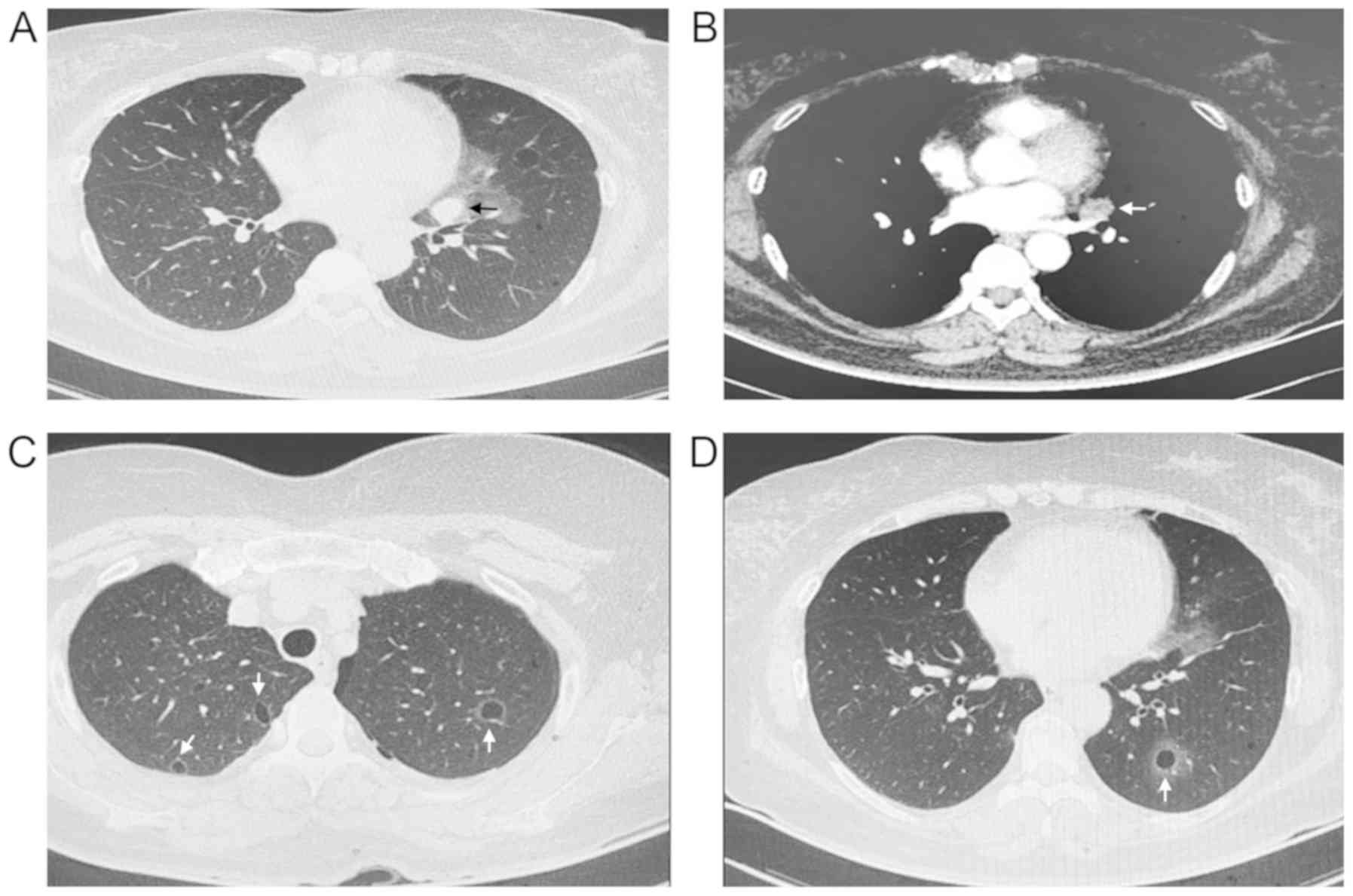
increased compared with the normal levels. The chest CT scan
revealed a solid nodule (2×2.5 cm) in the left upper lung and
multiple thin-walled cysts in the bilateral lung fields (Fig. 1).
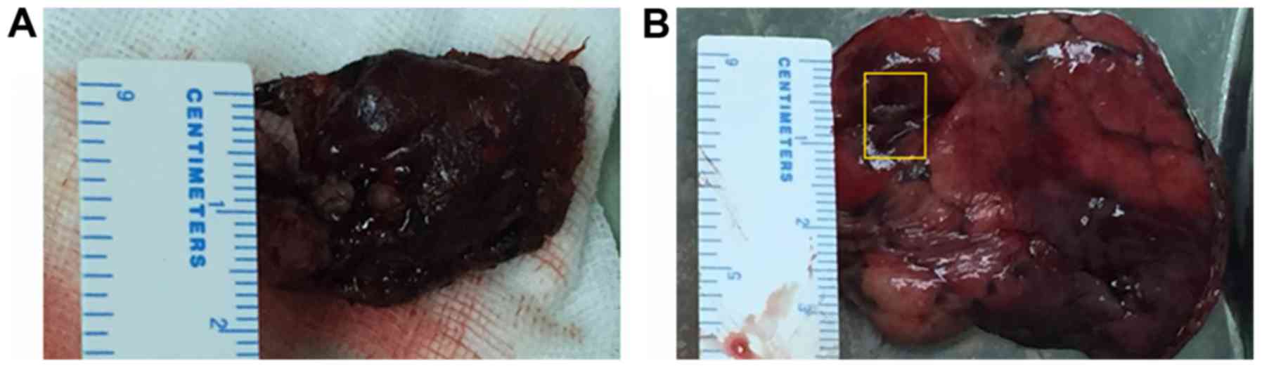
Video-assisted thoracoscopic surgery (VATS) was
performed in March 2017 to make a further diagnosis. Consequently,
resections of the solid nodule (2.5×2×0.8 cm) from the upper lobe
(Fig. 2A) and the apex of the left
lung, including the thin-walled cysts (Fig. 2B), were performed.
For histopathological and immunohistochemical
evaluation of the resected lung nodule, the material from the
surgical specimen was fixed in 10% neutral buffered formalin (cat.
no., HT 501128; Sigma-Aldrich; EMD Millipore) for 24 h, at room
temperature, embedded in paraffin, cut into 4 µm-thick sections,
and then hematoxylin and eosin (H&E) and immunohistochemical
(IHC) staining were performed.
In H&E staining, paraffin lung nodule sections
were deparaffinized in 2 changes of xylene, 10 min each, at room
temperature. Sections were re-hydrated in 2 changes of alcohol for
5 min each. Alcohol series were as follows: 95% alcohol for 2 min
and 70% alcohol for 2 min. After washing in running tap water for 5
min, sections were stained with hematoxylin solution (cat, no.,
3095771; Sigma-Aldrich; Merck KGaA) for 5 min, at room temperature.
After washing in water for 5 min, lung nodule sections were
differentiated in 1% acid alcohol for 30 sec and washed with
running tap water for 1 min. Slides were stained with 5% eosin
solution (cat, no. 318906; Sigma-Aldrich; Merck KGaA) for 1–3 min,
at room temperature. After washing in running tap water for 5 min,
lung nodule was dehydrated through 95% alcohol, 2 changes of
absolute alcohol for 5 min each. Finally, lung nodule sections were
cleared in 2 changes of xylene for 5 min each, and mounted with
xylene based mounting medium. H&E staining reaction was
observed using a standard light microscope (Leica, DM2000;
magnification 10×10; Leica Microsystems GmbH).
In IHC staining, the EnVision two-step
immunohistochemical staining technique (13) was used and the visualization system
was 3,3′-diaminobenzidine tetrahydrochloride (DAB). Sections were
dewaxed and hydrated. After endogenous peroxidase blocking with
EnVision Flex+ (cat, no., K8002; Dako; Agilent
Technologies, Inc.) for 5 min, at room temperature, the sections
were incubated with primary antibodies at an optimal dilution
(Table I), for 30 min, at room
temperature. After washing with PBS for 10 min, the sections were
incubated with a secondary antibody EnVision™ (cat, no., K8009;
Dako; Agilent Technologies, Inc.) for 30 min at room temperature.
After being washed with water, signal visualization was performed
with 3–3′diaminobenzidine DAB medium (D7034; Sigma-Aldrich; Merck
KGaA), for 5 min at room temperature. Finally, the sections were
washed and mounted. The immunohistochemical reaction was
interpreted using a standard light microscope (Leica, DM2000;
magnification 10×10; Leica Microsystems GMBH).
 | Table I.Primary antibodies used in
immunohistochemical analysis. |
Table I.
Primary antibodies used in
immunohistochemical analysis.
| Antibody | Dilution | Catalogue number | Supplier |
|---|
| CD10 | 1:00 | ab227659 | Abcam, Cambridge,
MA, USA |
| ER | 1:50 | sc787 | Santa Cruz
Biotechnology, CA, USA |
| PR | 1:50 | sc810 | Santa Cruz
Biotechnology, CA, USA |
| Vimentin | 1:200 | ab92547 | Abcam, Cambridge,
MA, USA |
| Bcl-2 | 1:50 | sc509 | Santa Cruz
Biotechnology, CA, USA |
| Inhibin | 1:50 | ab14087 | Abcam, Cambridge,
MA, USA |
| Desmin | 1:50 | sc23879 | Santa Cruz
Biotechnology, CA, USA |
| α-SMA | 1:200 | A5228 | Sigma-Aldrich,
Saint Louis, MO, USA |
| CK | 1:100 | Ab108388 | Abcam, Cambridge,
MA, USA |
| S-100 | 1:800 | ab52642 | Abcam, Cambridge,
MA, USA |
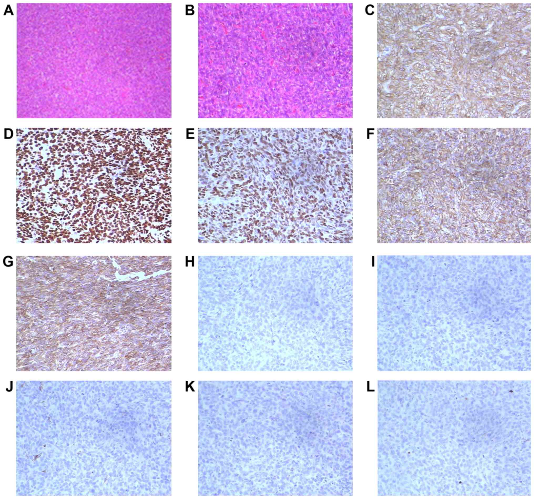
Upon pathological examination, the resected lung
nodule was determined to be a short spindle cell malignant tumor.
Hematoxylin and eosin staining indicated a proliferation of oval or
spindle cells and scattered arterioles (Fig. 3). Immunohistochemical examinations of
the tumor tissues revealed that they were strongly positive for
cluster of differentiation 10 (CD10), estrogen receptor (ER) and
progesterone receptor (PR). Furthermore, the tumor cells
demonstrated positivity for vimentin and Bcl-2, and were negative
for α-smooth muscle actin, desmin, creatine kinase (CK), S-100 and
inhibin (Fig. 3). The
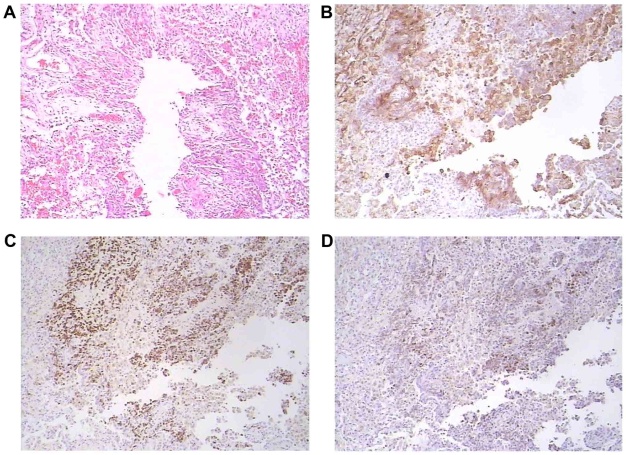
immunophenotype supported metastatic stromal sarcoma of the uterus.
A cystic specimen was observed in the left upper lobe of the lung
and the wall of the cyst was covered with oval cell tumors. The
cystic specimen was positively immunoreactive for ER, PR and CD10
(Fig. 4). The results of the
histological and immunohistochemical examinations of both the
resected lung nodule and the thin-walled cyst specimens
demonstrated that the low-grade ESS (the original uterine tumor)
had metastasized to the lungs.
After establishing the diagnosis, treatment with
megestrol was initiated, which was administered orally at a dose of
320 mg/day. Following 2 months of treatment, a number of the small
cysts disappeared, although the majority of them remained
unchanged. To date, the patient has continued with the megestrol
regimen and no new pulmonary metastatic lesions have been
identified.
Literature review
Materials and methods
The PubMed database was searched using ‘pulmonary
metastases of low-grade endometrial stromal sarcoma’ as the key
phrase and the full text results were downloaded. The inclusion
criteria were as follows: i) Female patients (without age
restriction) with a history of pathologically confirmed low-grade
ESS; ii) pathologically confirmed pulmonary metastasis of low-grade
ESS that had occurred at the time of the initial diagnosis of
low-grade ESS or during follow-up; ii) the article contained clear
and specific descriptions of the symptoms, imaging data,
pathological features, treatment and prognosis; and ii) the article
was published in English. No date limit was applied. Duplicate
reports and those that did not meet the inclusion criteria were
excluded.
A total of 35 cases described in articles that were
retrieved from the PubMed database were included in the present
study (8–12,14–26). The
present study retrospectively analyzed the clinical manifestations,
imaging data, pathological features, treatment and prognosis of the
36 cases of pulmonary metastases of low-grade ESS, which include
the 35 reported cases and the present case. The 36 cases are
summarized in Table II.
| Table II.Information for the 35 patients with
pulmonary metastasis of LGESS identified by literature review and
the current case. |
Table II.
Information for the 35 patients with
pulmonary metastasis of LGESS identified by literature review and
the current case.
| Author, year | Case no. | Diagnosis | Age at diagnosis of
LGESS, years | Gynecological
surgery | Time from
confirmation of LGESS to lung metastases, years | Respiratory
symptoms | Radiological
findings | Treatment | Follow-up | (Refs.) |
|---|
| Kim et al,
2004 | 1 | LGESS | 33 | Hysterectomy and
bilateral salpingo- oophorectomy | 4 | Dyspnea | Multiple
micronodules | Chemotherapy and
hormonal therapy | Alive with evidence
of disease | (10) |
| Satoh et al,
2003 | 2 | LGESS | 34 | Hysterectomy | 11 | Asymptomatic | Multiple
nodules | Lobectomy | Alive with | (14) |
|
|
|
|
|
|
|
|
|
| evidence of |
|
|
|
|
|
|
|
|
|
|
| disease |
|
| Murakami et
al, 2014 | 3 | LGESS | 46 | Hysterectomy | 11 | Pneumothoraces | Multiple
nodules, | Partial | Alive with | (15) |
|
|
|
|
|
|
|
| cavitary
lesions | resection | no evidence |
|
|
|
|
|
|
|
|
| and multiple
thin- | and hormonal | of disease |
|
|
|
|
|
|
|
|
| walled cysts | therapy |
|
|
| Miyamoto et
al, 2009 | 4 | LGESS | 45 | Hysterectomy | 23 | Asymptomatic | One mass | Lobectomy | Alive with | (16) |
|
|
|
|
|
|
|
| and two
nodules |
| no evidence |
|
|
|
|
|
|
|
|
|
|
| of disease |
|
| Lim et al,
2010 | 5 | LGESS | 30 | Hysterectomy | Prior to | Asymptomatic | Multiple
nodules | Hormonal | Alive with | (17) |
|
|
|
|
| and bilateral | surgery |
|
| therapy | no evidence |
|
|
|
|
|
| salpingo- |
|
|
|
| of disease |
|
|
|
|
|
| oophorectomy |
|
|
|
|
|
|
| Mahadeva et
al, 1999 | 6 | LGESS | 39 | None | At the same | Dyspnea, | Fine reticular | None | Succumbed | (18) |
|
|
|
|
|
| time as the | pneumothoraces | shadowing |
| to the |
|
|
|
|
|
|
| diagnosis of |
|
|
| disease |
|
|
|
|
|
|
| LGESS |
|
|
|
|
|
| Inayama et
al, 2000 | 7 | LGESS | 43 | Hysterectomy | 25 | Dry cough | Multiple
bilateral | None | Succumbed | (12) |
|
|
|
|
|
|
|
| nodules |
| to the |
|
|
|
|
|
|
|
|
|
|
| disease |
|
| Akhavan et
al, 2012 | 8 | LGESS | 39 | Hysterectomy | 4 | Asymptomatic | Multiple | Chemotherapy | Alive with | (19) |
|
|
|
|
|
|
|
| bilateral
nodules |
| evidence of |
|
|
|
|
|
|
|
|
|
|
| disease |
|
| Binesh et
al, 2013 | 9 | LGESS | 50 | Hysterectomy
and | Prior to | Dyspnea,
cough, | Two masses | None | Succumbed | (20) |
|
|
|
|
| bilateral
salpingo- | surgery | blood-tinged |
|
| to the |
|
|
|
|
|
| oophorectomy |
| sputum |
|
| disease |
|
| Kang et al,
2014 | 10 | LGESS | 35 | Myomectomy | 7 | Asymptomatic | Multiple
bilateral | Wedge | Alive with | (21) |
|
|
|
|
|
|
|
| nodules | resection and | evidence of |
|
|
|
|
|
|
|
|
|
| chemotherapy | disease |
|
| Abrams et
al, 1989 | 11 | LGESS | 34 | Hysterectomy | 27 | Asymptomatic | A cystic
nodule | Wedge | Alive with | (11) |
|
|
|
|
|
|
|
|
| resection | no evidence |
|
|
|
|
|
|
|
|
|
|
| of disease |
|
|
| 12 | LGESS | 33 | Hysterectomy
and | Prior to
surgery | Hemoptysis, | Multiple cysts | Chemotherapy | Succumbed |
|
|
|
|
|
| bilateral
salpingo- |
| pneumothoraces |
|
| to the |
|
|
|
|
|
| oophorectomy |
|
|
|
| disease |
|
| Itoh et al,
1997 | 13 | LGESS | 42 | Hysterectomy | 16 | Pneumothoraces | Multiple | Wedge
resection | Unknown | (9) |
|
|
|
|
|
|
| dry cough | thin-walled |
|
|
|
|
|
|
|
|
|
|
| cysts |
|
|
|
| Ota et al,
2002 | 14 | LGESS | 48 | Hysterectomy
and | 10 | Asymptomatic | Multiple
bilateral | Segmentectomy | Alive with | (22) |
|
|
|
|
| bilateral
salpingo- |
|
| nodules |
| no evidence |
|
|
|
|
|
| oophorectomy |
|
|
|
| of disease |
|
| Steele et
al, 1968 | 15 | LGESS | 35 | Hysterectomy | 5 | Chest pain, | An opacity | Pneumonectomy | Succumbed to | (23) |
|
|
|
|
|
|
| wheezing |
|
| the disease |
|
| Nakamura et
al, 2016 | 16 | LGESS | 52 | Hysterectomy
and | 6 | Asymptomatic | A mass | Hormonal | Alive with | (24) |
|
|
|
|
| bilateral
salpingo- |
|
|
| therapy, | evidence of |
|
|
|
|
|
| oophorectomy |
|
|
| surgery and | disease |
|
|
|
|
|
|
|
|
|
| chemotherapy |
|
|
| Chong et al,
2014 | 17 | LGESS | 65 | Hysterectomy
and | Prior to | Dyspnea, | Hematoxylin | Hormonal | Unknown | (25) |
|
|
|
|
| bilateral
salpingo- | surgery | pneumothorax | and eosin | therapy |
|
|
|
|
|
|
| oophorectomy |
|
| staining
indicated |
|
|
|
|
|
|
|
|
|
|
| oval or
spindle |
|
|
|
|
|
|
|
|
|
|
| cell
proliferation; |
|
|
|
|
|
|
|
|
|
|
| multiple |
|
|
|
|
|
|
|
|
|
|
| pulmonary |
|
|
|
|
|
|
|
|
|
|
| nodules and
small- |
|
|
|
|
|
|
|
|
|
|
| walled cysts |
|
|
|
| Spano et al,
2003 | 18 | LGESS | 44 | Hysterectomy | 3 | Asthenia | Bilateral | Hormonal | Alive with | (26) |
|
|
|
|
|
|
|
| pulmonary | therapy | no evidence |
|
|
|
|
|
|
|
|
| nodules |
| of disease |
|
|
| 19 | LGESS | 34 | Hysterectomy
and | 6 | Asymptomatic | Bilateral
pulmonary | Hormonal | Alive with no |
|
|
|
|
|
| bilateral
salpingo- |
|
| nodules | therapy | evidence of |
|
|
|
|
|
| oophorectomy |
|
|
|
| disease |
|
| Aubry et al,
2002 | 20 | LGESS | 40 | Hysterectomy | 11 | Asymptomatic | Multiple
nodules | Hormonal | Alive | (8) |
|
|
|
|
|
|
|
|
| therapy and | with no |
|
|
|
|
|
|
|
|
|
| wedge | evidence of |
|
|
|
|
|
|
|
|
|
| resection | disease |
|
|
| 21 | LGESS | 51 | Hysterectomy | 5 | Collarbone | Multiple
bilateral | Hormonal | Alive with |
|
|
|
|
|
|
|
| pain | nodules | therapy and | evidence of |
|
|
|
|
|
|
|
|
|
| wedge
resection | disease |
|
|
| 22 | LGESS | 33 | Hysterectomy | 5 | Asymptomatic | Solitary
nodule | Lobectomy | Alive with no |
|
|
|
|
|
|
|
|
|
|
| evidence of |
|
|
|
|
|
|
|
|
|
|
| disease |
|
|
| 23 | LGESS | 29 | Hysterectomy | 2.5 | Asymptomatic | Multiple
bilateral | Wedge | Alive with no |
|
|
|
|
|
|
|
|
| nodules and
cysts | resection | evidence of |
|
|
|
|
|
|
|
|
|
|
| disease |
|
|
| 24 | LGESS | 64 | Hysterectomy | 13 | Asymptomatic | Multiple
bilateral | Wedge | Alive with |
|
|
|
|
|
|
|
|
| nodules | resection | evidence of |
|
|
|
|
|
|
|
|
|
|
| disease |
|
|
| 25 | LGESS | 30 | Hysterectomy | 16 | Shortness of | Multiple
nodules | Hormonal | Alive with |
|
|
|
|
|
|
|
| breath, cough, | and pleural | therapy | evidence of |
|
|
|
|
|
|
|
| chest pain | effusion |
| disease |
|
|
| 26 | LGESS | 28 | Hysterectomy | 20 | Shortness of | Multiple
nodules | Lobectomy | Succumbed to |
|
|
|
|
|
|
|
| breath, cough, | and bilateral | and wedge | the disease |
|
|
|
|
|
|
|
| chest pain | pleural
effusion | resection |
|
|
|
| 27 | LGESS | 43 | Hysterectomy | 10 | Shortness of | Multiple
nodules | Chemo- | Alive with |
|
|
|
|
|
|
|
| breath, cough, |
| radiation and | no evidence |
|
|
|
|
|
|
|
| chest pain |
| wedge
resection | of disease |
|
|
| 28 | LGESS | 29 | Hysterectomy | 3 | Recurrent | Pleural | None | Alive with |
|
|
|
|
|
|
|
| pneumothoraces | thickening |
| evidence of |
|
|
|
|
|
|
|
|
| and cysts |
| disease |
|
|
| 29 | LGESS | 58 | Hysterectomy | 9 | Asymptomatic | Solitary | Wedge | Alive with |
|
|
|
|
|
|
|
|
| nodule | resection | no evidence |
|
|
|
|
|
|
|
|
|
|
| of disease |
|
|
| 30 | LGESS | 37 | Hysterectomy | 3 | Shortness of | Bilateral | Hormonal | Alive with |
|
|
|
|
|
|
|
| breath, chest
pain |
reticulonodular | therapy | evidence of |
|
|
|
|
|
|
|
|
| infiltrates |
| disease |
|
|
| 31 | LGESS | 59 | Hysterectomy | 8 | Asymptomatic | Solitary
nodule | Wedge | Alive with |
|
|
|
|
|
|
|
|
|
| resection | evidence of |
|
|
|
|
|
|
|
|
|
|
| disease |
|
|
| 32 | LGESS | 48 | Hysterectomy | 7 | Asymptomatic | Multiple
nodules | Hormonal | Alive with |
|
|
|
|
|
|
|
|
|
| therapy | evidence of |
|
|
|
|
|
|
|
|
|
|
| disease |
|
|
| 33 | LGESS | 49 | Hysterectomy | 4 | Asymptomatic | Multiple | Wedge | Unknown |
|
|
|
|
|
|
|
|
| bilateral
cysts | resection |
|
|
|
| 34 | LGESS | 36 | Hysterectomy | 7 | Right lower | Solitary
nodule | Hormonal | Alive with no |
|
|
|
|
|
|
|
| quadrant pain |
| therapy and | evidence of |
|
|
|
|
|
|
|
|
|
| wedge | disease |
|
|
|
|
|
|
|
|
|
| resection |
|
|
|
| 35 | LGESS | 31 | Hysterectomy | 15 | Asymptomatic | Multiple | Wedge | Alive with no |
|
|
|
|
|
|
|
|
| nodules | resection | evidence of |
|
|
|
|
|
|
|
|
|
|
| disease |
|
| Present case | 36 | LGESS | 53 | Hysterectomy
and | 1.5 | Chest
tightness, | A solid nodule | Hormonal | Alive with | – |
|
|
|
|
| bilateral
salpingo- |
| chest pain, | and multiple | therapy | evidence of |
|
|
|
|
|
| oophorectomy |
| pneumothoraces | thin-walled
cysts |
| disease |
|
Results
The 36 patients diagnosed with low-grade ESS had an
age range of 28–65 years. Among them, 25 patients (69.4%) were
initially diagnosed with low-grade ESS, and 11 patients (30.6%)
were initially misdiagnosed as not having low-grade ESS and then
received a modified diagnosis of low-grade ESS during treatment. A
total of 9 patients (25%) had undergone a hysterectomy and
bilateral adnexal resection, 25 patients (69.4%) had undergone a
hysterectomy alone, 1 patient (2.8%) had undergone tumor resection
and 1 patient (2.8%) had no history of surgery as the patient
refused surgery. Pulmonary metastases were identified in 5 patients
(13.9%) at the time of diagnosed with low-grade ESS, and in 31
patients (86.1%) following gynecological surgery. The time period
from confirmation of low-grade ESS to lung metastases was 1.5–27
years.
Of the 36 patients with pulmonary metastases of
low-grade ESS, dyspnea was experienced by 9 patients (25.0%), chest
pain was reported by 8 patients (22.2%), pneumothorax was
identified in 7 patients (19.4%), coughing was experienced by 6
patients (16.7%), hemoptysis was presented by 2 patients (5.6%) and
18 patients (50%) were asymptomatic. The most common pulmonary
symptom reported was dyspnea, followed by chest pain, pneumothorax
and coughing.
Among the 36 patients with pulmonary metastases of
low-grade ESS, 18 patients (50%) presented with multiple pulmonary
nodules, 6 patients (16.7%) had a solitary nodule or mass, 6
patients (16.7%) exhibited cystic lesions and 2 patients (5.6%) had
a reticular formation. Furthermore, 2 patients (5.6%) had multiple
nodules with pleural effusion, 2 patients (5.6%) exhibited multiple
pulmonary nodules with cystic lesions, 1 patient (2.8%) presented
with a solitary nodule with cystic lesions and 1 patient (2.8%) had
multiple nodules with cystic lesions and cavities.
The histology results for the 36 patients with
pulmonary metastases of low-grade ESS demonstrated that the lung
lesions were composed of short spindle cells arranged in
ill-defined whorls that were centered on numerous uniform
arterioles. The neoplastic cells were small with unremarkable
nuclear features and sparse cytoplasm. One tumor contained areas of
fibroblastic differentiation. Epithelioid features characterized by
sex cord-like differentiation were observed in 3 patients. The
immunohistochemical results revealed strong diffuse
immunoreactivity for ER and PR in almost all of the cases of
pulmonary metastasis. There was positive immunoreactivity for CD10
and vimentin and negative immunoreactivity for CD45, CK and S-100
in the majority of the specimens.
Pulmonary surgery in the form of a wedge resection
and/or lobectomy was performed in 21 patients. Among them, 5
patients received hormonal therapy following pulmonary surgery and
3 patients received chemotherapy following pulmonary surgery. A
total of 8 patients received hormonal therapy only and 2 patients
received chemotherapy only. Only 1 patient received chemotherapy
following hormonal therapy. In total, 4 patients did not receive
any treatment.
Follow-up data were available for 33 patients. Among
them, 6 patients succumbed to the disease. A total of 14 were alive
with no evidence of disease and 13 were alive with evidence of
stable disease.
Discussion
The present study describes the case of a female
with metastatic low-grade ESS who presented with simultaneous
cystic and solitary nodular lesions via chest CT imaging. The
patient had undergone a hysterectomy for low-grade ESS of the
uterus 1.5 years previously. The simultaneous presentation of
cystic and nodular lesions made the diagnosis problematic. VATS was
conducted for further diagnosis and, consequently, resections of
the solid nodule and the apex of the left lung, including the
thin-walled cysts, were performed. The pathological examination of
the resected lung specimens revealed a proliferation of tumor cells
with oval-shaped nuclei in both the nodular portion and the cystic
lesions. The cystic and solitary nodular lesions were considered to
be pulmonary metastasis of low-grade ESS.
To investigate the clinical and pathological
features, and enhance the awareness of pulmonary metastases in
patients with low-grade ESS, the 35 previously reported cases
identified in the literature and the current case were further
reviewed. Among these cases, the interval from hysterectomy to the
identification of pulmonary metastasis ranged from 1.5 to 27 years.
Patients with pulmonary metastases of low-grade ESS usually have no
specific symptoms. A number of patients have pulmonary metastasis
at the initial diagnosis of low-grade ESS and this may be
accompanied by corresponding respiratory symptoms (3,6,7). In the present study, the most common
pulmonary symptom was dyspnea, followed by chest pain, pneumothorax
and coughing. Asymptomatic pulmonary metastases were identified in
50% of the patients.
Pulmonary metastasis of low-grade ESS can manifest
as various patterns on CT scans (8).
The present study identified that the most common pattern of
pulmonary metastatic low-grade ESS was multiple pulmonary nodules,
which was observed in 50% of the patients reviewed. Unusual
presentations identified in the present study included a solitary
nodule, bilateral reticulonodular infiltrates and spontaneous
pneumothoraces associated with predominantly cystic lesions.
Although the majority of patients possessed multiple nodules, 16.7%
presented with a solitary nodule. Therefore, if a patient has a
history of low-grade ESS, the presence of a solitary nodule on a
chest CT scan should be closely monitored for lung metastasis.
Cystic metastases were identified in 16.7% of the patients with
pulmonary metastasis of ESS that were reviewed in the present
study. The thin-walled cysts may have been caused by the
proliferation of ESS cells in the airways. Tumor invasion in the
bronchial valve leads to pulmonary cysts. A solitary nodule and
cysts are individually known to reflect pulmonary metastasis of
low-grade ESS; however, the coexistence of these imaging features
is rare and can contribute to difficulty in making a diagnosis
(8). The present study reports a
case of metastatic low-grade ESS that simultaneously presented
cystic and solitary nodular lesions via a chest CT scan. The
mechanism underlying the coexistence of multiple lesions requires
investigation.
At present, the diagnosis of pulmonary metastasis of
low-grade ESS is based on the histopathological results (1). Histology is used to confirm that the
pulmonary metastasis has the pathological features of low-grade
ESS. Microscopically, low-grade ESS tumor cells are small and
consistently shaped, with short fusiform nuclei, swirled around
spiral arteriole-like vessels (1).
Tumor cells are similar to endometrial stromal cells in the
proliferative phase, with minimal cell atypia and usually low
mitotic activity (<5 mitoses/10 high power fields). The typical
microscopic feature of the tumor is tongue-like invasion of the
muscular layer and lymphatic vessels (8).
Immunohistochemical analysis of low-grade ESS
reveals positive expression of CD10, ER and PR. CD10 exhibits high
sensitivity and poor specificity, and requires staining with more
than two smooth muscle markers, including desmin, SMA or
h-caldesmon, to make a definite diagnosis (1). As low-grade ESS can be accompanied by
smooth muscle differentiation, smooth muscle markers may be focal
positive (8). Hwang et al
(27) revealed that the combined
application of CD10, ER, PR, h-caldesmon and transgelin
successfully distinguishes low-grade ESS from uterine
leiomyosarcoma; the identification of positive expression of CD10,
ER and PR, and negative expression of h-caldesmon and transgelin
increases the diagnostic accuracy for low-grade ESS.
Previous studies have expanded the understanding of
the molecular features of ESS. In low-grade ESS, the most common
chromosomal translocation is t(7;17)(p15;q21), which results in the
fusion of JAZF1 and SUZ12 genes (28). Other gene fusions include JAZF1-PHF1,
EPC1-PHF1, MEAF6-PHF1, ZC3H7-BCOR and MBTD1-CXorF67 (29–32).
These cytogenetic changes may be associated with pathogenesis, and
can be detected by fluorescence in situ hybridization and
reverse transcription-polymerase chain reaction. These
aforementioned molecular features may contribute to the diagnosis
of morphologically unclear cases.
At present, the treatment for patients with
low-grade ESS predominantly consists of surgery (33). The basic surgery for stage I–II
low-grade ESS involves a total hysterectomy and bilateral
adnexectomy, while patients with advanced stage (III–IV) low-grade
ESS can be treated with tumor cell inactivation therapies.
Postoperative adjuvant therapies for low-grade ESS can be of great
value. The 2016 National Comprehensive Cancer Network Clinical
Practice Guidelines for Uterine Tumors (34) recommend that only observation or
hormone therapy should be conducted in patients with stage I
low-grade ESS, hormone therapy with or without tumor-targeted
radiotherapy should be performed in patients with stages II–IVA,
and hormone therapy with or without palliative radiotherapy should
be conducted in those with stage IVB.
Low-grade ESS has a high reoccurrence rate, and the
optimal treatment for low-grade ESS with pulmonary metastasis has
not yet been established. It has been reported that patients could
benefit from further surgery, including the resection of distant
metastatic lesions (35). In the
cases reviewed in the present study, the surgeries were performed
in the form of wedge resection and/or lobectomy, and the majority
of patients exhibited a good prognosis. As low-grade ESS is
sensitive to hormone therapy, hormone therapy is also recommended
for patients with low-grade ESS that has recurred (34,36). The
recommended hormone therapy drugs include megestrol,
medroxyprogesterone and aromatase inhibitors, and
gonadotropin-releasing hormone analogs can also be used (34). Due to the lack of prospective
studies, the optimal dosage, drugs and treatment time of hormone
therapy are not clear. Low-grade ESS demonstrates a low response
rate to chemotherapy, so chemotherapy is only considered when
hormone therapy is ineffective (33). In addition, previous studies have
reported a number of potential therapeutic targets for low-grade
ESS, including platelet-derived growth factor receptor, vascular
endothelial growth factor receptor and histone deacetylases
(37); however, their clinical value
requires further investigation and confirmation.
In conclusion, the present study reports a case of
pulmonary metastatic low-grade ESS that simultaneously presented as
cystic and solitary nodular lesions. The coexistence of these
imaging features therefore indicates pulmonary metastasis of
low-grade ESS. The literature review demonstrated that pulmonary
metastases of low-grade ESS are not common but should be
disregarded. The clinical manifestations are not specific and
diagnosis is often difficult. The combination of clinical history,
imaging results and histological findings is essential for the
diagnosis of low-grade ESS with pulmonary metastasis. A combination
of surgery and adjuvant therapy may improve the treatment outcome.
As a rare disease, there is a lack of large sample research data on
low-grade ESS, and the optimal treatment strategy requires further
investigation.
Acknowledgements
Not applicable.
Funding
No funding was received.
Availability of data and materials
All data generated or analyzed during the present
study are included in this published article.
Authors' contributions
YX and ZL made substantial contributions to the
analysis and interpretation of patient data, and YX was a major
contributor in writing the manuscript. XG made substantial
contributions to the conception and design of the study.
Video-assisted thoracoscopic surgery was performed by JG. YL
performed the pathological examination. XS made contributions to
the acquisition of imaging data. All authors read and approved the
final manuscript.
Ethics approval and consent to
participate
Not applicable.
Patient consent for publication
The patient provided written informed consent for
publication.
Competing interests
The authors declare that they have no competing
interests.
Glossary
Abbreviations
Abbreviations:
|
ESS
|
endometrial stromal sarcoma
|
|
CT
|
computed tomography
|
|
VATS
|
video-assisted thoracoscopic
surgery
|
|
CD10
|
cluster of differentiation 10
|
|
ER
|
estrogen receptor
|
|
PR
|
progesterone receptor
|
|
SMA
|
α-smooth muscle actin
|
|
CK
|
creatine kinase
|
References
|
1
|
Ali RH and Rouzbahman M: Endometrial
stromal tumours revisited: An update based on the 2014 WHO
classification. J Clin Pathol. 68:325–332. 2015. View Article : Google Scholar : PubMed/NCBI
|
|
2
|
Prat J and Mbatani: Uterine sarcomas. Int
J Gynaecol Obstet. 131 (Suppl 2):S105–S110. 2015. View Article : Google Scholar : PubMed/NCBI
|
|
3
|
Xue WC and Cheung AN: Endometrial stromal
sarcoma of uterus. Best Pract Res Clin Obstet Gynaecol. 25:719–732.
2011. View Article : Google Scholar : PubMed/NCBI
|
|
4
|
Oliva E, Clement PB and Young RH:
Endometrial stromal tumors: An update on a group of tumors with a
protean phenotype. Adv Anat Pathol. 7:257–281. 2000. View Article : Google Scholar : PubMed/NCBI
|
|
5
|
Cohen I: Endometrial pathologies
associated with postmenopausal tamoxifen treatment. Gynecol Oncol.
94:256–266. 2004. View Article : Google Scholar : PubMed/NCBI
|
|
6
|
Ashraf-Ganjoei T, Behtash N, Shariat M and
Mosavi A: Low grade endometrial stromal sarcoma of uterine corpus,
a clinico-pathological and survey study in 14 cases. World J Surg
Oncol. 4:502006. View Article : Google Scholar : PubMed/NCBI
|
|
7
|
Pekindil G, Tuncyurek O, Orguc S, Inceboz
U, Kandiloglu AR and Caglar H: A case of endometrial stromal
sarcoma with curvilinear calcification. Gynecol Oncol. 98:318–321.
2005. View Article : Google Scholar : PubMed/NCBI
|
|
8
|
Aubry MC, Myers JL, Colby TV, Leslie KO
and Tazelaar HD: Endometrial stromal sarcoma metastatic to the
lung: A detailed analysis of 16 patients. Am J Surg Pathol.
26:440–449. 2002. View Article : Google Scholar : PubMed/NCBI
|
|
9
|
Itoh T, Mochizuki M, Kumazaki S, Ishihara
T and Fukayama M: Cystic pulmonary metastases of endometrial
stromal sarcoma of the uterus, mimicking lymphangiomyomatosis: A
case report with immunohistochemistry of HMB45. Pathol Int.
47:725–729. 1997. View Article : Google Scholar : PubMed/NCBI
|
|
10
|
Kim GY, Sung CO, Han J, Park JO and Lee
KS: Pulmonary metastases of uterine endometrial stromal sarcoma:
Diffuse micronodular and ground glass opacities: A case report. J
Korean Med Sci. 19:901–903. 2004. View Article : Google Scholar : PubMed/NCBI
|
|
11
|
Abrams J, Talcott J and Corson JM:
Pulmonary metastases in patients with low-grade endometrial stromal
sarcoma. Clinicopathologic findings with immunohistochemical
characterization. Am J Surg Pathol. 13:133–140. 1989. View Article : Google Scholar : PubMed/NCBI
|
|
12
|
Inayama Y, Shoji A, Odagiri S, Hirahara F,
Ito T, Kawano N and Nakatani Y: Detection of pulmonary metastasis
of low-grade endometrial stromal sarcoma 25 years after
hysterectomy. Pathol Res Pract. 196:129–134. 2000. View Article : Google Scholar : PubMed/NCBI
|
|
13
|
Qian ZD, Huang LL and Zhu XM: An
immunohistochemical study of CD83- and CD1a-positive dendritic
cells in the decidua of women with recurrent spontaneous abortion.
Eur J Med Res. 20:22015. View Article : Google Scholar : PubMed/NCBI
|
|
14
|
Satoh Y, Ishikawa Y, Miyoshi T, Mukai H,
Okumura S and Nakagawa K: Pulmonary metastases from a low-grade
endometrial stromal sarcoma confirmed by chromosome aberration and
fluorescence in-situ hybridization approaches: A case of recurrence
13 years after hysterectomy. Virchows Arch. 442:173–178.
2003.PubMed/NCBI
|
|
15
|
Murakami A, Hayashi T, Terao Y, Mori T,
Kumasaka T, Seyama K and Takahashi K: Cystic, nodular and cavitary
metastases to the lungs in a patient with endometrial stromal
sarcoma of the uterus. Intern Med. 53:1001–1005. 2014. View Article : Google Scholar : PubMed/NCBI
|
|
16
|
Miyamoto H, Jones CE, Raymond DP, Wandtke
JC, Strang JG, Bourne PA, Bonfiglio TA and Xu H: Pulmonary
metastases from uterine neoplasms after long tumour-free interval:
Four cases and review of the literature. Pathology. 41:234–241.
2009. View Article : Google Scholar : PubMed/NCBI
|
|
17
|
Lim MC, Lee S and Seo SS: Megestrol
acetate therapy for advanced low-grade endometrial stromal sarcoma.
Onkologie. 33:260–262. 2010. View Article : Google Scholar : PubMed/NCBI
|
|
18
|
Mahadeva R, Stewart S and Wallwork J:
Metastatic endometrial stromal sarcoma masquerading as pulmonary
lymphangioleiomyomatosis. J Clin Pathol. 52:147–148. 1999.
View Article : Google Scholar : PubMed/NCBI
|
|
19
|
Akhavan A, Shishebor F, Moghimi M and
Binesh F: Endometrial stromal sarcoma of uterus with metastasis to
the lung and brain. BMJ Case Rep. 2012(pii):
bcr20120069542012.PubMed/NCBI
|
|
20
|
Binesh F, Zahir ST, Akhavan A and Bovanlu
TR: Endometrial stromal sarcoma of the uterus presenting as
pulmonary metastasis. BMJ Case Rep. 2013(pii):
bcr20130085652013.PubMed/NCBI
|
|
21
|
Kang DO, Choi SI, Oh JY, Sim JK, Choi JH,
Choo JY, Hwang JW, Lee SH, Lee JH, Lee KY, et al: Endometrial
stromal sarcoma presented as an incidental lung mass with multiple
pulmonary nodules. Tuberc Respir Dis (Seoul). 76:131–135. 2014.
View Article : Google Scholar : PubMed/NCBI
|
|
22
|
Ota S, Shinagawa K, Ueoka H, Tada S,
Tabata M, Hamazaki S, Kondo E, Kiura K, Mannami T, Shibayama T, et
al: Spontaneous regression of metastatic endometrial stromal
sarcoma. Jpn J Clin Oncol. 32:71–74. 2002. View Article : Google Scholar : PubMed/NCBI
|
|
23
|
Steele SJ, Scott JM and Stephens TW:
Endometrial stromal sarcoma. Report of a case with a pulmonary
metastasis extending through the heart. Br J Surg. 55:943–945.
1968. View Article : Google Scholar : PubMed/NCBI
|
|
24
|
Nakamura K, Nakayama K, Ishikawa M,
Ishikawa N, Katagiri H, Katagiri A, Ishibashi T, Sato E, Iida K,
Sultana R and Kyo S: Letrozole as second-line hormonal treatment
for recurrent low-grade endometrial stromal sarcoma: A case report
and review of the literature. Oncol Lett. 12:3856–3860. 2016.
View Article : Google Scholar : PubMed/NCBI
|
|
25
|
Giin Chong S, Mitchell P, Fabre A and
McDonnell TJ: Recurrent pneumothoraces in a 65-year-old female: An
unusual case of cystic lung disease. Eur Respir Rev. 23:271–272.
2014. View Article : Google Scholar : PubMed/NCBI
|
|
26
|
Spano JP, Soria JC, Kambouchner M,
Piperno-Neuman S, Morin F, Morere JF, Martin A and Breau JL:
Long-term survival of patients given hormonal therapy for
metastatic endometrial stromal sarcoma. Med Oncol. 20:87–93. 2003.
View Article : Google Scholar : PubMed/NCBI
|
|
27
|
Hwang H, Matsuo K, Duncan K, Pakzamir E,
Pham HQ, Correa A, Fedenko A and Mhawech-Fauceglia P:
Immunohistochemical panel to differentiate endometrial stromal
sarcoma, uterine leiomyosarcoma and leiomyoma: Something old and
something new. J Clin Pathol. 68:710–717. 2015. View Article : Google Scholar : PubMed/NCBI
|
|
28
|
Conklin CM and Longacre TA: Endometrial
stromal tumors: The new WHO classification. Adv Anat Pathol.
21:383–393. 2014. View Article : Google Scholar : PubMed/NCBI
|
|
29
|
Micci F, Panagopoulos I, Bjerkehagen B and
Heim S: Consistent rearrangement of chromosomal band 6p21 with
generation of fusion genes JAZF1/PHF1 and EPC1/PHF1 in endometrial
stromal sarcoma. Cancer Res. 66:107–112. 2006. View Article : Google Scholar : PubMed/NCBI
|
|
30
|
Micci F, Gorunova L, Gatius S, Matias-Guiu
X, Davidson B, Heim S and Panagopoulos I: MEAF6/PHF1 is a recurrent
gene fusion in endometrial stromal sarcoma. Cancer Lett. 347:75–78.
2014. View Article : Google Scholar : PubMed/NCBI
|
|
31
|
Panagopoulos I, Thorsen J, Gorunova L,
Haugom L, Bjerkehagen B, Davidson B, Heim S and Micci F: Fusion of
the ZC3H7B and BCOR genes in endometrial stromal sarcomas carrying
an X;22-translocation. Genes Chromosomes Cancer. 52:610–618.
2013.PubMed/NCBI
|
|
32
|
Dewaele B, Przybyl J, Quattrone A, Finalet
Ferreiro J, Vanspauwen V, Geerdens E, Gianfelici V, Kalender Z,
Wozniak A, Moerman P, et al: Identification of a novel, recurrent
MBTD1-CXorf67 fusion in low-grade endometrial stromal sarcoma. Int
J Cancer. 134:1112–1122. 2014. View Article : Google Scholar : PubMed/NCBI
|
|
33
|
Amant F, Floquet A, Friedlander M,
Kristensen G, Mahner S, Nam EJ, Powell MA, Ray-Coquard I, Siddiqui
N, Sykes P, et al: Gynecologic cancer InterGroup (GCIG) consensus
review for endometrial stromal sarcoma. Int J Gynecol Cancer. 24
(Suppl 3):S67–S72. 2014. View Article : Google Scholar : PubMed/NCBI
|
|
34
|
Koh WJ, Greer BE, Abu-Rustum NR, Apte SM,
Campos SM, Cho KR, Chu C, Cohn D, Crispens MA, Dizon DS, et al:
Uterine Sarcoma, Version 1.2016: Featured updates to the NCCN
guidelines. J Natl Compr Canc Netw. 13:1321–1331. 2015. View Article : Google Scholar : PubMed/NCBI
|
|
35
|
Denschlag D, Thiel FC, Ackermann S, Harter
P, Juhasz-Boess I, Mallmann P, Strauss HG, Ulrich U, Horn LC,
Schmidt D, et al: Sarcoma of the Uterus. Guideline of the DGGG
(S2k-Level, AWMF registry No. 015/074, August 2015). Geburtshilfe
Frauenheilkd. 75:1028–1042. 2015. View Article : Google Scholar : PubMed/NCBI
|
|
36
|
Yamaguchi M, Erdenebaatar C, Saito F,
Motohara T, Miyahara Y, Tashiro H and Katabuchi H: Long-term
outcome of aromatase inhibitor therapy with Letrozole in patients
with advanced low-grade endometrial stromal sarcoma. Int J Gynecol
Cancer. 25:1645–1651. 2015. View Article : Google Scholar : PubMed/NCBI
|
|
37
|
Cuppens T, Tuyaerts S and Amant F:
Potential therapeutic targets in uterine sarcomas. Sarcoma.
2015:2432982015. View Article : Google Scholar : PubMed/NCBI
|