Introduction
Head and neck squamous cell carcinoma (HNSCC) was
reported as the sixth most common type of malignancy in humans,
constituting ~4% of all new cases in the United States in 2015
(1,2). According to a recent report (2018),
~600,000 patients were affected worldwide yearly and the incidence
rate has significantly increased (3). Improvements in clinical therapy have
not led to corresponding improvements in the prognosis of patients
with HNSCC. The 5-year survival rate of patients with HNSCC still
remains between 40 and 50%. Revealing the underlying mechanism of
HNSCC development could provide potential biomarkers or therapeutic
targets in HNSCC (4).
Several studies have focused on the mechanism of
HNSCC, and the new generation of sequencing technology provides a
rich resource for the study of significant genetic changes during
tumorigenesis and for the screening of potential diagnostic and
prognostic markers of cancer. For instance, it was reported that
actin-like protein 8 (ACTL8) was increasingly expressed in HNSCC
and regarded as an independent prognostic factor (5). The expression of neutrophil
gelatinase-associated lipocalin was lower in HNSCC than in normal
tissues, and was correlated with the tumorigenesis of HNSCC
(6). Calpain 6 expression was
significantly decreased in HNSCC and positively associated with the
survival rate of patients with the disease, thereby indicating the
role of calpain 6 as a tumor suppressor in HNSCC (7). However, due to the limited sample
sizes, previous studies may provide false predictions.
In the present study, integrated analysis was
performed to identify the key genes involved in the development of
HNSCC. Firstly, the differentially expressed genes (DEGs) between
HNSCC and normal tissues were screened, followed by Gene Ontology
(GO) enrichment analysis, Kyoto Encyclopedia of Genes and Genomes
(KEGG) enrichment analysis and protein-protein interaction (PPI)
network analysis. Finally, the key candidate DEGs were identified
according to Centiscape and log-rank survival analysis, and were
verified using the Gene Expression Ontology (GEO) datasets. These
key DEGs were identified as potential biomarkers for early
diagnosis and as therapeutic targets for HNSCC.
Materials and methods
Gene expression profile data and
identification of DEGs
The level-3 RNA sequence (RNA Seq) data (fragments
per kilobase of transcript per million mapped reads upper quartile
data) of HNSCC and corresponding normal tissue samples were
downloaded from the TCGA database (dataset no. :544; http://www.cancer.gov/tcga) (8) using the Genomic Data Commons
Application Programming Interface (9). A total of 544 samples from 500 patients
with HNSCC and 44 normal controls were collected in December 2018.
The normal controls included normal tissues from the oral cavity,
oral tongue, larynx, floor of the mouth and base of the tongue. The
raw data was downloaded and the log2 fold-change
(log2FC) was calculated using the Limma R package
(version 3.2.5; http://www.r-project.org/) to screen DEGs between
HNSCC and normal tissues. The following cut-off criteria were
applied: log2FC>2 and P<0.05. The adjustment of
P<0.5 was set as the threshold to adjust the P-value for
multiple comparisons.
For verification purposes, the microarray expression
dataset GSE6631 was downloaded (as minimum information about a
microarray experiment notation in mark-up language formatted family
files) from the GEO database (10).
The GSE6631 was based on the GPL8300 Platforms (Affymetrix Human
Genome U95 version 2 array) and included 44 HNCC samples and paired
normal samples (submission date, 2007; last updated, 2018)
(11). The sample information and
expression profile data were extracted by R package (version:3.2.5)
from GES6631. Statistical analyses were performed with GraphPad
Prism version 8.0 software (GraphPad Software, Inc.). Single
comparisons between two groups were performed using Paired
Student's t-test.
GO and pathway enrichment analyses of
DEGs
GO analysis and KEGG pathway enrichment of DEGs were
performed using the Database for Annotation, Visualization and
Integrated Discovery (DAVID) (version, 6.7; http://david-d.ncifcrf.gov/) to screen for possible
biological processes, cellular components, molecular functions and
signaling pathways of the involved DEGs (12). The resulting data were imported into
Cytoscape ClueGo software (version: 3.6.0) for visual analysis
(13). P<0.05 was considered as
statistically significant. The following parameter settings were
applied: Identifier, ‘official gene symbol.’; list type, ‘gene
list.’; species, ‘homo sapiens.’; count threshold, 2; and ease
threshold, 0.05.
PPI network construction and candidate
gene identification
The Search Tool for the Retrieval of Interacting
Genes/Proteins (STRING; version, 11.0; http://string-db.org) was used to construct the PPI
network (14). The minimum required
interaction score was set to a medium confidence of 0.4, and the
organism was set to ‘Homo sapiens’. The Cytoscape software
was then used to visualize the network. Cytoscape CentiScape
(version, 3.6.0; http://apps.cytoscape.org/apps/centiscape) (15) was used to screen candidate key
proteins in the network, according to the degree of centrality. The
genes with a node degree of ≥15 were considered as the candidate
key genes.
Association between candidate key
genes and clinicopathological parameters of HNSCC
By using the Gene Expression Profiling Interactive
Analysis (GEPIA) database (http://gepia.cancer-pku.cn/), the effect of candidate
key genes on overall survival (OS) rate was evaluated using
log-rank test and the Mantel-Cox test. P<0.05 for log-rank test
indicated statistical significance. Genes significantly associated
with HNSCC OS rate were considered as key genes. The following
parameter settings were applied: Group cut-off, ‘median.’; hazards
ratio, ‘yes.’; 95% confidence interval, ‘yes.’; and axis units,
‘months.’.
Comparison of key genes in HNSCC and
other types of cancer
In order to evaluate the specificity of the key
genes screened in the previous step to HNSCC, the expression of
these genes was examined in other tumor datasets from the GEPIA
database, including adrenocortical carcinoma, bladder urothelial
carcinoma (BLCA), breast invasive carcinoma, endocervical
adenocarcinoma (CESC), colon adenocarcinoma (COAD), esophageal
carcinoma (ESCA), liver hepatocellular carcinoma, lung squamous
cell carcinoma and lung adenocarcinoma (LUAD). These datasets came
from the TCGA and the GTEx projects (16). The unmatched normal and tumor tissues
were compared. The raw data were filtered based on the cut-offs
log2FC>2 and P<0.05.
Results
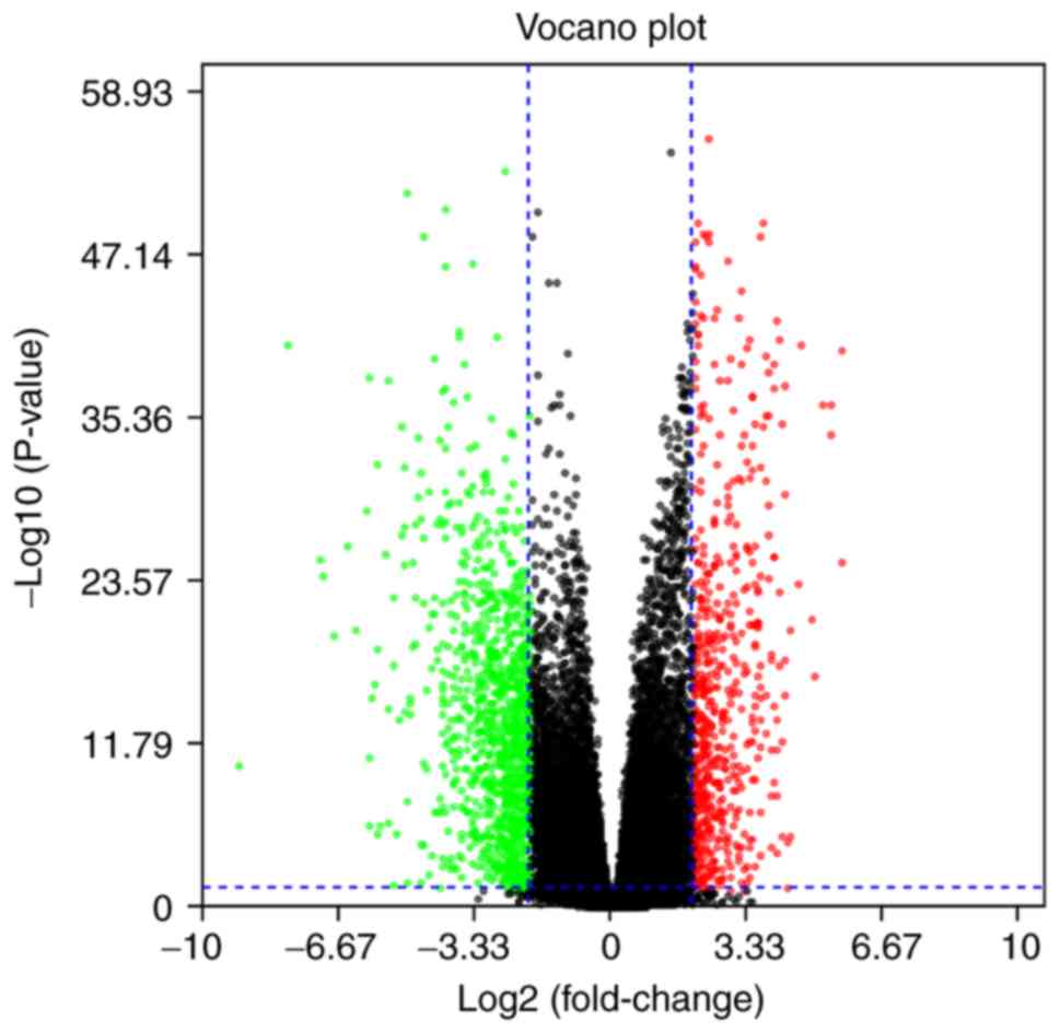
Identification of DEGs in HNSCC
RNAseq data of HNSCC and corresponding normal tissue
samples were downloaded from the TCGA database. The data was
screened by the Limma package, using P<0.05 and
log2FC>2 as the cut-off criteria, which identified
1,181 DEGs (Fig. 1), including 354
upregulated and 827 downregulated genes.
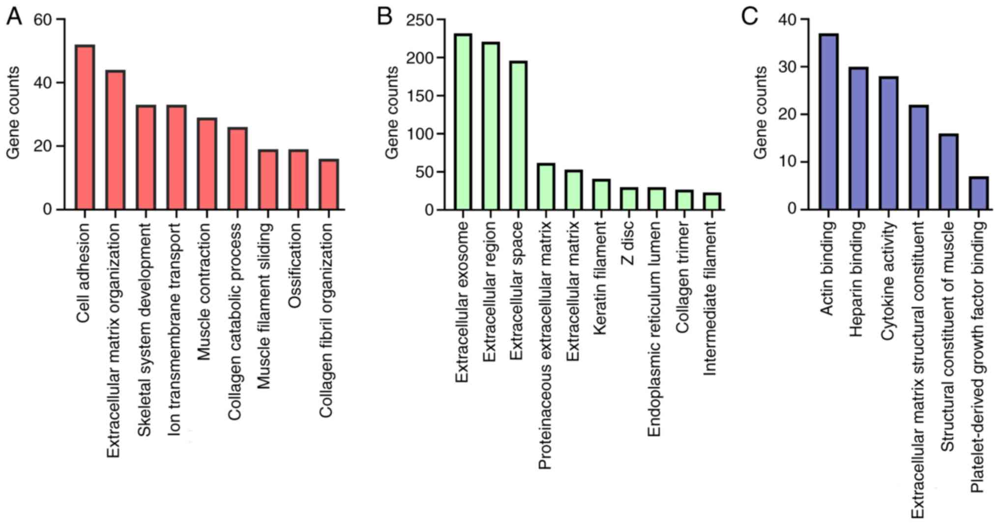
GO analysis and signaling pathway
enrichment of DEGs in HNSCC
The GO analysis of the 1,181 DEGs was performed
using the DAVID database, with the criterion set at P<0.05. The
DEGs were divided into three groups, namely, biological process,
cellular component and molecular function groups. As shown in
Fig. 2A, the main DEG-associated
biological functions were ‘cell adhesion’, ‘extracellular matrix
organization’, ‘skeletal system development’ and ‘ion transmembrane
transport’. As demonstrated in Fig.
2B, the cellular component analysis revealed that the selected
DEGs were mainly located at the ‘extracellular exosome’,
‘extracellular region’ and ‘extracellular space’. The molecular
function of the DEGs was mainly associated with ‘actin binding’,
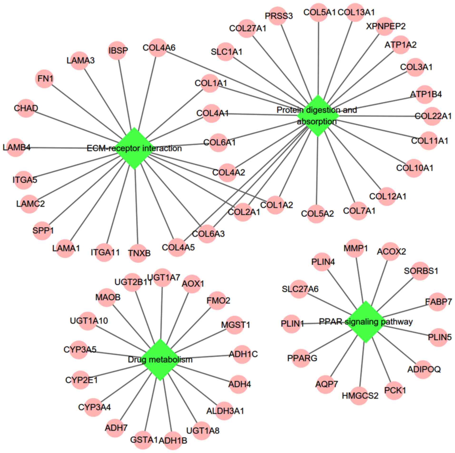
‘heparin binding’ and ‘cytokine activity’ (Fig. 2C). As demonstrated in Fig. 3, the KEGG pathways enriched by the
DEGs were mainly associated with ‘protein digestion and
absorption’, ‘extracellular matrix-receptor interaction’, ‘drug
metabolism’ and the ‘PPAR signaling pathway’.
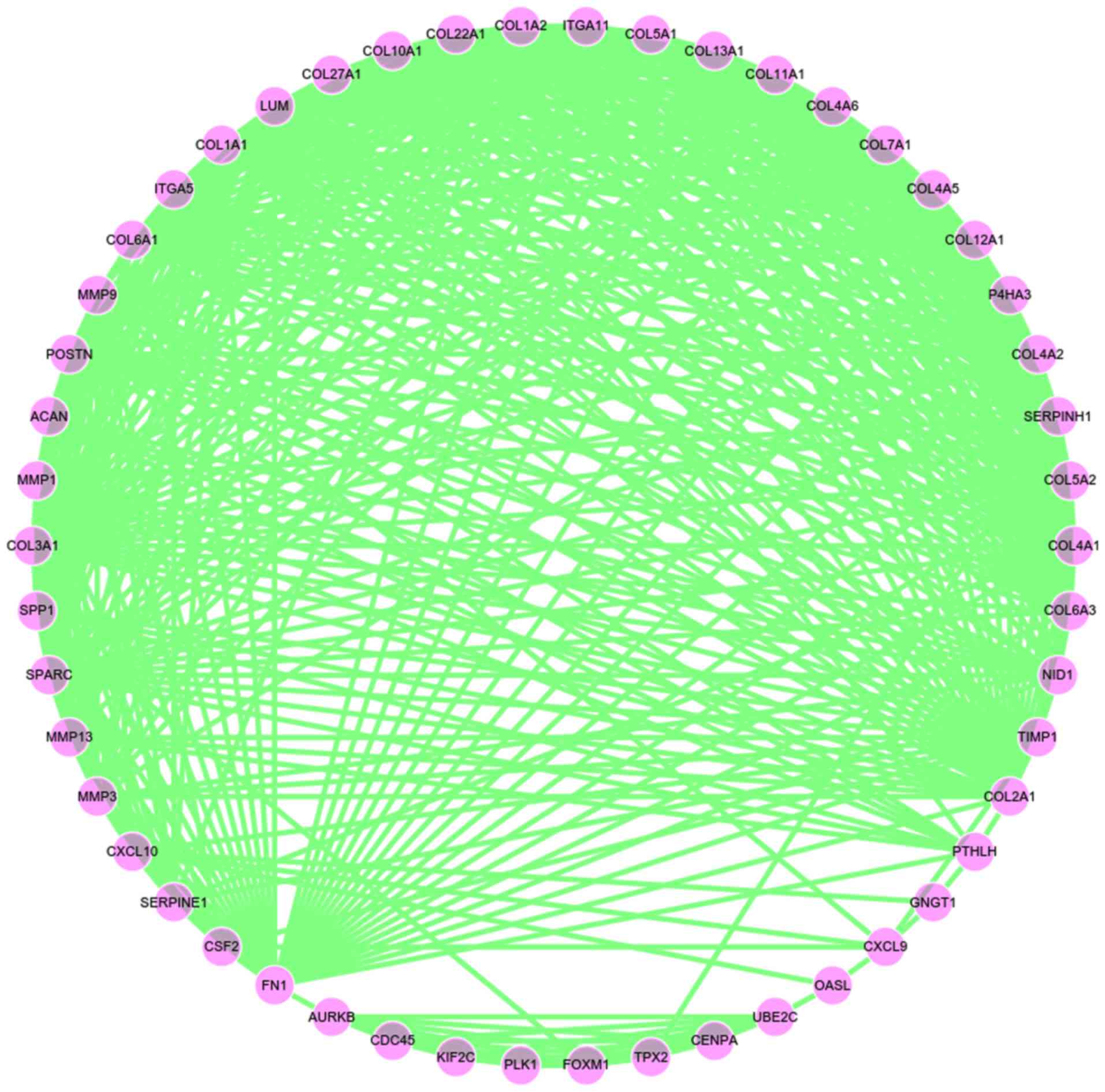
Key candidate DEG identification with
PPI network analysis
The PPI network of the DEG expression products was
constructed using Cytoscape software and the STRING database
(http://string-db.org). A total of 1,035 DEGs were
incorporated into the PPI network complex. The Cytoscape CentiScape
was used to screen candidate key genes in the network, with degree
of centrality ≥15 set as the inclusion criterion, which identified
50 genes that were included in the following analysis (Fig. 4).
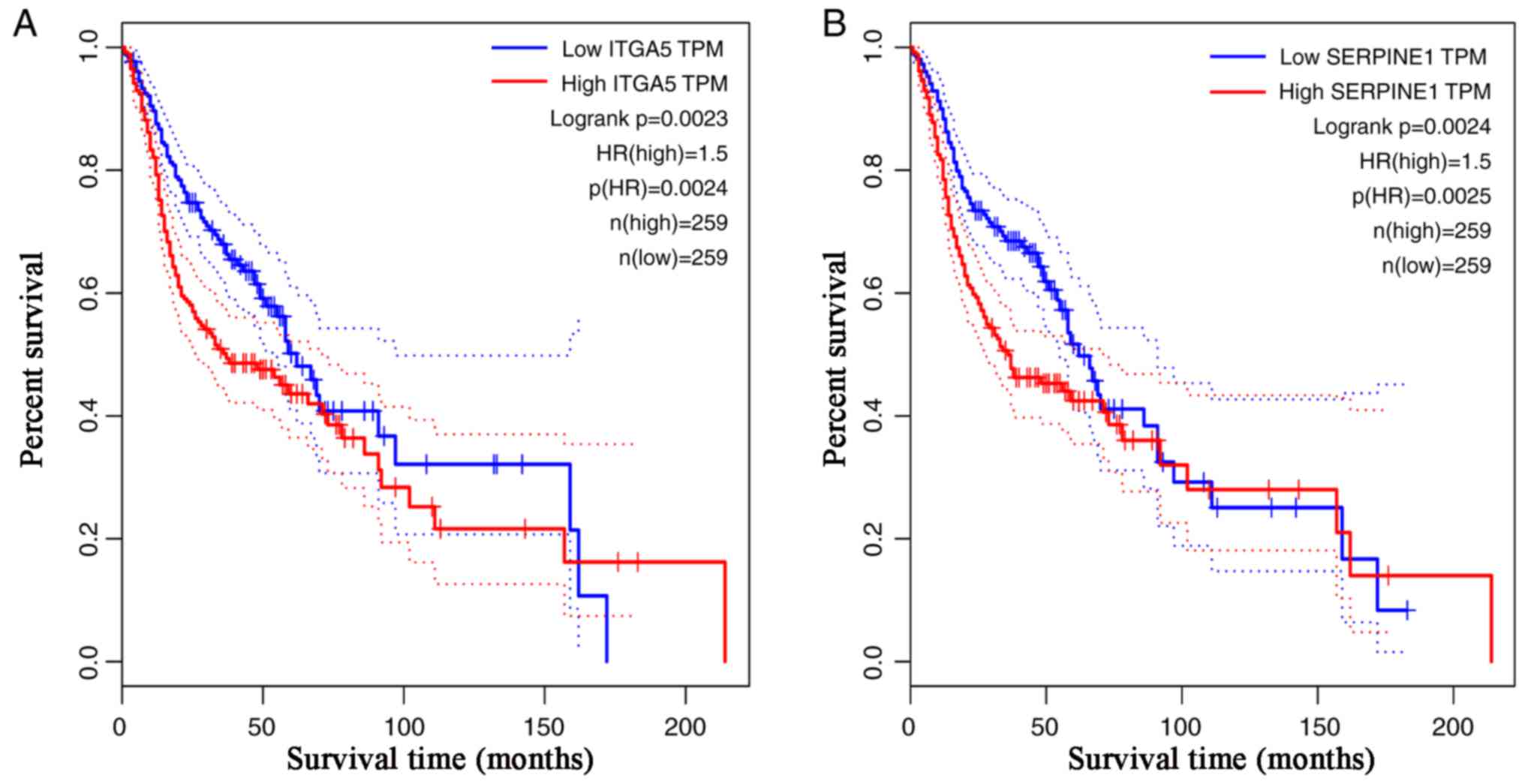
Log-rank survival analysis by the GEPIA database,
found 8 out of the 50 candidate key genes, including integrin α-5
(ITGA5) and serpin family E member 1 (SERPINE1), to be
significantly associated with HNSCC OS rate (P<0.05; Table I). If the threshold was set to
P<0.01, only ITGA5 and SERPINE1 were found to be associated with
the OS rate of HNSCC (Fig. 5). The
Cox proportional hazard ratios of ITGA5 and SERPINE1 were both
1.5.
 | Table I.Log regression analysis of differently
expressed genes with node degree of centrality ≥15. |
Table I.
Log regression analysis of differently
expressed genes with node degree of centrality ≥15.
| Gene symbol | Degree of
centrality | Log-rank,
P-value |
|---|
| COL1A1 | 45 | 0.30 |
| COL1A2 | 37 | 0.84 |
| FN1 | 34 | 0.11 |
| COL2A1 | 34 | 0.26 |
| COL3A1 | 33 | 0.56 |
| MMP9 | 32 | 0.93 |
| COL4A2 | 30 | 0.61 |
| COL4A1 | 30 | 0.62 |
| COL5A2 | 29 | 0.29 |
| COL4A5 | 28 | 0.85 |
| COL11A1 | 26 | 0.09 |
| COL7A1 | 26 | 0.10 |
| COL5A1 | 26 | 0.28 |
| SPARC | 26 | 0.30 |
| COL6A3 | 26 | 0.86 |
| SERPINH1 | 25 | 0.02 |
| COL6A1 | 25 | 0.22 |
| COL4A6 | 25 | 0.36 |
| TIMP1 | 24 | 0.04 |
| SPP1 | 24 | 0.04 |
| CSF2 | 23 | 0.02 |
| LUM | 23 | 0.63 |
| COL10A1 | 23 | 0.98 |
| GNGT1 | 22 | 0.15 |
| COL27A1 | 22 | 0.46 |
| ITGA5 | 21 | <0.01 |
| MMP1 | 21 | 0.04 |
| CXCL10 | 21 | 0.19 |
| CENPA | 21 | 0.30 |
| COL12A1 | 21 | 0.53 |
| P4HA3 | 20 | 0.06 |
| MMP13 | 20 | 0.09 |
| MMP3 | 20 | 0.23 |
| POSTN | 20 | 0.30 |
| PLK1 | 20 | 0.41 |
| COL13A1 | 20 | 0.44 |
| COL22A1 | 20 | 0.62 |
| FOXM1 | 19 | 0.55 |
| ACAN | 18 | 0.50 |
| AURKB | 18 | 0.63 |
| KIF2C | 17 | 0.29 |
| CDC45 | 17 | 0.90 |
| PTHLH | 17 | 0.92 |
| SERPINE1 | 16 | <0.01 |
| ITGA11 | 16 | 0.50 |
| TPX2 | 16 | 0.61 |
| OASL | 16 | 0.67 |
| NID1 | 15 | 0.04 |
| CXCL9 | 15 | 0.18 |
| UBE2C | 15 | 0.58 |
ITGA5 is highly expressed in HNSCC
specifically
To evaluate the specificity of the key genes
previously screened in HNSCC, their expression levels were
determined in other tumor datasets from the GEPIA database. The
expression of ITGA5 was only significantly increased in HNSCC and
decreased in BLCA, CESC, COAD and LUSC (Fig. 6A). However, SERPINE1 expression was
significantly increased in ESCA, as well as in HNSCC, with no other
significant changes in other tumors (Fig. 6B).
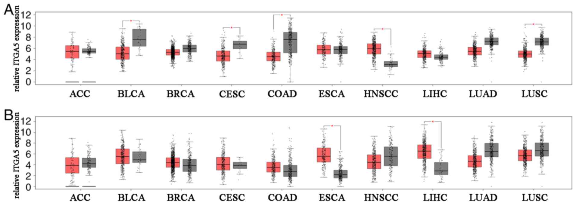 | Figure 6.Expression of ITGA5 and SERPINE1 in
different types of cancer. The expression levels of (A) ITGA5 and
(B) SERPINE1 were compared in different types of cancer in humans.
Box plots were drawn using the R software and the raw data from the
Gene Expression Profiling Interactive Analysis database. ITGA5
expression was only significantly increased in HNSCC, whereas it
was decreased in BLCA, CESC, COAD and LUSC. The expression of
SERPINE1 was significantly increased in HNSCC, as well as ESCA. The
red boxplots represented tumor samples and the grey boxplots
represented normal samples. ITGA5, integrin α-5; SERPINE1, serpin
family E member 1; HNSCC, head and neck squamous cell carcinoma;
ACC, adrenocortical carcinoma; BLCA, bladder urothelial carcinoma;
BRCA, breast invasive carcinoma; CESC, endocervical adenocarcinoma;
COAD, colon adenocarcinoma; ESCA, esophageal carcinoma; LIHC, liver
hepatocellular carcinoma; LUSC, lung squamous cell carcinoma; LUAD,
lung adenocarcinoma. |
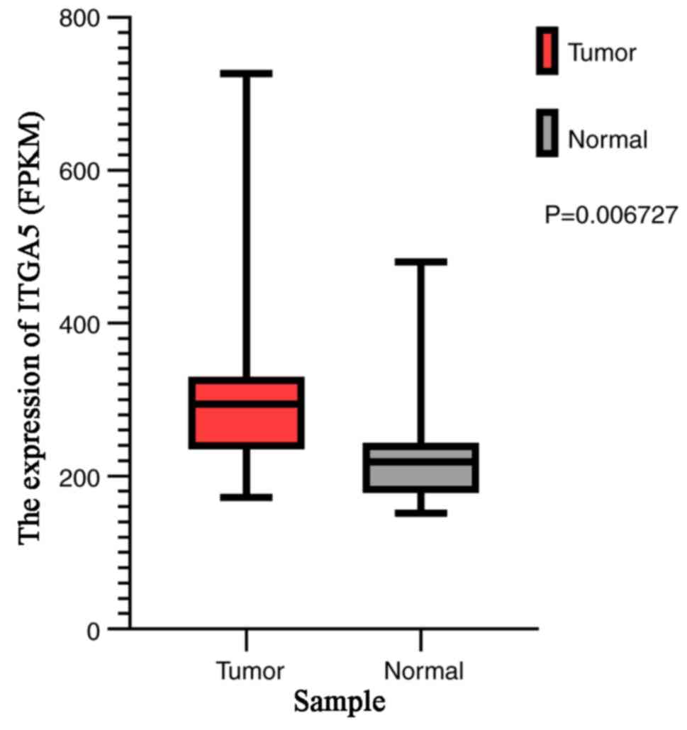
For verification, ITGA5 expression was evaluated in
HNSCC based on the microarray express dataset GSE6631. Compared
with ITGA5 expression in normal tissues, the expression in HNSCC
tissues was upregulated (P=0.007; Fig.
7).
Discussion
HNSCC is one of the most common types of
malignancies in humans; it is characterized by rapid progression, a
high migration capacity and high mortality rate. However, there are
almost no biomarkers or targets for the diagnosis and treatment of
HNSCC (1). Thus, the identification
of genes that are differentially expressed between tumor and normal
tissues will contribute to future studies on the pathogenesis of
HNSCC and will provide potential diagnostic biomarkers and
therapeutic targets.
In the present study, TCGA datasets of HNSCC were
integrated and analyzed using bioinformatics. Finally, a total of
1,181 DEGs were identified including 354 upregulated genes and 827
downregulated genes in the initial step. Subsequently, the 1,181
DEGs were analyzed based on GO terms and classified based on KEGG
signaling pathways. The GO analysis indicated that the DEGs were
mainly involved in ‘cell adhesion’, ‘extracellular matrix
organization’, ‘skeletal system development’, ‘ion transmembrane
transport’, ‘actin binding’, ‘heparin binding’ and ‘cytokine
activity’. The KEGG pathway analysis revealed that the DEGs were
mainly associated with ‘protein digestion and absorption’,
‘extracellular matrix-receptor interaction’, ‘drug metabolism’ and
the ‘PPAR signaling pathway’. In addition, the PPI network complex
was constructed and eight key genes, including ITGA5, SERPINE1,
serpin family H member 1, colony-stimulating factor 2, tissue
inhibitor of metalloproteinases 1, nidogen 1, secreted
phosphoprotein 1 and matrix metallopeptidase 1, were screened based
on centrality and log-rank survival analysis by GEPIA. The GEPIA
database contains genotype-tissue expression data, which increases
the sample size and accuracy of the analysis, and was therefore was
used to identify key DEGs. As a result, ITGA5 and SERPINE1 were
identified as key genes.
ITGA5 is an important member of the integrin family,
and its coding gene is located at the human chromosome 12q11-q13.
Integrin is an extracellular matrix receptor that acts as an
adhesive receptor for extracellular matrix proteins, including
fibrin, laminin and collagen (17).
ITGA5 forms the link between the extracellular matrix and
intracellular signal transduction, and also participates in a
variety of important physiological processes; it is also associated
with tumor occurrence, development, invasion and metastasis
(18). A study on hepatocellular
carcinoma (HCC) demonstrated a negative correlation between the
expression of ITGA5 and miR-128, and the downregulation of ITGA5
leading to the inhibition of HCC cell metastasis and stem cell-like
properties (19). However, the
expression of ITGA5 played diverse roles in breast cancer cells
(20). Expression in highly invasive
breast cancer cells was almost absent in comparison with that in
less invasive cells, thereby indicating the negative association
between ITGA5 and breast cancer metastasis (21,22). In
ovarian cancer cells, downregulation of ITGA5 induced by forced
expression of miR-17 significantly limited the adhesion, invasion
and tumorigenesis ability of cancer cells (23). ITGA5 was reported to be highly
expressed in glioblastoma compared with normal brain glial cells,
and its downregulation inhibited proliferation, invasion and
migration (24). ITGA5 expression
was also upregulated in oral squamous cell carcinoma (OSCC), a
common type of HNSCC (25).
Knockdown of ITGA5 inhibited the proliferation, migration and
invasion of OSCC cells, thereby indicating that ITGA5 could promote
OSCC progression.
SERPINE1, also known as plasminogen activator
inhibitor type I, is a primary inhibitor of plasminogen activators
and a marker of poor prognosis in cancer. In colorectal cancer,
SERPINE1 expression was upregulated and was significantly
associated with grading and microsatellite instability (26). In esophageal cancer, SERPINE1
expression was upregulated and significantly associated with age
range, which was consistent with the analysis of the present study
(27). SEPINE1 was highly expressed
in oral carcinomas compared with in the matched tumor adjacent
normal tissues (28), and its
overexpression resulted in increased proliferation and tumor
budding. Moreover, SEPINE1 was associated with poor
progression-free and cancer-specific survival in patients with head
and neck cancer (29,30).
In conclusion, analysis of GEPIA datasets
demonstrated the association between high expression of ITGA5 and
SERPINE1 and poor OS rate in patients with HNSCC, with identical
hazard ratio for both genes. The investigation of these genes was
conducted in other tumor datasets, in order to determine their
specificity to HNSCC. The expression of ITGA5 was significantly
increased in HNSCC only and decreased in BLCA, CESC, COAD and LUSC.
On the other hand, the expression of SERPINE1 was significantly
increased in ESCA, as well as in HNSCC. A limitation of the present
study was that the gene expression in HNSCC was only analyzed by
using the public database. In subsequent experiments, molecular
biology and cell biology methods will be utilized to further verify
these results. Overall, these findings suggest the potential of
ITGA5 as a diagnostic or prognostic marker and as a therapeutic
target in HNSCC.
Acknowledgements
Not applicable.
Funding
The present study was supported by the Medicine and
Health Science Technology Development Plan Project, Shandong
Province (grant no. 2017WSA15041), the National Natural Science
Foundation of China (grant nos. 81472530 and 81602374) and the
Natural Science Foundation of Shandong Province (grant no.
2016ZRA15065).
Availability of data and materials
All data generated or analyzed during the present
study are included in this published article.
Authors' contributions
BoZ performed data download, survival analysis and
drafted the manuscript. DW and KX performed the survival analysis,
GO analysis and pathway enrichment analysis. DY worked on PPI
network construction. ZM analyzed the association between the
candidate genes and prognosis, and also participated the design of
the study. BiZ was the major contributor in designing the present
study. All authors read and approved the final manuscript.
Ethics approval and consent to
participate
Not applicable.
Patient consent for publication
Not applicable.
Competing interests
The authors declare that they have no competing
interests.
References
|
1
|
Siegel RL, Miller KD and Jemal A: Cancer
statistics, 2018. CA Cancer J Clin. 68:7–30. 2018. View Article : Google Scholar : PubMed/NCBI
|
|
2
|
Cancer Genome Atlas Network, .
Comprehensive genomic characterization of head and neck squamous
cell carcinomas. Nature. 517:576–582. 2015. View Article : Google Scholar : PubMed/NCBI
|
|
3
|
Bray F, Ferlay J, Soerjomataram I, Siegel
RL, Torre LA and Jemal A: Global cancer statistics 2018: GLOBOCAN
estimates of incidence and mortality worldwide for 36 cancers in
185 countries. CA Cancer J Clin. 68:394–424. 2018. View Article : Google Scholar : PubMed/NCBI
|
|
4
|
Goossens N, Nakagawa S, Sun X and Hoshida
Y: Cancer biomarker discovery and validation. Transl Cancer Res.
4:256–269. 2015.PubMed/NCBI
|
|
5
|
Li B, Zhu J and Meng L: High expression of
ACTL8 is poor prognosis and accelerates cell progression in head
and neck squamous cell carcinoma. Mol Med Rep. 19:877–884.
2019.PubMed/NCBI
|
|
6
|
Wang L, Chen C, Li F, Hua Q, Chen S, Xiao
B, Dai M, Li M, Zheng A, Yu D, et al: Down-regulation of neutrophil
gelatinase-associated lipocalin in head and neck squamous cell
carcinoma correlated with tumorigenesis, not with metastasis. Int J
Clin Exp Pathol. 8:8857–8868. 2015.PubMed/NCBI
|
|
7
|
Xiang Y, Li F, Wang L, Zheng A, Zuo J, Li
M, Wang Y, Xu Y, Chen C, Chen S, et al: Decreased calpain 6
expression is associated with tumorigenesis and poor prognosis in
HNSCC. Oncol Lett. 13:2237–2243. 2017. View Article : Google Scholar : PubMed/NCBI
|
|
8
|
Cancer Genome Atlas Research Network, ;
Weinstein JN, Collisson EA, Mills GB, Shaw KR, Ozenberger BA,
Ellrott K, Shmulevich I, Sander C and Stuart JM: The Cancer Genome
Atlas Pan-Cancer analysis project. Nat Genet. 45:1113–1120. 2013.
View Article : Google Scholar : PubMed/NCBI
|
|
9
|
Wilson S, Fitzsimons M, Ferguson M, Heath
A, Jensen M, Miller J, Murphy MW, Porter J, Sahni H, Staudt L, et
al: Developing cancer informatics applications and tools using the
NCI genomic data commons API. Cancer Res. 77:e15–e18. 2017.
View Article : Google Scholar : PubMed/NCBI
|
|
10
|
Wilhite SE and Barrett T: Strategies to
explore functional genomics data sets in NCBI's GEO database.
Methods Mol Biol. 802:41–53. 2012. View Article : Google Scholar : PubMed/NCBI
|
|
11
|
Kuriakose MA, Chen WT, He ZM, Sikora AG,
Zhang P, Zhang ZY, Qiu WL, Hsu DF, McMunn-Coffran C, Brown SM, et
al: Selection and validation of differentially expressed genes in
head and neck cancer. Cell Mol Life Sci. 61:1372–1383. 2004.
View Article : Google Scholar : PubMed/NCBI
|
|
12
|
Huang da W, Sherman BT and Lempicki RA:
Systematic and integrative analysis of large gene lists using DAVID
bioinformatics resources. Nat Protoc. 4:44–57. 2009. View Article : Google Scholar : PubMed/NCBI
|
|
13
|
Bindea G, Mlecnik B, Hackl H, Charoentong
P, Tosolini M, Kirilovsky A, Fridman WH, Pagès F, Trajanoski Z and
Galon J: ClueGO: A cytoscape plug-in to decipher functionally
grouped gene ontology and pathway annotation networks.
Bioinformatics. 25:1091–1093. 2009. View Article : Google Scholar : PubMed/NCBI
|
|
14
|
Szklarczyk D, Gable AL, Lyon D, Junge A,
Wyder S, Huerta-Cepas J, Simonovic M, Doncheva NT, Morris JH, Bork
P, et al: STRING v11: Protein-protein association networks with
increased coverage, supporting functional discovery in genome-wide
experimental datasets. Nucleic Acids Res. 47:D607–D613. 2019.
View Article : Google Scholar : PubMed/NCBI
|
|
15
|
Scardoni G, Tosadori G, Faizan M, Spoto F,
Fabbri F and Laudanna C: Biological network analysis with
CentiScaPe: Centralities and experimental dataset integration.
F1000Res. 3:1392014. View Article : Google Scholar : PubMed/NCBI
|
|
16
|
Tang Z, Li C, Kang B, Gao G, Li C and
Zhang Z: GEPIA: A web server for cancer and normal gene expression
profiling and interactive analyses. Nucleic Acids Res. 45:W98–W102.
2017. View Article : Google Scholar : PubMed/NCBI
|
|
17
|
Meighan CM, Kelly VE, Krahe EC and Gaeta
AJ: α integrin cytoplasmic tails can rescue the loss of Rho-family
GTPase signaling in the C. elegans somatic gonad. Mech Dev.
136:111–122. 2015. View Article : Google Scholar : PubMed/NCBI
|
|
18
|
Morandi EM, Verstappen R, Zwierzina ME,
Geley S, Pierer G and Ploner C: ITGAV and ITGA5 diversely regulate
proliferation and adipogenic differentiation of human adipose
derived stem cells. Sci Rep. 6:288892016. View Article : Google Scholar : PubMed/NCBI
|
|
19
|
Zhao X, Wu Y and Lv Z: miR-128 modulates
hepatocellular carcinoma by inhibition of ITGA2 and ITGA5
expression. Am J Transl Res. 7:1564–1573. 2015.PubMed/NCBI
|
|
20
|
Fang Z, Yao W, Xiong Y, Zhang J, Liu L, Li
J, Zhang C and Wan J: Functional elucidation and
methylation-mediated downregulation of ITGA5 gene in breast cancer
cell line MDA-MB-468. J Cell Biochem. 110:1130–1141. 2010.
View Article : Google Scholar : PubMed/NCBI
|
|
21
|
Konstantinovsky S, Smith Y, Zilber S, Tuft
Stavnes H, Becker AM, Nesland JM, Reich R and Davidson B: Breast
carcinoma cells in primary tumors and effusions have different gene
array profiles. J Oncol. 2010:9690842010. View Article : Google Scholar : PubMed/NCBI
|
|
22
|
Xiao Y, Li Y, Tao H, Humphries B, Li A,
Jiang Y, Yang C, Luo R and Wang Z: Integrin alpha5 down-regulation
by miR-205 suppresses triple negative breast cancer stemness and
metastasis by inhibiting the Src/Vav2/Rac1 pathway. Cancer Lett.
433:199–209. 2018. View Article : Google Scholar : PubMed/NCBI
|
|
23
|
Gong C, Yang Z, Wu F, Han L, Liu Y and
Gong W: miR-17 inhibits ovarian cancer cell peritoneal metastasis
by targeting ITGA5 and ITGB1. Oncol Rep. 36:2177–2183. 2016.
View Article : Google Scholar : PubMed/NCBI
|
|
24
|
Feng L, Ma J, Ji H, Liu Y and Hu W:
miR-330-5p suppresses glioblastoma cell proliferation and
invasiveness through targeting ITGA5. Biosci Rep.
37:BSR201700192017. View Article : Google Scholar : PubMed/NCBI
|
|
25
|
Fan QC, Tian H, Wang Y and Liu XB:
Integrin-α5 promoted the progression of oral squamous cell
carcinoma and modulated PI3K/AKT signaling pathway. Arch Oral Biol.
101:85–91. 2019. View Article : Google Scholar : PubMed/NCBI
|
|
26
|
Mazzoccoli G, Pazienza V, Panza A, Valvano
MR, Benegiamo G, Vinciguerra M, Andriulli A and Piepoli A: ARNTL2
and SERPINE1: Potential biomarkers for tumor aggressiveness in
colorectal cancer. J Cancer Res Clin Oncol. 138:501–511. 2012.
View Article : Google Scholar : PubMed/NCBI
|
|
27
|
Klimczak-Bitner AA, Kordek R, Bitner J,
Musial J and Szemraj J: Expression of MMP9, SERPINE1 and miR-134 as
prognostic factors in esophageal cancer. Oncol Lett. 12:4133–4138.
2016. View Article : Google Scholar : PubMed/NCBI
|
|
28
|
Gao S, Nielsen BS, Krogdahl A, Sørensen
JA, Tagesen J, Dabelsteen S, Dabelsteen E and Andreasen PA:
Epigenetic alterations of the SERPINE1 gene in oral squamous cell
carcinomas and normal oral mucosa. Genes Chromosomes Cancer.
49:526–538. 2010.PubMed/NCBI
|
|
29
|
Pavón MA, Arroyo-Solera I, Téllez-Gabriel
M, León X, Virós D, López M, Gallardo A, Céspedes MV, Casanova I,
López-Pousa A, et al: Enhanced cell migration and apoptosis
resistance may underlie the association between high SERPINE1
expression and poor outcome in head and neck carcinoma patients.
Oncotarget. 6:29016–29033. 2015. View Article : Google Scholar : PubMed/NCBI
|
|
30
|
Arroyo-Solera I, Pavón MA, León X, López
M, Gallardo A, Céspedes MV, Casanova I, Pallarès V, López-Pousa A,
Mangues MA, et al: Effect of serpinE1 overexpression on the primary
tumor and lymph node, and lung metastases in head and neck squamous
cell carcinoma. Head Neck. 41:429–439. 2019.PubMed/NCBI
|