Introduction
Glioblastoma multiforme (GBM) is the most common and
lethal malignant primary brain tumor in the world, accounting for
14% of primary brain and other central nervous system tumors
(1). The degree of malignancy is
reflected by the uncontrollable cell proliferation, high
invasiveness, lack of apoptosis and increased angiogenesis
(2). The tumor is located under the
cortex and most of it occurs throughout the upper cerebral
hemisphere; the frontal lobe is the most common site of occurrence
(2). According to World Health
Organization classification in 2007, GBM was subcategorized as a
grade IV tumor (1), which is the
highest level in brain tumors. The median survival of patients with
GBM is 14 months and only 10% of patients survive >5 years,
which suggests that the prognosis is poor (3). In Europe, glioblastoma accounts for 49%
of gliomas, and almost 3,000 new cases are diagnosed every year
(4). Patients with glioblastoma are
mostly elderly, and ~30% of them are ≥70 years old. Currently, the
treatment for glioblastoma is surgery combined with radiotherapy
and chemotherapy or adjuvant therapy (chemotherapy) with
temozolomide. Although surgical resection can effectively reduce
intracranial pressure, it can also cause postoperative cerebral
edema and the incidence of neurological complications (5). However, as glioblastoma has a diffuse
infiltration and growth pattern, if a portion of the tumor is
located in important functional areas, such as language or motor
centers, in order not to aggravate the dysfunction of brain
function, the majority of tumors can only be partially resected and
cannot be completely separated from normal brain tissue (5). Although radiotherapy can work against
tumor cells of great quantity, radiation also causes damage to
normal brain tissue, and multiple treatments can lead to
glioblastoma developing tolerance (6). It has previously been reported that the
radioresistance of glioblastoma is mainly associated with DNA
damage repair mechanisms in GSCs (6). Although chemotherapy can effectively
treat the tumor, after long-term use, tumor cells also develop
resistance (7–9); solving drug resistance of the tumor is
a difficult problem in glioblastoma treatment, and a large number
of studies have been performed to identify potential solutions.
Since cancer stem cells, known as tumor-initiating
cells (TICs), were first identified in acute myelogenous leukemia
(10), tumor stem cells have become
a hotspot of research. In numerous types of tumor tissues, such as
myeloma, lung cancer and ovarian cancer, a small number of cells
have the ability of self-renewal, infinite proliferation and
multidirectional differentiation, with the basic characteristics of
stem cells, known as tumor stem cells (11–13).
They maintain the growth and proliferation of the tumor (14–16). In
2002, a type of cell with stem cell properties was identified in
glioma, which was subsequently termed cancer stem cell (17). The discovery of the cancer stem cell
provided a new idea for the treatment of tumors (18–20).
Therefore, multiple studies have been performed using GSCs and
demonstrated that they were closely associated with chemotherapy
and radiotherapy resistance (6–9,15,21,22). In
addition, GSCs were associated with promoting tumor angiogenesis
(23,24), hypoxia response (25,26),
tumor invasion and recurrence (27);
thus, GSCs serve an important role in tumor growth and development.
Therefore, GSCs may be a good target for the treatment of
glioblastoma. Natural products have always been an important source
of antitumor drugs (28,29), and isolated compounds have provided
an important value for the development of antitumor drugs, such as,
taxol and irinotecan. Valeriana jatamansi can relieve pain
and diarrhea, reduce temperature and anxiety (30). It is mainly distributed in the
southwest of China and used for the treatment of epigastric
distension pain, dysuria, diarrhea, dysentery, rheumatalgia, lower
back and knee tenderness and insomnia (31,32).
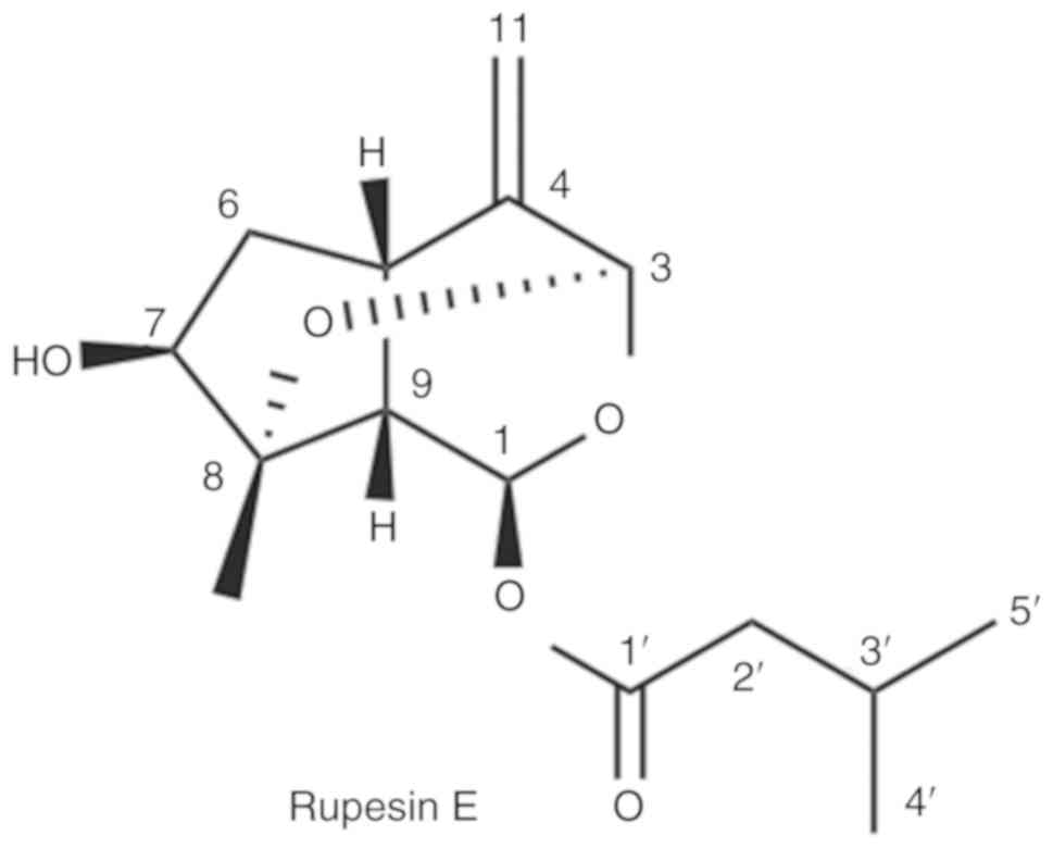
Rupesin E is an iridoid isolated from the roots and rhizomes of
Valeriana jatamansi. In a preliminary study, the screening
results indicated that rupesin E may be selectively sensitive to
GSCs. In order to confirm the activity and mechanism of rupesin E
against GSCs, further experiments were performed to determine the
proliferation, apoptotic, clone formation ability of GSCs after the
treatment of rupesin E.
Materials and methods
Materials
Rupesin E was provided by Dr Rongtao Li from Kunming
University of Science and Technology (Yunnan, China). Laminin (cat.
no. 23017-015; Gibco; Thermo Fisher Scientific, Inc.), B27
supplement (cat. no. 12587-010; Gibco; Thermo Fisher Scientific,
Inc.), Dulbecco's modified Eagle's Medium (DMEM; cat. no.
12800-017; Gibco, Thermo Fisher Scientific, Inc.), DMEM/F12 (cat.
no. 11330-032; Gibco; Thermo Fisher Scientific, Inc.), PBS (cat.
no. 18912-014; Gibco; Thermo Fisher Scientific, Inc.), fetal bovine
serum (cat. no. 10099-141; Gibco; Thermo Fisher Scientific, Inc.),
TryplE express (cat. no. 12604-021; Gibco) and
5-ethynyl-2′-deoxyuridine (EdU; Click-iT EdU Alexa Fluor 488
imaging kit; cat. no. C10337; Invitrogen) were purchased from
Thermo Fisher Scientific, Inc. BSA (cat. no. FC0077) was purchased
from MP Biomedicals, LLC. Goat serum (cat. no. C-0005) was
purchased from Shanghai Haoran Biotechnology Co., Ltd.
MTS[3-(4,5-dimethylthiazol-2-yl)-5-(3-carboxymethoxyphenyl)-2-(4-sulfophenyl)-2H-tetrazolium;
cat. no. G3581] was purchased from Promega Corporation. Tween-20
(cat. no. SLBX6047) was purchased from Sigma-Aldrich; Merck KGaA.
Low-gelling temperature agarose (cat. no. A9045) was purchased from
was purchased from Sigma-Aldrich; Merck KGaA. The Annexin V-FITC
Apoptosis Assays kit (cat. no. A005-4) was purchased from 7sea
biotech. The basic fibroblast growth factor (bFGF; cat. no.
AF-100-18B) and epidermal growth factor (EGF; cat. no. AF-100-15)
were purchased from PeproTech, Inc. Primary anti-cleaved caspase-3
antibody (rabbit anti-cleaved caspase-3; cat. no. 9661) was
obtained from Cell Signaling Technology, Inc. Anti-caspase-3
secondary antibody (Goat anti-rabbit IgG Fc Dylight-488; ab98462)
was obtained from Abcam. Primary nestin antibody (cat. no.
sc-23927) was purchased from Santa Cruz Biotechnology, Inc. Primary
glial fibrillary acidic protein antibody (GFAP; cat. no. 20334) was
purchased from Dako (Agilent Technologies, Inc). Primary GAPDH
antibody (cat. no. ab9484) was obtained from Abcam. Goat anti-mouse
IgG secondary antibody (cat. no. cw0102) was obtained from CWBio.
Goat anti-rabbit IgG secondary antibody (cat. no. A6154) was
obtained from Sigma-Aldrich (Merck KGaA). Flow cytometry (LSR
Fortessa) was obtained from Becton, Dickinson and Company. The
software used for the flow cytometry was the BD FACS Diva Software
v8.0.1 (Becton, Dickinson and Company). Automatic chemiluminescence
imaging analysis system (cat. no. Tanon-5200) was purchased from
Shanghai Tianneng Technology Co. Ltd. The digital camera
(Canon-1500D) was obtained from Canon, Inc.
Isolation and purification of Rupesin
E
Rupesin E was isolated from the roots and rhizomes
of Valeriana jatamansi by Dr Rongtao Li from Kunming
University of Science and Technology (Yunnan, China). Firstly, the
air-dried and powdered Valeriana jatamansi (25 kg) were
extracted with 95% ethanol (3×37 l, 24 h each time) at room
temperature and concentrated under vacuum to obtain a crude extract
(2.7 kg), which was suspended in water and extracted successively
with petroleum ether, ethyl acetate and n-butanol. The ethyl
acetate extract (340 g) was subjected to silica gel column
chromatography (CC; polyethylene/acetone; gradient, 1:0 to 0:1) to
yield the eluted fractions E1 - E7. E4 (40.2 g) was isolated by CC
over MCI gel (MCI gel is a highly porous styrene-divinylbenzene
polymer resin used as column packing material; methanol/water;
gradient, 30, 60, 70, 90 and 100%), silica gel (petroleum
ether/acetone 30:1 to 5:1), Sephadex LH-20 (chloroform/methanol
1:1) and semi-preparative high performance liquid chromatography
[HPLC; at 40°C; sample quantity, 50 µl; using a Zorbax SB-C18
column (5 µm; 4.6×150 mm; Agilent Technologies, Inc.); 20%
methanol/water; flow rate, 3 ml/min)] to yield rupesin E [181 mg,
with a retention time (tR; the time elapsed between
sample introduction at the beginning of the chromatogram and the
maximum signal of the given compound at the detector) of 14.3 min
and purity of 98.1%]. A total of 10 mg rupesin E was dissolved in 1
ml DMSO to make a 10 mg/ml stock solution and stored at −20°C,
protected from light. The purity of the compound was analyzed using
an Agilent 1260 HPLC system (at 40°C; sample quantity, 50 µl) with
a Zorbax SB-C18 (5 µm; 4.6 × 150 mm; Agilent Technologies, Inc.)
column. The solvent system was methanol/water, gradient at the flow
rate of 3 ml/min for 15 min. The structure of rupesin E was
identified by comparison of the spectroscopic data with previously
published values (33–36).
Cells and culture
The HAC (Human Astrocytes-cerebellar; cat. no. 1810)
cell line was purchased from ScienCell Research Laboratories, Inc.
and cultured in DMEM with 10% FBS. The three human GSC lines
(GSC-3#, GSC-12# and GSC-18#) used were established from three
different human glioblastoma samples by Kunming Institute of
Zoology (Yunnan, China) that was obtained from Yunnan Cancer
Hospital (Kunming, China) (37,38).
Tumor stem cells were cultured in GSC medium that consisted of
DMEM/F12, 1 × B27, 50 ng/ml bFGF and 50 ng/ml EGF supplemented with
100 U/ml penicillin and 100 µg/ml streptomycin. In addition, the
three human GSC lines were cultured in pre-coated culture dishes
with laminin, and the cells could attach and grow normally without
differentiation. Culture dishes were pre-coated with 10 µg/ml
laminin for 6–9 h at 37°C in a humidified incubator, dissociated
with TryplE express for 5 min at 37°C in a humidified incubator and
centrifuged at 800 × g, at 25°C for 5 min. The cells were
suspended, and 1×10⁶ cells/ml were seeded into the
pre-coated dishes and maintained at 37°C in a humidified incubator
with 5% CO2. Human patient-derived GSCs, termed GSC-3#,
GSC-12# and GSC-18#, were established as described before (39–42).
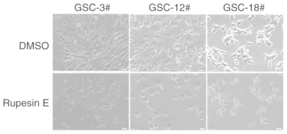
Morphological observation
The morphology of 10 µg/ml rupesin E-treated GSCs
(GSC-3#, GSC-12# and GSC-18#) at 72 h after treatment was examined
using an Olympus-IX71 light microscope (Olympus Corporation) and
images were captured using an Olympus-DP72 (Olympus Corporation;
magnification, ×40).
MTS assay
A total of 2×10⁴ HAC and 2×10⁴
GSCs (GSC-3#, GSC-12# and GSC-18#) in 150 µl per well were seeded
into 96-well plates and treated with 50 µl rupesin E at 80, 40, 20,
10, 5 and 2.5 µg/ml or 40, 20, 10, 5, 2.5 and 1.25 µg/ml,
respectively. In a screening experiment, the effect of rupesin E on
proliferation was stronger in glioma stem cells compared with that
in HAC cells for 72 h. Therefore, the four cell lines were treated
with different concentrations of rupesin E to obtain the
IC50 values. HAC and GSCs that were treated with the
same volume of DMSO (0.2%) were used as a normal control. All
groups of cells were incubated for 72 h at 37°C with 5%
CO2 after treatment with rupesin E. MTS reagent was
diluted 1:10 with fresh complete medium and mixed thoroughly
(43). The old medium was removed
and 100 µl/well fresh medium was added. Compared with GSCs, HAC has
higher metabolism and dehydrogenase activity, and can completely
react with substrate in a shorter time and thus, GSCs were
incubated for 1.5 h, whereas the HAC cells were incubated for 30
min, and the absorbance was measured using a BioTek synergy H1
microplate Hybrid Reader (BioTek Instruments, Inc.) at 490 nm. The
viability of cells treated with rupesin E was compared with the
control group treated with DMSO and expressed as a percentage. The
half-maximal inhibitory concentration (IC50) was
calculated using Graph Pad Prism v5 software (GraphPad Software,
Inc).
Cell proliferation assay
Cell proliferation assay was performed using
5-ethynyl-2′-deoxyuridine (EdU) immunofluorescence staining, as
previously reported (44). The
results were consistent in all three cell lines (GSC-3#, GSC-12#
and GSC-18#) and the two of these cell lines were randomly
selected. GSC-3# and GSC-18# were digested with TryplE express and
seeded onto a 24-well plate with 1×10⁵ cells/per well.
Rupesin E 10 µg/ml was added and the control wells were treated
with the same volume of DMSO (0.2%). The GSC-3# cells were treated
for 14 h and the GSC-18# cells were treated for 12 h at 37°C in a
humidified incubator. The GSCs were treated with EdU (10 µM) for 1
h at 37°C in a humidified incubator with 5% CO2.
Following incubation, the media was removed and 500 µl 4%
paraformaldehyde (PFA) was added to each well containing
coverslips. The plate was incubated for 15 min at room temperature,
following which the fixative was removed and the cells were washed
twice with 1 ml 3% BSA in PBS. Once the wash solution was removed,
500 µl 0.5% Triton X-100 in PBS was added to each well, incubated
at room temperature for 20 min and subsequently washed twice with
3% BSA. A total of 200 µl 1X Click-iT reaction mixture was added to
each well, the plate was briefly agitated to ensure that the
reaction mixture is distributed evenly over the coverslip and
incubated for 30 min at room temperature protected from light. The
reaction mixture was removed, and each well was washed twice with 1
ml 3% BSA in PBS and incubated with DAPI (10 mM; diluted 1:1,000 in
PBS) for 10 min in the dark. The cells were washed twice with PBS
and mounted onto a glass slide. Images were obtained using an
Olympus IX71 (Olympus Corporation) fluorescent microscope at the
magnifications ×20 and ×40.
Apoptosis assay
Apoptosis assay was performed using cleaved
caspase-3 immunofluorescence staining. GSC-3# and GSC-18# cells
(1×10⁵ cells/well) were plated in 24-well culture
dishes. The GSC-3# cells were treated with 10 µg/ml rupesin E for
39 h and the GSC-18# cells were treated with 10 µg/ml rupesin E for
14 h. A total of 500 µl 4% PFA was added to each well containing
the coverslips and incubated for 20 min, followed by washing with
0.3% Triton X-100 three times. The cells were blocked with 10% goat
serum (diluted in PBS) for 1 h at room temperature. Each well was
incubated with primary anti-cleaved caspase-3 antibody (rabbit
anti-cleaved caspase3; diluted 1:400 in PBS with 1% BSA and 0.25%
Tween-20) for 1 h at room temperature and washed with 0.2% Tween-20
four times. The wells were incubated with anti-caspase3 secondary
antibody (goat anti-rabbit IgG Fc Dylight-488; diluted 1:500 in PBS
buffer with 1% BSA and 0.5% Tween-20) for 1 h away from light at
room temperature. Each coverslip was washed with 0.2% Tween-20 4
times and incubated with DAPI (1:5,000 at 1 mg/ml) for 10 min in
the dark, and the slides were washed with PBS three times and
mounted onto glass slide. Images were obtained using an Olympus
IX71 (Olympus Corporation) fluorescent microscope at the
magnification of ×20 and ×40.
To further confirm these results, apoptosis assay
was also performed using Annexin V/propidium iodide (PI) staining
and analyzed using flow cytometry. GSC-3# cells (8×10⁵)
were plated in 6-well culture dishes. The cells were treated with
either 10 µg/ml rupesin E or the same volume of DMSO (0.2%) as a
control for 2, 4 and 8 h. Cells were digested using trypsin,
collected and centrifugated at 500 × g for 5 min at 25°C, following
which the supernatant was discarded, and the pellet was gently
resuspended with 1 ml PBS and centrifuged again at 500 × g for 5
min at 25°C. A total of 400 µl 1X binding buffer was added to
resuspend the cells, incubated with 5 µl Annexin V-FITC for 15 min
in the dark at 4°C, and 10 µl PI was added, placed on ice and
protected from light. Flow cytometry analysis was performed within
30 min.
Western blot analysis
GSC-3# cells were treated with or without 10 µg/ml
rupesin E (10 h), collected and lysed in RIPA buffer (cat. no.
R0010; Beijing Solarbio Science and Technology Co., Ltd.) with 0.1
mg/ml phenylmethylsulfonyl fluoride and phosphatase inhibitor. This
was followed by centrifugation at 20,000 × g for 30 min at 4°C. The
protein precipitate was removed and the protein in the supernatant
was quantified using the BCA assay kit (cat. no. P0010; Beyotime
Institute of Biotechnology), denatured at 95°C for 5 min and loaded
onto 10% SDS-PAGE (50 µg per lane). Following electrophoresis,
proteins were transferred to polyvinylidene fluoride membranes (EMD
Millipore). The membranes were blocked for 1.5 h at room
temperature in blocking buffer (5% skimmed milk in PBS with 0.2%
Tween-20) and incubated with the primary antibodies [Nestin and
glial fibrillary acidic protein (GFAP) were diluted to 1:500, GAPDH
to 1:1,000] overnight at 4°C. Following washing with 0.1% Tween-20
in PBS three times, the membranes were incubated with either goat
anti-rabbit IgG secondary antibody (1:10,000 dilution), or goat
anti-mouse IgG secondary antibodies (1:10,000 dilutions) for 2 h at
room temperature. Finally, the membranes were treated with
Automatic chemiluminescence imaging analysis system (Tanon-5200)
and exposed to ECL. Results were analyzed using Image J Software
(National Institutes of Health). GAPDH was used as a reference
gene.
Soft agar colony formation assay
The soft agar colony formation assay was performed
as previously described (45,46). The
low gelling agar (0.4 g) was mixed with 13 ml double distilled
water and sterilized by high-pressure steam sterilization (121°C,
15 min) to make 3.2% soft agar. To prepare the 0.8% bottom layer of
agar, 3.2% soft agar was diluted 1:4 in GSC medium and plated in a
6-well-plate at 1 ml/well. The plates were left at room temperature
to cool. To prepare the 0.4% upper layer of agar, GSC-3# and
GSC-18# were digested with TryplE express and 2×10⁴
cells/well were mixed with 0.8% agar to 0.4% concentration and
plated in a 6-well-plate at 1 ml/well, cooled and supplemented with
1 ml GSC medium. The plates were cultured in a cell incubator (at
37°C in a humidified incubator with 5% CO2) and fresh
medium was added every 3–4 days. After two weeks, once the clonal
spheres had reached a certain size (20 µm), they were treated with
20 µg/ml rupesin E every 3 days. The control groups were treated
with the same volume of 0.2% DMSO and fresh medium was added every
3 days. After two weeks, the GSC medium was removed and washed once
with PBS. The clonal spheres were fixed with 1 ml 4% PFA at room
temperature containing 0.005% crystal violet for 2 h. Images were
obtained using a digital camera and the clonal spheres were
counted.
Statistical analysis
All data were expressed as the means ± standard
error of the mean of three separate experiments. GraphPad Prism
v5.0 software (GraphPad Software, Inc.) was used for statistical
analysis. One-way analysis of variance followed by the least
significant difference post hoc test was used to determine
statistical significance. P<0.05 was considered to indicate a
statistically significant difference.
Results
Structure identification of rupesin
E
1H-NMR spectrum (deuterated acetone; 600
MHz) of rupesin E is presented in Fig.
S1 and 13C-NMR spectrum (deuterated acetone; 150
MHz) of compound rupesin E is presented in Fig. S2. The 1H-NMR and
13C-NMR data of Rupesin E are as follows and consistent
to those of the references (33–36).
Rupesin E, colorless oil,
C15H22O5, 1H-NMR
(CD3COCD3; 600 MHz): δH 6.28 (1H, d,
J=3.2 Hz, Ha−1), 5.03 (1H, s, H-3), 3.12–3.10
(1H, m, H-5), 2.08–2.00 (1H, overlap, Ha−6), 1.90–1.86
(1H, m, Hb−6), 3.85–3.80 (1H, m, H-7), 4.20 (1H, br s,
OH- C-7), 2.41–2.39 (1H, m, H-9), 1.36 (3H, s, H-10), 4.87 (1H, s,
Ha−11), 4.80 (1H, s, Hb−11), 2.19–2.11 (2H,
m, H-2′), 2.08–2.00 (1H, overlap, H-3′), 0.92 (3H, d, J=4.5
Hz, H-4′), 0.91 (3H, d, J=4.5 Hz, H-5′). 13C-NMR
(CD3COCD3; 150 MHz): δC 90.7 (CH,
C-1), 94.3 (CH, C-3), 150.4 (C, C-4), 37.5 (CH, C-5), 43.8
(CH2, C-6), 79.0 (CH, C-7), 83.0 (C, C-8), 42.5 (CH,
C-9), 19.2 (CH3, C-10), 107.2 (CH2, C-11),
171.9 (C, C-1′), 43.5 (CH2, C-2′), 26.1 (CH, C-3′), 22.5
(C, C-4′), 22.4 (CH, C-5′).
Rupesin E selectively inhibits the
viability of GSCs
In a screening experiment, the
proliferation-inhibiting efficacy of rupesin E (Fig. 1) against three human GSC lines
(GSC-3#, GSC-12# and GSC-18#) was evaluated. At 10 µg/ml, rupesin E
inhibited the proliferation of all GSCs effectively, based on
morphological observation (Fig. 2).
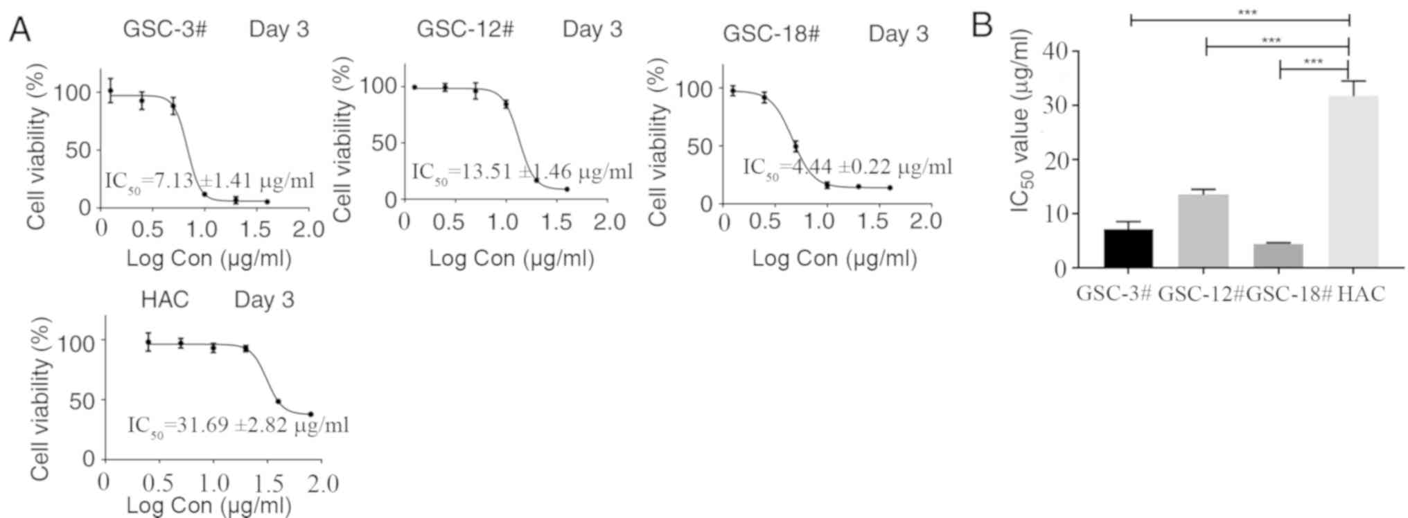
In addition, the inhibitory effect of rupesin E on the viability of
GSCs and normal human HAC cells was investigated using an MTS assay
at varying concentrations of rupesin E (40, 20, 10, 5, 2.5 and 1.25
µg/ml in GSCs; 80, 40, 20, 10, 5 and 2.5 µg/ml in HAC cells) after
treatment for 72 h. As shown in Fig.
3, rupesin E inhibited the viability of human GSCs and HAC
cells in a concentration-dependent manner. The half maximal
inhibitory concentration (IC50) values of rupesin E in
human GSCs (GSC-3#, GSC-12# and GSC-18#) were 7.13±1.41, 13.51±1.46
and 4.44±0.22 µg/ml at 72 h, respectively. For the HAC cells, the
IC50 value was 31.69±2.82 µg/ml for 72 h. These results
indicated that GSCs were more sensitive to rupesin E compared with
HAC cells.
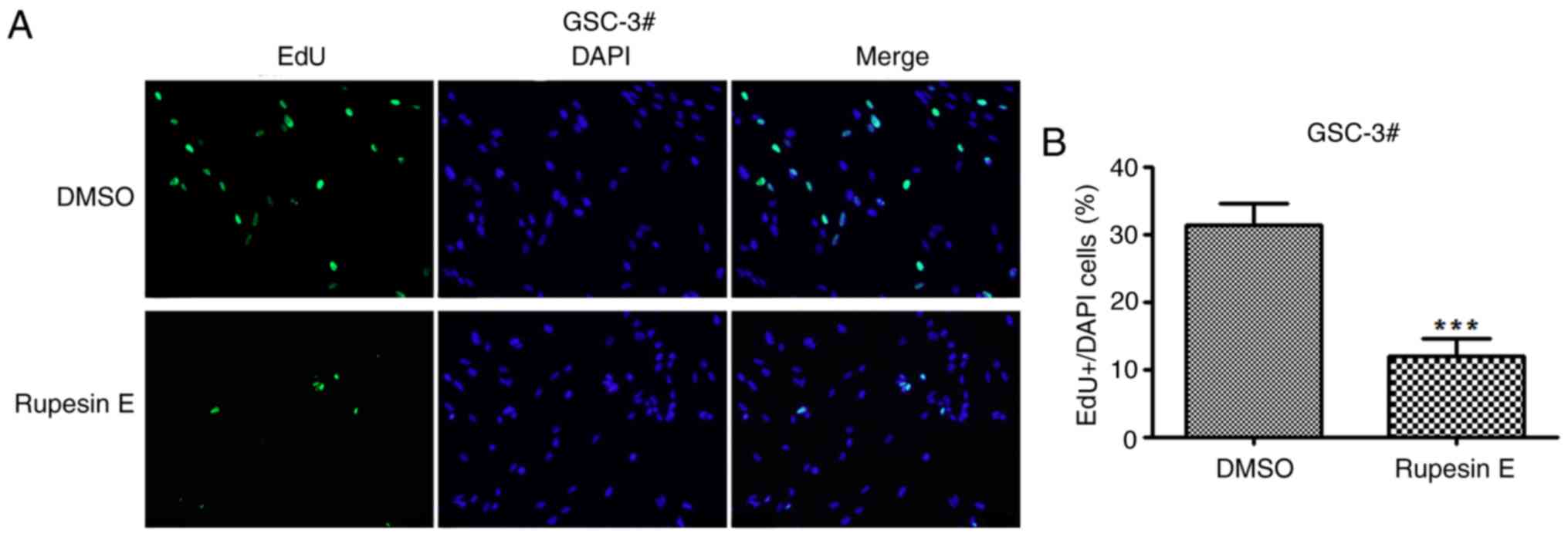
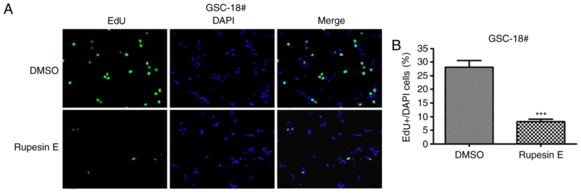
Rupesin E suppresses the proliferation
of GSCs
In the preliminary experiment, rupesin E was
identified to inhibit the proliferation of GSCs; however, its
mechanism of action is unknown. To determine whether rupesin E
suppressed cell proliferation by suppressing DNA synthesis, GSC-3#
and GSC-18# were subjected to EdU incorporation assay and treated
with rupesin E (10 µg/ml) for 14 and 12 h, respectively (Figs. 4 and 5). The results demonstrated that the
percentage of Edu-positive proliferative cells significantly
decreased in cells treated with rupesin E compared with that in the
control group. This indicated that rupesin E notably inhibited cell
proliferation through the suppression of DNA synthesis in the two
GSC lines.
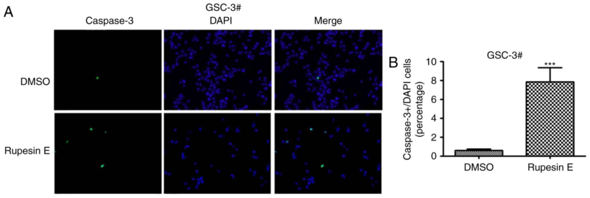
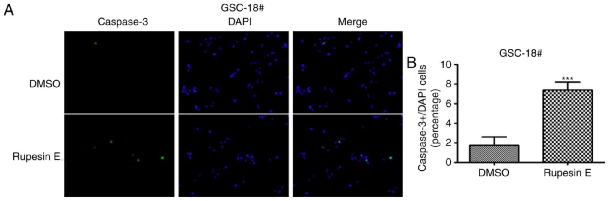
Rupesin E induces apoptosis of
GSCs
The treatment of rupesin E resulted in the decline
of cell viability in both GSC-3# and GSC-18#, and the cells became
spherical and detached, which suggested that rupesin E may induce
apoptosis in GSCs. To determine whether GSCs underwent apoptosis
after the treatment of rupesin E, the level of apoptosis in GSC-3#
and GSC-18# was measured using immunofluorescent staining. The
activation of caspase-3 occurs in the early stage of apoptosis,
which was significantly increased in both GSC-3# (Fig. 6) and GSC-18# (Fig. 7) treated with rupesin E (10 µg/ml)
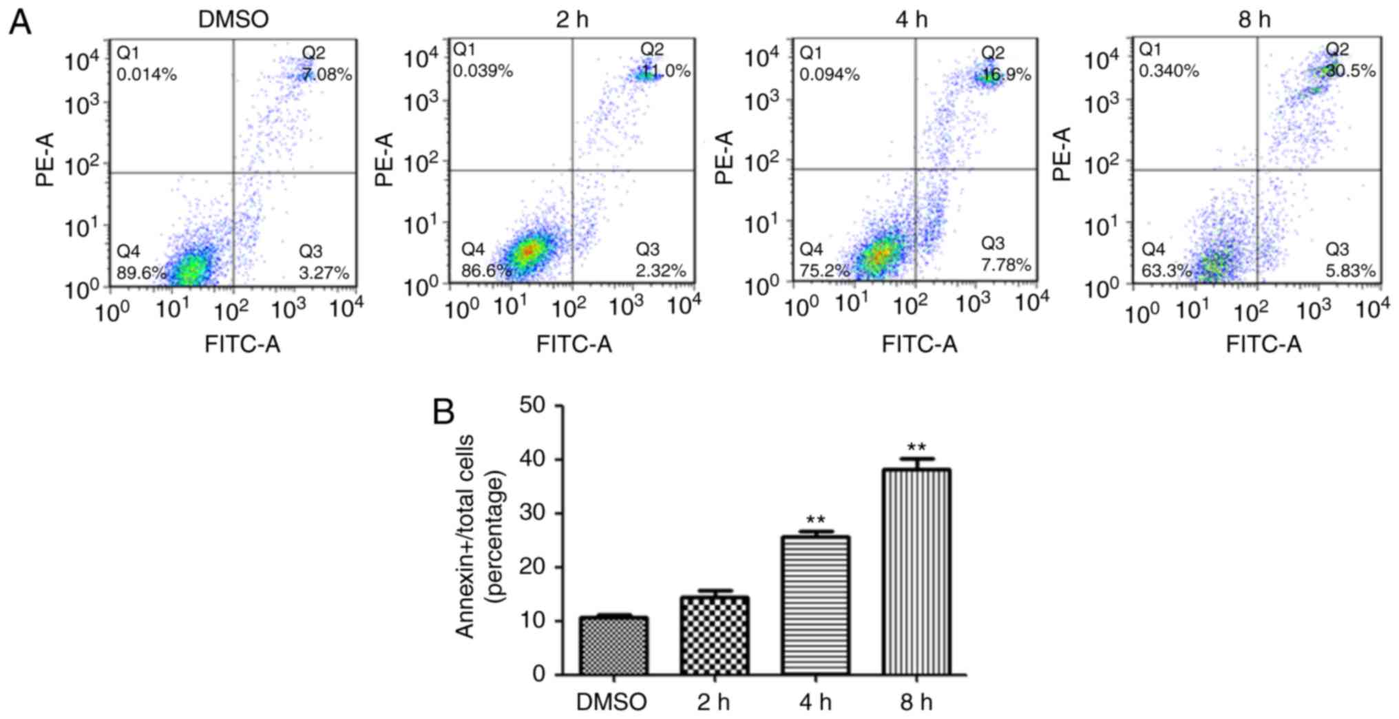
for 39 and 14 h, respectively. In addition, the level of apoptosis
was further measured using Annexin V/PI assay and flow cytometry in
GSC-3#. The result demonstrated that the proportions of Annexin
V-positive apoptotic cells were increased significantly in GSC-3#
compared with the control after 4 and 8 h treatment with rupesin E
(Fig. 8). These results revealed
that rupesin E induced apoptosis of GSC-3# and GSC-18#.
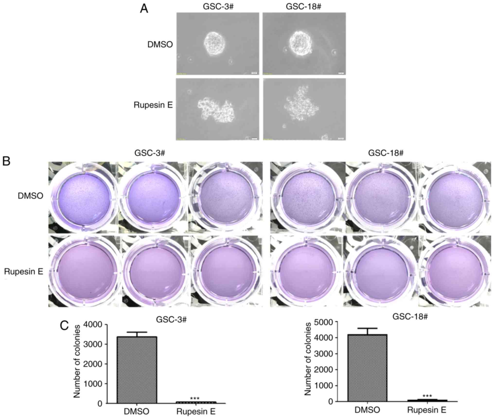
Rupesin E inhibits the colony
formation ability of GSCs
To examine whether rupesin E inhibited the
proliferative ability of GSCs, the colony formation assay was
performed to determine the ability of the single cell to divide
unlimitedly following the treatment of a cytotoxic agent in
vitro. GSC-3# and GSC-18# were seeded in the soft agar and once
the clonal spheres achieved a certain size (20 µm), rupesin E (20
µg/ml) was used to treat GSCs. The results demonstrated that
rupesin E significantly reduced the number of clonal spheres
(Fig. 9), thus reducing the ability
of GSCs to divide unlimitedly.
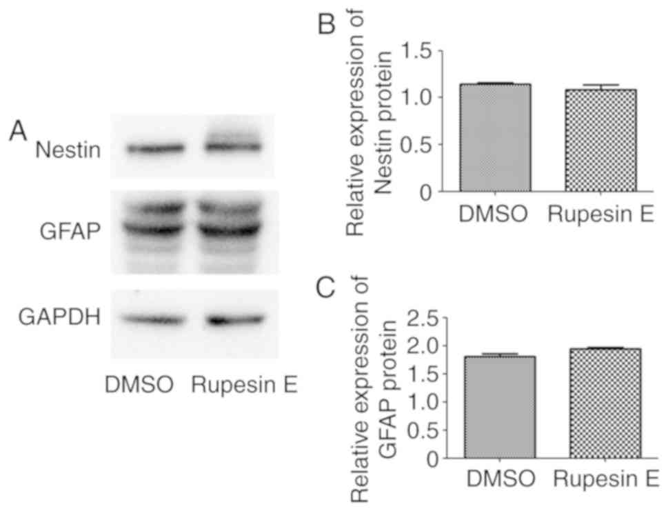
Rupesin E does not induce the
differentiation of GSCs
Nestin is a well-established marker of GSC stemness,
and GFAP is widely used as the differentiation marker of GSCs
(47). GSC-3# cells were treated
with 10 µg/ml rupesin E for 10 h, following which the protein
expression of nestin and GFAP were detected using western blot
analysis. The results demonstrated that after rupesin E treatment,
the expression of nestin did not significantly decline and the
expression of GFAP did not significantly increase (Fig. 10); these results indicated that
rupesin E could not induce GSC differentiation.
Discussion
The limitations of surgical treatment make it
impossible to completely remove tumors (5), and chemotherapy and radiotherapy are
only effective on rapidly dividing tumor cells. As the cancer stem
cells are tolerant to chemotherapy and radiotherapy, chemotherapy
and radiotherapy are ineffective against it (39). GSCs and normal stem cells exhibit the
same features, such as infinite proliferation, self-renewal and the
cloning expansion ability to form tumors, which may be important
reasons for tumor recurrence (17),
which was also demonstrated in the current study. This also
provides the theoretical basis for this study. Therefore, a large
number of studies aim to identify compounds that specifically
target glioma stem cells from small molecular compounds (48–50).
However, no drugs specifically targeting cancer stem cells have
been marketed to date. In the present study, a high-throughput
screening of 3,000 small-molecule compounds was conducted, and it
was found that rupesin E could selectively inhibit glioma stem
cells. Using the same concentration, rupesin E reduced
proliferation and induced apotosis of GSCs and had no effect on the
proliferation of HAC cells. In addition, rupesin E not only induced
GSC apoptosis, but also inhibited GSC ability of infinite
proliferation. Thus, rupesin E has the potential to be developed as
an antitumor agent, and combined with radiotherapy, chemotherapy
and surgical treatment, to completely remove the tumor.
Supplementary Material
Supporting Data
Acknowledgements
Not applicable.
Funding
The present study was partly supported by the
National Natural Science Foundation of China (grant nos. 81472862,
31560103 and U1602222).
Availability of data and materials
All data generated or analyzed during the present
study are included in this published article.
Authors' contributions
SQ and LQ performed the experiments and wrote the
manuscript. RL and XZ designed the study and edited the manuscript.
HL and XC collected and analyzed the data.
Ethics approval and consent for
publication
Not applicable
Patient consent for publication
Not applicable.
Competing interests
The authors declare that they have no competing
interests.
References
|
1
|
Louis DN, Ohgaki H, Wiestler OD, Cavenee
WK, Burger PC, Jouvet A, Scheithauer BW and Kleihues P: The 2007
WHO classification of tumours of the central nervous system. Acta
Neuropathol. 114:97–109. 2007. View Article : Google Scholar : PubMed/NCBI
|
|
2
|
Furnari FB, Fenton T, Bachoo RM, Mukasa A,
Stommel JM, Stegh A, Hahn WC, Ligon KL, Louis DN, Brennan C, et al:
Malignant astrocytic glioma: Genetics, biology, and paths to
treatment. Genes Dev. 21:2683–2710. 2007. View Article : Google Scholar : PubMed/NCBI
|
|
3
|
Scott CB, Scarantino C, Urtasun R, Movsas
B, Jones CU, Simpson JR, Fischbach AJ and Curran WJ Jr: Validation
and predictive power of radiation therapy oncology group (RTOG)
recursive partitioning analysis classes for malignant glioma
patients: A report using RTOG 90-06. Int J Radiat Oncol Biol Phys.
40:51–55. 1998. View Article : Google Scholar : PubMed/NCBI
|
|
4
|
Visser O, Ardanaz E, Botta L, Sant M,
Tavilla A and Minicozzi P; EUROCARE-5 Working Group, : Survival of
adults with primary malignant brain tumours in Europe; results of
the EUROCARE-5 study. Eur J Cancer. 51:2231–2241. 2015. View Article : Google Scholar : PubMed/NCBI
|
|
5
|
Alifieris C and Trafalis DT: Glioblastoma
multiforme: Pathogenesis and treatment. Pharmacol Ther. 152:63–82.
2015. View Article : Google Scholar : PubMed/NCBI
|
|
6
|
Bao S, Wu Q, McLendon RE, Hao Y, Shi Q,
Hjelmeland AB, Dewhirst MW, Bigner DD and Rich JN: Glioma stem
cells promote radioresistance by preferential activation of the DNA
damage response. Nature. 444:756–760. 2006. View Article : Google Scholar : PubMed/NCBI
|
|
7
|
Dean M, Fojo T and Bates S: Tumour stem
cells and drug resistance. Nat Rev Cancer. 5:275–284. 2005.
View Article : Google Scholar : PubMed/NCBI
|
|
8
|
Alisi A, Cho WC, Locatelli F and Fruci D:
Multidrug resistance and cancer stem cells in neuroblastoma and
hepatoblastoma. Int J Mol Sci. 14:24706–24725. 2013. View Article : Google Scholar : PubMed/NCBI
|
|
9
|
Chen J, Li Y, Yu TS, McKay RM, Burns DK,
Kernie SG and Parada LF: A restricted cell population propagates
glioblastoma growth after chemotherapy. Nature. 488:522–526. 2012.
View Article : Google Scholar : PubMed/NCBI
|
|
10
|
Genadry KC, Pietrobono S, Rota R and
Linardic CM: Soft tissue sarcoma cancer stem cells: An overview.
Front Oncol. 8:4752018. View Article : Google Scholar : PubMed/NCBI
|
|
11
|
Codony-Servat J, Verlicchi A and Rosell R:
Cancer stem cells in small cell lung cancer. Transl Lung Cancer
Res. 5:16–25. 2016.PubMed/NCBI
|
|
12
|
Johnsen HE, Bøgsted M, Schmitz A, Bødker
JS, El-Galaly TC, Johansen P, Valent P, Zojer N, Van Valckenborgh
E, Vanderkerken K, et al: The myeloma stem cell concept, revisited:
From phenomenology to operational terms. Haematologica.
101:1451–1459. 2016. View Article : Google Scholar : PubMed/NCBI
|
|
13
|
Parte SC, Batra SK and Kakar SS:
Characterization of stem cell and cancer stem cell populations in
ovary and ovarian tumors. J Ovarian Res. 11:692018. View Article : Google Scholar : PubMed/NCBI
|
|
14
|
Calabrese C, Poppleton H, Kocak M, Hogg
TL, Fuller C, Hamner B, Oh EY, Gaber MW, Finklestein D, Allen M, et
al: A perivascular niche for brain tumor stem cells. Cancer Cell.
11:69–82. 2007. View Article : Google Scholar : PubMed/NCBI
|
|
15
|
Koren E and Fuchs Y: The bad seed: Cancer
stem cells in tumor development and resistance. Drug Resist Updat.
28:1–12. 2016. View Article : Google Scholar : PubMed/NCBI
|
|
16
|
Lathia JD, Mack SC, Mulkearns-Hubert EE,
Valentim CL and Rich JN: Cancer stem cells in glioblastoma. Genes
Dev. 29:1203–1217. 2015. View Article : Google Scholar : PubMed/NCBI
|
|
17
|
Ignatova TN, Kukekov VG, Laywell ED,
Suslov ON, Vrionis FD and Steindler DA: Human cortical glial tumors
contain neural stem-like cells expressing astroglial and neuronal
markers in vitro. Glia. 39:193–206. 2002. View Article : Google Scholar : PubMed/NCBI
|
|
18
|
Bexell D, Svensson A and Bengzon J: Stem
cell-based therapy for malignant glioma. Cancer Treat Rev.
39:358–365. 2013. View Article : Google Scholar : PubMed/NCBI
|
|
19
|
Bovenberg MS, Degeling MH and Tannous BA:
Advances in stem cell therapy against gliomas. Trends Mol Med.
19:281–291. 2013. View Article : Google Scholar : PubMed/NCBI
|
|
20
|
Wang T, Shigdar S, Gantier MP, Hou Y, Wang
L, Li Y, Shamaileh HA, Yin W, Zhou SF, Zhao X and Duan W: Cancer
stem cell targeted therapy: Progress amid controversies.
Oncotarget. 6:44191–44206. 2015.PubMed/NCBI
|
|
21
|
Colak S and Medema JP: Cancer stem
cells-important players in tumor therapy resistance. FEBS J.
281:4779–4791. 2014. View Article : Google Scholar : PubMed/NCBI
|
|
22
|
Lage H: An overview of cancer multidrug
resistance: A still unsolved problem. Cell Mol Life Sci.
65:3145–3167. 2008. View Article : Google Scholar : PubMed/NCBI
|
|
23
|
Folkins C, Shaked Y, Man S, Tang T, Lee
CR, Zhu Z, Hoffman RM and Kerbel RS: Glioma tumor stem-like cells
promote tumor angiogenesis and vasculogenesis via vascular
endothelial growth factor and stromal-derived factor 1. Cancer Res.
69:7243–7251. 2009. View Article : Google Scholar : PubMed/NCBI
|
|
24
|
Bao S, Wu Q, Sathornsumetee S, Hao Y, Li
Z, Hjelmeland AB, Shi Q, McLendon RE, Bigner DD and Rich JN: Stem
cell-like glioma cells promote tumor angiogenesis through vascular
endothelial growth factor. Cancer Res. 66:7843–7848. 2006.
View Article : Google Scholar : PubMed/NCBI
|
|
25
|
Pietras A, Gisselsson D, Ora I, Noguera R,
Beckman S, Navarro S and Påhlman S: High levels of HIF-2alpha
highlight an immature neural crest-like neuroblastoma cell cohort
located in a perivascular niche. J Pathol. 214:482–488. 2008.
View Article : Google Scholar : PubMed/NCBI
|
|
26
|
Li Z, Bao S, Wu Q, Wang H, Eyler C,
Sathornsumetee S, Shi Q, Cao Y, Lathia J, McLendon RE, et al:
Hypoxia-inducible factors regulate tumorigenic capacity of glioma
stem cells. Cancer Cell. 15:501–513. 2009. View Article : Google Scholar : PubMed/NCBI
|
|
27
|
Plate KH and Risau W: Angiogenesis in
malignant gliomas. Glia. 15:339–347. 1995. View Article : Google Scholar : PubMed/NCBI
|
|
28
|
Mondal S, Bandyopadhyay S, Ghosh MK,
Mukhopadhyay S, Roy S and Mandal C: Natural products: Promising
resources for cancer drug discovery. Anticancer Agents Med Chem.
12:49–75. 2012. View Article : Google Scholar : PubMed/NCBI
|
|
29
|
Moselhy J, Srinivasan S, Ankem MK and
Damodaran C: Natural products that target cancer stem cells.
Anticancer Res. 35:5773–5788. 2015.PubMed/NCBI
|
|
30
|
Thies PW and Funke S: On the active
ingredients in baldrian. 1. Detection and isolation of isovalerian
acid esters with sedative effect from roots and rhizomes of various
valerian and kentranthus species. Tetrahedron Lett. 11:1155–1162.
1966.(In German). View Article : Google Scholar : PubMed/NCBI
|
|
31
|
Thies PW: Linarin-isovalerianate, a
currently unknown flavonoid from Valeriana wallichii D.C. 6. Report
on the active substances of Valeriana. Planta Med. 16:363–371.
1968.(In German). View Article : Google Scholar : PubMed/NCBI
|
|
32
|
Thies PW: Valerosidatum, ein
iridoidesterglycosid aus valeriana-arten 7. mitteilung über die
wirkstoffe des baldrians. Tetrahedron Lett. 11:2471–2474. 1970.
View Article : Google Scholar
|
|
33
|
Yang XP, Li EW, Zhang Q, Yuan CS and Jia
ZJ: Five new iridoids from Patrinia rupestris. Chem Biodivers.
3:762–770. 2006. View Article : Google Scholar : PubMed/NCBI
|
|
34
|
Dong FW, Jiang HH, Yang L, Gong Y, Zi CT,
Yang D, Ye CJ, Li H, Yang J, Nian Y, et al: Valepotriates from the
roots and rhizomes of Valeriana jatamansi jones as Novel
N-type calcium channel antagonists. Front Pharmacol. 9:8852018.
View Article : Google Scholar : PubMed/NCBI
|
|
35
|
Dong FW, Liu Yang, Wu ZK, Wei-Gao, Zi CT,
Dan Yang, Luo HR, Jun Zhou and Hu JM: Iridoids and sesquiterpenoids
from the roots of Valeriana jatamansi jones. Fitoterapia.
102:27–34. 2015. View Article : Google Scholar : PubMed/NCBI
|
|
36
|
Lin S, Chen T, Liu XH, Shen YH, Li HL,
Shan L, Liu RH, Xu XK, Zhang WD and Wang H: Iridoids and lignans
from Valeriana jatamansi. J Nat Prod. 73:632–638. 2010.
View Article : Google Scholar : PubMed/NCBI
|
|
37
|
Lee J, Kotliarova S, Kotliarov Y, Li A, Su
Q, Donin NM, Pastorino S, Purow BW, Christopher N, Zhang W, et al:
Tumor stem cells derived from glioblastomas cultured in bFGF and
EGF more closely mirror the phenotype and genotype of primary
tumors than do serum-cultured cell lines. Cancer cell. 9:391–403.
2006. View Article : Google Scholar : PubMed/NCBI
|
|
38
|
Wilson RJ, Thomas CD, Fox R, Roy DB and
Kunin WE: Spatial patterns in species distributions reveal
biodiversity change. Nature. 432:393–396. 2004. View Article : Google Scholar : PubMed/NCBI
|
|
39
|
Dai Z, Li SR, Zhu PF, Liu L, Wang B, Liu
YP, Luo XD and Zhao XD: Isocostunolide inhibited glioma stem cell
by suppression proliferation and inducing caspase dependent
apoptosis. Bioorg Med Chem Lett. 27:2863–2867. 2017. View Article : Google Scholar : PubMed/NCBI
|
|
40
|
Wang B, Dai Z, Yang XW, Liu YP, Khan A,
Yang ZF, Huang WY, Wang XH, Zhao XD and Luo XD: Novel
nor-monoterpenoid indole alkaloids inhibiting glioma stem cells
from fruits of Alstonia scholaris. Phytomedicine. 48:170–178. 2018.
View Article : Google Scholar : PubMed/NCBI
|
|
41
|
Wei X, Dai Z, Yang J, Khan A, Yu HF, Zhao
YL, Wang YF, Liu YP, Yang ZF, Huang WY, et al: Unprecedented sugar
bridged bisindoles selective inhibiting glioma stem cells. Bioorg
Med Chem. 26:1776–1783. 2018. View Article : Google Scholar : PubMed/NCBI
|
|
42
|
Yang XW, Dai Z, Wang B, Liu YP, Zhao XD
and Luo XD: Antitumor triterpenoid saponin from the fruits of
avicennia marina. Nat Prod Bioprospect. 8:347–353. 2018. View Article : Google Scholar : PubMed/NCBI
|
|
43
|
Chen J, Creed A, Chen AY, Huang H, Li Z,
Rankin GO, Ye X, Xu G and Chen YC: Nobiletin suppresses cell
viability through AKT pathways in PC-3 and DU-145 prostate cancer
cells. BMC Pharmacol Toxicol. 15:592014. View Article : Google Scholar : PubMed/NCBI
|
|
44
|
Ning H, Albersen M, Lin G, Lue TF and Lin
CS: Effects of EdU labeling on mesenchymal stem cells. Cytotherapy.
15:57–63. 2013. View Article : Google Scholar : PubMed/NCBI
|
|
45
|
Borowicz S, Van Scoyk M, Avasarala S,
Karuppusamy Rathinam MK, Tauler J, Bikkavilli RK and Winn RA: The
soft agar colony formation assay. J Vis Exp. e519982014.PubMed/NCBI
|
|
46
|
Franken NA, Rodermond HM, Stap J, Haveman
J and van Bree C: Clonogenic assay of cells in vitro. Nat Protoc.
1:2315–2319. 2006. View Article : Google Scholar : PubMed/NCBI
|
|
47
|
Bien-Möller S, Balz E, Herzog S, Plantera
L, Vogelgesang S, Weitmann K, Seifert C, Fink MA, Marx S, Bialke A,
et al: Association of glioblastoma multiforme stem cell
characteristics, differentiation, and microglia marker genes with
patient survival. Stem Cells Int. 2018:96282892018. View Article : Google Scholar : PubMed/NCBI
|
|
48
|
Danovi D, Folarin A, Gogolok S, Ender C,
Elbatsh AM, Engström PG, Stricker SH, Gagrica S, Georgian A, Yu D,
et al: A high-content small molecule screen identifies sensitivity
of glioblastoma stem cells to inhibition of polo-like kinase 1.
PLoS One. 8:e770532013. View Article : Google Scholar : PubMed/NCBI
|
|
49
|
Hothi P, Martins TJ, Chen L, Deleyrolle L,
Yoon JG, Reynolds B and Foltz G: High-throughput chemical screens
identify disulfiram as an inhibitor of human glioblastoma stem
cells. Oncotarget. 3:1124–1136. 2012. View Article : Google Scholar : PubMed/NCBI
|
|
50
|
Junca A, Villalva C, Tachon G, Rivet P,
Cortes U, Guilloteau K, Balbous A, Godet J, Wager M and
Karayan-Tapon L: Crizotinib targets in glioblastoma stem cells.
Cancer Med. 6:2625–2634. 2017. View Article : Google Scholar : PubMed/NCBI
|