Hepatocellular carcinoma (HCC) is one of the most
malignant tumors that pose a severe threat to human health. The
latest cancer statistics showed that the number of new liver cancer
cases and liver cancer-associated deaths worldwide in 2018 was
841,000 and 782,000, respectively (1). Liver cancer ranked sixth in terms of
new cancer cases and fourth in terms of cancer-associated deaths
worldwide in 2018 (1). The available
evidence indicates that immune escape of liver cancer cells plays a
vital role in the development of this malignancy (2,3) and
impairs the effectiveness of antitumor treatment (4). Therefore, effective blockage of the
occurrence of immune escape has become the focus of attention in
the prevention and treatment of HCC.
It is now known that the activation or inhibition of
immune cells in the body is regulated by positive and negative
signals (5,6). Among them, the interaction between
programmed cell death-1 (PD-1, also termed CD279) and programmed
death-ligand 1 (PD-L1, also termed CD274 and B7-H1) is the primary
negative immune regulatory signal, which inhibits the antitumor
immune activity of effector cells and mediates tumor immune escape
(7–10). Furthermore, immune checkpoint
blockers have recently emerged as a mainstream strategy for the
treatment of multiple solid tumors, including liver cancer
(11–14).
Hypoxia, a common phenomenon in the tumor
microenvironment, induces the expression of PD-L1 to promote immune
escape (15–17). A number of immune cells with
immunosuppressive activities, including tumor-associated regulatory
T cells (Tregs), myeloid-derived suppressor cells (MDSCs) and
tumor-associated macrophages (TAMs), are recruited to the tumor
tissue to form an immunosuppressive microenvironment (18–20).
Moreover, under hypoxic conditions, the expression of PD-L1 is
rapidly upregulated in these immunosuppressive cells in a
hypoxia-inducible factor 1α (HIF-1α)-dependent manner (21,22). In
this regard, the comprehensive analysis of the role and mechanism
of PD-L1 in hypoxia-induced immune escape is essential for the
improved treatment of liver cancer. The present review summarizes
the recent findings regarding the regulation of PD-L1-mediated
hypoxia-induced immune escape in HCC cells and discusses the
underlying mechanisms.
The compromised immune status of the body is
associated with the occurrence of liver cancer. When the immune
function is weakened or suppressed, the incidence of liver cancer
will increase significantly. Normally, once liver cancer cells are
formed in the body, the immune system can inhibit or kill these
cells in a variety of ways (23–25).
However, despite the immune surveillance and scavenger receptors,
it remains challenging to curb the occurrence and development of
liver cancer (26). The main reason
is that liver cancer cells may escape from the immune system attack
through various mechanisms. The PD-L1/PD-1 pathway, which promotes
cancer cell survival and proliferation, is a key mediator of the
immune escape of HCC cells (27–29).
Previous studies have shown that T cell-mediated
cellular immunity plays a pivotal role in the recognition and
killing of tumor cells (30,31). T cells recognize major
histocompatibility complexes that bear antigens derived from the
surface of cancer cells, which allows subsequent tumor recognition
and targeted killing (32).
Recently, it has been demonstrated that various mechanisms play a
role in increasing the expression of PD-L1 in tumor cells and in
the tumor microenvironment. PD-L1 is a transmembrane glycoprotein
composed of 290 amino acids, which belongs to the B7 family of
immune-regulatory ligands. The binding of PD-L1 to its PD-1
receptor suppresses T-cell migration, proliferation and secretion
of cytotoxic mediators, and restricts tumor cell killing, leading
to the occurrence of tumor cell immune escape (33). In the healthy immune system, the
PD-L1/PD-1 pathway plays a critical role in maintaining the balance
between protective immunity and immune tolerance. However, aberrant
activation of the PD-L1/PD-1 signaling pathway in the tumor
microenvironment is associated with the development of liver
cancer. A multivariate analysis showed that PD-L1 expression is an
independent predictor of postoperative recurrence of HCC (7,34).
Accumulating evidence has revealed that the elevated
level of PD-L1 in the tumor microenvironment constrains antitumor
immunity via the inhibition of antitumor effector cell function and
enhancement of the inhibitory activity of immunosuppressive cells
(12,16,35,36).
Cytotoxic T lymphocytes (CTLs) and natural killer (NK) cells are
the main local antitumor immune effector cells. Activated CTLs are
marked by granzyme B, which is the primary molecular mediator of
apoptosis (37). It has been shown
that the activation of the PD-1/PD-L1 signaling pathway restrains
CTL function by inducing apoptosis, anergy and exhaustion, and
promoting the secretion of immunosuppressive factors, leading to
the immune escape of tumor cells (38). Hepatoma tumor-infiltrating CTLs
express PD-1 molecules, which bind to PD-L1 that are expressed on
the surface of tumor cells, resulting in the depletion and
apoptosis of CTLs (39).
There are a large number of active immunosuppressive
cells in the tumor microenvironment, including Tregs, MDSCs and
TAMs (40). These immune cells form
a complex multi-cell population, which is an important part of the
tumor microenvironment. Indeed, various molecular interactions
between immune and cancer cells are considered a crucial step in
the direct or indirect induction of the occurrence and development
of tumors. These immunosuppressive cells also express a large
number of PD-L1 molecules, which induce apoptosis in CTLs by
binding to PD-1 (41). Tregs,
characterized by the expression of CD4, CD25 and Forkhead box
protein P3 (FOXP3), are the most characteristic immunosuppressive
cells. The inhibition of the immune response by Tregs is also
mediated by cell contact or the secretion of inhibitory cytokines,
such as interleukin (IL)-10 and transforming growth factor-β
(TGF-β) (42). A previous study
found that PD-L1 promoted Treg differentiation by converting
CD4+CD25+FOXP3− T cells to
CD4+CD25+FOXP3+ Tregs.
Furthermore, higher expression levels of PD-L1 on hepatic dendritic
cells were associated with an increased Treg cell induction
(43). Specific blocking of PD-L1 by
small interfering (si)RNA or monoclonal antibodies decreased the
production of CD4+CD25+FOXP3+
Tregs and induced Treg apoptosis (44). In a pig xenograft model, PD-L1 was
found to enhance Treg function and stimulate IL-10 production,
thereby further promoting the immune inhibitory function (45). Clinical data also showed that PD-L1
effectively stimulated the secretion of IL-10 in patients with
liver cancer, thereby further enhancing the immunosuppressive
effect of Tregs (46). Collectively,
these studies have shown that the PD-L1/PD-1 pathway inhibits the
antitumor function of CTLs, enhances the immunosuppressive activity
of Tregs, and promotes the secretion of immunosuppressive factors
by transmitting inhibitory signals, leading to the occurrence of
tumor immune escape.
Under hypoxic conditions, HIF-1α is a crucial
transcription factor that mediates the effect of hypoxia on the
adaptive regulation of tumor cells and the tumor microenvironment
(63–65). Under normoxic conditions, HIF-1α is
hydroxylated by prolyl hydroxylase (PHD) and ubiquitinated/degraded
by the von Hippel-Lindau E3 ubiquitin ligase complex. Under hypoxic
conditions, PHD activity is inhibited, and HIF-1α ubiquitination
and degradation are decreased, thereby stabilizing HIF-1α (66). Previous studies have shown that
HIF-1α is associated with PD-L1 expression (15,22).
Under hypoxic conditions, tumor cells, myeloid suppressor cells,
macrophages and dendritic cells all undergo rapid upregulation of
PD-L1 in a HIF-1α-dependent manner. Chromatin immunoprecipitation
and luciferase reporter assays showed that HIF-1α induced the
expression of PD-L1 by directly binding to the hypoxia response
element region of the PD-L1 promoter. Furthermore, the inhibition
of PD-L1 expression significantly decreased the secretion of IL-6
and IL-10 by MDSC, leading to the activation of T cells (22). Another in vitro study also
revealed that hypoxia stimulated the expression of PD-L1 in a
variety of human and murine tumor cells through HIF-1α (15). These studies demonstrate that hypoxia
induces PD-L1 expression by activating the HIF-1α cascade.
Accumulating evidence has indicated that the
essential mechanism underlying tumor immune escape is associated
with the presence of a large number of cytokines and growth factors
with immunosuppressive activities in the tumor microenvironment,
such as IL-6, vascular endothelial growth factor, TGF-β, IL-10,
IL-13, macrophage colony-stimulating factor and
granulocyte-macrophage colony-stimulating factor (67–69).
These cytokines stimulate immune inhibitory cells, including Tregs,
TAMs and MDSCs, and mediate the expression of a series of genes by
activating various signaling pathways. Among them, the STAT3 and
NF-κB pathways are essential hubs linking these cytokines to
cellular responses (70–73).
STAT3 is a member of the STAT family of
transcription factors. When cytokines in the tumor microenvironment
bind to their receptors, the Janus kinase and/or proto-oncogene
tyrosine-protein kinase Src will be activated and able to
phosphorylate STAT3. Following dimerization and nuclear
translocation, STAT3 will initiate the transcription of downstream
genes. A previous study found that STAT3 activation in tumor cells
induces the secretion of IL-6 and IL-10 cytokines, which results in
Treg proliferation. Moreover, STAT3 is also activated in Tregs and
further stimulates the expression of FOXP3, TGF-β and IL-10, which
inhibits CTLs and promotes the formation of an immunosuppressive
environment (70,74,75).
In addition, STAT3 and NF-κB are often coactivated
in tumor cells and play a vital role in the regulation of the
expression of cancer-promoting inflammatory genes (76). The coordination between STAT3 and
NF-κB is mainly manifested in the following aspects: i) Multiple
inflammatory factors, especially IL-6, induced by NF-κB are
essential activators of STAT3; ii) STAT3 directly interacts with
the NF-κB family member transcription factor p65 (RelA), leading to
its acetylation and inhibition of nuclear export, and constitutive
activation of NF-κB; iii) STAT3 and NF-κB co-regulate the
expression of a number of oncogenes and inflammatory genes; and iv)
the inflammatory factors induced by NF-κB and STAT3 form a positive
feedback loop to further activate NF-κB and STAT3 (77,78).
Notably, it has been shown that the expression of
HIF-1α is regulated by both NF-κB and STAT3. Under hypoxic
conditions, STAT3 is activated by phosphorylation, which not only
blocks HIF-1α degradation but also increases the synthesis of
HIF-1α (79). In human breast cancer
MCF-7 cells, the depletion of STAT3 by siRNA inhibited
CoCl2-induced HIF-1α nuclear accumulation (80). The NF-κB signaling pathway is also
activated under hypoxic conditions (81). Gel shift assay and chromatin
immunoprecipitation experiments confirmed that the NF-κB subunits
p50 and RelA bind to the promoter of HIF-1α and activate its
transcription (82). Since HIF-1α
transcriptionally induces PD-L1, these studies indicate that the
activation of the STAT3 and NF-κB pathways may indirectly stimulate
PD-L1 expression under hypoxic conditions.
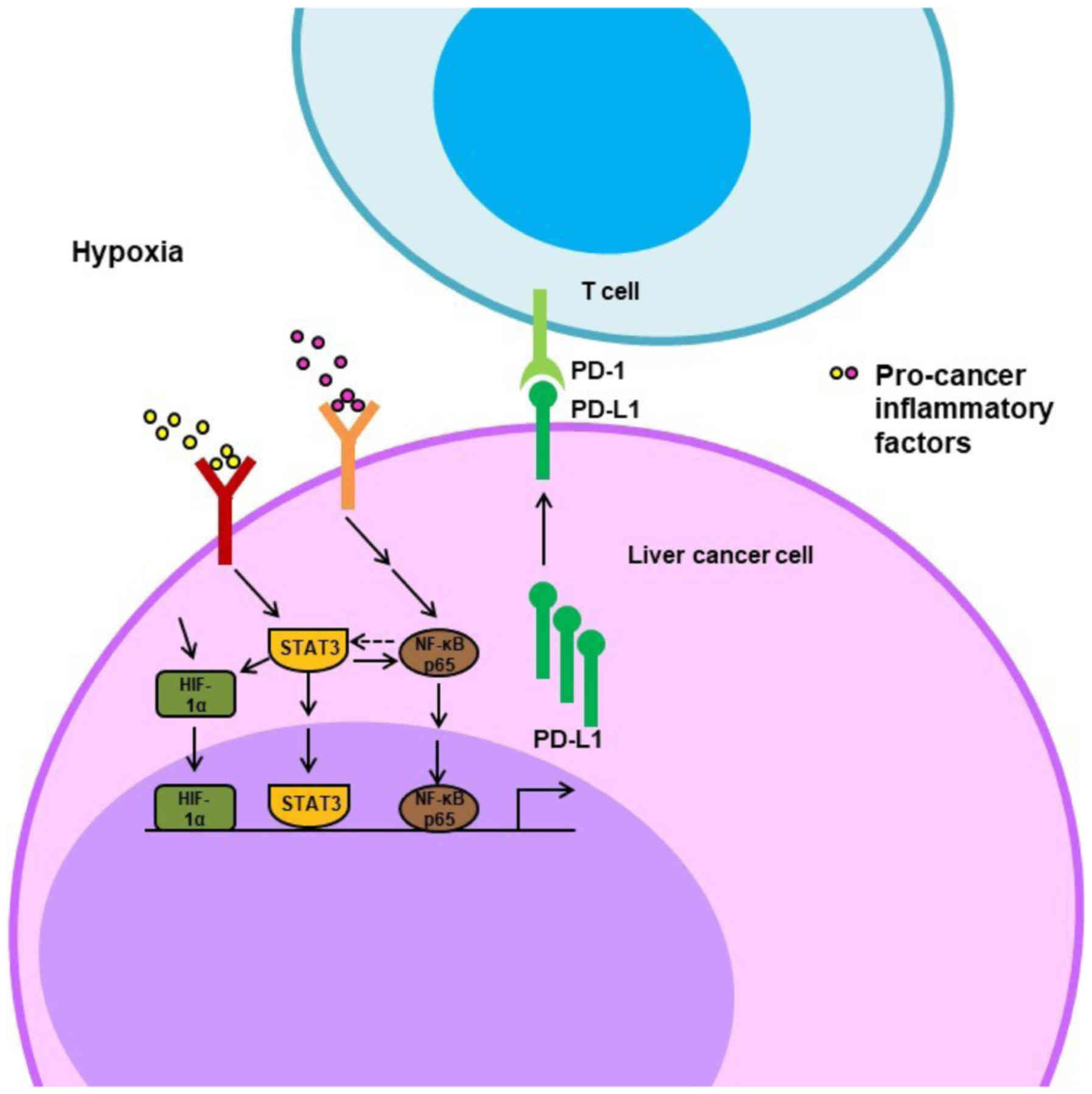
Furthermore, several studies have shown that the
STAT3 and NF-κB signaling pathways are also involved in the direct
regulation of PD-L1 at the transcriptional level (83–86). It
has been demonstrated that the co-culture of liver cancer cells
(BEL-7402 and SMMC-7721) with macrophages resulted in increased
PD-L1 mRNA and protein levels and that blocking either the NF-κB or
the STAT3 signaling pathway inhibited this co-culture effect on
PD-L1 expression (83). Another
study showed that EB virus latent membrane protein 1 (LMP1) induced
the expression of PD-L1 by the activation of NF-κB or STAT3; the
inhibition of one of these pathways notably decreased
LMP1-stimulated PD-L1 expression (84). Chromatin immunoprecipitation and
reporter assays revealed direct binding of STAT-3 and NF-κB to the
PD-L1 promoter, triggering PD-L1 transcription (85,86).
These studies indicate that the STAT3/NF-κB pathways directly and
indirectly regulate PD-L1 expression in the hypoxic
microenvironment (Fig. 2).
Immunotherapy is emerging as an appealing and
attractive strategy for the treatment of HCC. Novel immune
checkpoint inhibitors have revolutionized pharmacological treatment
options for cancer with remarkable clinical outcomes in a number of
human malignancies, including advanced HCC. It has been shown that
the inhibition of PD-L1 improves overall survival rates in patients
with HCC (87). Moreover, since
HIF-1α plays a vital role in regulating immune escape in the
hypoxic tumor microenvironment, a HIF-1α inhibitor is being
investigated for the treatment of HCC (88–91).
Several inhibitors of STAT3 and/or NF-κB are undergoing clinical
trials for HCC (92,93). In addition, due to the upregulation
of PD-L1 by STAT3, NF-κB and HIF-1α, a combination of a PD-L1
antibody with small molecule inhibitors of STAT3, NF-κB or HIF-1α
could be a more effective therapeutic strategy in advanced liver
cancer.
Immune escape is a key cause of tumor development.
Enhancing antitumor immunity of the body, as the core treatment
strategy, is being extensively studied in cancer care and research.
In the tumor hypoxic microenvironment, PD-L1 overexpression is a
crucial factor contributing to liver cancer immune escape and is
associated with the activation of the STAT3/NF-κB pathway and
HIF-1α. Therefore, the inhibition of STAT3 and NF-κB pathways or
HIF-1α should decrease PD-L1 expression and reverse immune escape.
Agents blocking STAT3, NF-κB or HIF-1α have great potential for
cancer immunotherapy, particularly in patients developing
resistance to PD-L1 and PD1 inhibitors.
Not applicable.
This work was supported in part by the National
Natural Science Foundation of China (grant no. 81873249), the Young
Taishan Scholars Program of Shandong Province (grant no.
tsqn201909200) and the Natural Science Foundation of Shandong
Province (grant. no. ZR2019MH058).
Not applicable.
SJ contributed to the conception of the study. QW
wrote the manuscript with support from SJ, TH and ZW. All authors
have read and approved the final version of the manuscript.
Not applicable.
Not applicable.
The authors declare that they have no competing
interests.
|
1
|
Bray F, Ferlay J, Soerjomataram I, Siegel
RL, Torre LA and Jemal A: Global cancer statistics 2018: GLOBOCAN
estimates of incidence and mortality worldwide for 36 cancers in
185 countries. CA Cancer J Clin. 68:394–424. 2018. View Article : Google Scholar : PubMed/NCBI
|
|
2
|
Han C, Jiang Y, Wang Z and Wang H: Natural
killer cells involved in tumour immune escape of hepatocellular
carcinomar. Int Immunopharmacol. 73:10–16. 2019. View Article : Google Scholar : PubMed/NCBI
|
|
3
|
Xie Y, Xiang Y, Sheng J, Zhang D, Yao X,
Yang Y and Zhang X: Immunotherapy for hepatocellular carcinoma:
Current advances and future expectations. J Immunol Res.
2018:87409762018. View Article : Google Scholar : PubMed/NCBI
|
|
4
|
Harding JJ, El Dika I and Abou-Alfa GK:
Immunotherapy in hepatocellular carcinoma: Primed to make a
difference? Cancer. 122:367–377. 2016. View Article : Google Scholar : PubMed/NCBI
|
|
5
|
Pardoll DM: The blockade of immune
checkpoints in cancer immunotherapy. Nat Rev Cancer. 12:252–264.
2012. View
Article : Google Scholar : PubMed/NCBI
|
|
6
|
Najafi M, Farhood B and Mortezaee K:
Contribution of regulatory T cells to cancer: A review. J Cell
Physiol. 234:7983–7993. 2019. View Article : Google Scholar : PubMed/NCBI
|
|
7
|
Herbst RS, Soria JC, Kowanetz M, Fine GD,
Hamid O, Gordon MS, Sosman JA, McDermott DF, Powderly JD, Gettinger
SN, et al: Predictive correlates of response to the anti-PD-L1
antibody MPDL3280A in cancer patients. Nature. 515:563–567. 2014.
View Article : Google Scholar : PubMed/NCBI
|
|
8
|
Shergold AL, Millar R and Nibbs RJB:
Understanding and overcoming the resistance of cancer to PD-1/PD-L1
blockade. Pharmacol Res. 145:1042582019. View Article : Google Scholar : PubMed/NCBI
|
|
9
|
Pio R, Ajona D, Ortiz-Espinosa S,
Mantovani A and Lambris JD: Complementing the cancer-immunity
cycle. Front Immunol. 10:7742019. View Article : Google Scholar : PubMed/NCBI
|
|
10
|
Prestipino A and Zeiser R: Clinical
implications of tumor-intrinsic mechanisms regulating PD-L1. Sci
Transl Med. 11(pii): eaav48102019. View Article : Google Scholar : PubMed/NCBI
|
|
11
|
Iñarrairaegui M, Melero I and Sangro B:
Immunotherapy of hepatocellular carcinoma: Facts and hopes. Clin
Cancer Res. 24:1518–1524. 2018. View Article : Google Scholar : PubMed/NCBI
|
|
12
|
Hamanishi J, Mandai M, Matsumura N, Abiko
K, Baba T and Konishi I: PD-1/PD-L1 blockade in cancer treatment:
Perspectives and issues. Int J Clin Oncol. 21:462–473. 2016.
View Article : Google Scholar : PubMed/NCBI
|
|
13
|
Mocan T, Sparchez Z, Craciun R, Bora CN
and Leucuta DC: Programmed cell death protein-1 (PD-1)/programmed
death-ligand-1 (PD-L1) axis in hepatocellular carcinoma: Prognostic
and therapeutic perspectives. Clin Transl Oncol. 21:702–712. 2019.
View Article : Google Scholar : PubMed/NCBI
|
|
14
|
Ho CM, Chen HL, Hu RH and Lee PH:
Harnessing immunotherapy for liver recipients with hepatocellular
carcinoma: A review from a transplant oncology perspective. Ther
Adv Med Oncol. 11:17588359198434632019. View Article : Google Scholar : PubMed/NCBI
|
|
15
|
Barsoum IB, Smallwood CA, Siemens DR and
Graham CH: A mechanism of hypoxia-mediated escape from adaptive
immunity in cancer cells. Cancer Res. 74:665–674. 2014. View Article : Google Scholar : PubMed/NCBI
|
|
16
|
Chen J, Jiang CC, Jin L and Zhang XD:
Regulation of PD-L1: A novel role of pro-survival signalling in
cancer. Ann Oncol. 27:409–416. 2016. View Article : Google Scholar : PubMed/NCBI
|
|
17
|
Guo R, Li Y, Wang Z, Bai H, Duan J, Wang
S, Wang L and Wang J: Hypoxia-inducible factor-1α and nuclear
factor-κB play important roles in regulating programmed cell death
ligand 1 expression by epidermal growth factor receptor mutants in
non-small-cell lung cancer cells. Cancer Sci. 110:1665–1675. 2019.
View Article : Google Scholar : PubMed/NCBI
|
|
18
|
Facciabene A, Peng X, Hagemann IS, Balint
K, Barchetti A, Wang LP, Gimotty PA, Gilks CB, Lal P, Zhang L and
Coukos G: Tumour hypoxia promotes tolerance and angiogenesis via
CCL28 and T(reg) cells. Nature. 475:226–230. 2011. View Article : Google Scholar : PubMed/NCBI
|
|
19
|
Terry S, Buart S and Chouaib S: Hypoxic
stress-induced tumor and immune plasticity, suppression, and impact
on tumor heterogeneity. Front Immunol. 8:16252017. View Article : Google Scholar : PubMed/NCBI
|
|
20
|
Laoui D, Van Overmeire E, Di Conza G,
Aldeni C, Keirsse J, Morias Y, Movahedi K, Houbracken I, Schouppe
E, Elkrim Y, et al: Tumor hypoxia does not drive differentiation of
tumor-associated macrophages but rather fine-tunes the M2-like
macrophage population. Cancer Res. 74:24–30. 2014. View Article : Google Scholar : PubMed/NCBI
|
|
21
|
Labiano S, Palazon A and Melero I: Immune
response regulation in the tumor microenvironment by hypoxia. Semin
Oncol. 42:378–386. 2015. View Article : Google Scholar : PubMed/NCBI
|
|
22
|
Noman MZ, Desantis G, Janji B, Hasmim M,
Karray S, Dessen P, Bronte V and Chouaib S: PD-L1 is a novel direct
target of HIF-1α, and its blockade under hypoxia enhanced
MDSC-mediated T cell activation. J Exp Med. 211:781–790. 2014.
View Article : Google Scholar : PubMed/NCBI
|
|
23
|
Reig M, Boix L, Mariño Z, Torres F, Forns
X and Bruix J: Liver cancer emergence associated with antiviral
treatment: An immune surveillance failure? Semin Liver Dis.
37:109–118. 2017. View Article : Google Scholar : PubMed/NCBI
|
|
24
|
Flecken T, Schmidt N, Hild S, Gostick E,
Drognitz O, Zeiser R, Schemmer P, Bruns H, Eiermann T, Price DA, et
al: Immunodominance and functional alterations of tumor-associated
antigen-specific CD8+ T-cell responses in hepatocellular
carcinoma. Hepatology. 59:1415–1426. 2014. View Article : Google Scholar : PubMed/NCBI
|
|
25
|
Schreiber RD, Old LJ and Smyth MJ: Cancer
immunoediting: Integrating immunity's roles in cancer suppression
and promotion. Science. 331:1565–1570. 2011. View Article : Google Scholar : PubMed/NCBI
|
|
26
|
Owusu Sekyere S, Schlevogt B, Mettke F,
Kabbani M, Deterding K, Wirth TC, Vogel A, Manns MP, Falk CS,
Cornberg M, et al: HCC Immune surveillance and antiviral therapy of
hepatitis C virus infection. Liver Cancer. 8:41–65. 2019.
View Article : Google Scholar : PubMed/NCBI
|
|
27
|
Kudo M: Systemic therapy for
hepatocellular carcinoma: Latest advances. Cancers (Basel).
10(pii): E4122018. View Article : Google Scholar : PubMed/NCBI
|
|
28
|
Sahin B: Enlighting the shadow for
advanced hepatocellular carcinoma: Immunotherapy with immune
checkpoint inhibitors. J Gastrointest Cancer. 48:288–290. 2017.
View Article : Google Scholar : PubMed/NCBI
|
|
29
|
Waidmann O: Recent developments with
immunotherapy for hepatocellular carcinoma. Expert Opin Biol Ther.
18:905–910. 2018. View Article : Google Scholar : PubMed/NCBI
|
|
30
|
Farhood B, Najafi M and Mortezaee K:
CD8+ cytotoxic T lymphocytes in cancer immunotherapy: A
review. J Cell Physiol. 234:8509–8521. 2019. View Article : Google Scholar : PubMed/NCBI
|
|
31
|
Kosti P, Maher J and Arnold JN:
Perspectives on chimeric antigen receptor T-cell immunotherapy for
solid tumors. Front Immunol. 9:11042018. View Article : Google Scholar : PubMed/NCBI
|
|
32
|
Topalian SL, Drake CG and Pardoll DM:
Immune checkpoint blockade: A common denominator approach to cancer
therapy. Cancer Cell. 27:450–461. 2015. View Article : Google Scholar : PubMed/NCBI
|
|
33
|
Chen Y, Wang Q, Shi B, Xu P, Hu Z, Bai L
and Zhang X: Development of a sandwich ELISA for evaluating soluble
PD-L1 (CD274) in human sera of different ages as well as
supernatants of PD-L1+ cell lines. Cytokine. 56:231–238.
2011. View Article : Google Scholar : PubMed/NCBI
|
|
34
|
Gao Q, Wang XY, Qiu SJ, Yamato I, Sho M,
Nakajima Y, Zhou J, Li BZ, Shi YH, Xiao YS, et al: Overexpression
of PD-L1 significantly associates with tumor aggressiveness and
postoperative recurrence in human hepatocellular carcinoma. Clin
Cancer Res. 15:971–979. 2009. View Article : Google Scholar : PubMed/NCBI
|
|
35
|
Lian S, Xie R, Ye Y, Xie X, Li S, Lu Y, Li
B, Cheng Y, Katanaev VL and Jia L: Simultaneous blocking of CD47
and PD-L1 increases innate and adaptive cancer immune responses and
cytokine release. EBioMedicine. 42:281–295. 2019. View Article : Google Scholar : PubMed/NCBI
|
|
36
|
Sun C, Mezzadra R and Schumacher TN:
Regulation and function of the PD-L1 checkpoint. Immunity.
48:434–452. 2018. View Article : Google Scholar : PubMed/NCBI
|
|
37
|
Barry M, Heibein JA, Pinkoski MJ, Lee SF,
Moyer RW, Green DR and Bleackley RC: Granzyme B short-circuits the
need for caspase 8 activity during granule-mediated cytotoxic
T-lymphocyte killing by directly cleaving Bid. Mol Cell Biol.
20:3781–3794. 2000. View Article : Google Scholar : PubMed/NCBI
|
|
38
|
Sanmamed MF and Chen L: Inducible
expression of B7-H1 (PD-L1) and its selective role in tumor site
immune modulation. Cancer J. 20:256–261. 2014. View Article : Google Scholar : PubMed/NCBI
|
|
39
|
Zhao Q, Xiao X, Wu Y, Wei Y, Zhu LY, Zhou
J and Kuang DM: Interleukin-17-educated monocytes suppress
cytotoxic T-cell function through B7-H1 in hepatocellular carcinoma
patients. Eur J Immunol. 41:2314–2322. 2011. View Article : Google Scholar : PubMed/NCBI
|
|
40
|
Andersen MH: The balance players of the
adaptive immune system. Cancer Res. 78:1379–1382. 2018. View Article : Google Scholar : PubMed/NCBI
|
|
41
|
Galdiero MR, Garlanda C, Jaillon S, Marone
G and Mantovani A: Tumor associated macrophages and neutrophils in
tumor progression. J Cell Physiol. 228:1404–1412. 2013. View Article : Google Scholar : PubMed/NCBI
|
|
42
|
Campbell DJ and Koch MA: Phenotypical and
functional specialization of FOXP3+ regulatory T cells.
Nat Rev Immunol. 11:119–130. 2011. View Article : Google Scholar : PubMed/NCBI
|
|
43
|
Liu H, Bakthavatsalam R, Meng Z, Li Z, Li
W, Perkins JD and Reyes J: PD-L1 signal on liver dendritic cells is
critical for Foxp3(+)CD4(+)CD25(+) Treg and liver tolerance
induction in mice. Transplant Proc. 45:1853–1855. 2013. View Article : Google Scholar : PubMed/NCBI
|
|
44
|
Beswick EJ, Pinchuk IV, Das S, Powell DW
and Reyes VE: Expression of the programmed death ligand 1, B7-H1,
on gastric epithelial cells after Helicobacter pylori exposure
promotes development of CD4+ CD25+
FoxP3+ regulatory T cells. Infect Immun. 75:4334–4341.
2007. View Article : Google Scholar : PubMed/NCBI
|
|
45
|
Ding Q, Lu L, Zhou X, Zhou Y and Chou KY:
Human PD-L1-overexpressing porcine vascular endothelial cells
induce functionally suppressive human
CD4+CD25hiFoxp3+ Treg cells. J Leukoc Biol.
90:77–86. 2011. View Article : Google Scholar : PubMed/NCBI
|
|
46
|
Geng L, Deng J, Jiang G, Song P, Wang Z,
Jiang Z, Zhang M and Zheng S: B7-H1 up-regulated expression in
human hepatocellular carcinoma tissue: Correlation with tumor
interleukin-10 levels. Hepatogastroenterology. 58:960–964.
2011.PubMed/NCBI
|
|
47
|
Sormendi S and Wielockx B: Hypoxia pathway
proteins as central mediators of metabolism in the tumor cells and
their microenvironment. Front Immunol. 9:402018. View Article : Google Scholar : PubMed/NCBI
|
|
48
|
Deng J, Li J, Sarde A, Lines JL, Lee YC,
Qian DC, Pechenick DA, Manivanh R, Le Mercier I, Lowrey CH, et al:
Hypoxia-induced VISTA promotes the suppressive function of
myeloid-derived suppressor cells in the tumor microenvironment.
Cancer Immunol Res. 7:1079–1090. 2019. View Article : Google Scholar : PubMed/NCBI
|
|
49
|
Hayashi Y, Yokota A, Harada H and Huang G:
Hypoxia/pseudohypoxia-mediated activation of hypoxia-inducible
factor-1α in cancer. Cancer Sci. 110:1510–1517. 2019. View Article : Google Scholar : PubMed/NCBI
|
|
50
|
Chouaib S, Umansky V and Kieda C: The role
of hypoxia in shaping the recruitment of proangiogenic and
immunosuppressive cells in the tumor microenvironment. Contemp
Oncol (Pozn). 22:7–13. 2018.PubMed/NCBI
|
|
51
|
Guo X, Xue H, Shao Q, Wang J, Guo X, Chen
X, Zhang J, Xu S, Li T, Zhang P, et al: Hypoxia promotes
glioma-associated macrophage infiltration via periostin and
subsequent M2 polarization by upregulating TGF-beta and M-CSFR.
Oncotarget. 7:80521–80542. 2016. View Article : Google Scholar : PubMed/NCBI
|
|
52
|
Vaupel P and Multhoff G:
Hypoxia-/HIF-1α-driven factors of the tumor microenvironment
impeding antitumor immune responses and promoting malignant
progression. Adv Exp Med Biol. 1072:171–175. 2018. View Article : Google Scholar : PubMed/NCBI
|
|
53
|
Kumar V and Gabrilovich DI:
Hypoxia-inducible factors in regulation of immune responses in
tumour microenvironment. Immunology. 143:512–519. 2014. View Article : Google Scholar : PubMed/NCBI
|
|
54
|
Gabrilovich DI and Nagaraj S:
Myeloid-derived suppressor cells as regulators of the immune
system. Nat Rev Immunol. 9:162–174. 2009. View Article : Google Scholar : PubMed/NCBI
|
|
55
|
Rodriguez PC, Ochoa AC and Al-Khami AA:
Arginine metabolism in myeloid cells shapes innate and adaptive
immunity. Front Immunol. 8:932017. View Article : Google Scholar : PubMed/NCBI
|
|
56
|
Munder M, Choi BS, Rogers M and Kropf P:
L-arginine deprivation impairs Leishmania major-specific T-cell
responses. Eur J Immunol. 39:2161–2172. 2009. View Article : Google Scholar : PubMed/NCBI
|
|
57
|
Bronte V and Zanovello P: Regulation of
immune responses by L-arginine metabolism. Nat Rev Immunol.
5:641–654. 2005. View Article : Google Scholar : PubMed/NCBI
|
|
58
|
Hu CE, Gan J, Zhang RD, Cheng YR and Huang
GJ: Up-regulated myeloid-derived suppressor cell contributes to
hepatocellular carcinoma development by impairing dendritic cell
function. Scand J Gastroenterol. 46:156–164. 2011. View Article : Google Scholar : PubMed/NCBI
|
|
59
|
Hoechst B, Voigtlaender T, Ormandy L,
Gamrekelashvili J, Zhao F, Wedemeyer H, Lehner F, Manns MP, Greten
TF and Korangy F: Myeloid derived suppressor cells inhibit natural
killer cells in patients with hepatocellular carcinoma via the
NKp30 receptor. Hepatology. 50:799–807. 2009. View Article : Google Scholar : PubMed/NCBI
|
|
60
|
Hoechst B, Ormandy LA, Ballmaier M, Lehner
F, Krüger C, Manns MP, Greten TF and Korangy F: A new population of
myeloid-derived suppressor cells in hepatocellular carcinoma
patients induces CD4(+)CD25(+)Foxp3(+) T cells. Gastroenterology.
135:234–243. 2008. View Article : Google Scholar : PubMed/NCBI
|
|
61
|
Amann T, Bataille F, Spruss T, Mühlbauer
M, Gäbele E, Schölmerich J, Kiefer P, Bosserhoff AK and Hellerbrand
C: Activated hepatic stellate cells promote tumorigenicity of
hepatocellular carcinoma. Cancer Sci. 100:646–653. 2009. View Article : Google Scholar : PubMed/NCBI
|
|
62
|
Ju MJ, Qiu SJ, Fan J, Xiao YS, Gao Q, Zhou
J, Li YW and Tang ZY: Peritumoral activated hepatic stellate cells
predict poor clinical outcome in hepatocellular carcinoma after
curative resection. Am J Clin Pathol. 131:498–510. 2009. View Article : Google Scholar : PubMed/NCBI
|
|
63
|
Soni S and Padwad YS: HIF-1 in cancer
therapy: Two decade long story of a transcription factor. Acta
Oncol. 56:503–515. 2017. View Article : Google Scholar : PubMed/NCBI
|
|
64
|
Xia Y, Jiang L and Zhong T: The role of
HIF-1α in chemo-/radioresistant tumors. Onco Targets Ther.
11:3003–3011. 2018. View Article : Google Scholar : PubMed/NCBI
|
|
65
|
Petrova V, Annicchiarico-Petruzzelli M,
Melino G and Amelio I: The hypoxic tumour microenvironment.
Oncogenesis. 7:102018. View Article : Google Scholar : PubMed/NCBI
|
|
66
|
Günter J, Ruiz-Serrano A, Pickel C, Wenger
RH and Scholz CC: The functional interplay between the HIF pathway
and the ubiquitin system-more than a one-way road. Exp Cell Res.
356:152–159. 2017. View Article : Google Scholar : PubMed/NCBI
|
|
67
|
Rabinovich GA, Gabrilovich D and Sotomayor
EM: Immunosuppressive strategies that are mediated by tumor cells.
Annu Rev Immunol. 25:267–296. 2007. View Article : Google Scholar : PubMed/NCBI
|
|
68
|
Bian X, Xiao YT, Wu T, Yao M, Du L, Ren S
and Wang J: Microvesicles and chemokines in tumor microenvironment:
Mediators of intercellular communications in tumor progression. Mol
Cancer. 18:502019. View Article : Google Scholar : PubMed/NCBI
|
|
69
|
Gun SY, Lee SWL, Sieow JL and Wong SC:
Targeting immune cells for cancer therapy. Redox Biol.
25:1011742019. View Article : Google Scholar : PubMed/NCBI
|
|
70
|
Wang Y, Shen Y, Wang S, Shen Q and Zhou X:
The role of STAT3 in leading the crosstalk between human cancers
and the immune system. Cancer Lett. 415:117–128. 2018. View Article : Google Scholar : PubMed/NCBI
|
|
71
|
Huynh J, Chand A, Gough D and Ernst M:
Therapeutically exploiting STAT3 activity in cancer-using tissue
repair as a road map. Nat Rev Cancer. 19:82–96. 2019. View Article : Google Scholar : PubMed/NCBI
|
|
72
|
Loh CY, Arya A, Naema AF, Wong WF, Sethi G
and Looi CY: Signal transducer and activator of transcription
(STATs) proteins in cancer and inflammation: Functions and
therapeutic implication. Front Oncol. 9:482019. View Article : Google Scholar : PubMed/NCBI
|
|
73
|
Capece D, Verzella D, Tessitore A, Alesse
E, Capalbo C and Zazzeroni F: Cancer secretome and inflammation:
The bright and the dark sides of NF-κB. Semin Cell Dev Biol.
78:51–61. 2018. View Article : Google Scholar : PubMed/NCBI
|
|
74
|
Yu H, Kortylewski M and Pardoll D:
Crosstalk between cancer and immune cells: Role of STAT3 in the
tumour microenvironment. Nat Rev Immunol. 7:41–51. 2007. View Article : Google Scholar : PubMed/NCBI
|
|
75
|
Kitamura H, Ohno Y, Toyoshima Y, Ohtake J,
Homma S, Kawamura H, Takahashi N and Taketomi A:
Interleukin-6/STAT3 signaling as a promising target to improve the
efficacy of cancer immunotherapy. Cancer Sci. 108:1947–1952. 2017.
View Article : Google Scholar : PubMed/NCBI
|
|
76
|
Kunnumakkara AB, Sailo BL, Banik K, Harsha
C, Prasad S, Gupta SC, Bharti AC and Aggarwal BB: Chronic diseases,
inflammation, and spices: How are they linked? J Transl Med.
16:142018. View Article : Google Scholar : PubMed/NCBI
|
|
77
|
Yu H, Pardoll D and Jove R: STATs in
cancer inflammation and immunity: A leading role for STAT3. Nat Rev
Cancer. 9:798–809. 2009. View Article : Google Scholar : PubMed/NCBI
|
|
78
|
Fan Y, Mao R and Yang J: NF-κB and STAT3
signaling pathways collaboratively link inflammation to cancer.
Protein Cell. 4:176–185. 2013. View Article : Google Scholar : PubMed/NCBI
|
|
79
|
Gao P, Niu N, Wei T, Tozawa H, Chen X,
Zhang C, Zhang J, Wada Y, Kapron CM and Liu J: The roles of signal
transducer and activator of transcription factor 3 in tumor
angiogenesis. Oncotarget. 8:69139–69161. 2017.PubMed/NCBI
|
|
80
|
Cascio S, D'Andrea A, Ferla R, Surmacz E,
Gulotta E, Amodeo V, Bazan V, Gebbia N and Russo A: miR-20b
modulates VEGF expression by targeting HIF-1 alpha and STAT3 in
MCF-7 breast cancer cells. J Cell Physiol. 224:242–249.
2010.PubMed/NCBI
|
|
81
|
Van Welden S, Selfridge AC and Hindryckx
P: Intestinal hypoxia and hypoxia-induced signalling as therapeutic
targets for IBD. Nat Rev Gastroenterol Hepatol. 14:596–611. 2017.
View Article : Google Scholar : PubMed/NCBI
|
|
82
|
Belaiba RS, Bonello S, Zahringer C,
Schmidt S, Hess J, Kietzmann T and Gorlach A: Hypoxia up-regulates
hypoxia-inducible factor-1alpha transcription by involving
phosphatidylinositol 3-kinase and nuclear factor kappaB in
pulmonary artery smooth muscle cells. Mol Biol Cell. 18:4691–4697.
2007. View Article : Google Scholar : PubMed/NCBI
|
|
83
|
Chen J, Li G, Meng H, Fan Y, Song Y, Wang
S, Zhu F, Guo C, Zhang L and Shi Y: Upregulation of B7-H1
expression is associated with macrophage infiltration in
hepatocellular carcinomas. Cancer Immunol Immunother. 61:101–108.
2012. View Article : Google Scholar : PubMed/NCBI
|
|
84
|
Fang W, Zhang J, Hong S, Zhan J, Chen N,
Qin T, Tang Y, Zhang Y, Kang S, Zhou T, et al: EBV-driven LMP1 and
IFN-γ up-regulate PD-L1 in nasopharyngeal carcinoma: Implications
for oncotargeted therapy. Oncotarget. 5:12189–12202. 2014.
View Article : Google Scholar : PubMed/NCBI
|
|
85
|
Wölfle SJ, Strebovsky J, Bartz H, Sähr A,
Arnold C, Kaiser C, Dalpke AH and Heeg K: PD-L1 expression on
tolerogenic APCs is controlled by STAT-3. Eur J Immunol.
41:413–424. 2011. View Article : Google Scholar : PubMed/NCBI
|
|
86
|
Huang G, Wen Q, Zhao Y, Gao Q and Bai Y:
NF-κB plays a key role in inducing CD274 expression in human
monocytes after lipopolysaccharide treatment. PLoS One.
8:e616022013. View Article : Google Scholar : PubMed/NCBI
|
|
87
|
Kudo M: Immuno-oncology in hepatocellular
carcinoma: 2017 update. Oncology. 93 (Suppl 1):S147–S159. 2017.
View Article : Google Scholar
|
|
88
|
Lin D and Wu J: Hypoxia inducible factor
in hepatocellular carcinoma: A therapeutic target. World J
Gastroenterol. 21:12171–12178. 2015. View Article : Google Scholar : PubMed/NCBI
|
|
89
|
Wu L, Fu Z, Zhou S, Gong J, Liu CA, Qiao Z
and Li S: HIF-1α and HIF-2α: Siblings in promoting angiogenesis of
residual hepatocellular carcinoma after high-intensity focused
ultrasound ablation. PLoS One. 9:e889132014. View Article : Google Scholar : PubMed/NCBI
|
|
90
|
Liang Y, Zheng T, Song R, Wang J, Yin D,
Wang L, Liu H, Tian L, Fang X, Meng X, et al: Hypoxia-mediated
sorafenib resistance can be overcome by EF24 through Von
Hippel-Lindau tumor suppressor-dependent HIF-1α inhibition in
hepatocellular carcinoma. Hepatology. 57:1847–1857. 2013.
View Article : Google Scholar : PubMed/NCBI
|
|
91
|
Liu F, Wang P, Jiang X, Tan G, Qiao H,
Jiang H, Krissansen GW and Sun X: Antisense hypoxia-inducible
factor 1alpha gene therapy enhances the therapeutic efficacy of
doxorubicin to combat hepatocellular carcinoma. Cancer Sci.
99:2055–2061. 2008.PubMed/NCBI
|
|
92
|
Brambilla L, Genini D, Laurini E, Merulla
J, Perez L, Fermeglia M, Carbone GM, Pricl S and Catapano CV:
Hitting the right spot: Mechanism of action of OPB-31121, a novel
and potent inhibitor of the signal transducer and activator of
transcription 3 (STAT3). Mol Oncol. 9:1194–1206. 2015. View Article : Google Scholar : PubMed/NCBI
|
|
93
|
Ciombor KK, Feng Y, Benson AB III, Su Y,
Horton L, Short SP, Kauh JS, Staley C, Mulcahy M, Powell M, et al:
Phase II trial of bortezomib plus doxorubicin in hepatocellular
carcinoma (E6202): A trial of the Eastern Cooperative Oncology
Group. Invest New Drugs. 32:1017–1027. 2014. View Article : Google Scholar : PubMed/NCBI
|