Introduction
Hepatocellular carcinoma (HCC) is a common malignant
tumor worldwide, with increasing incidence rate and mortality rate.
More than 700,000 new cases emerge each year, and its mortality
rate ranks third among malignant tumors (1,2). There
is often no specific clinical manifestation in the early stage, so
the vast majority of HCC patients tend to be diagnosed in the late
stage. Once distant metastasis occurs, its prognosis deteriorates
significantly, and the survival rate after surgery will sharply
decline. To date, metastasis and recurrence have constituted major
causes of death for HCC patients (3,4).
Long non-coding ribonucleic acids (lncRNAs) are RNAs
that cannot encode proteins, with approximately 200 nucleotides in
length (5,6). Originally considered as ‘background
noise’ or ‘junk DNA’, lncRNAs were deemed as useless RNAs, which
exert no influence on the transcription and translation of proteins
(7,8). However, recent years have seen
increasing number of studies that have pointed out that lncRNAs
play an important role in the biological process as necessary
regulators to promote or inhibit the growth of transcribed tumor
cells or to spur tumor metastasis (9–12). Long
non-coding RNA SNHG7 (lncRNA-SNHG7), which is 2176 bp-long and
located on chromosome 9q34.3, is a recognized bidirectional lncRNA.
It was reported that it could guide snoRNA to undergo
post-translational modification (13). Furthermore, it was a ribosomal RNA
involved in modifying the growth and proliferation of cells.
Abnormal expression of snoRNA is a potential factor for
carcinogenesis. It was reported that lncRNA-SNHG7 was able to
affect the regulation of proliferation, metastasis and invasion and
suppress apoptosis of malignant tumor cells including lung cancer
(14), pancreatic cancer (15), esophageal cancer (16) and gastric cancer (17). However, its role in HCC has remained
unclear.
The present study explored the expression of
lncRNA-SNHG7 and its clinical significance in HCC with the aim of
contributing to the clarification of the biological mechanism of
the tumor as well as providing valuable targets for corresponding
diagnosis and treatment.
Patients and methods
Collection of tissue specimens
This study was approved by the Ethics Committee of
Affiliated Hospital of Weifang Medical University (Weifang, China).
Signed informed consents were obtained from all participants before
entry to the study. The HCC tissues and para-carcinoma tissues
(>5 cm away from cancer tissues) were harvested from 100
patients undergoing radical or palliative resection from April 2016
to December 2018, and the personal information and detailed
clinical data of patients, including patient's sex, age, AFP
levels, HBsAg levels, tumor size, tumor number, lymph node
metastasis and clinical stage were collected intact. Patients
treated by radiotherapy and chemotherapy were excluded. The tissues
were pathologically diagnosed with HCC, and the freshly-resected
specimens were immediately cryopreserved in liquid nitrogen until
quantitative real-time polymerase chain reaction (qRT-PCR) was
performed.
QRT-PCR analysis
RNAs in primary HCC tissues and normal adjacent
tissues were extracted according to the operation steps given in
the specification of TRIzol reagent (Invitrogen; Thermo Fisher
Scientific, Inc.), and lncRNA-SNHG7 gene was subjected to PCR based
on the specification of reverse transcription kit. The conditions
of PCR were as follows: pre-denaturation at 95°C for 10 min,
followed by a total of 40 cycles for 15 sec at 95°C, 1 min at 60°C
and 30 sec at 72°C. The relative expression level of lncRNA-SNHG7
was calculated by 2−∆∆Ct method. Primer sequences used
in the study are shown below: SNHG7 F: 5′-GTGTGTCCCTTGGTGGAGAG-3′;
R: TCCCAGAT ACCAGCGAAGGA-3′. GAPDH F: 5′-AGAAGGCTGGGGCTCATTTG-3′;
R: 5′-AGGGGCCATCCACAGTCTTC-3′.
Patient follow-up
The follow-up was conducted by phone or outpatient
visit to record the survival of patients. The deadline for
follow-up was 7 years. The total survival period was from the date
of onset to the date of the final follow-up or death, in
months.
Statistical analysis
Statistical Product and Service Solutions (SPSS)
19.0 software (IBM Corp.) was selected for the processing of result
data, and measurement data were expressed as mean ± standard
deviation (mean ± SD) or median. Paired t-test was used to compare
the expression level of lncRNA-SNHG7 in primary HCC tissues and
normal adjacent tissues. Group χ2 test was adopted to
analyze the associations of the expression of lncRNA-SNHG7 in HCC
tissues with the clinicopathological features of patients. Overall
survival (OS) and progression-free survival (PFS) of the patients
was evaluated via Kaplan-Meier survival analysis and the intergroup
differences were analyzed by log rank test. Cox proportional hazard
regression model was, respectively, chosen for single factor
analysis and multiple factor analysis of survival. P<0.05 was
considered to indicate a statistically significant difference.
Results
Expression of lncRNA-SNHG7 in HCC
tissues
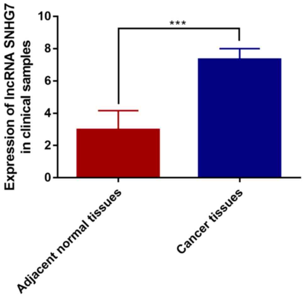
Expression of lncRNA-SNHG7 in 100 pairs of cancer
tissues and para-normal tissues was detected. The results showed
that the expression of lncRNA-SNHG7 in HCC tissues was much higher
than that of para-normal tissues, the difference was statistically
significant (P<0.001) (Fig. 1).
These results are consistent with lncRNA-SNHG7 described in
literature.
The HCC samples were divided into SNHG7-high
expression group (n=53) and SNHG7-low expression group (n=47) based
on the mean expression level of lncRNA-SNHG7 from qRT-PCR analysis.
The relationship between lncRNA-SNHG7 expression and
clinicopathological features of patients was further analyzed, and
it was found that there was no statistical difference between
lncRNA-SNHG7 expression and patient's sex, age, AFP levels, HBsAg
levels or tumor size (P>0.05). However, statistical difference
was found between lncRNA-SNHG7 expression and tumor number, lymph
node metastasis and clinical stage (Table I).
 | Table I.lncRNA-SNHG7 expression and clinical
features of patients with HCC. |
Table I.
lncRNA-SNHG7 expression and clinical
features of patients with HCC.
|
|
| lncRNA-SNHG7 |
|
|---|
|
|
|
|
|
|---|
| Features | No. | High | Low | P-value |
|---|
| No. | 100 | 53 | 47 |
|
| Sex |
|
|
| 0.930 |
| Male | 66 | 34 | 32 |
|
|
Female | 34 | 19 | 15 |
|
| Age (years) |
|
|
| 0.815 |
|
<60 | 43 | 23 | 20 |
|
| ≥60 | 57 | 30 | 27 |
|
| AFP |
|
|
| 0.894 |
|
<20 | 28 | 16 | 12 |
|
| ≥20 | 72 | 37 | 35 |
|
| HBsAg |
|
|
| 0.922 |
|
Positive | 60 | 31 | 29 |
|
|
Negative | 40 | 22 | 18 |
|
| Tumor size (cm) |
|
|
| 0.886 |
|
<5 | 39 | 20 | 19 |
|
| ≥5 | 61 | 33 | 28 |
|
| Tumor number |
|
|
| 0.001 |
|
Solitary | 69 | 25 | 44 |
|
|
Multiple | 31 | 28 | 3 |
|
| Lymph node
metastasis |
|
|
| 0.008 |
|
Absence | 62 | 24 | 38 |
|
|
Presence | 38 | 29 | 9 |
|
| Clinical stage |
|
|
| 0.012 |
| I +
II | 67 | 30 | 37 |
|
| III +
IV | 33 | 23 | 10 |
|
Effect of lncRNA-SNHG7 on the
prognosis of patients with HCC
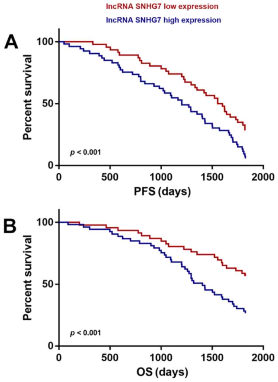
Correlation between lncRNA-SNHG7 expression and
survival time of patients was evaluated using the Kaplan-Meier
method. The results revealed that the patients with high expression
of lncRNA-SNHG7 had worse progression-free survival (PFS) and
overall survival (OS) time compared to SNHG7-low expression
patients. The results were statistically significant (P<0.001)
(Fig. 2), indicating high expression
of lncRNA-SNHG7 in HCC patients predicted poor prognosis.
Univariate and multivariate analyses
of lncRNA-SNHG7 expression and HCC clinicopathological data
Univariate Cox proportional hazards regression model
analysis was used to analyze pathological parameters of HCC. The
results revealed that tumor number, lymph node metastasis, clinical
stage and lncRNA-SNHG7 expression level were statistically
significant risk factors.
The above four factors with significant influence
were incorporated into the multivariate Cox proportional hazard
model for further analysis (Table
II). Similarly to tumor number, lymph node metastasis and
clinical stage. Expression of lncRNA-SNHG7 was an independent
prognosis index of patients with HCC (P<0.05). These findings
suggested that lncRNA-SNHG7 might play an important role in the
progression of HCC.
| Table II.Univariate and multivariate analyses
of postoperative prognosis in patients with gastric cancer. |
Table II.
Univariate and multivariate analyses
of postoperative prognosis in patients with gastric cancer.
|
| Univariate
analysis | Multivariate
analysis |
|---|
|
|
|
|
|---|
| Features | Hazard ratio/CI
(95%) | P-value | Hazard ratio/CI
(95%) | P-value |
|---|
| Sex |
0.983/0.754–1.248 | 0.868 |
|
|
| Age |
1.103/0.813–1.199 | 0.913 |
|
|
| Tumor size |
1.627/0.917–2.425 | 0.071 |
|
|
| Tumor position |
0.955/0.600–1.136 | 0.492 |
|
|
| Tumor
differentiation |
2.120/1.138–3.869 | 0.033 |
2.006/1.091–3.106 | 0.049 |
| T stage |
1.455/0.920–2.861 | 0.195 |
|
|
| N stage |
3.374/1.821–5.429 | 0.007 |
3.173/1.720–4.851 | 0.030 |
| M stage |
3.987/2.349–4.780 | 0.012 |
3.294/2.322–4.234 | 0.027 |
| TNM stage |
4.102/3.076–5.371 | 0.003 |
3.722/2.926–5.110 | 0.008 |
| lncRNACADM1-AS1
expression level |
2.994/1.670–4.051 | 0.009 |
2.584/1.621–3.880 | 0.015 |
Discussion
HCC is characterized by insidious onset and rapid
progress. The vast majority of patients are diagnosed in the middle
and late stages, thus missing the best time for surgery. Even with
radical resection, the postoperative recurrence and metastasis
still significantly impair the long-term survival of patients
(18,19). In order to realize early detection of
HCC and improve the prognosis of patients, the cellular and
molecular biological mechanisms leading to HCC invasion and
metastasis have attracted extensive attention and study.
Sequencing of the human genome indicated that
protein-encoding genes account for <3% of all sequences in the
human genome, and >80% of the sequences are frequently
transcribed into RNAs without protein-encoding functions, called
non-coding RNAs. They are a complex network that regulates gene
expression and a key to the regulation of many essential biological
functions of tumor cells (10,20,21).
They are categorized into short non-coding RNAs and lncRNAs based
on different sequence lengths (12).
lncRNAs cannot be translated into proteins, but they could affect
gene transcription, protein modification after translation and
epigenetic regulation (22,23). A growing number of studies have shown
that lncRNAs are abnormally expressed in various types of tumors,
which imply their potential value in the early diagnosis of certain
tumors and were considered as markers for early diagnosis.
lncRNA-JADE was found with a key functional link that connects the
DNA damage response (DDR) to histone H4 acetylation, and that
dysregulation of lncRNA-JADE might contribute to breast
tumorigenesis (24). The positive
lncRNA-HOTAIR/HER2 correlation was associated with gastric cancer
development. High expression of HOTAIR was associated with shorter
overall survival of gastric cancer patients (25). LncRNA MALAT-1 was positively related
to Bcl-2 expression in lung cancer and weak MALAT-1 expression
patients with resected lung cancer demonstrated adverse prognosis
(26).
Various studies have pointed out that lncRNA-SNHG7
is highly expressed in various malignant tumors and is a potential
molecular marker. Qi et al (27) indicated that through miR-503/cyclin
D1 pathway, lncRNA-SNHG7 promoted the development of prostate
cancer. Colorectal cancer patients with high expression of
lncRNA-SNHG7 had a significantly poor prognosis (28). Further results indicated that
lncRNA-SNHG7 facilitated the proliferation and metastasis of tumor
cells by regulating GALNT7 expression and PI3K/Akt/mTOR pathway.
She et al (14) found that
lncRNA-SNHG7 was highly expressed in lung cancer tissues, and the
inhibition of lncRNA-SNHG7 expression could promote apoptosis of
lung cancer cells. The role of lncRNA-SNHG7 in lung cancer was
through the regulation of FAIM2 gene. The above studies indicated
that lncRNA-SNHG7 might become a potential target for the treatment
of tumors. However, so far, studies on the expression and
biological functions of lncRNA-SNHG7 in tumor tissues are
relatively few, and there are no relevant reports involving the
field of HCC.
It was discovered in this study that the expression
level of lncRNA-SNHG7 in HCC tissues was much higher than that in
normal adjacent tissues, suggesting that the inhibition of
lncRNA-SNHG7 expression might be related to the occurrence and
procession of HCC. The correlation of the expression of
lncRNA-SNHG7 with clinicopathological features of HCC patients
found that the high expression of lncRNA-SNHG7 in HCC tissues was
related to tumor number, lymph node metastasis and clinical stage
but not associated with patient's sex, age, AFP levels, HBsAg
levels and tumor size.
Multidisciplinary team (MDT) diagnosis and treatment
of HCC patients by various treatment methods has gradually become a
characteristic trend of HCC treatment (29). In clinical practice, certain
indicators that were capable of accurately predicting the prognosis
would promote the identification of people with strong recurrence
tendency and provide the corresponding basis for prevention and
treatment, thus preventing recurrence as early as possible. This
study analyzed the clinicopathological features of 100 HCC
patients, and revealed that high expression of lncRNA-SNHG7 in HCC
patients indicated a poor prognosis. Moreover, the results of
univariate analysis and multivariate analysis model showed that
lncRNA-SNHG7 expression was an influential factor for the prognosis
of HCC patients. Clinical detection of lncRNA-SNHG7 expression
level might assist in making the prognosis of HCC patients.
In conclusion, lncRNA-SNHG7 might contribute to the
development of HCC and serve as a clinical biomarker and a
therapeutic target for HCC patients.
Acknowledgements
Not applicable.
Funding
No funding was received.
Availability of data and materials
All data generated or analyzed during this study are
included in this published article.
Authors' contributions
AS and XC designed the study and performed the
experiments, AS and JM collected the data, XC and XH analyzed the
data, AS and XC prepared the manuscript. All authors read and
approved the final manuscript.
Ethics approval and consent to
participate
This study was approved by the Ethics Committee of
Affiliated Hospital of Weifang Medical University (Weifang, China).
Signed informed consents were obtained from the patients and/or the
guardians.
Patient consent for publications
Not applicable.
Competing interests
The authors declare that they have no competing
interests.
References
|
1
|
Siegel RL, Miller KD and Jemal A: Cancer
statistics, 2018. CA Cancer J Clin. 68:7–30. 2018. View Article : Google Scholar : PubMed/NCBI
|
|
2
|
Braillon A: Hepatocellular carcinoma.
Lancet. 380:469–471. 2012. View Article : Google Scholar : PubMed/NCBI
|
|
3
|
Lau WY and Lai EC: Hepatocellular
carcinoma: Current management and recent advances. Hepatobiliary
Pancreat Dis Int. 7:237–257. 2008.PubMed/NCBI
|
|
4
|
El-Serag HB and Rudolph KL: Hepatocellular
carcinoma: Epidemiology and molecular carcinogenesis.
Gastroenterology. 132:2557–2576. 2007. View Article : Google Scholar : PubMed/NCBI
|
|
5
|
Mercer TR, Dinger ME and Mattick JS: Long
non-coding RNAs: Insights into functions. Nat Rev Genet.
10:155–159. 2009. View
Article : Google Scholar : PubMed/NCBI
|
|
6
|
Wilusz JE, Sunwoo H and Spector DL: Long
noncoding RNAs: Functional surprises from the RNA world. Genes Dev.
23:1494–1504. 2009. View Article : Google Scholar : PubMed/NCBI
|
|
7
|
Kelley D and Rinn J: Transposable elements
reveal a stem cell-specific class of long noncoding RNAs. Genome
Biol. 13:R1072012. View Article : Google Scholar : PubMed/NCBI
|
|
8
|
Bonasio R and Shiekhattar R: Regulation of
transcription by long noncoding RNAs. Annu Rev Genet. 48:433–455.
2014. View Article : Google Scholar : PubMed/NCBI
|
|
9
|
Gupta RA, Shah N, Wang KC, Kim J, Horlings
HM, Wong DJ, Tsai MC, Hung T, Argani P, Rinn JL, et al: Long
non-coding RNA HOTAIR reprograms chromatin state to promote cancer
metastasis. Nature. 464:1071–1076. 2010. View Article : Google Scholar : PubMed/NCBI
|
|
10
|
Esteller M: Non-coding RNAs in human
disease. Nat Rev Genet. 12:861–874. 2011. View Article : Google Scholar : PubMed/NCBI
|
|
11
|
Eddy SR: Non-coding RNA genes and the
modern RNA world. Nat Rev Genet. 2:919–929. 2001. View Article : Google Scholar : PubMed/NCBI
|
|
12
|
Ørom UA, Derrien T, Beringer M, Gumireddy
K, Gardini A, Bussotti G, Lai F, Zytnicki M, Notredame C, Huang Q,
et al: Long noncoding RNAs with enhancer-like function in human
cells. Cell. 143:46–58. 2010. View Article : Google Scholar : PubMed/NCBI
|
|
13
|
Chaudhry MA: Small nucleolar RNA host
genes and long non-coding RNA responses in directly irradiated and
bystander cells. Cancer Biother Radiopharm. 29:135–141. 2014.
View Article : Google Scholar : PubMed/NCBI
|
|
14
|
She K, Huang J, Zhou H, Huang T, Chen G
and He J: lncRNA-SNHG7 promotes the proliferation, migration and
invasion and inhibits apoptosis of lung cancer cells by enhancing
the FAIM2 expression. Oncol Rep. 36:2673–2680. 2016. View Article : Google Scholar : PubMed/NCBI
|
|
15
|
Cheng D, Fan J, Ma Y, Zhou Y, Qin K, Shi M
and Yang J: LncRNA SNHG7 promotes pancreatic cancer proliferation
through ID4 by sponging miR-342-3p. Cell Biosci. 9:282019.
View Article : Google Scholar : PubMed/NCBI
|
|
16
|
Xu LJ, Yu XJ, Wei B, Hui HX, Sun Y, Dai J
and Chen XF: LncRNA SNHG7 promotes the proliferation of esophageal
cancer cells and inhibits its apoptosis. Eur Rev Med Pharmacol Sci.
22:2653–2661. 2018.PubMed/NCBI
|
|
17
|
Wang MW, Liu J, Liu Q, Xu QH, Li TF, Jin S
and Xia TS: LncRNA SNHG7 promotes the proliferation and inhibits
apoptosis of gastric cancer cells by repressing the P15 and P16
expression. Eur Rev Med Pharmacol Sci. 21:4613–4622.
2017.PubMed/NCBI
|
|
18
|
Altekruse SF, McGlynn KA and Reichman ME:
Hepatocellular carcinoma incidence, mortality, and survival trends
in the United States from 1975 to 2005. J Clin Oncol. 27:1485–1491.
2009. View Article : Google Scholar : PubMed/NCBI
|
|
19
|
Chang CH, Chau GY, Lui WY, Tsay SH, King
KL and Wu CW: Long-term results of hepatic resection for
hepatocellular carcinoma originating from the noncirrhotic liver.
Arch Surg. 139:320–326. 2004. View Article : Google Scholar : PubMed/NCBI
|
|
20
|
Stefani G and Slack FJ: Small non-coding
RNAs in animal development. Nat Rev Mol Cell Biol. 9:219–230. 2008.
View Article : Google Scholar : PubMed/NCBI
|
|
21
|
Griffiths-Jones S, Moxon S, Marshall M,
Khanna A, Eddy SR and Bateman A: Rfam: Annotating non-coding RNAs
in complete genomes. Nucleic Acids Res. 33:D121–D124. 2005.
View Article : Google Scholar : PubMed/NCBI
|
|
22
|
Zhang Q and Jeang KT: Long non-coding RNAs
(lncRNAs) and viral infections. Biomed Pharmacother. 3:34–42.
2013.PubMed/NCBI
|
|
23
|
Carlevaro-Fita J, Rahim A, Guigó R, Vardy
LA and Johnson R: Cytoplasmic long noncoding RNAs are frequently
bound to and degraded at ribosomes in human cells. RNA. 22:867–882.
2016. View Article : Google Scholar : PubMed/NCBI
|
|
24
|
Wan G, Hu X, Liu Y, Han C, Sood AK, Calin
GA, Zhang X and Lu X: A novel non-coding RNA lncRNA-JADE connects
DNA damage signalling to histone H4 acetylation. EMBO J.
32:2833–2847. 2013. View Article : Google Scholar : PubMed/NCBI
|
|
25
|
Liu XH, Sun M, Nie FQ, Ge YB, Zhang EB,
Yin DD, Kong R, Xia R, Lu KH, Li JH, et al: Lnc RNA HOTAIR
functions as a competing endogenous RNA to regulate HER2 expression
by sponging miR-331-3p in gastric cancer. Mol Cancer. 13:922014.
View Article : Google Scholar : PubMed/NCBI
|
|
26
|
Schmidt LH, Görlich D, Spieker T, Rohde C,
Schuler M, Mohr M, Humberg J, Sauer T, Thoenissen NH, Huge A, et
al: Prognostic impact of Bcl-2 depends on tumor histology and
expression of MALAT-1 lncRNA in non-small-cell lung cancer. J
Thorac Oncol. 9:1294–1304. 2014. View Article : Google Scholar : PubMed/NCBI
|
|
27
|
Qi H, Wen B, Wu Q, Cheng W, Lou J, Wei J,
Huang J, Yao X and Weng G: Long noncoding RNA SNHG7 accelerates
prostate cancer proliferation and cycle progression through cyclin
D1 by sponging miR-503. Biomed Pharmacother. 102:326–332. 2018.
View Article : Google Scholar : PubMed/NCBI
|
|
28
|
Li Y, Zeng C, Hu J, Pan Y, Shan Y, Liu B
and Jia L: Long non-coding RNA-SNHG7 acts as a target of miR-34a to
increase GALNT7 level and regulate PI3K/Akt/mTOR pathway in
colorectal cancer progression. J Hematol Oncol. 11:892018.
View Article : Google Scholar : PubMed/NCBI
|
|
29
|
Siddique O, Yoo ER, Perumpail RB,
Perumpail BJ, Liu A, Cholankeril G and Ahmed A: The importance of a
multidisciplinary approach to hepatocellular carcinoma. J
Multidiscip Healthc. 10:95–100. 2017. View Article : Google Scholar : PubMed/NCBI
|