Introduction
Colorectal cancer (CRC) is one of the most common
human cancers and is associated with high morbidity and mortality
(1). Particularly in cases of
advanced rectal cancer, preoperative radiotherapy (PRT) and
chemoradiotherapy (PCRT) improve local control and long-term
disease-free survival, compared with surgery alone (2,3).
Furthermore, PCRT has been one of the standard therapies for rectal
cancer regarding anus preservation and is used for the prevention
of local recurrence and presurgical downstaging (4). In practice, the efficiency of PCRT
varies between individuals; pathologic complete response has been
reported in the range of 15 to 20% (5–7). Various
clinical factors have been reported as predictors of histological
regression or tumor downstaging in rectal cancer, such as the
circumferential extent of the tumor, distance from the anal verge,
and serum levels of carcinoembryonic antigen (CEA) (8–10).
However, the validation of these predictors remains
insufficient.
The phosphorylation of histone H2AX (into γ-H2AX) is
induced by DNA double-strand breaks (DSB). As tumor cells are
usually deficient in DNA damage response (DDR) pathways, it has
been suggested that constitutive expression of histone γ-H2AX might
indicate the disruption of DDR pathways and genomic instability
(11). γ-H2AX expression gradually,
but significantly, increases during tumor progression in human CRC
(12). In vitro, increasing
levels of γ-H2AX, after irradiation, have been correlated with
radiosensitivity in 18 human cell lines (13). Additionally, high γ-H2AX expression
is associated with poor prognosis in CRC patients (14). Therefore, we hypothesized that the
expression level of γ-H2AX is a predictor of radiosensitivity in
rectal cancer. In this study, we sought to clarify the relationship
between γ-H2AX expression and radiosensitivity in CRC, using in
vivo and in vitro experiments.
Materials and methods
Data set analysis
The Oncomine™ Research Platform (Thermo Fisher
Scientific, Inc.) was used in this study. The mRNA expression
levels of H2AX were investigated in each cohort study. The detailed
methodology of these studies is available in the references
(15–22).
Cell culture
Human CRC cell lines WiDr and DLD-1 were purchased
from JCRB (Japanese Collection of Research Bio Resources) Cell
Bank. Both lines were authenticated by short tandem repeat (STR)
sequence profiling by JCRB. STR examination showed that the WiDr
was identical to HT-29 (23). All
cells were cultured in RPMI-1640 (HyClone; GE Healthcare Life
Sciences) supplemented with 10% (v/v) heat-inactivated fetal bovine
serum at 37°C, in a humidified atmosphere containing 5%
CO2.
Patient samples
Six pairs of CRC tissues and adjacent normal mucosa
tissues, and eleven pairs of endoscopic biopsy samples from CRC
tissues and adjacent normal mucosa, were obtained from patients who
had undergone surgical resection of their tumor between 2013 and
2015 at Osaka Medical College Hospital (Takatsuki, Osaka, Japan).
Collection and investigation of the samples were approved by the
research Ethics Committee of Osaka Medical College (approval no.
1280, 2 September 2013) in accordance with the Declaration of
Helsinki. Before treatment, each patient provided written, informed
consent regarding the use of their tissues in our research. All
tissue sample pairs were collected from the same patient. Detailed
clinical information is shown in Tables
I and II. Pathological staging
of the cancers was performed according to postoperative
pathological reports, using guidelines for the treatment of
colorectal cancer from the Japanese Society for Cancer of the Colon
and Rectum 2010 (24). Each ‘grade
of effect’, induced by PCRT, was evaluated histologically by our
hospital's pathologist, using surgically resected specimens. The
criteria for the assessment of response to PCRT are defined as
follows: Grade 0 (no effect): No tumor cell necrosis or
degeneration was observed. Grade 1 (mild effect): Tumor cell
necrosis or degeneration is present in less than one third of the
entire lesion (minimal effect) or in more than one third but less
than two thirds of the entire lesion (mild effect). Grade 2
(moderate effect): Although prominent tumor cell necrosis,
degeneration, lytic change, and/or disappearance is present in more
than two thirds of the entire lesion, viable tumor cells remain.
Grade 3 (marked effect): Necrosis and/or lytic change is present
throughout the entire lesion, accompanied by replacement of
fibrosis, and viable tumor cells were not observed. Assessment was
performed on as many pathological specimens as possible, including
those prepared from the section of the whole tumor at the point of
maximum diameter (25).
 | Table I.Clinical and pathological features of
patients with CRC without any preoperative therapy. |
Table I.
Clinical and pathological features of
patients with CRC without any preoperative therapy.
| Case | Age | Sex | Location | Type | Tumor diameter
(mm) | Pathology | Tumora | Nodea |
Metastasisa | Stagea |
|---|
| 1 | 62 | M | A | 1 | 23×18 | tub1 | 2 | 0 | 0 | I |
| 2 | 62 | M | S | 2 | 64×38 | tub2, tub1 | 3 | 0 | 0 | IIA |
| 3 | 58 | M | R | 2 | 53×50 |
tub1=pap>tub2 | 3 | 1a | 1 | IV |
| 4 | 67 | M | T | 3 | 56×54 | tub2>tub1 | 3 | 1a | 1 | IV |
| 5 | 68 | F | R | 2 | 54×44 | tub2,
tub1>por2 | 3 | 1a | 0 | IIIB |
| 6 | 58 | M | R | 0 | 15×12 | tub1, tub2 | 1 | 0 | 0 | I |
| Table II.Clinical and pathological features of
patients with rectal cancer receiving preoperative
chemoradiotherapy. |
Table II.
Clinical and pathological features of
patients with rectal cancer receiving preoperative
chemoradiotherapy.
| Case | Age | Sex | Type | Pathology | Tumora | Nodea |
Metastasisa | Stagea |
|---|
| A, Grade 1a +
1b |
| 1 | 41 | M | 2 | tub1, tub2 | 3 | 1 | 0 | IIIB |
| 2 | 77 | M | 3 | tub1, tub2 | 3 | 1 | 0 | IIIB |
| 3 | 62 | F | 3 | tub2 | 3 | 0 | 0 | IIA |
| B, Grade 2 |
| 1 | 72 | M | 3 | tub1, tub2 | 3 | 1 | 0 | IIIB |
| 2 | 67 | F | 2 | tub1 | 3 | 1 | 0 | IIIB |
| 3 | 74 | M | 3 | tub2 | 3 | 0 | 0 | IIA |
| 4 | 62 | M | 2 | tub2>tub1 | 3 | 0 | 0 | IIA |
| C, Grade 3 |
| 1 | 57 | M | 2 | tub2>por | 3 | 2a | 0 | IIIB |
| 2 | 65 | M | 2 | tub1 | 3 | 0 | 0 | IIA |
| 3 | 67 | M | 2 | tub2 | 3 | 1 | 0 | IIIB |
| 4 | 71 | M | 2 | tub1, pap | 3 | 0 | 0 | IIA |
Irradiation time course
experiments
CRC cells were seeded in 6-well plates at a
concentration of 0.4×10⁴ cells per well (10-30%
confluence) the day before irradiation. After irradiation at 10 Gy,
cells were incubated for 24, 48, 72 and 96 h, and the effects were
assessed.
Gene silencing in irradiation
experiments
siRNA (siR) for H2AX was purchased from Santa Cruz
Biotechnology. Silencer negative control siRNA (Invitrogen; Thermo
Fisher Scientific, Inc.) was used as a control for nonspecific
effects. Cells were transfected with 10 nM siRNAs using
Lipofectamine™ RNAiMAX (Invitrogen; Thermo Fisher Scientific, Inc.)
according to the manufacturer's protocol. After 24 h of
transfection, the cells were irradiated with 10 Gy and subsequently
collected after 72 h.
Cell viability
MTT
(3-(4,5-dimethylthiazol-2-yl)-2,5-diphenyltetrazolium-bromide (MTT)
solution was purchased from Sigma-Aldrich; Merck KGaA. The detailed
protocol is described in a previous report (26). Absorbance at 540 nm was measured
using an SH-1000 Lab microplate reader (Corona Electric Co.,
Ltd.).
Immunohistochemistry (IHC)
Detailed protocol information is described in our
previous reports (26,27). Anti-phospho-histone H2A.X (Ser139)
antibody (EMD Millipore) was used. Images were taken with a BZ-x700
microscope (Keyence Co.).
Western blot analysis
Detailed protocol information is described in our
previous reports (26–28). The primary antibodies used were as
follows: anti-phospho-histone H2A.X (Merck Millipore; 1:1,000) and
anti-β-actin (Cell Signaling Technology, Inc.; 1:1,000).
HRP-conjugated horse anti-mouse and anti-rabbit IgGs (Cell
Signaling Technology; 1:1,000) were used as secondary antibodies.
Immunoblots were detected and visualized using Fusion-FX7 (Vilber
Lourmat).
Statistical analysis
Each experiment was performed in triplicate. The
data are presented as the mean ± SE. All statistical analyses were
performed using JMP® 12 (SAS Institute Inc.).
Statistical differences between the mean values of multiple groups
were determined using analysis of variance followed by Student's
t-test or one-way analysis of variance (ANOVA). Tukey-Kramer test
was performed post hoc following one-way ANOVA. P-values <0.05
indicated statistical significance.
Results
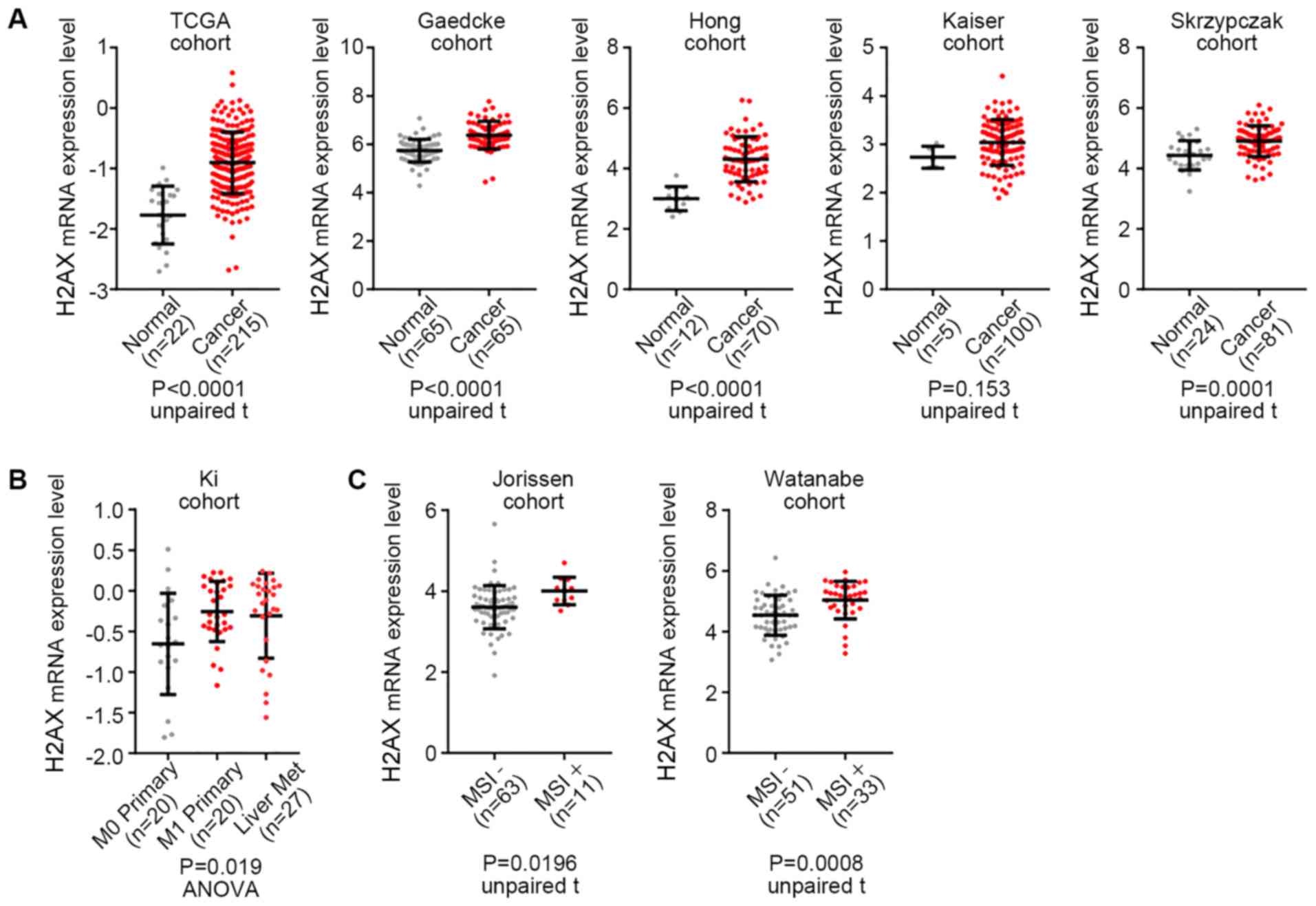
Significant upregulation of H2AX mRNA
in CRC tissues
The expression of γ-H2AX is increased in advanced
CRC and is associated with poor prognosis (12,14).
First, the mRNA levels of H2AX were investigated using datasets
from cohort studies. Our dataset analysis showed that the mRNA
levels of H2AX were significantly upregulated in CRC tissues
compared to those in normal tissues, except for one cohort study
(Fig. 1A). The expression levels of
H2AX in CRC patients with distant metastasis (M1 Primary),
including liver metastasis (Liver Met), was higher than that in
patients without metastasis (M0 Primary; Fig. 1B). Interestingly, H2AX mRNA levels in
the group positive for microsatellite instability (MSI) were higher
than those in the MSI-negative group (Fig. 1C). These results suggest that
upregulation of H2AX mRNA is associated with the development of
CRC, in a similar way to the expression of γ-H2AX.
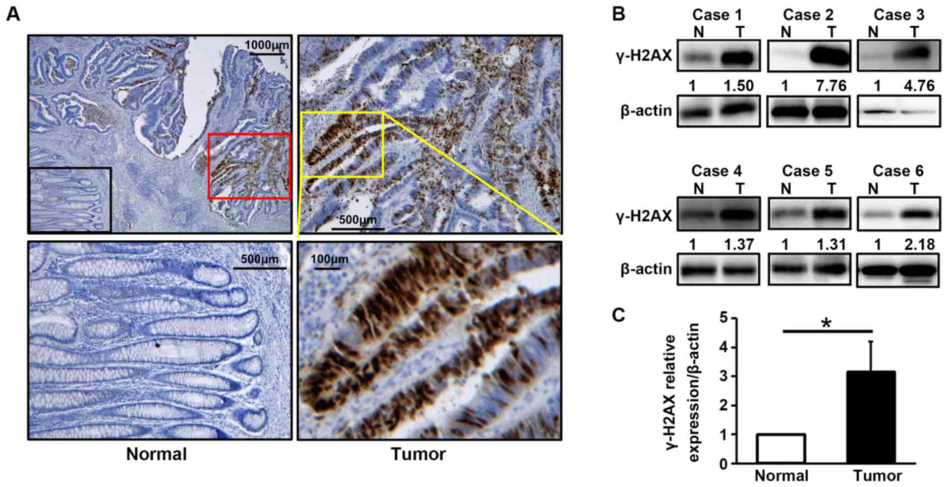
The protein expression of γ-H2AX was
upregulated in CRC specimens compared to adjacent normal
tissues
We examined the protein expression of γ-H2AX in
advanced CRC tissue without preoperative therapy (Table I). IHC showed that almost all nuclei
in cancer cells were stained, indicating strong expression of
γ-H2AX compared to that in adjacent normal tissues (Fig. 2A). The same tendency was observed
during western blot analysis (Fig. 2B
and C). These findings support the results of a previous study
(12), indicating that the
expression levels of γ-H2AX were upregulated in advanced CRC
tissue.
The suppression of γ-H2AX facilitated
the inhibition of cell viability induced by irradiation in CRC
cells
To assess the protective effect of γ-H2AX against
irradiation, changes in the expression levels of γ-H2AX were
measured after irradiation treatment, in two CRC cell lines. As
shown in Fig. 3A and B, the
expression level of γ-H2AX increased in correlation with
irradiation-induced inhibition of cell viability, in a
time-dependent manner. Subsequently, we examined the effects of
combination treatment (irradiation and knockdown of H2AX by siRNA)
in these cell lines. As expected, additional inhibition of growth
was observed with the combination therapy, compared to that with
irradiation or siR-H2AX alone, in both cell lines (Fig. 3C and D). These results imply that the
expression of γ-H2AX is associated with radiosensitivity in CRC
cells.
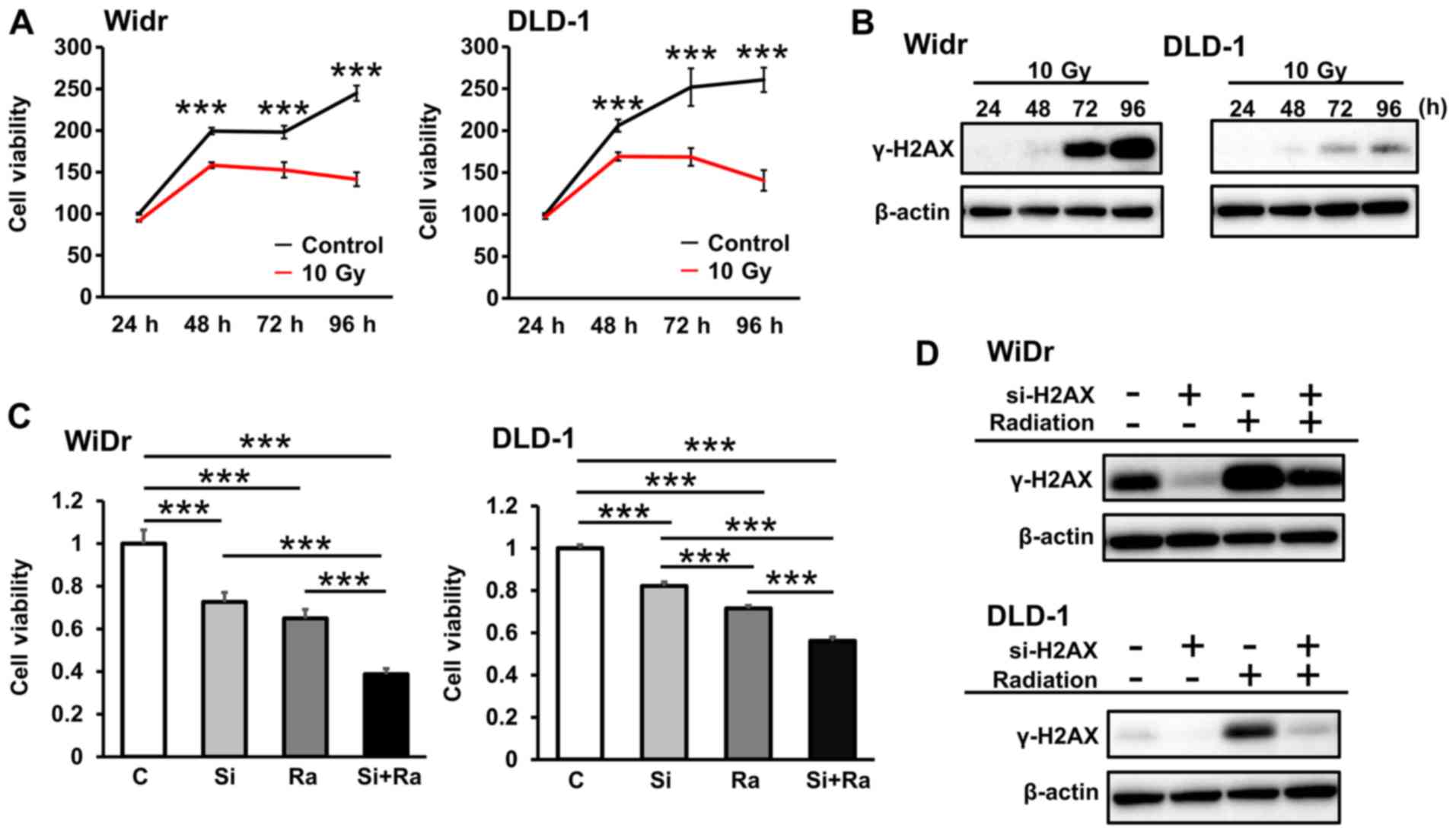 | Figure 3.Association between irradiation and
γ-H2AX expression in CRC cells. (A) Cell viabilities at 24, 48, 72
and 96 h after 10 Gy irradiation in two CRC cell lines. (B) The
protein expression of γ-H2AX in irradiation-treated CRC cells. The
experimental conditions were the same as in (A). (C) Cell viability
after irradiation and siR-H2AX combination treatment in CRC cells.
The effects were assessed 72 h after irradiation. C, control; Si,
siR-H2AX; Ra, irradiation. (D) The protein expression levels of
γ-H2AX after single or combination treatment. The experimental
conditions were the same as in (C). ***P<0.001 as indicated.
H2AX, H2A histone family member X; CRC, colorectal cancer; siR or
Si, small interfering RNA; C, control; Ra, irradiation. |
Sensitivity to preoperative
chemoradiotherapy was enhanced in the low γ-H2AX-expression group
of patients with advanced rectal cancer
Finally, we investigated the role of γ-H2AX in
vivo. We examined the response to PCRT in biopsy samples, based
on preoperative inspection (Table
II). As shown in Fig. 4A and B,
the expression levels of γ-H2AX in grade 2 or grade 3 tissues were
significantly lower than those of the grade 1 group. These results
suggest that low expression of γ-H2AX enhances radiosensitivity in
CRC cells (Fig. 4C).
 | Figure 4.Association between preoperative
chemoradiation therapy and γ-H2AX expression in clinical rectal
cancer specimens. (A) Representative protein expression of γ-H2AX
in each grade of preoperative chemoradiation therapy. (B)
Quantification western blot analysis results in 11 patients with
advanced rectal cancer. (C) The effectiveness of radiotherapy
depends on γ-H2AX expression in CRC. CRC cell sensitivity to
radiotherapy with low expressions of γ-H2AX is high. Namely, the
expression of γ-H2AX is associated with the potential for
radiotherapy resistance. *P<0.05 as indicated. H2AX, H2A histone
family member X; CRC, colorectal cancer; grade 1a, minimal effect;
grade 1b, mild effect; grade 2, moderate effect; grade 3, marked
effect in rectal cancer patients; N, normal, T, tumor. |
Discussion
In this study, we found that the mRNA levels of H2AX
and the expression of γ-H2AX in CRC tissue were both higher than in
normal tissues. γ-H2AX has also been reported as a diagnostic and
prognostic marker in other types of cancer, such as cancer of the
breast, bladder, and ovary (29–31).
These reports support our results, and suggest that H2AX,
especially in its phosphorylated form, may be a gene which is
universally associated with cancer. Importantly, the expression
levels of H2AX seemed to be related to MSI. Recently, MSI has been
recognized as one of the key mechanisms of carcinogenesis due to
lack of mismatch repair (MMR), and has been associated with immune
checkpoint blockade therapy using pembrolizumab (32). DSB immediately phosphorylates H2AX to
form γ-H2AX (33), and γ-H2AX is a
known sensitive marker for DSB (11). Hence, the results met our
expectations, and the elucidation of the detailed association
between the roles of H2AX (non-phosphorylated form) and the
acquisition of MSI remains an important issue.
In this study, we also evaluated the potential of
γ-H2AX to predict the effectiveness of preoperative radiotherapy.
The endogenous expression levels of γ-H2AX in WiDr cells without
any therapy was higher than that in DLD-1 cells, and the effect of
γ-H2AX suppression on cell viability in WiDr cells was stronger
than that in DLD-1 cells after irradiation. Taken together, these
findings imply that the expression level of γ-H2AX is important for
radiosensitivity, and CRC cells with an elevated expression of
γ-H2AX possess a certain tolerance to irradiation. Various
predictive factors for radiotherapy have been previously reported,
such as p53, ki67, Bax, Bcl-2, cyclooxygenase-2 and CD133 (34–37).
However, a bona fide predictive marker for radiotherapy has yet to
be established, and further research is required to identify
reliable candidates.
In addition, the underlying mechanism which
associates γ-H2AX with cell death, after irradiation, is unknown.
Several molecular mechanisms have been reported, relating to genes
associated with cell death and γ-H2AX. It has been shown that the
inhibition of caspase-4 activation interferes with γ-H2AX in CRC
cells (38). The relationship with
poly(ADP-ribose) polymerase (PARP), which is activated by DNA
damage similar to H2AX, is extremely important because
PARP-inhibitors are, clinically, expected to become novel
anticancer drugs (39). In addition,
epigenetic regulation should be considered. For example,
microRNA-138 regulates DNA damage by targeting H2AX (40), and H2AX phosphorylation regulates
apoptosis in lung cancer cells via the microRNA-3196/PUMA pathway
(41).
The present study had some limitations. First, the
number of cases used in the investigation of sensitivity to
preoperative chemoradiotherapy was small (only eleven cases). To
enhance the reliability of our findings, larger studies and
confirmatory studies are needed. Second, we also have to consider
the influence of chemotherapy on γ-H2AX expression. In many cases,
radiotherapy is not performed alone, as a preoperative therapy for
CRC. Fluorouracil and oxaliplatin, which are generally used in
preoperative chemoradiotherapy, induce DNA damage. In this study,
we attempted to focus on the association between H2AX and
radiosensitivity, and avoided an extremely complicated experimental
system. Third, regarding the experiments using CRC cells, the
association of γ-H2AX with radiosensitivity was examined in a
limited environment. Although a comparative investigation in a
xenografted mouse model, using CRC cells with either high- or low-
γ-H2AX expression, might support our findings, we selected human
specimens because of concerns that the immune response would be
insufficient in an animal model. Fourth, our findings of an
association between H2AX and MSI were preliminary results.
Considering recent advances in immune checkpoint blockade therapy,
it is time to clarify the detailed mechanisms of this
association.
In conclusion, the expression levels of H2AX and
γ-H2AX were upregulated in CRC cells. Moreover, this upregulation
may be associated with MSI. In radiotherapy, sensitivity was
enhanced by the suppression of γ-H2AX, and γ-H2AX showed potential
as a novel predictive marker of the effectiveness of preoperative
radiotherapy in CRC patients.
Acknowledgements
The authors would like to thank Ms. Akiko Miyamoto
at the Laboratory of General and Gastroenterological Surgery, as
well as Mr. Rintaro Oide and Mr. Teruo Ueno at the Osaka Medical
College Research and Development Center for their technical
support. The authors would also like to acknowledge Professor Asako
Nakamura (Department of Biological Sciences, College of Science,
Ibaraki University) for advice.
Funding
The current study was supported by the Japan Society
for the Promotion of Science KAKENHI (grant no. 17K10680) and by
Osaka Medical College (OMC) Internal Research.
Availability of data and materials
All data generated or analyzed during this study are
included in this published article.
Authors' contributions
SK, KTas and KTani conceived and designed the
current study. SK performed in vivo experiments, NK
performed in vitro experiments and KK analyzed datasets. TT,
SK, NK, KTas, KTani, YInom, YIm, RT, YInou, MK, KK, KU, MY, JO,
KTana and SWL interpreted and analyzed the data. SK, KTani, MY,
KTana and JO provided materials and funding. SK, KTas, KTani wrote
and revised the manuscript. KU supervised the current study. All
authors read and approved the final manuscript.
Ethics approval and consent to
participate
The present study was approved by the research
Ethics Committee of Osaka Medical College (approval no. 1280; 2nd
September 2013) and was conducted in accordance with the Helsinki
Declaration. Before treatment, each patient provided written
informed consent regarding the use of their tissues in this
research.
Patient consent for publication
Not applicable.
Competing interests
The authors declare that they have no competing
interests.
Glossary
Abbreviations
Abbreviations:
|
CRC
|
colorectal cancer
|
|
DDR
|
DNA damage response
|
|
DMSO
|
dimethyl sulfoxide
|
|
DSB
|
double-strand breaks
|
|
IHC
|
immunohistochemistry
|
|
PARP
|
poly (ADP-ribose) polymerase
|
|
PCRT
|
preoperative chemoradiotherapy
|
|
siRNA
|
small interfering RNA
|
References
|
1
|
Arnold M, Sierra MS, Laversanne M,
Soerjomataram I, Jemal A and Bray F: Global patterns and trends in
colorectal cancer incidence and mortality. Gut. 66:683–691. 2017.
View Article : Google Scholar : PubMed/NCBI
|
|
2
|
Chetty R and McCarthy AJ: Neoadjuvant
chemoradiation and rectal cancer. J Clin Pathol. 72:97–101. 2019.
View Article : Google Scholar : PubMed/NCBI
|
|
3
|
Häfner MF and Debus J: Radiotherapy for
colorectal cancer: Current standards and future perspectives. Visc
Med. 32:172–177. 2016. View Article : Google Scholar : PubMed/NCBI
|
|
4
|
Sauer R, Becker H, Hohenberger W, Rödel C,
Wittekind C, Fietkau R, Martus P, Tschmelitsch J, Hager E, Hess CF,
et al German Rectal Cancer Study Group, : Preoperative versus
postoperative chemoradiotherapy for rectal cancer. N Engl J Med.
351:1731–1740. 2004. View Article : Google Scholar : PubMed/NCBI
|
|
5
|
Aschele C, Cionini L, Lonardi S, Pinto C,
Cordio S, Rosati G, Artale S, Tagliagambe A, Ambrosini G, Rosetti
P, et al: Primary tumor response to preoperative chemoradiation
with or without oxaliplatin in locally advanced rectal cancer:
Pathologic results of the STAR-01 randomized phase III trial. J
Clin Oncol. 29:2773–2780. 2011. View Article : Google Scholar : PubMed/NCBI
|
|
6
|
Rödel C, Liersch T, Becker H, Fietkau R,
Hohenberger W, Hothorn T, Graeven U, Arnold D, Lang-Welzenbach M,
Raab HR, et al German Rectal Cancer Study Group, : Preoperative
chemoradiotherapy and postoperative chemotherapy with fluorouracil
and oxaliplatin versus fluorouracil alone in locally advanced
rectal cancer: Initial results of the German CAO/ARO/AIO-04
randomised phase 3 trial. Lancet Oncol. 13:679–687. 2012.
View Article : Google Scholar : PubMed/NCBI
|
|
7
|
Cho E, Park IJ, Yeom SS, Hong SM, Lee JB,
Kim YW, Kim MJ, Lim HM, Lim SB, Yu CS, et al: A Multigene model for
predicting tumor responsiveness after preoperative
chemoradiotherapy for rectal cancer. Int J Radiat Oncol Biol Phys.
105:834–842. 2019. View Article : Google Scholar : PubMed/NCBI
|
|
8
|
Yoon SM, Kim DY, Kim TH, Jung KH, Chang
HJ, Koom WS, Lim SB, Choi HS, Jeong SY and Park JG: Clinical
parameters predicting pathologic tumor response after preoperative
chemoradiotherapy for rectal cancer. Int J Radiat Oncol Biol Phys.
69:1167–1172. 2007. View Article : Google Scholar : PubMed/NCBI
|
|
9
|
Park JS, Huh JW, Park YA, Cho YB, Yun SH,
Kim HC, Lee WY and Chun HK: A circumferential resection margin of 1
mm is a negative prognostic factor in rectal cancer patients with
and without neoadjuvant chemoradiotherapy. Dis Colon Rectum.
57:933–940. 2014. View Article : Google Scholar : PubMed/NCBI
|
|
10
|
Wallin U, Rothenberger D, Lowry A, Luepker
R and Mellgren A: CEA - a predictor for pathologic complete
response after neoadjuvant therapy for rectal cancer. Dis Colon
Rectum. 56:859–868. 2013. View Article : Google Scholar : PubMed/NCBI
|
|
11
|
Bonner WM, Redon CE, Dickey JS, Nakamura
AJ, Sedelnikova OA, Solier S and Pommier Y: GammaH2AX and cancer.
Nat Rev Cancer. 8:957–967. 2008. View
Article : Google Scholar : PubMed/NCBI
|
|
12
|
Oka K, Tanaka T, Enoki T, Yoshimura K,
Ohshima M, Kubo M, Murakami T, Gondou T, Minami Y, Takemoto Y, et
al: DNA damage signaling is activated during cancer progression in
human colorectal carcinoma. Cancer Biol Ther. 9:246–252. 2010.
View Article : Google Scholar : PubMed/NCBI
|
|
13
|
Klokov D, MacPhail SM, Banáth JP, Byrne JP
and Olive PL: Phosphorylated histone H2AX in relation to cell
survival in tumor cells and xenografts exposed to single and
fractionated doses of X-rays. Radiother Oncol. 80:223–229. 2006.
View Article : Google Scholar : PubMed/NCBI
|
|
14
|
Lee YC, Yin TC, Chen YT, Chai CY, Wang JY,
Liu MC, Lin YC and Kan JY: High expression of phospho-H2AX predicts
a poor prognosis in colorectal cancer. Anticancer Res.
35:2447–2453. 2015.PubMed/NCBI
|
|
15
|
Cancer Genome Atlas Network, .
Comprehensive molecular characterization of human colon and rectal
cancer. Nature. 487:330–337. 2012. View Article : Google Scholar : PubMed/NCBI
|
|
16
|
Gaedcke J, Grade M, Jung K, Camps J, Jo P,
Emons G, Gehoff A, Sax U, Schirmer M, Becker H, et al: Mutated KRAS
results in overexpression of DUSP4, a MAP-kinase phosphatase, and
SMYD3, a histone methyltransferase, in rectal carcinomas. Genes
Chromosomes Cancer. 49:1024–1034. 2010. View Article : Google Scholar : PubMed/NCBI
|
|
17
|
Hong Y, Downey T, Eu KW, Koh PK and Cheah
PY: A ‘metastasis-prone’ signature for early-stage mismatch-repair
proficient sporadic colorectal cancer patients and its implications
for possible therapeutics. Clin Exp Metastasis. 27:83–90. 2010.
View Article : Google Scholar : PubMed/NCBI
|
|
18
|
Kaiser S, Park YK, Franklin JL, Halberg
RB, Yu M, Jessen WJ, Freudenberg J, Chen X, Haigis K, Jegga AG, et
al: Transcriptional recapitulation and subversion of embryonic
colon development by mouse colon tumor models and human colon
cancer. Genome Biol. 8:R1312007. View Article : Google Scholar : PubMed/NCBI
|
|
19
|
Skrzypczak M, Goryca K, Rubel T, Paziewska
A, Mikula M, Jarosz D, Pachlewski J, Oledzki J and Ostrowski J:
Modeling oncogenic signaling in colon tumors by multidirectional
analyses of microarray data directed for maximization of analytical
reliability. PLoS One. 5:e130912010. View Article : Google Scholar : PubMed/NCBI
|
|
20
|
Ki DH, Jeung HC, Park CH, Kang SH, Lee GY,
Lee WS, Kim NK, Chung HC and Rha SY: Whole genome analysis for
liver metastasis gene signatures in colorectal cancer. Int J
Cancer. 121:2005–2012. 2007. View Article : Google Scholar : PubMed/NCBI
|
|
21
|
Jorissen RN, Lipton L, Gibbs P, Chapman M,
Desai J, Jones IT, Yeatman TJ, East P, Tomlinson IP, Verspaget HW,
et al: DNA copy-number alterations underlie gene expression
differences between microsatellite stable and unstable colorectal
cancers. Clin Cancer Res. 14:8061–8069. 2008. View Article : Google Scholar : PubMed/NCBI
|
|
22
|
Watanabe T, Kobunai T, Toda E, Yamamoto Y,
Kanazawa T, Kazama Y, Tanaka J, Tanaka T, Konishi T, Okayama Y, et
al: Distal colorectal cancers with microsatellite instability (MSI)
display distinct gene expression profiles that are different from
proximal MSI cancers. Cancer Res. 66:9804–9808. 2006. View Article : Google Scholar : PubMed/NCBI
|
|
23
|
Chen TR, Drabkowski D, Hay RJ, Macy M and
Peterson W Jr: WiDr is a derivative of another colon adenocarcinoma
cell line, HT-29. Cancer Genet Cytogenet. 27:125–134. 1987.
View Article : Google Scholar : PubMed/NCBI
|
|
24
|
Watanabe T, Itabashi M, Shimada Y, Tanaka
S, Ito Y, Ajioka Y, Hamaguchi T, Hyodo I, Igarashi M, Ishida H, et
al Japanese Society for Cancer of the Colon and Rectum, : Japanese
Society for Cancer of the Colon and Rectum (JSCCR) guidelines 2010
for the treatment of colorectal cancer. Int J Clin Oncol. 17:1–29.
2012. View Article : Google Scholar : PubMed/NCBI
|
|
25
|
Japanese Society for Cancer of the Colon
and Rectum, . Japanese Classification of Colorectal, Appendiceal,
and Anal Carcinoma: The 3d English Edition (Secondary Publication).
J Anus Rectum Colon. 3:175–195. 2019. View Article : Google Scholar : PubMed/NCBI
|
|
26
|
Kawaguchi N, Tashiro K, Taniguchi K, Kawai
M, Tanaka K, Okuda J, Hayashi M and Uchiyama K: Nogo-B
(Reticulon-4B) functions as a negative regulator of the apoptotic
pathway through the interaction with c-FLIP in colorectal cancer
cells. Biochim Biophys Acta Mol Basis Dis. 1864:2600–2609. 2018.
View Article : Google Scholar : PubMed/NCBI
|
|
27
|
Matsuo K, Taniguchi K, Hamamoto H, Ito Y,
Futaki S, Inomata Y, Shima T, Asakuma M, Lee SW, Tanaka K, et al:
Delta-like 3 localizes to neuroendocrine cells and plays a pivotal
role in gastrointestinal neuroendocrine malignancy. Cancer Sci.
110:3122–3131. 2019. View Article : Google Scholar : PubMed/NCBI
|
|
28
|
Taniguchi K, Wada SI, Ito Y, Hayashi J,
Inomata Y, Lee SW, Tanaka T, Komura K, Akao Y, Urata H, et al:
α-Aminoisobutyric acid-containing amphipathic helical
peptide-cyclic RGD conjugation as a potential drug delivery system
for MicroRNA replacement therapy in vitro. Mol Pharm. 16:4542–4550.
2019. View Article : Google Scholar : PubMed/NCBI
|
|
29
|
Djuzenova CS, Elsner I, Katzer A,
Worschech E, Distel LV, Flentje M and Polat B: Radiosensitivity in
breast cancer assessed by the histone gamma-H2AX and 53BP1 foci.
Radiat Oncol. 8:982013. View Article : Google Scholar : PubMed/NCBI
|
|
30
|
Fernández MI, Gong Y, Ye Y, Lin J, Chang
DW, Kamat AM and Wu X: γ-H2AX level in peripheral blood lymphocytes
as a risk predictor for bladder cancer. Carcinogenesis.
34:2543–2547. 2013. View Article : Google Scholar : PubMed/NCBI
|
|
31
|
Mei L, Hu Q, Peng J, Ruan J, Zou J, Huang
Q, Liu S and Wang H: Phospho-histone H2AX is a diagnostic and
prognostic marker for epithelial ovarian cancer. Int J Clin Exp
Pathol. 8:5597–5602. 2015.PubMed/NCBI
|
|
32
|
Le DT, Durham JN, Smith KN, Wang H,
Bartlett BR, Aulakh LK, Lu S, Kemberling H, Wilt C, Luber BS, et
al: Mismatch repair deficiency predicts response of solid tumors to
PD-1 blockade. Science. 357:409–413. 2017. View Article : Google Scholar : PubMed/NCBI
|
|
33
|
Wakasugi M, Sasaki T, Matsumoto M, Nagaoka
M, Inoue K, Inobe M, Horibata K, Tanaka K and Matsunaga T:
Nucleotide excision repair-dependent DNA double-strand break
formation and ATM signaling activation in mammalian quiescent
cells. J Biol Chem. 289:28730–28737. 2014. View Article : Google Scholar : PubMed/NCBI
|
|
34
|
Kim NK, Park JK, Lee KY, Yang WI, Yun SH,
Sung J and Min JS: p53, BCL-2, and Ki-67 expression according to
tumor response after concurrent chemoradiotherapy for advanced
rectal cancer. Ann Surg Oncol. 8:418–424. 2001. View Article : Google Scholar : PubMed/NCBI
|
|
35
|
Chang HJ, Jung KH, Kim DY, Jeong SY, Choi
HS, Kim YH, Sohn DK, Yoo BC, Lim SB and Kim DH: Bax, a predictive
marker for therapeutic response to preoperative chemoradiotherapy
in patients with rectal carcinoma. Hum Pathol. 36:364–371. 2005.
View Article : Google Scholar : PubMed/NCBI
|
|
36
|
Kobayashi H, Hashiguchi Y, Ueno H, Shinto
E, Kajiwara Y and Mochizuki H: Absence of cyclooxygenase-2 protein
expression is a predictor of tumor regression in rectal cancer
treated with preoperative short-term chemoradiotherapy. Dis Colon
Rectum. 50:1354–1362. 2007. View Article : Google Scholar : PubMed/NCBI
|
|
37
|
Shinto E, Hashiguchi Y, Ueno H, Kobayashi
H, Ishiguro M, Mochizuki H, Yamamoto J and Hase K: Pretreatment
CD133 and cyclooxygenase-2 expression as the predictive markers of
the pathological effect of chemoradiotherapy in rectal cancer
patients. Dis Colon Rectum. 54:1098–1106. 2011. View Article : Google Scholar : PubMed/NCBI
|
|
38
|
Kang SJ, Lee YJ, Kang SG, Cho S, Yoon W,
Lim JH, Min SH, Lee TH and Kim BM: Caspase-4 is essential for
saikosaponin a-induced apoptosis acting upstream of caspase-2 and
γ-H2AX in colon cancer cells. Oncotarget. 8:100433–100448. 2017.
View Article : Google Scholar : PubMed/NCBI
|
|
39
|
Redon CE, Nakamura AJ, Zhang YW, Ji JJ,
Bonner WM, Kinders RJ, Parchment RE, Doroshow JH and Pommier Y:
Histone gammaH2AX and poly(ADP-ribose) as clinical pharmacodynamic
biomarkers. Clin Cancer Res. 16:4532–4542. 2010. View Article : Google Scholar : PubMed/NCBI
|
|
40
|
Yang H, Luo J, Liu Z, Zhou R and Luo H:
MicroRNA-138 regulates DNA damage response in small cell lung
cancer cells by directly targeting H2AX. Cancer Invest. 33:126–136.
2015. View Article : Google Scholar : PubMed/NCBI
|
|
41
|
Xu C, Zhang L, Duan L and Lu C:
MicroRNA-3196 is inhibited by H2AX phosphorylation and attenuates
lung cancer cell apoptosis by downregulating PUMA. Oncotarget.
7:77764–77776. 2016. View Article : Google Scholar : PubMed/NCBI
|