Introduction
Endometrial cancer (EC) is the most common
gynecological malignancy in high-income countries, and it generally
is associated with a good prognosis due to early-stage diagnosis
(1). In patients with EC, the
survival rates are associated with tumor stage, the depth of
myometrial invasion and the histological type of cancer, including
the cancer cell grade of differentiation. Following surgery and
tumor removal, the key factors associated with prognosis are
histological grade and clinical cancer stage, and these are
important for determining further treatment decisions. EC has been
traditionally divided into type 1 and 2 based on microscopic
findings, and these are associated with different clinical
outcomes. Furthermore, ~85% of newly-diagnosed cases are
histologically endometrioid type endometrial cancer (EEC) and
belong to the type 1 group, whereas type 2 cancers, such as serous
carcinoma, are found in 3–10% of cases. Endometrial clear cell
carcinoma accounts for <5% of diagnoses and both latter types
belong to the group of non-endometrioid endometrial cancers (NEECs)
(2). In recent years, this
simplistic pathological classification of EC types has been
challenged by high-quality molecular data available from large
genome databases. For example, four clinically distinct EC types
are defined by The Cancer Genome Atlas database. These types have
been selected on the basis of their overall gene mutational status,
which includes specific p53, polymerase ε (a DNA polymerase
involved in DNA replication and repair) and phosphatase and tensin
homolog protein mutations, along with microsatellite instability
and histology assessments (3).
Additionally, novel histologic categories of endometrial
hyperplasia (EH) types, with clear prognostic implications as
possible EC precursors, have been proposed (4). Furthermore, it is becoming evident from
an epidemiologic point of view that metabolic factors may serve an
important role in EC (5). Although
consensus in various aspects of EC diagnosis and treatment has been
recently reached by the European Society for Medical Oncology, the
European Society of Gynaecological Oncology and the European
Society of Radiation Oncology (6),
there is a need for novel, robust, prognostic and predictive
biomarkers of EC and EH that could be used in the near future to
improve therapeutic decisions.
One notable histological parameter is tumor budding
(TB), and its prognostic significance was first described by Hase
et al (7) in 1993 in
colorectal cancer. TB is defined as single cells or clusters of up
to four cells at the margin of the tumor front (7). This specific phenomenon has been
observed in various types of cancer in which the invasive parts of
the tumors send finger-like projections called ‘buds’ into adjacent
tissues (8). During local cancer
growth, some of these cell clusters detach from the main tumor body
and invade the neighboring stroma. This phenomenon is regarded as a
histological basis of metastasis formation and further tumor
invasion. In colorectal cancer, TB has been demonstrated to be a
novel prognostic factor that may be used to better define the risk
of adverse outcomes (8).
Additionally, Yamaguchi et al (9) have revealed that TB is a distinct
morphological feature that has biologic and prognostic significance
in adenocarcinoma of the lung. Gujam et al (10) have found that, in patients with
invasive ductal breast cancer, TB is a significant predictor of
survival. Furthermore, it is independent of adverse pathological
characteristics and components of the tumor microenvironment
(10). Lugli et al (11) have proposed a three-tier system that
should be used along with budding count in order to facilitate risk
stratification in patients with colorectal cancer. Since TB and
tumor grade are not the same and TB is now a well-described and
standardized prognostic factor, these authors concluded that TB
should be included in guidelines and protocols for colorectal
cancer reporting (11).
Interestingly, this histopathological feature can be identified by
usual routine pathological examination in different types of cancer
(12).
TB may be further divided into peritumoral budding
(PTB), where tumor buds are counted at the tumor front, and
intratumoral budding (ITB), where clustered cancer cells
representing tumor buds are observed and counted in the tumor
center (13). PTB can only be
assessed in endoscopic or surgical resection specimens, whereas ITB
can be assessed in both colorectal cancer biopsies and resection
specimens. Both ITB and PTB have been considered to be
morphological markers of epithelial-mesenchymal transition (EMT)
(14). EMT can be found in
physiological and pathological conditions, and it has been defined
as the transformation of an epithelial cell into a spindle cell
(14). Using immunohistochemistry
(IHC), the loss of membrane E-cadherin expression and the
appearance of mesenchymal cell markers can be demonstrated
(15). Notably, an association
between loss of E-cadherin and TB has been identified in EC
(16). TB can be studied with the
use of IHC and specific markers, such as E-cadherin or laminin. The
latter is the main active element of various basal membranes,
including the perivascular basal lamina (17). Laminin promotes attachment,
spreading, scattering and migration of non-tumorigenic epithelial
cells. Previous studies have revealed that the expression levels of
laminin subchain, namely laminin-5γ2-chain (L5γ2), could be a
specific marker for invasive tumors because it is frequently
expressed as a monomer in several types of cancer cells in
association with a lack of simultaneous expression of other laminin
chains, such as L5-α3 and L5-β3 (18,19).
Furthermore, immunohistochemical experiments have revealed that
laminin 5-γ2 is expressed at the invasive front of TB cells
(20).
Most solid tumors that grow beyond 2–3 mm in size
require angiogenesis (21). Normal
endometrium secretes angiogenic factors, including mainly vascular
endothelial growth factor, during the menstrual cycle and in early
pregnancy (22). Elevated
concentrations of proangiogenic factors secreted by malignant
tumors, such as EC, along with activation of tissue matrix
metalloproteinases induce the formation of the microvascular
network (23). This increased
vascularity is frequently observed in aggressive EC and could be
used as a specific target in anticancer therapy (24). Blood vessels in cancer exhibit
various structural and functional abnormalities, including unusual
leakiness which enables the dissemination of tumor cells into the
bloodstream. Furthermore, malignant tumor microvessel density (MVD)
is heterogeneous; the highest values are found in the invading
tumor edge, where the density may be 4–10 times greater than inside
the tumor. Additionally, the arrangement of vessels in the center
of a tumor is much more chaotic than at its edges (25). Both MVD and TB can be examined by
light microscopy and histopathological examination with or without
IHC. These methods are much cheaper to perform than molecular
analyses and are readily available in most pathology units. Based
on a sufficient number of standardized cases, they may offer novel
indications for a more accurate classification of the removed
endometrial tumors.
To the best of our knowledge, no previous studies
have attempted to associate tumor angiogenesis with TB and with
various clinical and pathological parameters in patients with EC.
Therefore, the present pilot study aimed to analyze the association
between TB and selected clinicopathological features in female
patients with EC.
Materials and methods
Patients and tumor samples
The present study included 137 female patients,
among them 117 had EEC and 3 had NEEC. Additionally, the present
study included 8 cases of simple EH and 9 cases of atypical
endometrial hyperplasia (AEH). The patients received surgery at the
Ist Department of Gynecological Oncology and Gynecology of the
Medical University of Lublin (Lublin, Poland) between January 2011
and January 2014.
Data collected included patient age at diagnosis,
tumor stage and histological grade. Postmenopausal status was
considered as when a woman had no periods for 12 consecutive months
prior to surgery. The histological type and grade of the tumors was
classified according to the criteria of the World Health
Organization (4). Malignant tumor
staging was established according to the International Federation
of Gynecology and Obstetrics (FIGO 2009) criteria (26). In some, but not all, patients,
typical coexisting diseases, such as obesity, diabetes and/or
hypertension, were noted. According to previously published data
(5,6,24,25,27),
the present study did not regard these diseases as potentially
confounding variables of both tumor angiogenesis and/or TB. All
patients included in the present study were treated with a total
abdominal or laparoscopic hysterectomy with adnexectomy with or
without pelvic lymph node resection according to the FIGO
guidelines. All analyzed samples were obtained by excision. None of
the patients in the study groups were treated with cytostatics,
since the material was always collected after surgery but before
any chemotherapy was initiated.
The median age of the patients was 63±9.7 years
(range, 40–83 years) at the time of diagnosis, which was
representative of the general population with EC. The Medical
University of Lublin Ethics Committee approved the study protocol.
All participants were informed of the nature of the study and
provided informed oral consent.
IHC
Immunohistochemical analysis was performed using
paraffin wax-embedded representative tumor tissue sections fixed in
10% neutral buffered formalin. Sections (4-µm thick) of the
formalin-fixed, paraffin-embedded tissues were mounted on silanized
slides (Dako; Agilent Technologies, Inc.). The slides were then
air-dried, and the tissues were deparaffinized and rehydrated. The
following primary antibodies were used: CD34 (mouse, monoclonal IgG
Class II, Clone QBEnd 10, DAKO Cytomation; catalog number M7165;
dilution 1:50); laminin-5, gamma-2 chain Clone 4G1 (catalog
number:M7262; non-conjugated mouse monoclonal antibody DAKO
Cytomation dilution1:50) for tumor budding index (TBI) calculation.
Following deparaffinization, rehydration and antigen retrieval with
the Target Retrieval solution at pH=9.0 (Dako; Agilent
Technologies, Inc.), three cycles of heating in a microwave oven (5
min each; 750 W) were performed. Tissue sections were incubated
with the primary antibody for 1 h at room temperature. After
washing in Wash Buffer (Dako; Agilent Technologies, Inc.), the
slides were incubated with the secondary antibody conjugated to
streptavidin-biotin-peroxidase complex (Dako REAL EnVision
Detection System, Peroxidase/DAB+, Rabbit/Mouse - PL Code: K5007.
and a color reaction was developed using 3′-3-diaminobenzidine
tetrahydrochloride (Dako; Agilent Technologies, Inc.) according to
the manufacturer's protocol. The sections were counterstained with
Mayer's hematoxylin. For each case, the negative control was
applied by replacing the antibody with PBS or nonimmune serum.
Variables
MVD
The average number of microvessels within selected
tumor areas was determined according to the Weidner method
(28) as previously described
(27). Briefly, tumor sections were
first examined at low magnifications (×40 and ×100) to identify the
most vascular areas of the invasive front ‘hotspots’, i.e., the
area(s) with most intense CD34 staining and the highest apparent
density of microvessels. Subsequently, as a rule, 10 fields were
examined, except in a few cases where less tumor tissue was
available. The counts were expressed as the average of all fields
examined at high magnification [x200; high power field (HPF)]. MVD
was presented as the mean number of vessels per one HPF.
TB and TBI
TB was defined as dissociated single cancer cells or
clusters of up to four cancer cells with cytoplasmic L5γ2
immunostaining, ahead of the invasive tumor front. First, the area
was scanned at low power (×100) to identify the region displaying
maximal budding. Subsequently, tumor buds were counted in
high-power fields (×400; 0.49 mm2) in the area at the
invasive front. The present study compared two widespread methods
of quantification of tumor buds. First, as proposed by Ueno et
al (8), the assessment was
performed by counting buds in a region of interest spanning one
microscope high-power field (1-HPF) and displaying maximal budding.
Classification was dichotomic and scored as ‘negative’ (<5 buds)
or ‘positive’ (≥5 buds). The ‘10-HPF’ method proposed by
Karamitopoulou et al (29)
was the second method of counting tumor buds used in the present
study. According to this method, the average number of the 10
counts was taken as the final TBI.
Statistical analysis
Statistical analysis was performed using Statistica
software v.10.0 (StatSoft, Inc.). The association between
categorical variables was examined by Pearson's χ2 test
or Fisher's exact test, as deemed appropriate. The Shapiro-Wilk
test was used to assess the normality of data distribution.
Mann-Whitney nonparametric tests were used to compare categorical
with continuous tumor variables when there were two categories,
whereas the Kruskal-Wallis nonparametric test was used when there
were more than two categories. P<0.05 was considered to indicate
a statistically significant difference.
Results
Most of the examined patients were postmenopausal
(126/137; 92%) and the mean body mass index (BMI) in the study
group was 31.7±6.1 (range, 21.8–44.6). Table I presents selected clinical and
pathological features of the studied population. There were
statistically significant differences in age between the group of
patients with EC and the group of patients with benign endometrial
lesions (H, 9.1; P=0.01 as per Kruskal-Wallis test by ranks). The
differences in BMI values between EC/NEEC and EH/AEH groups were
not statistically significant (P=0.3). Histological analysis of the
removed specimens revealed 25% cases of low grade (G1) EC; most of
the remaining malignancies were grade 2 and only 12% of ECs were
undifferentiated high grade (G3) or undifferentiated. The majority
of the endometrial cancers were classified as clinical FIGO stage I
(73 cases; 61%), and 30 cases (25%) were FIGO stage II. The
morphologic features that best distinguished endometrial
hyperplasia or EC from normal endometrium included glandular
crowding that was well distinguished from normal glands and
abnormal architecture of the glands, with their long axes pointing
in different directions or being parallel to the endometrial
surface. Other histological features used for the discrimination
between malignant and benign lesions included irregularly-shaped
glands that were dilated, densely packed non-secretory glands,
including budding or branching glands and nuclear atypia in cases
of atypical hyperplasia, and EC with cribriform or confluent glands
in cases of carcinoma. Selected clinical and pathological features
of the study population are presented in Table I.
 | Table I.Selected clinical and pathological
features of the studied population. |
Table I.
Selected clinical and pathological
features of the studied population.
| Feature | N (%) |
|---|
| Type of endometrial
pathology |
| EC | 117 (85.4) |
|
NEEC | 3 (2.2) |
| EH | 8 (5.8) |
|
AEH | 9 (6.6) |
| Histopathological
grading |
| G1 | 35 (29) |
| G2 | 74 (61) |
| G3 | 11 (10) |
| Clinical staging
(FIGO) |
| I | 70 (58) |
| II | 33 (27) |
|
III | 16 (13) |
| IV | 1 (1) |
MVD-CD34 assessment
Table II shows
median values of MVD-CD34 according to clinical and histological
features in endometrial lesions. In the patients included in the
present study, the median value (MD) of MVD-CD34 was 19/HPF (range,
13–29). Statistically significant differences were identified
between patients with malignant lesions and patients with
nonmalignant endometrial lesions (P=0.01). The present study
revealed that the median MVD-CD34 in patients with EEC was higher
than that in patients with EH/AEH and NEEC. MVD was associated with
histological grade and well-differentiated tumors in which MVD was
significantly lower than in undifferentiated tumors (MD, 14 vs.
24.9 in G1 and G3, respectively; P=0.001). Age, menopausal status
and BMI were not associated with tumor MVD. MVD, as assessed using
the anti-CD34 antibody, did not differ between the groups with low
and high clinical FIGO stage of EC (MD, 18.4 in the low-stage
group; and MD, 17.6 in the high-stage group; P=0.2).
| Table II.Median values of MVD-CD34 according
to clinical and histological features in endometrial lesions. |
Table II.
Median values of MVD-CD34 according
to clinical and histological features in endometrial lesions.
| Feature | Median value
(range) | P-value |
|---|
| All patients | 19 (13–29);
min-max 2–49 |
|
| Type of endometrial
lesion |
| H=11.2; P=0.01 |
| EC
(n=117; 85.4%) | 19 (13.8–28.8) |
|
| NEEC
(n=3; 2.2%) | 10 (2.1–14.8) |
|
| EH
(n=8; 5.8%) | 16 (8.4–30) |
|
| AEH
(n=9; 6.6%) | 9 (7–18.9) |
|
| Menopausal
status |
| Z=0.6; P=0.6 |
| Before
menopause (n=11; 8%) | 20.7
(10.8–27.8) |
|
| After
menopause (n=126; 92%) | 18.8
(12.7–28.9) |
|
| Histopathological
grading |
| H=13.4;
P=0.001 |
| G1
(n=31; 25%) | 14 (11.8–20.8) |
|
| G2
(n=77; 63%) | 18.6
(14.7–30.9) |
|
| G3
(n=15; 12%) | 24.9
(19.1–33.7) |
|
| Clinical stage of
the disease (FIGO staging) |
| H=4.2; P=0.2 |
| I
(n=70; 60%) | 18.4
(14.3–27.8) |
|
| II
(n=30; 25%) | 23.6
(15.3–33.7) |
|
| III+IV
(n=17; 13%) | 17.6
(14.6–23.7) |
|
TB assessment using laminin L5γ2
expression
Laminin L5γ2 expression in tumor buds was identified
in 120 (84%) patients with endometrial lesions, and 89 of them were
classified as TB-positive. Table
III shows the results of TB quantification in relation to
clinical and histopathological features in endometrial lesions
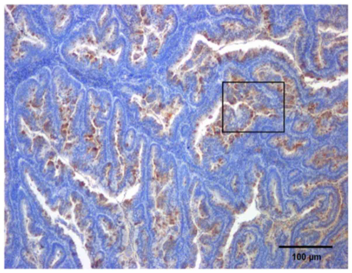
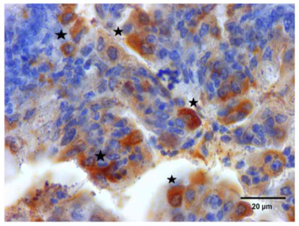
assessed using the ‘1-HPF’ scoring method. Examples of laminin
5γ2-chain expression in tumor buds in EC are shown in Figs. 1 and 2. More TB-positive lesions were observed in
patients with EC compared with in patients with non-malignant
hyperplasia (P=0.003). Table IV
presents median values of TBI according to clinical and
histopathological features in endometrial lesions assessed using
the ‘10-HPF’ scoring method. The median value of TBI was 9.2
(range, 1.2–16.8) and it was significantly associated with
malignant endometrial lesions (P=0.002). Benign endometrial lesions
had a TBI ranging among 0.4 for EH, 1.1 for AEH and 14.1 for NEEC.
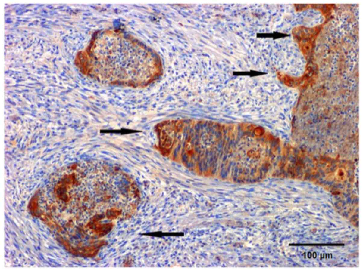
Figs. 3 and 4 present typical patterns of TB in the
invasive front of EC. The median TBI was 10.7 in women with EC.
Most of the high grade endometrial tumors (G3) were positive for
the TB phenomenon (13/15; P=0.006). Additionally, high TBI values
were more often observed in high-grade tumors than in low grade
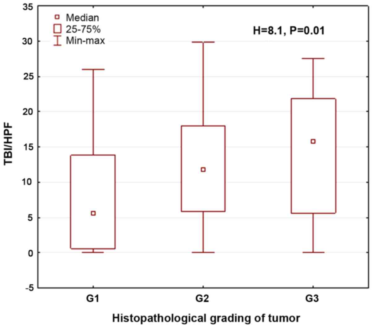
malignant tumors (P=0.01). Fig. 5
presents median values of TBI according to histological grade of
EC. The median values of TBI in patients with G2 and G3 EC were
12.1 and 16.2, respectively. These indices were markedly higher
than TBI values in low grade tumors which had a median TBI of 4.5.
No significant associations between TB and the clinical stage of EC
were found. However, advanced malignant endometrial tumors (FIGO
stage III and IV) tended to be TB-positive more often (13/17). The
median values of TBI did not differ between low clinical FIGO stage
(I and II) and high clinical stage (III and IV) tumors. The TBI
values were 8.8 in low FIGO stage EC and 10.3 in high-stage EC
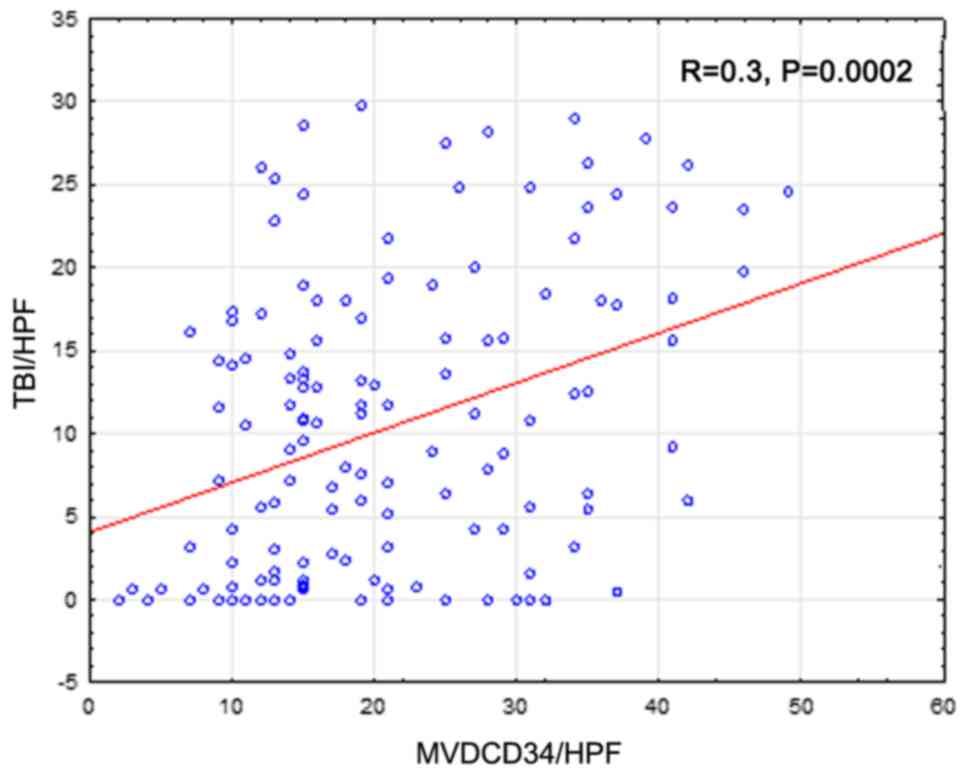
(P=0.2). TBI was markedly associated with MVD (P=0.0002) and
TB-positive tumors had a markedly higher MVD than TB-negative EC.
Furthermore, menopausal status was associated with TB, and
TB-positive tumors were found more frequently in postmenopausal
patients (P=0.03). In the group of premenopausal patients, the TBI
was significantly higher compared with that in postmenopausal women
(TBI, 0.3 vs. 11.1, respectively; P<0.005). No association was
identified between TBI and patient age (P=0.1) or BMI (P=0.12).
Fig. 6 presents the median values of
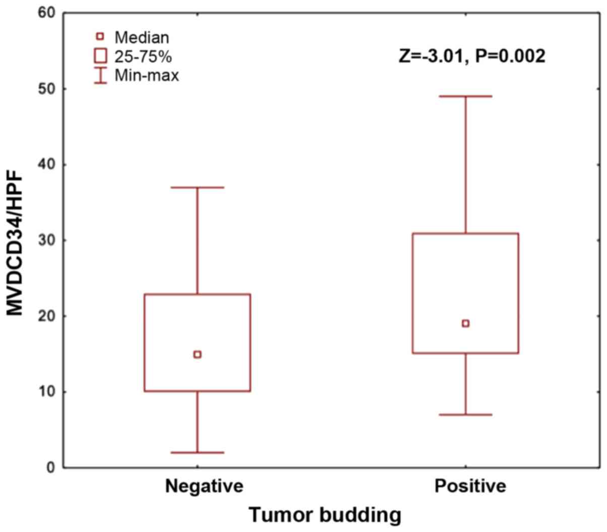
MVD in TB positive and negative ECs. The association between MVD
and TBI in EC is presented in Fig.
7.
| Table III.The results of tumor budding
quantification in relation to clinical and histopathological
features in endometrial lesions (1-HPF scoring method). |
Table III.
The results of tumor budding
quantification in relation to clinical and histopathological
features in endometrial lesions (1-HPF scoring method).
| Feature | Negative (≤5
buddsHPF) N=47 (34.6%) | Positive (<5
buddsHPF) N=89 (65.4%) | P-values
(χ2 or Z) |
|---|
| Type of endometrial
pathology |
|
| 18.8; P=0.0003 |
| EC
(n=117; 85.4%) | 33 (28%) | 83 (72%) |
|
| NEEC
(n=3; 2.2%) | 1 (33%) | 2 (67%) |
|
| EH
(n=8; 5.8%) | 8 (100%) | 0 (0%) |
|
| AEH
(n=9; 6.6%) | 5 (56%) | 4 (44%) |
|
| Menopausal
status |
|
| 4.4; P=0.03 |
| Before
menopause (n=11; 8%) | 7 (15%) | 4 (5%) |
|
| After
menopause (n=126; 92%) | 40 (85%) | 85 (95%) |
|
| Histopathological
grade of tumor (grading) |
|
| 10.2; P=0.006 |
| G1
(n=31; 25%) | 15 (44%) | 16 (18%) |
|
| G2
(n=77; 63%) | 18 (53%) | 59 (67%) |
|
| G3
(n=15; 12%) | 1 (3%) | 13 (15%) |
|
| Clinical stage of
the desease (FIGO staging) |
|
| 2.4; P=0.4 |
| I
(n=73; 61%) | 24 (70%) | 48 (57%) |
|
| II
(n=30; 25%) | 6 (18%) | 24 (28%) |
|
| III+IV
(n=17; 14%) | 4 (12%) | 13 (15%) |
|
| Microvessel density
(MVD-CD34) |
| Median
(range) | 14.4
(9.9–22.7) | 19.1
(14.8–30.8) | −3.01; P=0.002 |
| BMI |
| Median
(range) | 31.6
(26.5–35.6) | 31
(26.9–34.8) | 0.17; P=0.8 |
| Table IV.Median values of Tumor Budding Index
(TBI) acording to clinical and histopathological features in
endometrial neoplasia (10-HPF scoring method). |
Table IV.
Median values of Tumor Budding Index
(TBI) acording to clinical and histopathological features in
endometrial neoplasia (10-HPF scoring method).
| Feature | Median (range) | P-value |
|---|
| All groups | 9.2 (1.2–16.8);
min-max: 0–29.8 |
|
| Histological type
of endometrial lesion |
| H=15.2;
P=0.002 |
| EC
(n=117; 85.4%) | 10.7
(3.2–17.7) |
|
| NEEC
(n=3; 2.2%) | 14.1 (0–28) |
|
| EH
(n=8; 5.8%) | 0.4 (0–1.7) |
|
| AEH
(n=9; 6.6%) | 1.1 (0.5–13.1) |
|
| Menopausal
status |
| Z=3.3;
P=0.0009 |
| Before
menopause n=11 (8%) | 0.3 (0–6.1) |
|
| After
menopause n=126 (92%) | 11.1
(2.3–17.7) |
|
| Histological grade
of tumor (grading) |
| H=8.1; P=0.01 |
| G1
(n=32; 23%) | 4.5 (0.6–13.5) |
|
| G2
(n=77; 56%) | 12.1
(5.8–17.9) |
|
| G3
(n=15; 11%) | 16.2
(5.8–21.6) |
|
| Clinical stage of
the desease (FIGO staging) |
| H=5.1; P=0.2 |
| I
(n=82; 60%) | 8.8 (2.5–15.7) |
|
| II
(n=27; 20%) | 14.5
(5.5–18.8) |
|
| III+IV
(n=13; 9%) | 10.3 (2.6–18.7 |
|
Discussion
Most EC cases are diagnosed in early stages, but
15–20% of women with aggressive cancer types have an increased risk
of occult malignancy dissemination and tumor recurrence despite
chemo- and radiotherapy (30).
Currently, tumor staging according to the FIGO criteria remains the
basic method used to stratify women with EC into prognostic groups
that could benefit from different types of surgery and chemo- or
radiotherapy. Factors controlling growth of EC and its interactions
with the surrounding uterine stromal microenvironment have recently
gained increasing attention. Little is known about the regulation
of TB and MVD in EC. Furthermore, in most of the studied cancer
types such as lung, breast, colorectal and endometrial endometrioid
cancers, the presence of tumor budding phenomenon was associated
with lower survival rates (8–11).
A putative connection between TB and neoangiogenesis
at the invasive tumor front has not been investigated yet. The
present study revealed that TB, in terms of L5γ2 expression,
increased gradually in endometrial lesions as they progressed from
benign endometrial hyperplasia to AEH and finally to EC (EEC and
NEEC). When examining tumor sections stained for L5γ2, clusters of
undifferentiated malignant cells were observed in the tumor stroma,
and these were located mainly in close proximity and ahead of the
invasive front of the tumor. It was speculated that L5γ2 expression
in tumor buds at the invasive front of endometrial neoplasia may be
associated with the process of tumor differentiation. The present
results indicated that there was a link between intratumoral MVD
and endometrial tumor cell proliferation or TB. This was expected,
since an adequate blood vascular system is required for effective
tumor cell growth. Furthermore, when activated, endothelial cells
can release various paracrine growth factors important for cancer
cells, such as collagenases, urokinases and plasminogen activators
(25). These factors enable tumors
to spread into adjacent tissues and lymphovascular spaces.
Tumor buds could be regarded as a more invasive
subpopulation of cells disseminated from the mass of the tumor.
Therefore, they may have acquired the ability to invade the
lymphatic system and metastasize to distant nodes. This hypothesis
is in line with the results of a previous study by Koyuncuoglu
et al (16) which reported a
high prognostic value of TB in both endometrioid and
non-endometrioid EC. In this study, cytokeratin C11 was used for
improved visualization of numerous buds fused with stromal
fibroblasts, yielding three- to four-fold higher tumor bud
calculations compared with those using only histological
hematoxylin and eosin (H&E) staining. TB was detected by both
H&E and cytokeratin C11 staining methods in 95 patients with
primary EC. The authors demonstrated that a high TB count was
strongly associated with undifferentiated tumors, advanced stage
and decreased postoperative survival. Park et al (12) recently demonstrated that TB is
associated with depth of invasion and higher FIGO grade in patients
with EC, suggesting reduced histologic differentiation,
lymphovascular invasion and lymph node involvement. The presence of
TB is an independent parameter for the prediction of lymphovascular
invasion in multivariate analysis and a significant factor for the
prediction of lymph node metastasis in univariate analysis
(12). Another similar study by
Huang et al (31)
demonstrated the prognostic significance of TB in early-stage
cervical cancer (ESCC). They revealed that TB is an independent and
unfavorable prognostic factor for patients with ESCC. The authors
have suggested that following radical surgery, TB assessment could
be promising for improved recurrence risk stratification. Marangon
et al (20) evaluated L5γ2
expression in 57 patients with oral squamous cell carcinoma (OSCC)
and its association with the intensity of TB and the density of
stromal myofibroblasts. In their study, higher laminin-5 γ2
expression was associated with high-intensity TB and with a higher
density of stromal myofibroblasts, suggesting that TB is associated
with the establishment of an invasive phenotype of neoplastic cells
and a permissive environment for tumor invasion in OSCC. Several
lines of evidence suggest that the presence of TB may indicate the
process of EMT, which is commonly associated with increased
expression of molecules related to tumor invasion, such as matrix
metalloproteinases, presence of L5γ2 in tumor cells and activation
of the Wnt signaling pathway (32,33).
Despite numerous apparent molecular differences
between EC types, histological criteria with or without selected
IHC markers are still widely used for the initial diagnosis of a
tumor type. However, since this cancer is a heterogeneous disease,
there is a need to effectively stratify patients according to
further treatment. A comprehensive characterization of the
endometrial malignant tumors and their microenvironment is required
for improved prediction of the effects of current treatment
methods. However, the main obstacle is the absence of a consensus
methodology, which is why it remains problematic to evaluate the
real prognostic significance of TB in these malignant tumors. The
most frequently used staining technique is the H&E method which
may have some limitations and cannot without difficulty
discriminate between real buds and other structures, where tissue
disintegration gives the false impression of budding and these
fragments should be excluded from counting (13,19).
Another important issue is associated with the
counting method that should be applied. The present results
indicated that the observed significant associations between
clinical and histological variables were comparable for both
counting methods. Nevertheless, it seems that quantification of 10
HPF should be used for optimal viewing of surgical samples, while
the more restrictive ‘1 HPF’ method may be reserved for small
samples, such as endometrial biopsies (29).
To the best of our knowledge, the present study is
one of the few that have reported the possible significance of TB
estimation in patients with EC. The present results indicated for
the first time that L5γ2 expression in tumor buds could be useful
for the microscopic assessment of EC cell invasion. Previously, it
has been demonstrated that the invasive front of the lesions
exhibits a striking disorganization at the tumor architecture which
may have been related to EMT and stem cell activation (32–34). In
particular, the changes included loss of glandular features for
differentiated cancer and loss of the trabecular characteristic for
undifferentiated carcinoma (9). Most
likely, this phenomenon helps to mobilize the EC cells from the
main tumor mass, which is followed by invasion of host tissues
through movement and their angiogenic activity (35). Our previous study revealed that the
invasive potential of EC is related to the angiogenic phenotype of
tumor tissues, and the ability of a tumor to develop its own
microvascular network (27). In the
current study, it was observed that, in EC, MVD increased with an
increase in the number of TB structures. An implication of these
observations is the possibility that at least in some endometrial
malignant tumors, neoangiogenesis as assessed by MVD and TB are
associated, and could be used as potential prognostic factors.
In patients with malignant endometrial lesions, a
possibility of biopsy material examination is usually obtained
prior to the decision on the type of surgery being made. It is not
yet known if samples from the uterine cavity could be sufficient
for both histology and TB assessment. Recently, Almangush et
al (36) have reviewed all
published reports on TB in diagnostic biopsies and matching cancer
surgical specimens. They found that not only did all these studies
show that evaluation of TB is easily applicable, but also that
there is a significant association between the expression of TB in
both surgical specimens and their corresponding biopsies specimens.
Furthermore, the assessment of the TB phenomenon in diagnostic
biopsies enabled a more accurate prognosis of lymphatic spread
beyond the uterus and decreased survival of patients with EC.
Unfortunately, to the best of our knowledge, there has been no
study that compared TB in endometrial biopsies and material
obtained after hysterectomy.
The strengths of the present study include the
relatively large group of patients who received surgery for EC at
one institution. We also are aware that the group with endometrial
hyperplasias was much smaller and that it's usually much better to
have 60–65 participants in both conditions rather than 17 in one
condition and 120 in the other - even though the total number of
participants is much greater in the second set-up. However, simple
randomization can cause serious imbalances and in fact,
theoretically, it's possible to end up with no participants in one
of the groups. Prior to the present study, it was difficult to make
predictions about if and how EMT and TB could influence
neoangiogenesis in malignant endometrial tumors. This was the first
study to report an association between the phenomenon of TB in EC
and MVD assessment. The presented results could be important in
furthering our understanding of the role of malignant EC cell
interactions in the uterine stroma. Furthermore, since EMT
inhibitors are already available, future studies should address the
question if TB measurements could serve as potential markers for
targeted anticancer therapy in patients with EC.
There were potential sources of bias in the present
study, and the findings of the present study were subject to at
least three limitations. First, the intensity of TB was arbitrarily
categorized into low and high intensity. TB counts are also prone
to subjective and interobserver variability. However, in the
present study, only one experienced researcher (SC) was responsible
for IHC microscopic preparations, detection, counting and reporting
of the relevant data. Therefore, interobserver variability was not
a possible confounding factor. Second, the present study used only
a small number of different types of benign endometrial lesions. It
was attempted to show the results in a relatively large group of EC
cases, but it was also considered interesting to make comparisons
with several cases of endometrial hyperplasia, both simple and
atypical. It was considered unnecessary to discard data in order to
perfectly balance the datasets, although the simple randomization
used in the present study had less power, i.e. a lower chance to
find systematic differences between the studied conditions. Third,
despite investigating 120 EC cases, the present analysis remained
hampered by a lack of survival analysis due to the relatively small
number of non-endometroid EC cases. An explanation for omitting
this parameter is that the survival data have already been
published in two other studies (12,16), and
that the survival analysis in cases of EC must take into
consideration >10 years since the collection of data. All the
aforementioned limitations mean that the findings of the present
study need to be interpreted cautiously.
Despite the relatively limited sample size, the
present study also offers valuable insights into EC
microangiogenesis and its possible association with the TB
phenomenon. As has been suggested in colorectal cancer, TB may be
applied in the future as an additional quantitative prognostic
factor to facilitate the management of patients with EC in three
possible clinical scenarios. First, if TB and/or increased MVD are
identified in uterine endometrial samples prior to surgery, the
patients could benefit from pelvic lymph node dissection. Second,
the presence of intensive high-grade TB may be considered as an
additional indication that neoadjuvant chemotherapy could increase
the chances of survival of a patient. Third, the results of
preoperative endometrial biopsy and the finding of intensive TB
could be used to recommend neoadjuvant chemotherapy for patients
and maybe, if validated, could predict the regression of these
malignant tumors (37,38). TB grade could potentially help
discriminate patients into groups with worse or better prognosis,
even in cases of advanced-stage EC. Further studies are required to
examine the molecular factors and mechanisms of TB and its possible
association with microangiogenesis at the invasive front of EC.
In summary, it was concluded that TB assessment
using laminin expression combined with MVD measurements using a
CD34 antibody provided novel insights into whether these markers
could be novel and valuable indicators of tumor aggressiveness in
patients with EC. Additionally, it was hypothesized that the
present results highlight the potential usefulness of the TB
phenomenon and appeared to identify the behavior of aggressive EC.
An implication of this is the possibility that both markers
combined could be further applied in patients with endometrial
malignant tumors to facilitate improved and personalized treatment
planning.
Acknowledgements
The authors would like to thank Dr Jadwiga
Sierocińska-Sawa, Head of the Department of Pathology of the
Clinical Hospital No. 1 in Lublin (Lublin, Poland), for her
excellent expert assistance with the tumor immunopathological
studies and Professor Agata Smolen, Head of the Department of
Biostatistics of the Medical University of Lublin for her
assistance with the statistical analysis. This abstract was
presented at the 2016 International Gynecological Cancer Society
Meeting in Lisbon, Portugal (October 29–31, 2016), and was
published at 16th Biennial Meeting of the International Gynecologic
Cancer Society, International Journal of Gynecologic Cancer 2016;
26:1-1193.
Funding
The current study was supported in part by a grant
(grant no. DS119) from the Medical University of Lublin.
Availability of data and materials
The datasets used and/or analyzed during the current
study are available from the corresponding author on reasonable
request.
Authors' contributions
TK was involved in project development, data
collection, data analysis, manuscript preparation and editing. TL
participated in project development, data collection, data analysis
and manuscript preparation. NS was involved project development,
data analysis and manuscript editing. GG developed the project and
analyzed the data. SC performed data collection,
immunohistochemical studies and data analysis. MC was involved in
project development, data collection and manuscript editing. AC
participated in project development, data analysis, manuscript
editing and supervision. All authors read and approved the final
manuscript.
Ethics approval and consent to
participate
The present study was approved by the Bioethical
Committee of the Medical University of Lublin. Oral patient consent
was obtained for participation.
Patient consent for publication
Oral patient consent was obtained for
publication.
Competing interests
The authors declare that they have no competing
interests.
Glossary
Abbreviations
Abbreviations:
|
AEH
|
atypical endometrial hyperplasia
|
|
BMI
|
body mass index
|
|
CD34
|
cluster differentiation 34
(microvessels marker used in immunohistochemistry)
|
|
EC
|
endometrial cancer
|
|
EEC
|
endometrioid endometrial cancer
|
|
EH
|
endometrial hyperplasia
|
|
EMT
|
epithelial-mesenchymal transition
|
|
ESCC
|
early-stage cervical cancer
|
|
FIGO
|
International Federation of
Gynecologists and Obstetricians
|
|
G1, G2, G3
|
histological grades of malignant
tumors
|
|
H&E
|
histological hematoxylin and eosin
staining
|
|
HPF
|
high power field of a microscope
|
|
IHC
|
immunohistochemistry
|
|
ITB
|
intratumoral budding
|
|
L5γ2
|
laminin subchain 5γ2
|
|
MD
|
median value
|
|
MVD-CD34
|
microvessel density assessed with CD34
expression
|
|
MVD
|
microvessel density
|
|
NEEC
|
non-endometrioid endometrial
cancer
|
|
OSCC
|
oral squamous cell carcinoma
|
|
PTB
|
peritumoral budding
|
|
TB
|
tumor budding
|
|
TBI
|
tumor budding index
|
References
|
1
|
Torre LA, Islami F, Siegel RL, Ward EM and
Jemal A: Global cancer in women: Burden and trends. Cancer
Epidemiol Biomarkers Prev. 26:444–457. 2017. View Article : Google Scholar : PubMed/NCBI
|
|
2
|
Siegel RL, Miller KD and Jemal A: Cancer
statistics, 2018. CA Cancer J Clin. 68:7–30. 2018. View Article : Google Scholar : PubMed/NCBI
|
|
3
|
Suarez AA, Felix AS and Cohn DE: Bokhman
redux: Endometrial cancer ‘types’ in the 21st century. Gynecol
Oncol. 144:243–249. 2017. View Article : Google Scholar : PubMed/NCBI
|
|
4
|
Emons G, Beckmann MW, Schmidt D and
Mallmann P; Uterus commission of the Gynecological Oncology Working
Group (AGO), : New WHO Classification of Endometrial Hyperplasias.
Geburtshilfe Frauenheilkd. 75:135–136. 2015. View Article : Google Scholar : PubMed/NCBI
|
|
5
|
Felix AS, Yang HP, Bell DW and Sherman ME:
Epidemiology of endometrial carcinoma: Etiologic importance of
hormonal and metabolic influences. Adv Exp Med Biol. 943:3–46.
2017. View Article : Google Scholar : PubMed/NCBI
|
|
6
|
Colombo N, Creutzberg C, Amant F, Bosse T,
González-Martín A, Ledermann J, Marth C, Nout R, Querleu D, Mirza
MR, et al ESMO-ESGO-ESTRO Endometrial Consensus Conference Working
Group, : ESMO-ESGO-ESTRO consensus conference on endometrial
cancer: Diagnosis, treatment and follow-up. Int J Gynecol Cancer.
26:2–30. 2016. View Article : Google Scholar : PubMed/NCBI
|
|
7
|
Hase K, Shatney C, Johnson D, Trollope M
and Vierra M: Prognostic value of tumor ‘budding’ in patients with
colorectal cancer. Dis Colon Rectum. 36:627–635. 1993. View Article : Google Scholar : PubMed/NCBI
|
|
8
|
Ueno H, Murphy J, Jass JR, Mochizuki H and
Talbot IC: Tumour ‘budding’ as an index to estimate the potential
of aggressiveness in rectal cancer. Histopathology. 40:127–132.
2002. View Article : Google Scholar : PubMed/NCBI
|
|
9
|
Yamaguchi Y, Ishii G, Kojima M, Yoh K,
Otsuka H, Otaki Y, Aokage K, Yanagi S, Nagai K, Nishiwaki Y, et al:
Histopathologic features of the tumor budding in adenocarcinoma of
the lung: Tumor budding as an index to predict the potential
aggressiveness. J Thorac Oncol. 5:1361–1368. 2010. View Article : Google Scholar : PubMed/NCBI
|
|
10
|
Gujam FJ, McMillan DC, Mohammed ZM,
Edwards J and Going JJ: The relationship between tumour budding,
the tumour microenvironment and survival in patients with invasive
ductal breast cancer. Br J Cancer. 113:1066–1074. 2015. View Article : Google Scholar : PubMed/NCBI
|
|
11
|
Lugli A, Kirsch R, Ajioka Y, Bosman F,
Cathomas G, Dawson H, El Zimaity H, Fléjou JF, Hansen TP, Hartmann
A, et al: Recommendations for reporting tumor budding in colorectal
cancer based on the International Tumor Budding Consensus
Conference (ITBCC) 2016. Mod Pathol. 30:1299–1311. 2017. View Article : Google Scholar : PubMed/NCBI
|
|
12
|
Park JY, Hong DG, Chong GO and Park JY:
Tumor budding is a valuable diagnostic parameter in prediction of
disease progression of endometrial endometrioid carcinoma. Pathol
Oncol Res. 25:723–730. 2019. View Article : Google Scholar : PubMed/NCBI
|
|
13
|
Lugli A, Vlajnic T, Giger O,
Karamitopoulou E, Patsouris ES, Peros G, Terracciano LM and Zlobec
I: Intratumoral budding as a potential parameter of tumor
progression in mismatch repair-proficient and mismatch
repair-deficient colorectal cancer patients. Hum Pathol.
42:1833–1840. 2011. View Article : Google Scholar : PubMed/NCBI
|
|
14
|
Grigore AD, Jolly MK, Jia D, Farach-Carson
MC and Levine H: Tumor budding: The name is EMT. Partial EMT. J
Clin Med. 5:E512016. View Article : Google Scholar : PubMed/NCBI
|
|
15
|
Gurzu S, Turdean S, Kovecsi A, Contac AO
and Jung I: Epithelial-mesenchymal, mesenchymal-epithelial, and
endothelial-mesenchymal transitions in malignant tumors: An update.
World J Clin Cases. 3:393–404. 2015. View Article : Google Scholar : PubMed/NCBI
|
|
16
|
Koyuncuoglu M, Okyay E, Saatli B, Olgan S,
Akin M and Saygili U: Tumor budding and E-Cadherin expression in
endometrial carcinoma: Are they prognostic factors in endometrial
cancer? Gynecol Oncol. 125:208–213. 2012. View Article : Google Scholar : PubMed/NCBI
|
|
17
|
Hallmann R, Horn N, Selg M, Wendler O,
Pausch F and Sorokin LM: Expression and function of laminins in the
embryonic and mature vasculature. Physiol Rev. 85:979–1000. 2005.
View Article : Google Scholar : PubMed/NCBI
|
|
18
|
Miyazaki K: Laminin-5 (laminin-332):
Unique biological activity and role in tumor growth and invasion.
Cancer Sci. 97:91–98. 2006. View Article : Google Scholar : PubMed/NCBI
|
|
19
|
Masuda R, Kijima H, Imamura N, Aruga N,
Nakazato K, Oiwa K, Nakano T, Watanabe H, Ikoma Y, Tanaka M, et al:
Laminin-5γ2 chain expression is associated with tumor cell
invasiveness and prognosis of lung squamous cell carcinoma. Biomed
Res. 33:309–317. 2012. View Article : Google Scholar : PubMed/NCBI
|
|
20
|
Marangon Junior H, Rocha VN, Leite CF, de
Aguiar MC, Souza PE and Horta MC: Laminin-5 gamma 2 chain
expression is associated with intensity of tumor budding and
density of stromal myofibroblasts in oral squamous cell carcinoma.
J Oral Pathol Med. 43:199–204. 2014. View Article : Google Scholar : PubMed/NCBI
|
|
21
|
Folkman J: Role of angiogenesis in tumor
growth and metastasis. Semin Oncol. 29 (Suppl 16):15–18. 2002.
View Article : Google Scholar : PubMed/NCBI
|
|
22
|
Demir R, Yaba A and Huppertz B:
Vasculogenesis and angiogenesis in the endometrium during menstrual
cycle and implantation. Acta Histochem. 112:203–214. 2010.
View Article : Google Scholar : PubMed/NCBI
|
|
23
|
Mahecha AM and Wang H: The influence of
vascular endothelial growth factor-A and matrix metalloproteinase-2
and −9 in angiogenesis, metastasis, and prognosis of endometrial
cancer. OncoTargets Ther. 10:4617–4624. 2017. View Article : Google Scholar
|
|
24
|
Viallard C and Larrivée B: Tumor
angiogenesis and vascular normalization: Alternative therapeutic
targets. Angiogenesis. 20:409–426. 2017. View Article : Google Scholar : PubMed/NCBI
|
|
25
|
Nagy JA and Dvorak HF: Heterogeneity of
the tumor vasculature: The need for new tumor blood vessel
type-specific targets. Clin Exp Metastasis. 29:657–662. 2012.
View Article : Google Scholar : PubMed/NCBI
|
|
26
|
Zalewski K, Doniec J, Baranowski W and
Bidziński M: The revised FIGO staging system for uterine
malignancies. Ginekol Pol. 8:778–782. 2010.
|
|
27
|
Czekierdowski A, Czekierdowska S, Czuba B,
Cnota W, Sodowski K, Kotarski J and Zwirska-Korczala K: Microvessel
density assessment in benign and malignant endometrial changes. J
Physiol Pharmacol. 59 (Suppl 4):45–51. 2008.PubMed/NCBI
|
|
28
|
Weidner N: Tumour vascularity and
proliferation: Clear evidence of a close relationship. J Pathol.
189:297–299. 1999. View Article : Google Scholar : PubMed/NCBI
|
|
29
|
Karamitopoulou E, Zlobec I, Kölzer V,
Kondi-Pafiti A, Patsouris ES, Gennatas K and Lugli A: Proposal for
a 10-high-power-fields scoring method for the assessment of tumor
budding in colorectal cancer. Mod Pathol. 26:295–301. 2013.
View Article : Google Scholar : PubMed/NCBI
|
|
30
|
de Boer SM, Powell ME, Mileshkin L,
Katsaros D, Bessette P, Haie-Meder C, Ottevanger PB, Ledermann JA,
Khaw P, Colombo A, et al PORTEC study group, : Toxicity and quality
of life after adjuvant chemoradiotherapy versus radiotherapy alone
for women with high-risk endometrial cancer (PORTEC-3): An
open-label, multicentre, randomised, phase 3 trial. Lancet Oncol.
17:1114–1126. 2016. View Article : Google Scholar : PubMed/NCBI
|
|
31
|
Huang B, Cai J, Xu X, Guo S and Wang Z:
High-grade tumor budding stratifies early-stage cervical cancer
with recurrence risk. PLoS One. 11:e01663112016. View Article : Google Scholar : PubMed/NCBI
|
|
32
|
Zlobec I and Lugli A: Epithelial
mesenchymal transition and tumor budding in aggressive colorectal
cancer: Tumor budding as oncotarget. Oncotarget. 1:651–661. 2010.
View Article : Google Scholar : PubMed/NCBI
|
|
33
|
Mirantes C, Espinosa I, Ferrer I, Dolcet
X, Prat J and Matias-Guiu X: Epithelial-to-mesenchymal transition
and stem cells in endometrial cancer. Hum Pathol. 44:1973–1981.
2013. View Article : Google Scholar : PubMed/NCBI
|
|
34
|
Koelzer VH, Zlobec I, Berger MD, Cathomas
G, Dawson H, Dirschmid K, Hädrich M, Inderbitzin D, Offner F, Puppa
G, et al: Tumor budding in colorectal cancer revisited: Results of
a multicenter interobserver study. Virchows Arch. 466:485–493.
2015. View Article : Google Scholar : PubMed/NCBI
|
|
35
|
Mazurek A and Kuć P:
Angiogenesis-prognostic factor in patients with endometrial cancer.
Ginekol Pol. 76:838–845. 2005.(In Polish). PubMed/NCBI
|
|
36
|
Almangush A, Youssef O, M. Pirinen M,
Sundström J, Leivo I and Mäkitie AA: Does evaluation of tumour
budding in diagnostic biopsies have a clinical relevance? A
systematic review. Histopathology. 74:536–544. 2019. View Article : Google Scholar : PubMed/NCBI
|
|
37
|
Papa A, Zaccarelli E, Caruso D, Vici P,
Benedetti Panici P and Tomao F: Targeting angiogenesis in
endometrial cancer - new agents for tailored treatments. Expert
Opin Investig Drugs. 25:31–49. 2016. View Article : Google Scholar : PubMed/NCBI
|
|
38
|
Lheureux S and Oza AM: Endometrial
cancer-targeted therapies myth or reality? Review of current
targeted treatments. Eur J Cancer. 59:99–108. 2016. View Article : Google Scholar : PubMed/NCBI
|