Introduction
Cutaneous T-cell lymphomas (CTCLs) are a group of
heterogeneous lymphoproliferative diseases (1), with malignant cells possessing
skin-homing properties (2). The
World Health Organization-European Organization for Research and
Treatment of Cancer classification for cutaneous lymphomas,
published by Willemze et al (3), segregates CTCLs into sixteen forms
(including subtypes). However, the present study only examined the
most common manifestations of the disease, namely mycosis fungoides
(MF) and Sézary syndrome (SS). MF is a rare indolent type of skin
tumor following a relatively benign course, characterized by the
accumulation of peripheral CD4+/CD45R0+
helper T cells in the skin (4).
These T cells are present on parts of the skin as patches similar
to those typical for eczema, atopic dermatitis and especially
psoriasis (5), thus making its early
diagnosis challenging, and the disease may obscurely progress to a
cutaneous nodular or even tumor stage (6). When the latter is accompanied by
systemic involvement, MF transforms to its ‘leukemic form’, SS
(although MF is not always a prerequisite for SS) (1,7–11).
In order to evaluate the stage of the disease, a
number of examinations must be performed according to the algorithm
developed by the International Society for Cutaneous Lymphomas,
such as complete physical examination, skin and lymph node biopsies
and blood and radiological tests (12). Depending on the diagnosis, treatment
may vary from skin-directed therapies using topical
corticosteroids, bexarotene, Psoralen-UVA, UVB, topical
chemotherapy and photodynamic therapy, to a systemic approach using
intravenous chemotherapy, interferon-α and histone deacetylase
inhibitors, with stem cell transplantation as an option for very
advanced cases (12,13). However, it is of paramount importance
to consider that these guidelines are based on a limited database
with very few randomized trials performed This is due to the
frequency of CTCL occurrence, as primary cutaneous lymphomas make
≈5% of non-Hodgkin's lymphomas (14,15), and
CTCLs in turn make ≈65% of primary cutaneous lymphomas (16). Therefore, it is clear that the
incidence of CTCLs is scarce, which is why both the USA Food and
Drug Administration and the European Medicines Agency classify CTCL
as a rare disease (17). In
addition, CTCL is registered under the codes ORPHA:2584 for MF and
ORPHA:3162 for SS in the Orphanet database (18). Thus, it is unlikely for an orphan
disease to be the subject of numerous clinical trials. Further
insight into the pathogenesis and the specific characteristics of
the disease is required in order to develop novel therapeutic
modalities.
An important feature of patients with a progressive
stage of CTCL, especially those with erythrodermic cutaneous T-cell
lymphoma (a group to which SS belongs as a leukemic variant of
CTCL) is that they often have decreased levels of normal blood T
cells, which is also seen in patients with advanced acquired
immunodeficiency syndrome (AIDS) (19,20).
This is because the malignant T cell clone expands at the expense
of normal T cells, creating a deficiency in the number of the
latter (21,22). Since the immune system is compromised
in these patients, one should proceed carefully in the selection of
novel therapeutic agents, opting for those that are well tolerated,
in order to avoid immune collapse, given the fact that current
therapies exhibit a marked immunotoxicity (23–29).
This problem becomes more obvious considering that patients with
CTCL are more susceptible to infections due to the impaired skin
barrier caused by tumors and/or lesions (30).
Considering the aforementioned points, curcumin, a
natural compound derived from the rhizome of the plant Curcuma
longa L., with proven anti-inflammatory, antioxidant,
antimicrobial and antineoplastic properties (31), and with minimal toxicity (32), remains a favorable candidate for the
treatment of CTCL. Numerous studies regarding the cytotoxic effect
of curcumin on CTCL cell lines have already been published
(33–35). Additionally, this polyphenolic
compound possesses antimicrobial activity (36–38) as
it inhibits the growth of highly pathogenic Staphylococcus
aureus strains, which are the cause of the most common
infections affecting patients with CTCL (39,40) and
are probably a liable factor in the malignant progression of this
disease (41). Curcumin is absorbed
poorly after oral administration (42). This can be explained since this
natural polyphenolic compound is practically insoluble in water, is
photodegradable and has a very fast metabolism and a short
half-life (43). Overall, it is
clear that curcumin administered orally results in very low plasma
concentrations (44,45). With low bioavailability being a major
drawback for developing curcumin as a therapeutic agent, a number
of studies are trying to improve the pharmacokinetics of natural
substances by using various nano-formulations as transport vehicles
(46–51).
In the present study, curcumin was incorporated in
micelles based on one or two copolymers: Pluronic®P-123
or a mixture of Pluronic®P-123 and
Pluronic®F-127 in a 1:1 ratio. The present study aimed
to compare these micellar formulations to the commonly used ethanol
(EtOH) solution of curcumin, by analyzing the antineoplastic
efficacy and internalization rate. Throughout the present report,
the aforementioned solutions will be abbreviated as CRM (EtOH), CRM
(P123) and CRM (P123/F127). In order to elucidate the molecular
mode of action, the effect of curcumin on key signal transduction
proteins associated with tissue inflammation, cell proliferation
and survival was analyzed.
Materials and methods
Chemicals and reagents
Curcumin (molecular weight, 489,722 g/mol; cat. no.
C1386), absolute ethanol (cat. no. 46139) and the MTT dye (cat. no.
M2128) were purchased from Sigma-Aldrich; Merck KGaA.
Pluronic®P-123
(PEO20PPO70PEO20) and
Pluronic®F-127
(PEO101PPO56PEO101) were provided
by BASF SE.
Cell lines and culture conditions
All three human cell lines, namely HuT-78 (for SS;
cat. no. TIB-161™), MJ (for MF; cat. no. CRL-8294™) and HH (cat.
no. CRL-2105™), are derived from CTCL and were purchased from the
American Type Culture Collection. The cell lines were tested for
mycoplasma infection using an EZdetect™ PCR kit (cat. no.
CCK022-25R) for mycoplasma detection, obtained from HiMedia
Laboratories Pvt. Ltd.. Two types of the recommended culture media
were used for each cell line, with the only difference being the
presence or absence of phenol red and were purchased from Gibco;
Thermo Fisher Scientific, Inc. For the HuT-78 and MJ cell lines,
the culture media used were Iscove's Modified Dulbecco's Media
(IMDM) with (cat. no. 1852716) or without phenol red (cat. no.
1929922), both supplemented with 20% FBS (cat. no. P160706;
PAN-Biotech GmbH) and 5% L-glutamine (cat. no. 1978288; Gibco;
Thermo Fisher Scientific, Inc.). For the HH cell line, the culture
media used were RPMI-1640 with (cat. no. 1924313) or without phenol
red (cat. no. 1945343) supplemented with 10% FBS and 5%
L-glutamine. The culture conditions were 37°C, 5% CO2,
with the incubation period after treatment being 24 h.
Preparation and characterization of
micelles
Curcumin was incorporated into mixed micelles based
on Pluronic®P-123 or a mixture of
Pluronic®P-123 and Pluronic®F-127 triblock
copolymers as previously described (38). Briefly, curcumin and both copolymers
were simultaneously dissolved in methanol. Subsequently, the
methanol was completely evaporated and the film was dispersed in
purified water to give aqueous micellar dispersion. The freshly
prepared dispersion was filtered (0.22 µm) and the fractions
collected after rinsing the filter with ethanol were
spectrophotometrically evaluated at 428 nm (Thermo Fisher
Scientific, Inc.) for the presence of non-encapsulated curcumin.
The size and zeta-potential of the curcumin-loaded mixed micelles
CRM (P123/F127) were examined using photon correlation spectroscopy
and electrophoretic laser Doppler velocimetry (Zetamaster analyzer;
Malvern Instruments, Ltd.).
Optimization of the MTT-dye reduction
assay
Phenol red (phenolsulfonphthalein) is a substance
used as a pH indicator in numerous cell culture media. Its color
exhibits a gradual transition from yellow (λmax=443 nm)
to red (λmax=570 nm) over a pH range from 6.8 to 8.2; at
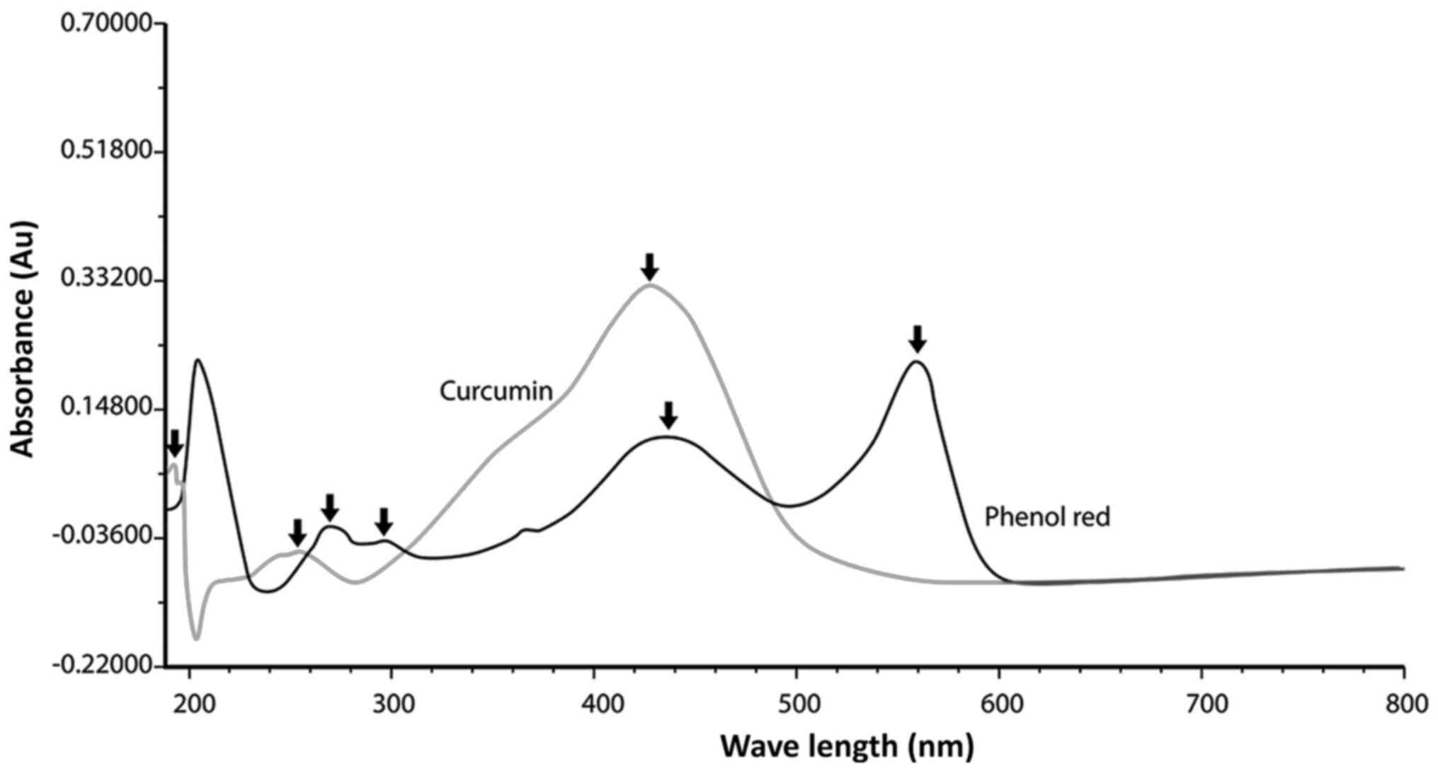
pH >8.2, phenol red turns bright pink (52). Curcumin and phenol red have
absorption maxima of 428 and 436 nm respectively, and a
spectrophotometric analysis was performed with both substances, in
order to detect the presence of overlapping absorbance peaks that
may lead to false interpretation of the cytotoxic results (Fig. 1). The spectrometer used for this
analysis was a HP UV/VIS-spectrometer (Diode array detector;
wavelength range, 190–820 nm; wavelength accuracy, ±2 nm) supplied
by Agilent Technologies.
Optimized MTT assay for the evaluation
of cell survival
The cell survival rate was measured using the MTT
dye reduction assay as described by Mosmann (53), with slight protocol modifications as
previously described (54). This
method is based on the reduction of the yellow tetrazolium salt MTT
to a violet MTT-formazan by the mitochondrial succinate
dehydrogenase in viable cells (55).
Briefly, cells from the three CTCL cell lines (HuT-78, HH and MJ)
were seeded in 96-well plates with 100 µl/well and a density of
0.35×10⁶ cells/ml. After 24 h of incubation at 37°C with
5% CO2, the cells were treated with different
concentrations (100, 80, 60, 40, 20, 10, 5, 2.5, 1.25 and 0.625 µM)
of CRM (EtOH) and CRM (P123/F127) and placed back in the incubator.
After 24, 48 and 72 h, MTT solution (5 mg/ml in PBS) was added. The
cells were further incubated for 3 h and 30 min at 37°C with 5%
CO2. The formazan crystals formed by the cells from the
metabolism of MTT were dissolved by adding 110 µl/well 95%
2-propanol and 5% HCl. Absorbance was measured using a photometer
(Anthos 2001; Anthos Labtec Instruments GmbH) at 540 nm, using a
reference filter of 690 nm. For every concentration, ≥8 wells were
used. In a well containing 100 µl of the respective culture media,
10 µl of MTT and 110 µl 95% 2-propanol (96%) and 5% HCl (37%) was
used as blank. The cell survival rate was calculated as a
percentage of the untreated control, using GraphPad Prism software
(version 6.01 for Windows, GraphPad Software, Inc.).
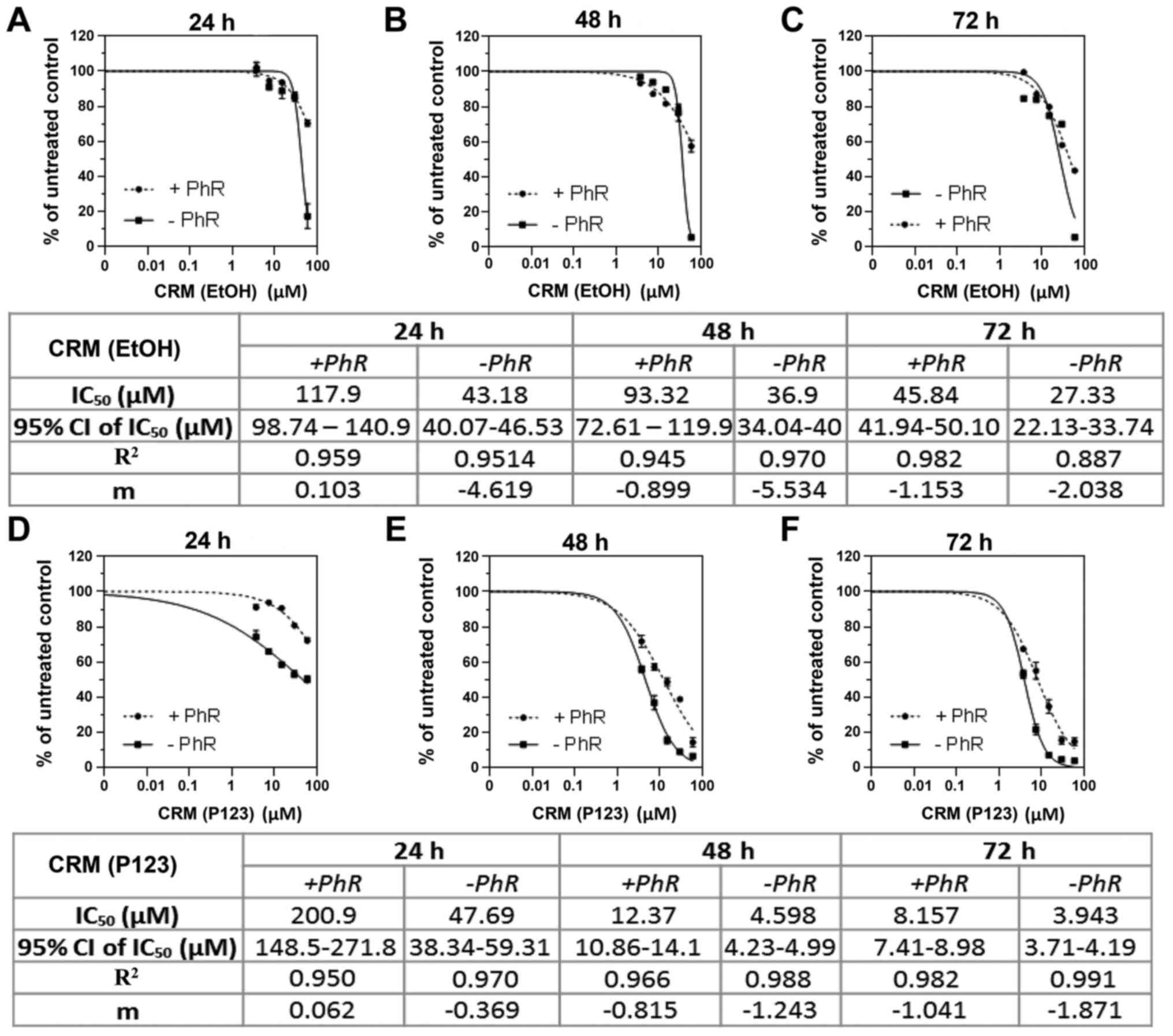
IC25, IC50 and IC75 values of
curcumin were calculated from the concentration-response curves
using the non-linear regression mathematical equation
Y=100/(1+10^((LogIC50-X)*HillSlope))) available in the software
under section ‘Dose-response inhibition’ as ‘log(inhibitor) vs.
normalized response, Variable slope’. Least square fit was used as
fitting model. For presentation of the data points ‘mean ± SD’ was
chosen. The Pearson correlation coefficient was also calculated for
the data. The values presented on Fig.
2 are the values of the square of r computed from the sum of
the squares of the distances of the points from the best-fit curve
determined by nonlinear regression.
Spectrophotometric measurement for the
evaluation of curcumin internalization
To compare the internalization rate of the two
different curcumin formulations into HuT-78, HH and MJ cells, a
spectrophotometric analysis was performed. Briefly, cells were
treated with the two different curcumin formulations and after 1–3
and 24 h, the culture medium was collected after separation from
the cells by centrifugation (277 × g at room temperature for 5
min). Subsequently, 4.5 ml solvent mixture of acetonitrile:
Methanol (1:1 v/v) was added to the tubes containing the culture
media. The samples were sonicated (35 kHz) at 20°C for 5 min and
centrifuged to separate the precipitate (277 × g at room
temperature for 5 min). The supernatant mixtures were filtered and
subjected to spectrophotometric determination against blank
solution that was similarly prepared. This method was performed to
determine the amount of curcumin in the cell culture media. The
more curcumin was contained in the culture media, the less was
internalized into the cells during the incubation. The method's
analysis options used in this experiment were as follows:
Multicomponent analysis, Beer's Law Calibration Curve type, Least
Squares fit algorithm, derivative order of 0, polynomial degree of
0, one smoothing point, data interval of 2, 428 nm analytical
wavelength and 25°C temperature. The reagents used included:
Curcumin reference substance (RS), acetonitrile and methanol, all
supplied by Sigma-Aldrich; Merck KGaA.
Western blotting
In order to observe protein expression changes
induced by curcumin, immunoblot analyses were performed. Cells from
the three CTCL cell lines were seeded at a density of
2×10⁶ in 25 cm2 culture flasks followed by
curcumin treatment with various concentrations [respective
IC25, IC50 and IC75 values for CRM
(EtOH) and 2.5, 5, 10 and 20 µM CRM (P123/F127) on HuT-78 cells].
The incubation period was 24 h (37°C, 5% CO2). The
content of the flasks was harvested and lysed with RIPA buffer (150
mM NaCl, 1% Triton X-100, 0.5% Na deoxycholate, 0.1% SDS and 50 mM
Tris; Thermo Fisher Scientific, Inc.), supplemented with complete
protease inhibitor cocktail tablets and
Na2VO3 (10 µM). The Bradford assay (56) was performed to estimate the protein
concentration of each sample using Carl Roth®
Roti®-Nanoquant (cat. no. K880.2) and BSA (cat. no.
8076.1) as a calibrating protein, both supplied by Carl Roth Gmbh
& Co. Kg. The lysates were mixed with the required amount of
NuPAGE® LDS Sample Buffer (4X; cat. no. 2083421;
Invitrogen; Thermo Fisher Scientific, Inc.). After 5 min at 99°C,
the samples (20 µl each) were separated on FastGene®
PAGE 4–20% gels (cat. no. G34121812; Nippon Genetics Europe GmbH)
via SDS-PAGE. The proteins were transferred onto PVDF membranes
(cat. no. 88520; Thermo Fisher Scientific, Inc.) blocked for 1 h at
room temperature (1X TBS supplemented with 5% skim milk powder and
0.1% Tween-20) and blotted for the target proteins using the
following primary monoclonal antibodies (mAbs): Bad (Rabbit mAb;
cat. no. 9239; dilution 1:1,000), Bax (Rabbit mAb; cat. no. 5023;
dilution 1:1,000), Bcl-2 (Rabbit mAb; cat. no. 3498; dilution
1:1,000) ALK (Rabbit mAb; cat. no. 3633; 1:2,000), Phospho-Janus
kinase (p-Jak)3 (Tyr980/981; Rabbit mAb; cat. no. 5031; dilution
1:1,000), Phospho-Janus kinase (p-Jak)2 (Tyr1007; Rabbit mAb; cat.
no. 3771; dilution 1:1,000), mTOR (Rabbit mAb; cat. no. 2983;
dilution 1:1,000), Phospho-mTOR (Rabbit mAb; cat. no. 5536;
dilution 1:1,000), p-Stat3 (Tyr705; XP® Rabbit mAb; cat.
no. 9145; dilution 1:1,000), p-Stat5 (Tyr694; XP® Rabbit
mAb; 4322; 1:1,000), p-phospholipase (PLC γ1 (Tyr783; Rabbit mAb;
cat. no. 14008; dilution 1:1,000), Raptor (Rabbit mAb; cat. no.
2280; dilution 1:1,000), Rictor (Rabbit mAb; cat. no. 2114;
dilution 1:1,000), p-glycogen synthase kinase (GSK)-3β (Ser9; cat.
no. 9336; dilution 1:1,000), p21 Waf1/Cip1 (Rabbit mAb; cat. no.
2947; dilution 1:1,000), p-NF-κB p65 (Ser536; Rabbit mAb; cat. no.
3033; dilution 1:1,000), all from Cell Signaling Technology, Inc.,
and Wilms' tumor 1 (WT-1) (Mouse mAb;cat. no. sc-7385; dilution
1:500) supplied by Santa Cruz Biotechnology, Inc. HRP-conjugated
secondary anti-rabbit (cat. no. 7074) and anti-mouse (cat. no.
7076) IgG antibodies (Cell Signaling Technology, Inc.; both at
1:2,000 dilution) were applied and a Pierce™ ECL Western Blotting
Substrate (cat. no. 32209) supplied by Thermo Fisher Scientific,
Inc. was used to visualize the immunoblots. Normalization of
protein levels was achieved using the expression levels of β-actin
(cat. no. sc-47778; dilution 1:1,000) supplied by Santa Cruz
Biotechnology, Inc.and densitometry was performed using ImageJ
software (1.52p; Java 1.8.0_112; 64-bit; National Institutes of
Health) (57).
ELISA measurement of NF-κB
transcription factor activation
Comparison of the expression levels of total and
phosphorylated human NF-κB p65 was performed in CTCL cells treated
either with micellar curcumin, CRM (P123/F127), or with the
standard ethanolic solution of curcumin, CRM (EtOH) using two
concentrations for each formulation, 2.5 and 5 µM. The cells were
incubated at 37°C supplied with 5% CO2 for 24 h. The
procedure was performed using a NFκB p65 (Total/Phospho) ELISA kit
(cat. no. ADI-EKS-446) supplied by Enzo Life Sciences, Inc.,
according to the manufacturer's protocol. The obtained data was
analyzed using the protein array analyzer written for ImageJ,
designed by Gilles Carpentier, 2008 (58).
Statistical analysis
All experiments were performed in triplicate. The
Student's t-test was used to compare the control and the treated
groups in Fig. 3. Multiple
comparisons were performed using one-way ANOVA followed by the post
hoc Tukey's test (Fig. 4). P<0.05
was considered to indicate a statistically significant difference.
The data are presented as the mean ± standard deviation. All
experiments were performed using GraphPad Prism software (version
6.01 for Windows; GraphPad Software, Inc.).
Results
Optimization of the MTT assay for
curcumin solutions
Fig. 1 shows that
phenol red, a common pH-indicator present in a wide range of
culture media, may interfere with the results obtained when
measuring the cytotoxic efficacy of curcumin on suspension cell
cultures using the MTT dye reduction assay. This is due to the
relatively contiguous absorbance peaks of both substances (428 nm
for curcumin and 436 nm for phenol red). Considering the prominence
of curcumin in scientific studies, the present result may be of
great importance in the optimization of colorimetric assays that
investigate this natural substance.
MTT tests with and without phenol
red
As expected, the results of the experiments on
cytotoxicity exhibited marked differences in the IC50
values in the presence or absence of phenol red (Fig. 2). These differences were visible
throughout all studied time intervals (24, 48 and 72 h) and did not
depend on the curcumin formulation used, CRM (EtOH) or CRM (P123).
Since the empty Pluronic®P-123 poloxamers exhibited a
high toxicity (Fig. S1), the
present study aimed to identify an improved nano-formulation for
subsequent experiments. The objective of the current study was to
compare solely the cytotoxic properties of curcumin between
different formulations and not the cytotoxic effects of the carrier
itself.
Characterization of the micellar
solution consisting of Pluronic®P-123 and
Pluronic®F-127 (1:1 v/v)
The preparation and loading of curcumin into the
micellar solution consisting of Pluronic®P-123 and
Pluronic®F-127 (1:1 v/v) was performed using the film
hydration method, which has been reported as the most appropriate
procedure in a previous study (38).
The resulting micelles had a slightly negative zeta-potential (~7
mV) and a small particle diameter (~55 nm), which was considered a
prerequisite for their stability. In addition, the small diameter
was considered advantageous for the intracellular transport of
curcumin-loaded micelles. The novel nano-formulation did not
exhibit considerable cytotoxic activity, resulting in a more
appropriate nano-formulation for the present study of the specific
curcumin cytotoxicity (Fig.
S2).
Comparison of the antiproliferative
efficacy of curcumin in ethanol and micellar solutions
The comparison between the antiproliferative
efficacies of the novel micellar solution of curcumin and the
standard ethanolic one was performed via MTT dye reduction assay
(Table I). The results clearly
demonstrated that CRM (P123/F127) exhibited a clear superiority
over the cytotoxic efficacy of the CRM (EtOH) in all cell lines.
For instance, regarding the most resistant cell line, HuT-78, after
24 h of treatment the IC50 value of CRM (P123/F127) was
29.76 µM compared with that of CRM (EtOH) which was significantly
higher (43.18 µM). A more profound difference was observed when
comparing the IC50 values after 48 h, 6.58 µM; CRM
(P123/F127)/36.90 µM; CRM (EtOH) and 72 h, 3.91 µM; CRM
(P123/F127)/27.33 µM; CRM (EtOH).
 | Table I.Cytotoxic comparison between the two
formulations of curcumin on the three cell lines after 24, 48 and
72 h of treatment. |
Table I.
Cytotoxic comparison between the two
formulations of curcumin on the three cell lines after 24, 48 and
72 h of treatment.
|
|
| IC50
(95% CI), µM |
|---|
|
|
|
|
|---|
| Cell line | Curcumin
formulation | 24 h | 48 h | 72 h |
|---|
| HuT-78 | CRM (EtOH) | 43.18
(40.07–43.18) | 36.90
(34.04–40.00) | 27.33
(22.13–33.74) |
|
| CRM
(P123/F127) | 29.76
(23.65–38.02) | 6.58
(5.80–7.40) | 3.91
(3.65–4.18) |
| HH | CRM (EtOH) | 23.61
(21.45–25.99) | 19.40
(18.60–20.24) | 14.74
(12.94–17.17) |
|
| CRM
(P123/F127) | 4.13
(1.81–8.02) | 5.07
(3.763–6.25) | 1.24
(0.68–2.22) |
| MJ | CRM (EtOH) | 30.45
(28.28–32.79) | 33.28
(29.00–38.19) | 16.09
(15.09–17.17) |
|
| CRM
(P123/F127) | 26.16
(15.35–46.81) | 4.16
(2.73–5.76) | 2.93
(2.74–3.14) |
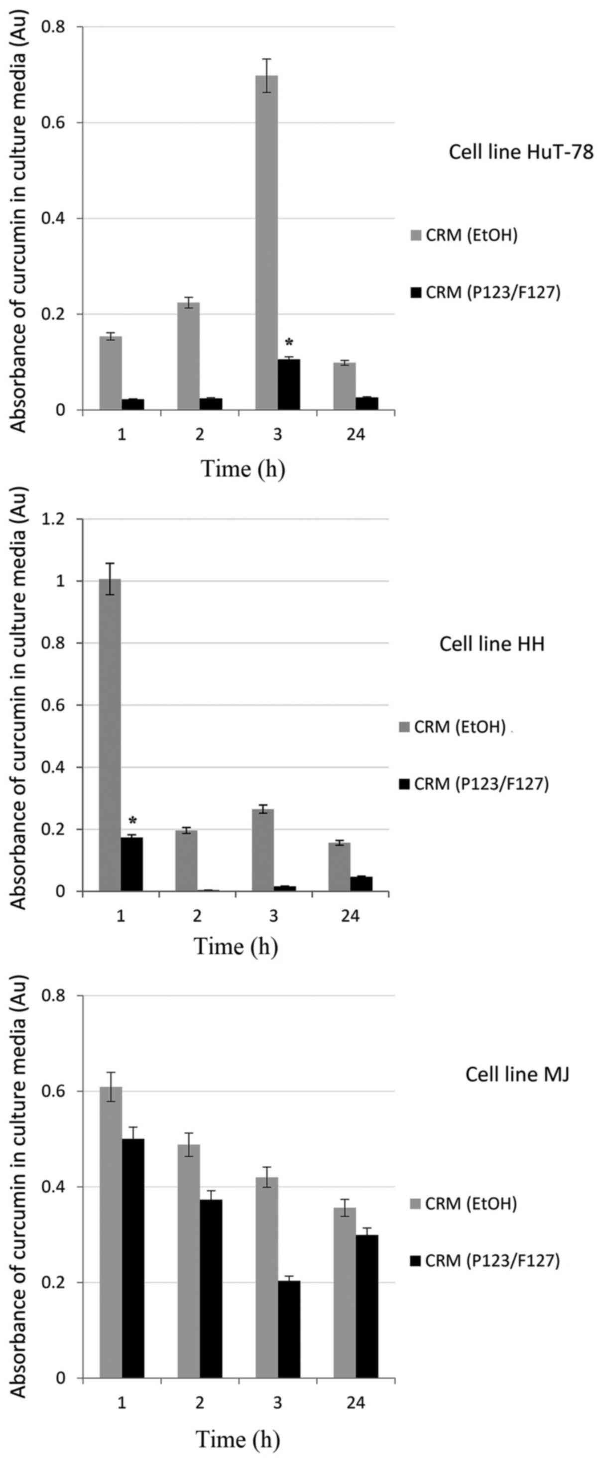
Comparison of the internalization rate
of curcumin in ethanol and micellar solutions
In order to obtain information regarding the speed
of internalization of both curcumin formulations, spectrophotometry
was performed using a UV/Vis-spectrometer. The results from the
spectrophotometric analysis of remaining curcumin amounts in the
culture media from both formulations after different time intervals
are shown in Fig. 3. CRM (P123/F127)
penetrated the cells faster, hence the lower concentration of
curcumin in the supernatant media, compared with CRM (EtOH).
According to the Lambert-Beer law, the absorbance of a
light-absorbing material (such as curcumin) must be proportional to
its concentration in a solution (59). The higher the concentration of
curcumin in the culture media, the lower its concentration in the
CTCL cells. Therefore, CRM (P123/F127) appeared to have a faster
internalization rate than CRM (EtOH).
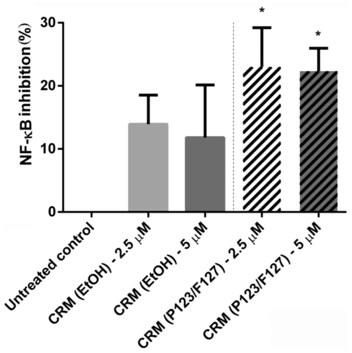
NF-κB inhibition between curcumin in
ethanol and micellar solutions
NF-κB is a transcription factor involved in the
regulation of immune and inflammatory responses (60,61).
Similarly to numerous neoplasms (62,63),
CTCL is also known to have constitutive NF-κB activation (64–66),
which functions by inhibiting cell death through the
transcriptional induction of genes encoding anti-apoptotic
proteins, thus making it a desirable therapeutic target. In order
to detect changes in the activation levels of NF-κB induced by
curcumin, a NF-κB p65 (Total/Phospho) ELISA kit was used in the
present study. Chemiluminescent signals from samples, after
normalization to the untreated control (set to 0%), revealed that
NF-κB inhibition in HuT-78 cells was significantly higher using
both concentrations of CRM (P123/F127) compared with using the
respective concentrations of CRM (EtOH) (Fig. 4). Notably, the amount of curcumin
released from the micelles after 24 h of incubation equals up to
38% of the initial concentration (38), a fact that makes the micellar
formulation even more potent.
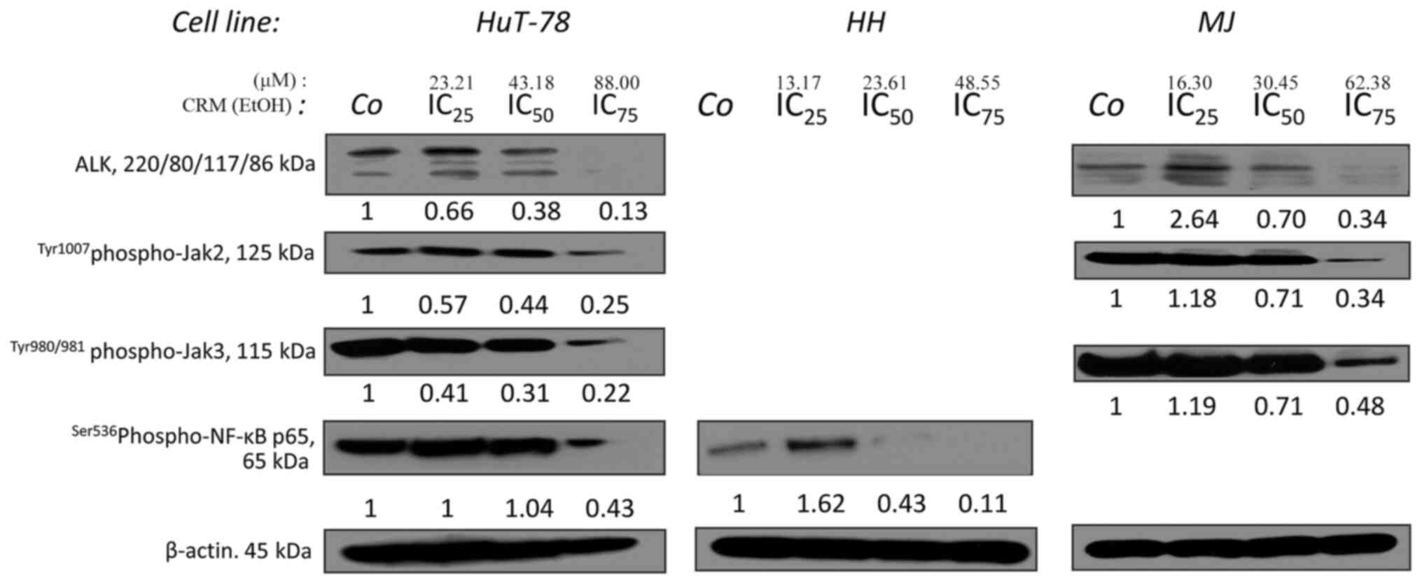
Inhibition of p-NF-κB p65, ALK, p-Jak2
and p-Jak3 by curcumin
In accordance with the aforementioned results, CRM
(EtOH) downregulated the phosphorylated form of NF-κB p65, as
measured via western blotting in HuT-78 and HH cells (Fig. 5). p-NF-κB p65 was also downregulated
in MJ cells (data not shown). Additionally, ALK downregulation and
dephosphorylation of p-Jak2 and p-Jak3 was observed in a
concentration-dependent manner in HuT-78 and MJ cells (Fig. 5).
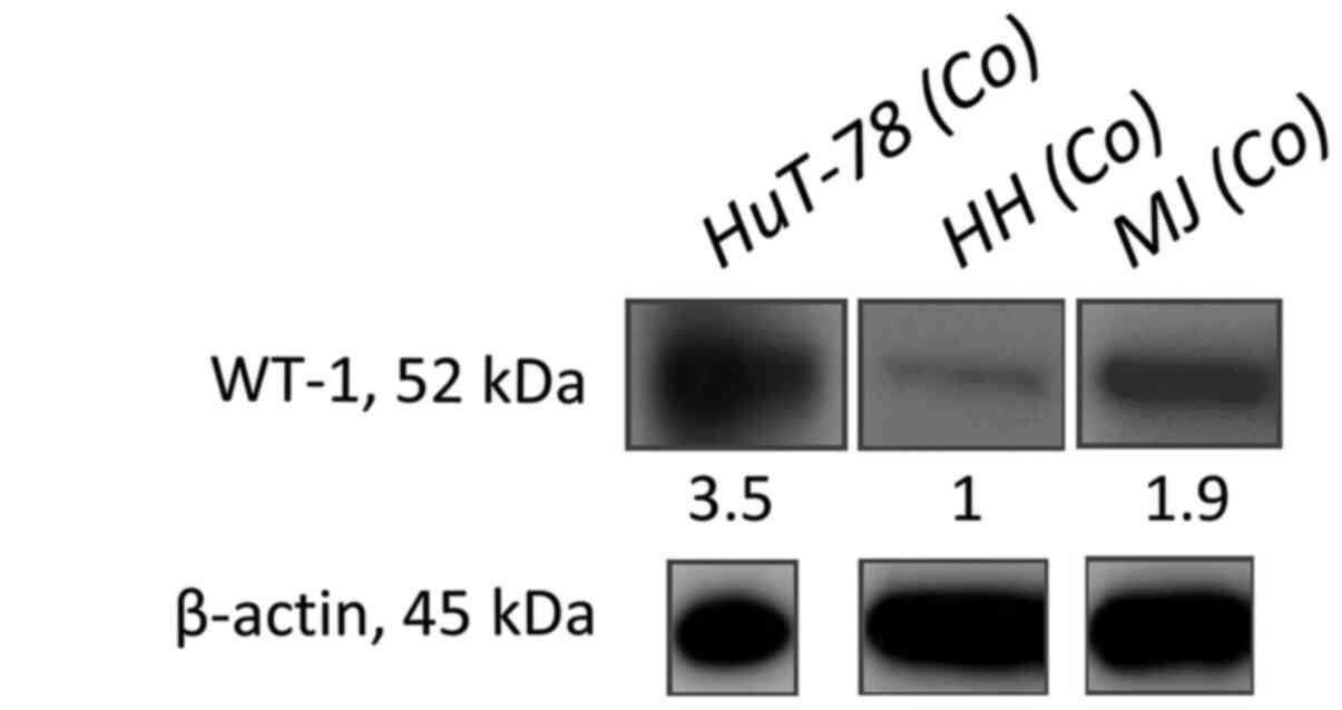
Immunoblot analysis to detect the
presence of WT-1 using only control groups
WT-1 is a transcription factor that in humans is
encoded by the WT-1 gene on chromosome 11p (67–69). It
is commonly detected in malignant non-differentiated cells and its
upregulation is a poor prognostic factor as it negatively affects
the clinical outcome in a variety of tumors, including
non-Hodgkin's lymphoma (70). It is
used as a possible marker for residual disease in acute myeloid
leukemia (AML) after chemotherapy (71). To the best of our knowledge, the
present study aimed for the first time to discover the expression
levels of WT-1 in all studied CTCL cell lines. WT-1 was
overexpressed in HuT-78 cells, followed by an intermediate
expression in MJ cells and a low one in HH cells (Fig. 6). Notably, the present finding,
compared with the cytotoxic experiments, revealed that WT-1 may
serve an important role in cell resistance against cytotoxic
agents, such as curcumin. This result shifted the focus of the
present study to the more resistant cell lines, mainly HuT-78 and
to some extent MJ cells.
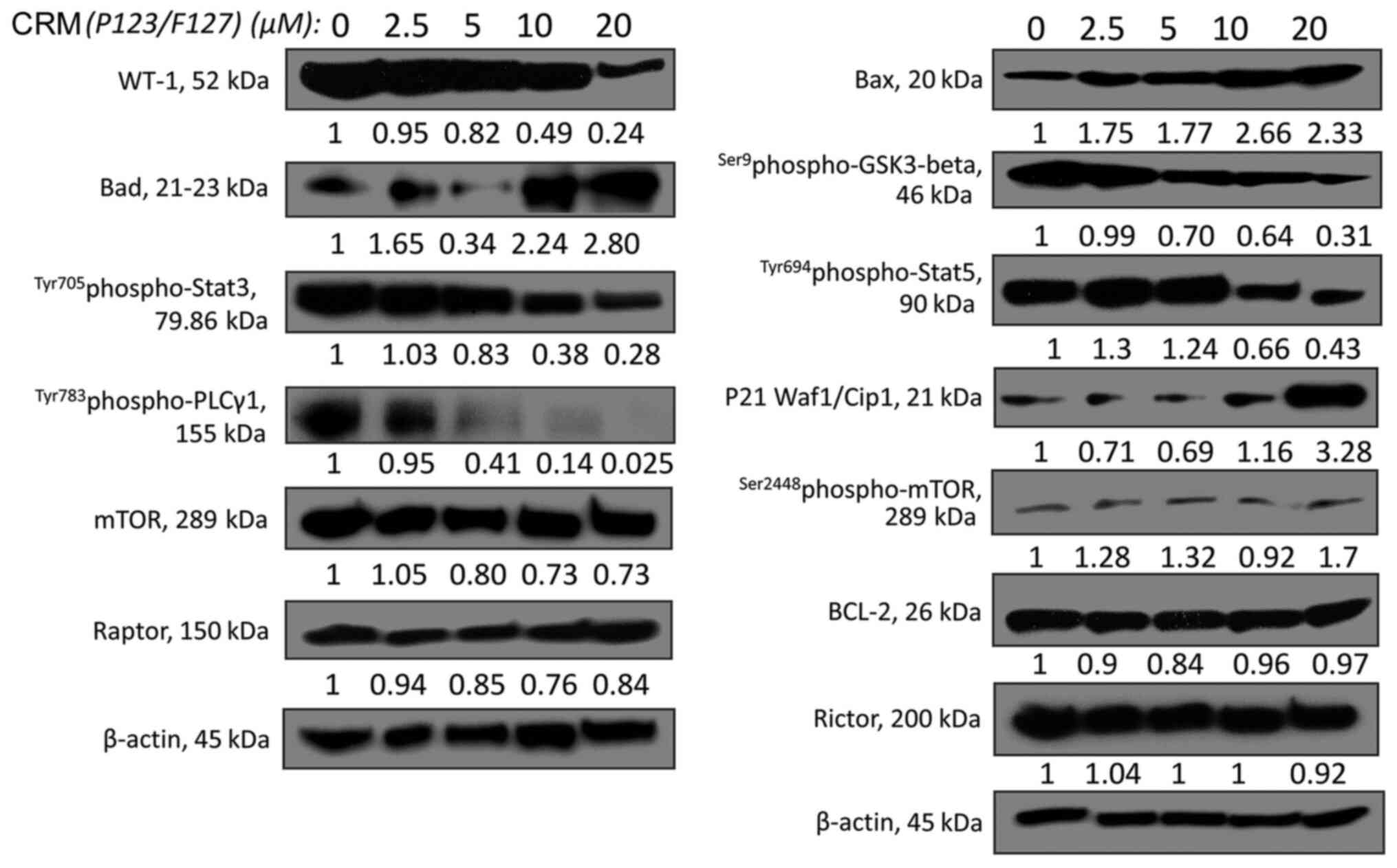
Further analysis of the signal
transduction changes triggered by CRM (P123/F127) in HuT-78
cells
Subsequent experiments aimed to further investigate
the signal transduction changes induced by the novel nanosized
curcumin in the most resistant cell line, HuT-78, which in the
current study represented patients with SS, the leukemic form of
CTCL. CRM (P123/F127) upregulated the pro-apoptotic factors Bad,
Bax and p21 Waf1/Cip1, while it had no effect on the anti-apoptotic
protein BCL-2. CRM (P123/F127) downregulated the transcription
factor WT-1, p-STAT3, p-STAT5, p-PLCγ1 and p-GSK3-β. Lastly, it had
no effect on selected proteins belonging to the mTOR signaling
pathway, namely mTOR, p-mTOR, Raptor and Rictor (Fig. 7).
Discussion
In an era where targeted therapies (together with
immunotherapy) are on the rise for cancer treatment (72–76), the
identification of natural substances with antineoplastic properties
and minimal toxicity, such as curcumin, may improve existing
therapeutic approaches. In the present study, the cytotoxic
efficacy and internalization rate of curcumin was enhanced by its
incorporation into a novel nanosized system consisting of an
aqueous solution of Pluronic®P-123 and
Pluronic®F-127 (1:1 v/v). This enhancement was reflected
at the molecular level by the inhibition of NF-κB p65, which was
induced to a greater extent by the curcumin-loaded micelles than by
the standard ethanol solution of curcumin, measured using a
specific ELISA. The inhibition of NF-κB p65 is associated with a
reduction of inflammatory changes and tumor cell drug resistance
(66,77–80). The
ELISA method represents a faster and easier alternative to the
electrophoresis mobility shift assay, giving the opportunity to
quantify the activation of the transcription factor NF-κB.
Dephosphorylation of p65 NF-κB by CRM (EtOH) as measured via
western blotting was in accordance with the result obtained using
the specific ELISA NFκB p65 (Total/Phospho) kit, as both tests
demonstrated downregulation of NF-κB.
To the best of our knowledge western blotting
revealed for the first time that another transcription factor,
WT-1, was expressed in CTCL cells and may be involved in the
molecular pathogenesis of CTCL. Notably, in the present study WT-1
expression was found to be directly associated with cytotoxic
sensitivity to curcumin, with the HuT-78 cell line which expressed
high levels of WT-1 being the most resistant, followed by MJ and
lastly HH cells. CRM (P123/F127) suppressed the relatively high
expression levels of WT-1 in HuT-78 cells, originating from the
leukemic variant of the disease, known as SS. The present result
may prove to be of great importance if a link between the
upregulation of this transcription factor and a poor prognostic
outcome is identified, similar to that between WT-1 and AML
(71,81–83).
Furthermore, WT-1 downregulation may reflect the curcumin-induced
lymphoma cell differentiation.
The evidence of the association between ALK, a
receptor tyrosine kinase, and various types of human cancer is well
established, with ALK upregulation being present in a variety of
human tumors and cell lines such as those of non-small-cell lung
cancer (NSCLC), melanoma, glioblastoma and lymphoma (84). ALK-positivity in CTCL is rare and
associated with an aggressive course (85). The activation of the ALK protein is
due to a chromosomal translocation, mainly t(2;5)(p23;q35), leading
to a fusion protein with constitutive kinase activity, which is
associated with an aggressive course of CTCL (85). In the present study, curcumin
downregulated ALK, as well as the phosphorylated form of one of the
downstream effectors of ALK, PLCγ, which may serve an important
role in the progression of the disease. Bona fide gain of function
mutations of PLCγ1, the PLCγ form predominantly expressed by T
cells (86), lead to the
constitutive activation of proximal and distal PLCγ1 signaling
cascades, including the activation of NF-κB, with the inhibition of
this pathway resulting in decreased CTCL cell proliferation and
viability (87–89).
In accordance with the aforementioned findings,
upregulation of the pro-apoptotic proteins Bad, Bax and p21
Waf1/Cip1 by curcumin was also observed in the present study.
Furthermore, curcumin inhibited important proteins involved in the
JAK-STAT signaling pathway, which have an essential role in the
pathogenesis of CTCL (89,90), and the phosphorylated form of GSK-3β.
The association between the inhibition of JAK and GSK-3β to promote
cell death in CTCL cells has been described in previous studies.
Pérez et al (91)
demonstrated that ruxolitinib (a specific JAK inhibitor) exerts a
dose-dependent cytotoxicity on CTCL cells, while Rovedo et
al (92) revealed that the
inhibition of GSK-3β increases the cytotoxic activity of
enzastaurin (a protein kinase Cβ inhibitor) against CTCL cells.
This inhibition probably affects cell proliferation and survival
via the already established association between GSK-3β and NF-κB
in vitro, as well as in vivo (92).
Considering the emerging primary or secondary
mechanisms of resistance developed by neoplasms to mAbs and small
molecule inhibitors, such as C481S point mutation, PLCγ2
gain-of-function mutation, constitutive PI3K/Akt and NF-κB
signaling pathways activation (93–96), as
well as novel molecular data (demonstrated in the present study)
clarifying the pleiotropic mode of action of curcumin, this natural
substance may be used in combination with targeted therapies. More
specifically, curcumin may be used to augment the therapeutic
efficacy of currently used targeted agents, such as crizotinib (ALK
inhibitor), ruxolitinib (JAK inhibitor), denosumab and bortezomib
(NF-κB inhibitors), especially in patients with SS, the leukemic
form of the disease that frequently requires aggressive systemic
chemotherapy. In addition, the use of a combination of curcumin and
ruxolitinib for skin application has a logical foundation, firstly
because ruxolitinib is already used topically for the treatment of
skin diseases, such as vitiligo and atopic dermatitis (97–100),
and secondly due to the fact that ruxolitinib inhibits the growth
of CTCL cells in vitro (91).
Similar combination treatment schedules may become even more
feasible by incorporating curcumin into nanoparticles, such as the
novel nanosized aqueous solution of Pluronic®P-123 and
Pluronic®F-127 identified in the present study. The
latter may serve as effective curcumin-delivery system and improve
its pharmacokinetic and cytotoxic properties, making the substance
more potent and suitable for systemic and topical application on
patch lesions or skin tumors associated with CTCL.
In conclusion, experimental findings of the present
study indicated that curcumin may be used as a prospective
single-agent compound or as a partner in combination schedules for
CTCL, especially after inclusion in nanosized drug delivery
systems. Further studies on curcumin combinations with other
complementary acting antineoplastic agents in CTCL models are
warranted.
Supplementary Material
Supporting Data
Acknowledgements
The authors of the present study would like to thank
Professor Chem. Eng. Alexander Dimitrov Kroumov, Department of
Biotechnology, The Stephan Angeloff Institute of Microbiology,
Bulgarian Academy of Sciences (Sofia, Bulgaria), for reviewing the
statistical analyses of all experiments. A number of the cytotoxic
experiments were performed using laboratory equipment donated by
the Alexander von Humboldt Foundation (Grant ‘Equipment Subsidies’,
Certificate of Donation from Department 3.4/V-8151/14038) to Maya
M. Zaharieva, Ph.D., for establishing a ‘Laboratory for in vitro
cytotoxicity and signal transduction’ at the Department of
Infectious Microbiology, The Stephan Angeloff Institute of
Microbiology, Bulgarian Academy of Sciences (Sofia, Bulgaria).
Funding
The present study was supported by a research grant
for ‘Doctoral candidates and young academics and scientists’ by the
German Academic Exchange Service (DAAD), the Erasmus+ program and
the Bulgarian National Science Foundation [grant no. DN 03/3
(2016–2020)].
Availability of data and materials
The datasets used and/or analyzed during the present
study are available from the corresponding author upon reasonable
request.
Authors' contributions
AGXT conceived, designed and performed the
experiments. MMZ co-designed and helped with the performance of the
cytotoxic experiments. MHM helped with the performance of the
western blot experiments. IPET designed and performed the
spectrophotometric experiments for the detection of the overlapping
absorbances of curcumin and phenol red, as well as for the
detection of the amount of curcumin in the cell culture media. KY
designed and loaded the copolymeric micelles with curcumin. MRB and
SMK helped with the conception of the study, its supervision, and
reviewed the first version of the manuscript before its submission.
All authors have read and approved the final version of the
manuscript.
Ethics approval and consent to
participate
Not applicable.
Patient consent for publication
Not applicable.
Competing interests
The authors declare that they have no competing
interests.
References
|
1
|
Hwang ST, Janik JE, Jaffe ES and Wilson
WH: Mycosis fungoides and Sézary syndrome. Lancet. 371:945–957.
2008. View Article : Google Scholar : PubMed/NCBI
|
|
2
|
Willemze R, Kerl H, Sterry W, Berti E,
Cerroni L, Chimenti S, Diaz-Peréz JL, Geerts ML, Goos M, Knobler R,
et al: EORTC classification for primary cutaneous lymphomas: As
proposal from the cutaneous lymphoma study group of the European
organization for research and treatment of cancer. Blood.
90:354–371. 1997.PubMed/NCBI
|
|
3
|
Willemze R, Jaffe ES, Burg G, Cerroni L,
Berti E, Swerdlow SH, Ralfkiaer E, Chimenti S, Diaz-Perez JL,
Duncan LM, et al: WHO-EORTC classification for cutaneous lymphomas.
Blood. 105:3768–3785. 2005. View Article : Google Scholar : PubMed/NCBI
|
|
4
|
Burg G, Kempf W, Cozzio A, Feit J,
Willemze R, S Jaffe E, Dummer R, Berti E, Cerroni L, Chimenti S, et
al: WHO/EORTC classification of cutaneous lymphomas 2005:
Histological and molecular aspects. J Cutan Pathol. 32:647–674.
2005. View Article : Google Scholar : PubMed/NCBI
|
|
5
|
Girardi M, Heald PW and Wilson LD: The
pathogenesis of mycosis fungoides. N Engl J Med. 350:1978–1988.
2004. View Article : Google Scholar : PubMed/NCBI
|
|
6
|
Manso R, Martínez-Magunacelaya N,
Eraña-Tomás I, Monsalvez V, Rodríguez-Peralto JL, Ortiz-Romero PL,
Santonja C, Cristóbal I, Piris MA and Rodríguez-Pinilla SM: Mycosis
fungoides progression could be regulated by microRNAs. PLoS One.
13:e01984772018. View Article : Google Scholar : PubMed/NCBI
|
|
7
|
Criscione VD and Weinstock MA: Incidence
of cutaneous T-cell lymphoma in the United States, 1973–2002. Arch
Dermatol. 143:854–859. 2007. View Article : Google Scholar : PubMed/NCBI
|
|
8
|
Hutchinson CB, Stoecker M, Wang FF,
Papalas J, Sebastian S, Burchette J, Datto M and Wang E: Molecular
detection of circulating Sezary cells in patients with mycosis
fungoides: Could it predict future development of secondary Sezary
syndrome? A single-institution experience. Leuk Lymphoma.
53:868–877. 2012. View Article : Google Scholar : PubMed/NCBI
|
|
9
|
Wu XS, Lonsdorf AS and Hwang ST: Cutaneous
T-cell lymphoma: Roles for chemokines and chemokine receptors. J
Invest Dermatol. 129:1115–1119. 2009. View Article : Google Scholar : PubMed/NCBI
|
|
10
|
Yamashita T, Abbade LP, Marques ME and
Marques SA: Mycosis fungoides and Sezary syndrome: Clinical,
histopathological and immunohistochemical review and update. An
Bras Dermatol. 87:817–830. 2012. View Article : Google Scholar : PubMed/NCBI
|
|
11
|
Yumeen S and Girardi M: Insights into the
molecular and cellular underpinnings of cutaneous T cell lymphoma.
Yale J Biol Med. 93:111–121. 2020.PubMed/NCBI
|
|
12
|
Prince HM, Whittaker S and Hoppe RT: How I
treat mycosis fungoides and Sézary syndrome. Blood. 114:4337–4353.
2009. View Article : Google Scholar : PubMed/NCBI
|
|
13
|
Wollina U: Cutaneous T cell lymphoma:
Update on treatment. Int J Dermatol. 51:1019–1036. 2012. View Article : Google Scholar : PubMed/NCBI
|
|
14
|
Esche BA and Fitzpatrick PJ: Cutaneous
malignant lymphoma. Int J Radiat Oncol Biol Phys. 12:2111–2115.
1986. View Article : Google Scholar : PubMed/NCBI
|
|
15
|
Zucca E and Cavalli F: Extranodal
lymphomas. Ann Oncol. 11 (Suppl 3):S219–S222. 2000. View Article : Google Scholar
|
|
16
|
Sokołowska-Wojdyło M, Olek-Hrab K and
Ruckemann-Dziurdzińska K: Primary cutaneous lymphomas: Diagnosis
and treatment. Postepy Dermatol Alergol. 32:368–383. 2015.
View Article : Google Scholar : PubMed/NCBI
|
|
17
|
Richter T, Nestler-Parr S, Babela R, Khan
ZM, Tesoro T, Molsen E and Hughes DA; International Society for
Pharmacoeconomics and Outcomes Research Rare Disease Special
Interest Group, : Rare disease terminology and definitions-A
systematic global review: Report of the ISPOR rare disease special
interest group. Value Health. 18:906–914. 2015. View Article : Google Scholar : PubMed/NCBI
|
|
18
|
Orphanet, . Orphanet: An online rare
disease and orphan drug data base. © INSERM 1999. simplehttp://www.orpha.net2019
|
|
19
|
Okoye AA and Picker LJ: CD4(+) T-cell
depletion in HIV infection: Mechanisms of immunological failure.
Immunol Rev. 254:54–64. 2013. View Article : Google Scholar : PubMed/NCBI
|
|
20
|
Vidya Vijayan KK, Karthigeyan KP, Tripathi
SP and Hanna LE: Pathophysiology of CD4+ T-Cell
Depletion in HIV-1 and HIV-2 Infections. Front Immunol. 8:5802017.
View Article : Google Scholar : PubMed/NCBI
|
|
21
|
Krejsgaard T, Odum N, Geisler C, Wasik MA
and Woetmann A: Regulatory T cells and immunodeficiency in mycosis
fungoides and Sézary syndrome. Leukemia. 26:424–432. 2012.
View Article : Google Scholar : PubMed/NCBI
|
|
22
|
Heald P, Yan SL and Edelson R: Profound
deficiency in normal circulating T cells in erythrodermic cutaneous
T-cell lymphoma. Arch Dermatol. 130:198–203. 1994. View Article : Google Scholar : PubMed/NCBI
|
|
23
|
Coondoo A, Phiske M, Verma S and Lahiri K:
Side-effects of topical steroids: A long overdue revisit. Indian
Dermatol Online J. 5:416–425. 2014. View Article : Google Scholar : PubMed/NCBI
|
|
24
|
Farber EM, Abel EA and Cox AJ: Long-term
risks of psoralen and UV-A therapy for psoriasis. Arch Dermatol.
119:426–431. 1983. View Article : Google Scholar : PubMed/NCBI
|
|
25
|
Farol LT and Hymes KB: Bexarotene: A
clinical review. Expert Rev Anticancer Ther. 4:180–188. 2004.
View Article : Google Scholar : PubMed/NCBI
|
|
26
|
Nurgali K, Jagoe RT and Abalo R:
Editorial: Adverse effects of cancer chemotherapy: Anything new to
improve tolerance and reduce sequelae? Front Pharmacol. 9:2452018.
View Article : Google Scholar : PubMed/NCBI
|
|
27
|
Sleijfer S, Bannink M, Van Gool AR, Kruit
WH and Stoter G: Side effects of interferon-alpha therapy. Pharm
World Sci. 27:423–431. 2005. View Article : Google Scholar : PubMed/NCBI
|
|
28
|
Subramanian S, Bates SE, Wright JJ,
Espinoza-Delgado I and Piekarz RL: Clinical toxicities of histone
deacetylase inhibitors. Pharmaceuticals (Basel). 3:2751–2767. 2010.
View Article : Google Scholar : PubMed/NCBI
|
|
29
|
Vidula N, Villa M, Helenowski IB,
Jovanovic BD, Meagher R, Mehta J, Singhal S, Winter JN, Frankfurt
O, Altman JK, et al: Adverse events during hematopoietic stem cell
infusion: Analysis of the infusion product. Clin Lymphoma Myeloma
Leuk. 15:e157–e162. 2015. View Article : Google Scholar : PubMed/NCBI
|
|
30
|
Tsambiras PE, Patel S, Greene JN, Sandin
RL and Vincent AL: Infectious complications of cutaneous T-cell
lymphoma. Cancer Control. 8:185–188. 2001. View Article : Google Scholar : PubMed/NCBI
|
|
31
|
Sharma RA, Gescher AJ and Steward WP:
Curcumin: The story so far. Eur J Cancer. 41:1955–1968. 2005.
View Article : Google Scholar : PubMed/NCBI
|
|
32
|
Hsu CH and Cheng AL: Clinical studies with
curcumin. Adv Exp Med Biol. 595:471–480. 2007. View Article : Google Scholar : PubMed/NCBI
|
|
33
|
Zhang C, Li B, Zhang X, Hazarika P,
Aggarwal BB and Duvic M: Curcumin selectively induces apoptosis in
cutaneous T-cell lymphoma cell lines and patients' PBMCs: Potential
role for STAT-3 and NF-kappaB signaling. J Invest Dermatol.
130:2110–2119. 2010. View Article : Google Scholar : PubMed/NCBI
|
|
34
|
Khan MA, Gahlot S and Majumdar S:
Oxidative stress induced by curcumin promotes the death of
cutaneous T-cell lymphoma (HuT-78) by disrupting the function of
several molecular targets. Mol Cancer Ther. 11:1873–1883. 2012.
View Article : Google Scholar : PubMed/NCBI
|
|
35
|
Yosifov DY, Kaloyanov KA, Guenova ML,
Prisadashka K, Balabanova MB, Berger MR and Konstantinov SM:
Alkylphosphocholines and curcumin induce programmed cell deathin
cutaneous T-cell lymphoma cell lines. Leuk Res. 38:49–56. 2014.
View Article : Google Scholar : PubMed/NCBI
|
|
36
|
Devaraj S, Jagannathan N and Neelakantan
P: Antibiofilm efficacy of photoactivated curcumin, triple and
double antibiotic paste, 2% chlorhexidine and calcium hydroxide
against Enterococcus fecalis in vitro. Sci Rep. 6:247972016.
View Article : Google Scholar : PubMed/NCBI
|
|
37
|
Trochopoulos A, Ivanov E, Yakub G, Rashkov
I, Manolova N, Momekova D, Zaharieva MM, Najdenski H, Berger MR and
Konstantinov S: Antineoplastic potential of curcumin loaded
polymeric formulations against human malignant cells. Humboldt
Union in Bulgaria; Sofia, Bulgaria: pp. 44–57. 2018
|
|
38
|
Zaharieva MM, Kroumov AD, Dimitrova L,
Tsvetkova I, Trochopoulos A, Konstantinov SM, Berger MR, Momchilova
M, Yoncheva K and Najdenski HM: Micellar curcumin improves the
antibacterial activity of the alkylphosphocholines erufosine and
miltefosine against pathogenic Staphyloccocus aureus.
Biotechnol Biotechnological Equip. 33:38–53. 2019. View Article : Google Scholar
|
|
39
|
Axelrod PI, Lorber B and Vonderheid EC:
Infections complicating mycosis fungoide and sézary syndrome. JAMA.
267:1354–1358. 1992. View Article : Google Scholar : PubMed/NCBI
|
|
40
|
Bonin S, Tothova SM, Barbazza R, Brunetti
D, Stanta G and Trevisan G: Evidence of multiple infectious agents
in mycosis fungoides lesions. Exp Mol Pathol. 89:46–50. 2010.
View Article : Google Scholar : PubMed/NCBI
|
|
41
|
Willerslev-Olsen A, Krejsgaard T, Lindahl
LM, Bonefeld CM, Wasik MA, Koralov SB, Geisler C, Kilian M, Iversen
L, Woetmann A and Odum N: Bacterial toxins fuel disease progression
in cutaneous T-cell lymphoma. Toxins (Basel). 5:1402–1421. 2013.
View Article : Google Scholar : PubMed/NCBI
|
|
42
|
Kunwar A, Barik A, Pandey R and
Priyadarsini KI: Transport of liposomal and albumin loaded curcumin
to living cells: An absorption and fluorescence spectroscopic
study. Biochim Biophys Acta. 1760:1513–1520. 2006. View Article : Google Scholar : PubMed/NCBI
|
|
43
|
Anand P, Kunnumakkara AB, Newman RA and
Aggarwal BB: Bioavailability of curcumin: Problems and promises.
Mol Pharm. 4:807–818. 2007. View Article : Google Scholar : PubMed/NCBI
|
|
44
|
Aggarwal BB, Sundaram C, Malani N and
Ichikawa H: Curcumin: The Indian solid gold. Adv Exp Med Biol.
595:1–75. 2007. View Article : Google Scholar : PubMed/NCBI
|
|
45
|
Goel A, Kunnumakkara AB and Aggarwal BB:
Curcumin as ‘Curecumin’: From kitchen to clinic. Biochem Pharmacol.
75:787–809. 2008. View Article : Google Scholar : PubMed/NCBI
|
|
46
|
Mohanty C and Sanjeeb K: The in vitro
stability and in vivo pharmacokinetics of curcumin prepared as an
aqueous nanoparticulate formulation. Biomaterials. 31:6597–6611.
2010. View Article : Google Scholar : PubMed/NCBI
|
|
47
|
Song Z, Feng R, Sun M, Guo C, Gao Y, Li L
and Zhai G: Curcumin-loaded PLGA-PEG-PLGA triblock copolymeric
micelles: Preparation, pharmacokinetics and distribution in vivo. J
Colloid Interface Sci. 354:116–123. 2011. View Article : Google Scholar : PubMed/NCBI
|
|
48
|
Li R, Qiao X, Li Q, He R, Ye M, Xiang C,
Lin X and Guo D: Metabolic and pharmacokinetic studies of curcumin,
demethoxycurcumin and bisdemethoxycurcumin in mice tumor after
intragastric administration of nanoparticle formulations by liquid
chromatography coupled with tandem mass spectrometry. J Chromatogr
B Analyt Technol Biomed Life Sci. 879:2751–2758. 2011. View Article : Google Scholar : PubMed/NCBI
|
|
49
|
Yallapu MM, Jaggi M and Chauhan SC:
Curcumin nanoformulations: A future nanomedicine for cancer. Drug
Discov Today. 17:71–80. 2012. View Article : Google Scholar : PubMed/NCBI
|
|
50
|
Jelezova I, Drakalska E, Momekova D,
Shalimova N, Momekov G, Konstantinov S, Rangelov S and Pispas S:
Curcumin loaded pH-sensitive hybrid lipid/block copolymer nanosized
drug delivery systems. Eur J Pharm Sci. 78:67–78. 2015. View Article : Google Scholar : PubMed/NCBI
|
|
51
|
Mehanny M, Hathout RM, Geneidi AS and
Mansour S: Exploring the use of nanocarrier systems to deliver the
magical molecule; Curcumin and its derivatives. J Control Release.
225:1–30. 2016. View Article : Google Scholar : PubMed/NCBI
|
|
52
|
Klement JF, Rice NR, Car BD, Abbondanzo
SJ, Powers GD, Bhatt PH, Chen CH, Rosen CA and Stewart CL:
IkappaBalpha deficiency results in a sustained NF-kappaB response
and severe widespread dermatitis in mice. Mol Cell Biol.
16:2341–2349. 1996. View Article : Google Scholar : PubMed/NCBI
|
|
53
|
Mosmann T: Rapid colorimetric assay for
cellular growth and survival: Application to proliferation and
cytotoxicity assays. J Immunol Methods. 65:55–63. 1983. View Article : Google Scholar : PubMed/NCBI
|
|
54
|
Konstantinov SM, Eibl H and Berger MR:
BCR-ABL influences the antileukaemic efficacy of
alkylphosphocholines. Br J Haematol. 107:365–374. 1999. View Article : Google Scholar : PubMed/NCBI
|
|
55
|
Van Meerloo J, Kaspers GJ and Cloos J:
Cell sensitivity assays: The MTT assay. Methods Mol Biol.
731:237–245. 2011. View Article : Google Scholar : PubMed/NCBI
|
|
56
|
Bradford MM: A rapid and sensitive method
for the quantitation of microgram quantities of protein utilizing
the principle of protein-dye binding. Anal Biochem. 72:248–254.
1976. View Article : Google Scholar : PubMed/NCBI
|
|
57
|
Schneider CA, Rasband WS and Eliceiri KW:
NIH image to imageJ: 25 years of image analysis. Nat Methods.
9:671–675. 2012. View Article : Google Scholar : PubMed/NCBI
|
|
58
|
Abramoff MD, Magalhães PJ and Ram SJ:
Image processing with ImageJ. Biophot Int. 11:36–42. 2004.
|
|
59
|
Swinehart DF: The Beer-Lambert Law. J Chem
Educ. 39:3331962. View Article : Google Scholar
|
|
60
|
Hayden MS, West AP and Ghosh S: NF-kappaB
and the immune response. Oncogene. 25:6758–6780. 2006. View Article : Google Scholar : PubMed/NCBI
|
|
61
|
Liu T, Zhang L, Joo D and Sun SC: NF-κB
signaling in inflammation. Signal Transduct Target Ther.
2:170232017. View Article : Google Scholar : PubMed/NCBI
|
|
62
|
Xia Y, Shen S and Verma IM: NF-κB, an
active player in human cancers. Cancer Immunol Res. 2:823–830.
2014. View Article : Google Scholar : PubMed/NCBI
|
|
63
|
Baldwin AS: Regulation of cell death and
autophagy by IKK and NF-κB: Critical mechanisms in immune function
and cancer. Immunol Rev. 246:327–345. 2012. View Article : Google Scholar : PubMed/NCBI
|
|
64
|
Juvekar A, Manna S, Ramaswami S, Chang TP,
Vu HY, Ghosh CC, Celiker MY and Vancurova I: Bortezomib induces
nuclear translocation of IκBα resulting in gene-specific
suppression of NF-κB-dependent transcription and induction of
apoptosis in CTCL. Mol Cancer Res. 9:183–194. 2011. View Article : Google Scholar : PubMed/NCBI
|
|
65
|
Kiessling MK, Klemke CD, Kaminski MM,
Galani IE, Krammer PH and Gulow K: Inhibition of constitutively
activated nuclear factor-kappaB induces reactive oxygen species-
and iron-dependent cell death in cutaneous T-cell lymphoma. Cancer
Res. 69:2365–2374. 2009. View Article : Google Scholar : PubMed/NCBI
|
|
66
|
Sors A, Jean-Louis F, Pellet C, Laroche L,
Dubertret L, Courtois G, Bachelez H and Michel L: Down-regulating
constitutive activation of the NF-kappaB canonical pathway
overcomes the resistance of cutaneous T-cell lymphoma to apoptosis.
Blood. 107:2354–2363. 2006. View Article : Google Scholar : PubMed/NCBI
|
|
67
|
Call KM, Glaser T, Ito CY, Buckler AJ,
Pelletier J, Haber DA, Rose EA, Kral A, Yeger H, Lewis WH, et al:
Isolation and characterization of a zinc finger polypeptide gene at
the human chromosome 11 Wilms' tumor locus. Cell. 60:509–520. 1990.
View Article : Google Scholar : PubMed/NCBI
|
|
68
|
Gessler M, Poustka A, Cavenee W, Neve RL,
Orkin SH and Bruns GA: Homozygous deletion in Wilms tumours of a
zinc-finger gene identified by chromosome jumping. Nature.
343:774–778. 1990. View Article : Google Scholar : PubMed/NCBI
|
|
69
|
Huang A, Campbell CE, Bonetta L,
McAndrews-Hill MS, Chilton-MacNeill S, Coppes MJ, Law DJ, Feinberg
AP, Yeger H and Williams BR: Tissue, developmental, and
tumor-specific expression of divergent transcripts in Wilms tumor.
Science. 250:991–994. 1990. View Article : Google Scholar : PubMed/NCBI
|
|
70
|
Ujj Z, Buglyo G, Udvardy M, Vargha G, Biro
S and Rejto L: WT1 overexpression affecting clinical outcome in
non-hodgkin lymphomas and adult acute lymphoblastic leukemia.
Pathol Oncol Res. 20:565–570. 2014. View Article : Google Scholar : PubMed/NCBI
|
|
71
|
Casalegno-Garduno R, Schmitt A, Spitschak
A, Greiner J, Wang L, Hilgendorf I, Hirt C, Ho AD, Freund M and
Schmitt M: Immune responses to WT1 in patients with AML or MDS
after chemotherapy and allogeneic stem cell transplantation. Int J
Cancer. 138:1792–1801. 2016. View Article : Google Scholar : PubMed/NCBI
|
|
72
|
Baudino TA: Targeted cancer therapy: The
next generation of cancer treatment. Curr Drug Discov Technol.
12:3–20. 2015. View Article : Google Scholar : PubMed/NCBI
|
|
73
|
Kumar M, Nagpal R, Hemalatha R, Verma V,
Kumar A, Singh S, Marotta F, Jain S and Yadav H: Targeted cancer
therapies: The future of cancer treatment. Acta Biomed. 83:220–233.
2012.PubMed/NCBI
|
|
74
|
Wraith DC: The future of immunotherapy: A
20-year perspective. Front Immunol. 8:16682017. View Article : Google Scholar : PubMed/NCBI
|
|
75
|
Yu Y and Cui J: Present and future of
cancer immunotherapy: A tumor microenvironmental perspective. Oncol
Lett. 16:4105–4113. 2018.PubMed/NCBI
|
|
76
|
Zhang H and Chen J: Current status and
future directions of cancer immunotherapy. J Cancer. 9:1773–1781.
2018. View Article : Google Scholar : PubMed/NCBI
|
|
77
|
DiDonato JA, Mercurio F and Karin M: NF-κB
and the link between inflammation and cancer. Immunol Rev.
246:379–400. 2012. View Article : Google Scholar : PubMed/NCBI
|
|
78
|
Godwin P, Baird AM, Heavey S, Barr MP,
O'Byrne KJ and Gately K: Targeting nuclear factor-kappa B to
overcome resistance to chemotherapy. Front Oncol. 3:1202013.
View Article : Google Scholar : PubMed/NCBI
|
|
79
|
Moore MM, Chua W, Charles KA and Clarke
SJ: Inflammation and cancer: Causes and consequences. Clin
Pharmacol Ther. 87:504–508. 2010. View Article : Google Scholar : PubMed/NCBI
|
|
80
|
Hoesel B and Schmid JA: The complexity of
NF-κB signaling in inflammation and cancer. Mol Cancer. 12:862013.
View Article : Google Scholar : PubMed/NCBI
|
|
81
|
Candoni A, Toffoletti E, Gallina R,
Simeone E, Chiozzotto M, Volpetti S and Fanin R: Monitoring of
minimal residual disease by quantitative WT1 gene expression
following reduced intensity conditioning allogeneic stem cell
transplantation in acute myeloid leukemia. Clin Transplant.
25:308–316. 2011. View Article : Google Scholar : PubMed/NCBI
|
|
82
|
Nowakowska-Kopera A, Sacha T, Florek I,
Zawada M, Czekalska S and Skotnicki AB: Wilms' tumor gene 1
expression analysis by real-time quantitative polymerase chain
reaction for monitoring of minimal residual disease in acute
leukemia. Leuk Lymphoma. 50:1326–1332. 2009. View Article : Google Scholar : PubMed/NCBI
|
|
83
|
Spassov BV, Stoimenov AS, Balatzenko GN,
Genova ML, Peichev DB and Konstantinov SM: Wilms' tumor protein and
FLT3-internal tandem duplication expression in patients with de
novo acute myeloid leukemia. Hematology. 16:37–42. 2011. View Article : Google Scholar : PubMed/NCBI
|
|
84
|
Hallberg B and Palmer RH: Mechanistic
insight into ALK receptor tyrosine kinase in human cancer biology.
Nat Rev Cancer. 13:685–700. 2013. View Article : Google Scholar : PubMed/NCBI
|
|
85
|
Wehkamp U, Oschlies I, Nagel I, Brasch J,
Kneba M, Günther A, Klapper W and Weichenthal M: ALK-positive
primary cutaneous T-cell-lymphoma (CTCL) with unusual clinical
presentation and aggressive course. J Cutan Pathol. 42:870–877.
2015. View Article : Google Scholar : PubMed/NCBI
|
|
86
|
Stoica GE, Kuo A, Aigner A, Sunitha I,
Souttou B, Malerczyk C, Caughey DJ, Wen D, Karavanov A, Riegel AT
and Wellstein A: Identification of anaplastic lymphoma kinase as a
receptor for the growth factor pleiotrophin. J Biol Chem.
276:16772–16779. 2001. View Article : Google Scholar : PubMed/NCBI
|
|
87
|
Patel VM, Flanagan CE, Martins M, Jones
CL, Butler RM, Woollard WJ, Bakr FS, Yoxall A, Begum N, Katan M, et
al: Frequent and persistent PLCG1 mutations in sezary cells
directly enhance PLCү1 activity and stimulate NFKB, AP-1, and NFAT
signaling. J Invest Dermatol. 140:380–389.e4. 2020. View Article : Google Scholar : PubMed/NCBI
|
|
88
|
Smith-Garvin JE, Koretzky GA and Jordan
MS: T cell activation. Annu Rev Immunol. 27:591–619. 2009.
View Article : Google Scholar : PubMed/NCBI
|
|
89
|
Vaque JP, Gomez-Lopez G, Monsalvez V,
Varela I, Martínez N, Pérez C, Domínguez O, Graña O,
Rodríguez-Peralto JL, Rodríguez-Pinilla SM, et al: PLCG1 mutations
in cutaneous T-cell lymphomas. Blood. 123:2034–2043. 2014.
View Article : Google Scholar : PubMed/NCBI
|
|
90
|
Kiessling MK, Oberholzer PA, Mondal C,
Karpova MB, Zipser MC, Lin WM, Girardi M, Macconaill LE, Kehoe SM,
Hatton C, et al: High-throughput mutation profiling of CTCL samples
reveals KRAS and NRAS mutations sensitizing tumors toward
inhibition of the RAS/RAF/MEK signaling cascade. Blood.
117:2433–2440. 2011. View Article : Google Scholar : PubMed/NCBI
|
|
91
|
Pérez C, González-Rincón J, Onaindia A,
Almaráz C, García-Díaz N, Pisonero H, Curiel-Olmo S, Gómez S,
Cereceda L, Madureira R, et al: Mutated JAK kinases and deregulated
STAT activity are potential therapeutic targets in cutaneous T-cell
lymphoma. Haematologica. 100:e450–e453. 2015. View Article : Google Scholar : PubMed/NCBI
|
|
92
|
Rovedo MA, Krett NL and Rosen ST:
Inhibition of glycogen synthase kinase-3 increases the cytotoxicity
of enzastaurin. J Invest Dermatol. 131:1442–1449. 2011. View Article : Google Scholar : PubMed/NCBI
|
|
93
|
Balaji S, Ahmed M, Lorence E, Yan F, Nomie
K and Wang M: NF-κB signaling and its relevance to the treatment of
mantle cell lymphoma. J Hematol Oncol. 11:832018. View Article : Google Scholar : PubMed/NCBI
|
|
94
|
Merolle MI, Ahmed M, Nomie K and Wang ML:
The B cell receptor signaling pathway in mantle cell lymphoma.
Oncotarget. 9:25332–25341. 2018. View Article : Google Scholar : PubMed/NCBI
|
|
95
|
Rosenthal A: Small molecule inhibitors in
chronic lymphocytic lymphoma and B cell non-hodgkin lymphoma. Curr
Hematol Malig Rep. 12:207–216. 2017. View Article : Google Scholar : PubMed/NCBI
|
|
96
|
Valla K, Flowers CR and Koff JL: Targeting
the B cell receptor pathway in non-Hodgkin lymphoma. Expert Opin
Investig Drugs. 27:513–522. 2018. View Article : Google Scholar : PubMed/NCBI
|
|
97
|
Kim BS, Howell MD, Sun K, Papp K, Nasir A
and Kuligowski ME; INCB 18424-206 Study Investigators, : Treatment
of atopic dermatitis with ruxolitinib cream (JAK1/JAK2 inhibitor)
or triamcinolone cream. J Allergy Clin Immunol. 145:572–582. 2020.
View Article : Google Scholar : PubMed/NCBI
|
|
98
|
Owens S and Howell MD: Ruxolitinib cream
suppresses Th2 inflammation in adult patients with atopic
dermatitis. J Allergy Clin Immunol. 143:AB1282019. View Article : Google Scholar
|
|
99
|
Rosmarin D, Pandya AG, Lebwohl M, Grimes
P, Hamzavi I, Gottlieb AB, Butler K, Kuo F, Sun K, Ji T, et al:
Ruxolitinib cream for treatment of vitiligo: A randomised,
controlled, phase 2 trial. Lancet. 396:110–120. 2020. View Article : Google Scholar : PubMed/NCBI
|
|
100
|
Rothstein B, Joshipura D, Saraiya A, Abdat
R, Ashkar H, Turkowski Y, Sheth V, Huang V, Au SC, Kachuk C, et al:
Treatment of vitiligo with the topical Janus kinase inhibitor
ruxolitinib. J Am Acad Dermatol. 76:1054–1060.e1. 2017. View Article : Google Scholar : PubMed/NCBI
|