Introduction
Colon adenocarcinoma (COAD) is the third leading
cause of morbidity (46.9 per 100,000 men and 35.6 per 100,000
women) and mortality (17.7 per 100,000 men and 12.4 per 100,000
women) among malignant neoplasms worldwide, according to statistics
from 2013 (1). Alcohol and processed
meat consumption, advanced age, family history and tumor metastasis
are closely associated with the incidence of COAD (2). Although surgical resection,
chemotherapy and radiotherapy have been widely used to treat colon
cancer, most patients with advanced disease exhibit drug
resistance, leading to a particularly poor prognosis (3,4).
Increasing evidence has suggested that left-sided colon cancer
(LCC), which arises from the embryonic midgut, and right-sided
colon cancer (RCC), which originates from the hindgut, exhibit
distinct differences in embryonic origin, biology, anatomy, genetic
mutations and alterations, and clinical outcomes, and this may be
the major cause of the poor prognosis following surgery and medical
procedures in COAD (5). Therefore,
identification of tumor-driven genes underlying the progression of
LCC and RCC is of great importance for monitoring the progression
and improving the prognosis of patients with COAD.
The cecum, ascending colon, hepatic flexure and
transverse colon are anatomically referred to as the right colon,
while the descending colon and sigmoid colon are classified as the
left colon (6). Cancer located in
the right and left colon is defined as RCC or LCC, respectively.
The incidence of RCC is associated with sex, age, cancer history
and insulin resistance, whereas LCC is closely associated with a
low fiber diet, heavy smoking and alcoholism (7). Cancer located in the right colon has a
prevalence toward being of a more advanced tumor stage and having a
large tumor size, and often metastasizes to the lymph nodes or
peritoneal region (8). Poor survival
rate is observed in patients with RCC compared with LCC (9). LCC is associated with a reduced risk of
death, which may be due to the observation that patients with LCC
more commonly present with early-stage disease, and this results in
the disparity in prognosis according to tumor site (10). Numerous differences in molecular
signaling pathways have been reported between RCC and LCC (11). Notably, using molecular biology
techniques, numerous molecular events that cause distinct efficacy
of molecular targeted agents, such as TP53, KRAS, CpG Island
Methylator Phenotype (CIMP) and microsatellite instability (MSI),
in patients with LCC and RCC have been revealed in recent years
(12). For example, deregulated
genes for CIMP high and MSI tumors are often downregulated. In
contrast, genes that are deregulated in TP53 are likely to be
upregulated compared to normal paired samples. However, to the best
of our knowledge, the specific molecules that underlie the
difference between LCC and RCC have not yet been determined.
Guanine nucleotide binding-protein γ subunit 4
(GNG4) is a member of the G-protein trimer complex, and was first
identified as the brain-specific subunit (13). In the human brain, GNG4 is more
highly expressed in the hippocampus compared with in other brain
regions, and GNG4 expression is reduced with advanced age, which
suggests that GNG4 is associated with cognitive decline (14). A previous study has suggested that
GNG4 is hypermethylated and notably decreased in glioblastoma (GBM)
(15). In addition to its roles in
the brain, GNG4 is involved in pathological processes, it activates
signaling pathways in acute myocardial infarction, and it may
contribute to the prevention and treatment of recurrent
cardiovascular events (16).
Additionally, it has been demonstrated that GNG4 is a hub gene that
has a high degree of connectivity in colon cancer (17). According to bioinformatics analysis,
the hub gene GNG4 has a clinical diagnostic value in patients with
colorectal cancer (18). Therefore,
GNG4 is an important regulator during tumor progression,
particularly in colon cancer. However, whether there is molecular
heterogeneity of GNG4 in LCC and RCC remains unclear.
The present study was aimed to investigate the
molecular heterogeneity of GNG4 in LCC and RCC.
Additionally, the association between GNG4 expression and
clinical characteristics including disease stage and overall
survival (OS) rate was also assessed in LCC and RCC of Caucasian
and Chinese patients. These findings demonstrated that GNG4 may be
used as an effective prognostic factor for LCC and RCC.
Materials and methods
Tissue microarray
A tissue and cDNA microarray containing 78 colon
carcinoma and paired para-carcinoma tissues was purchased from
Shanghai Outdo Biotech Co., Ltd. Among the 78 cases, 32 were tumors
located in the left colon and the other 46 were tumors located in
the right colon. All experiments were approved by the Ethics
Committee of the Affiliated Cancer Hospital of Fujian Medical
University (Fuzhou, China).
Immunohistochemistry (IHC)
For IHC staining, 5 µm thick paraffin embedded
sections were dewaxed in an oven for 30 min at 60°C. After
deparaffinization in xylene and rehydration in gradient ethanol,
antigens were retrieved with 0.01 M citrate salt solution (pH 6.0)
using high pressure method (100°C) for 15 min. Following washing
with PBS plus Tween-20 and blocking with 5% BSA (cat. no. A8010;
Beijing Solarbio Science & Technology Co., Ltd.) for 1 h at
room temperature, the slides were incubated with primary antibody
against GNG4 (cat. no. ab238868; 1:250; Abcam) at 4°C overnight.
Following incubation with the secondary antibody (cat. no. ab6721;
1:2,000; Abcam) for 15 min at room temperature, antigen-antibody
complexes were visualized using 3,3-diaminobenzidine reagent (cat.
no. ZL1-9081; OriGene Technologies, Inc.). The results were
observed under a biological inverted light microscope (×5 and ×20
magnifications; IX51; Olympus Corporation). Comprehensive analysis
included measuring staining intensity and the number of positive
cells as follows: Negative (−), 0%; weakly positive (+), <20%;
moderately positive (++), 20–50%; and strongly positive (+++),
>50%.
Reverse transcription-quantitative PCR
(RT-qPCR)
cDNA extracted from 78 cases of colon carcinoma,
corresponding to the same samples as the tissue microarray, was
purchased from Shanghai Outdo Biotech Co., Ltd. Samples were plated
in 96-well plates and subsequently, RT-qPCR was performed using the
SYBR Green kit (Roche Diagnostics GmbH) according to manufacturers
protocols. GNG4 expression was normalized to GAPDH expression. The
primer sequences were shown in Table
SI. Relative gene expression levels were calculated using the
2−∆∆Cq method (19).
Western blotting
A total of 15 clinical samples from patients aged
between 38 and 62 years (13 men and 2 women; mean age,
50.47±2.035), including 5 normal colon tissues, 5 left-sided colon
tumor tissues and 5 right-sided colon tumor tissues obtained from
resection were collected at The Affiliated Cancer Hospital of
Fujian Medical University between October 2019 and February 2020.
All these patients had been diagnosed with colon cancer previously.
Written informed consent was obtained from all subjects, and the
present study was approved by the Ethics Committee of The
Affiliated Cancer Hospital of Fujian Medical University. Total
protein was extracted from normal colon and tumor tissues using
RIPA protein lysate (cat. no. P0013B; Beyotime Institute of
Biotechnology). Protein concentration was determined using BCA
Protein Assay Kit (23225; Thermo Fisher Scientific, Inc.). Equal
amounts of protein (30 µg) were loaded and separated on 12% gels by
SDS-PAGE. Subsequently, protein was transferred to an activated
PVDF membrane (IPVH00010; EMD Millipore). The membrane was blocked
with 5% skimmed milk dissolved in PBS-Tween-20 (0.1%) at room
temperature (~25°C) for 1 h. Next, primary antibody against GNG4
(cat. no. PA5-103877; 1:800; Thermo Fisher Scientific, Inc.) was
added for an incubation at 4°C overnight. β-actin (cat. no. BM0627;
1:5,000; Boster Biological Technology) served as the internal
control. Following incubation with goat anti-rabbit secondary
antibody conjugated to HRP (cat. no. BA1054; 1:5,000; Boster
Biological Technology) at room temperature (~25°C) for an
additional 1 h, the membrane was incubated with ECL visualization
reagent (34065; Thermo Fisher Scientific, Inc.) and the bands were
visualized using the Imagequant LAS 4000 mini machine (Cytiva).
Data collection and analysis
The samples collected from TCGA were divided into
LCC (n=181) and RCC (n=258). No information in anatomic neoplasm
was excluded from the dataset. The mRNA expression levels of
GNG4 in normal colon tissues and colon cancer tissues were
collected from the Oncomine database (https://www.oncomine.org/resource/login.html) and the
Gene Expression Profiling Interactive Analysis (GEPIA) version 2
database (20). GNG4 expression in
LCC (n=99) and RCC (n=167) excluding the normal samples was
determined using the raw data from The Cancer Genome Atlas (TCGA,
n=524; http://portal.gdc.cancer.gov/) and
analyzed using GraphPad v6.0 software (GraphPad Software, Inc.).
The samples collected from the Oncomine database included 12 normal
colon samples and 70 COAD samples. The association between GNG4
expression and disease stage in patients with COAD was analyzed
with one-way ANOVA (followed by Tukeys post hoc test) using raw
data downloaded from the GEPIA database. In the tissue microarray,
the association between GNG4 and disease stage was determined based
on the IHC staining results. The disease stage information was
mentioned in the tissue microarray.
Survival analysis
OS rate was analyzed using GEPIA on the basis of
GNG4 expression status. The difference in OS rate between patients
with LCC and RCC was compared using the raw data from TCGA and
analyzed with GraphPad v6.0 software. The data from the tissue
microarray were used to evaluate the OS rate in Chinese patients
with tumors in the left and right colon.
Statistical analysis
The association between pathological stage and GNG4
expression in tissue microarray was analyzed with GraphPad Prism
6.0 (GraphPad Software, Inc.) using χ2 and Fishers exact
tests. The association analysis between tumor stage and GNG4 status
in patients with COAD was performed using one-way ANOVA, followed
by Tukeys post hoc test. Histological staining was evaluated using
Image Pro Plus 6.0 software (Media Cybernetics). GNG4 gene
expression and relative protein expression for tissue microarray
were compared with GraphPad Prism using two-tailed t-tests
(unpaired) and one-way ANOVA followed by Tukeys post hoc test,
respectively. Kaplan-Meier plotter was used to generate the
survival curves using GraphPad Prism software (version 6.0;
GraphPad Software, Inc.), and data were compared between groups
using the log-rank (Mantel-Cox) test (univariate Cox regression
analysis). P<0.05 was considered to indicate a statistically
significant difference.
Results
GNG4 is highly expressed in White
patients with COAD
To determine GNG4 expression in White patients with
COAD included in TCGA database, the present study analyzed the
transcriptional levels of GNG4 in 275 patients with COAD and 349
normal controls. The mean expression level of GNG4 was increased
2.2-fold in the tumor group compared with that in the normal group
(Fig. 1A). Subsequently, the
association between GNG4 and disease stage of COAD was assessed.
The results indicated that GNG4 status exhibited no difference when
comparing patients with COAD in stages I and II. However, GNG4
expression significantly increased in disease stages III and IV
compared with that in low disease stages (I and II) (stage I vs.
III, P=0.0428; stage I vs. IV, P<0.01; stage II vs. III,
P<0.01; and stage II vs. IV, P<0.01), with the highest levels
observed in stage IV (stage III vs. IV, P=0.0427) (Fig. 1B). GNG4 expression was divided into
two groups, using as a cut-off the median method which was obtained
from GEPIA 2.0. Additionally, patients with COAD and high GNG4
expression (n=135) had a decreased survival rate compared with
patients with low GNG4 expression (n=135) (Fig. 1C). Therefore, high expression levels
of GNG4 were positively associated with pathological grade and
prognosis in White patients with COAD.
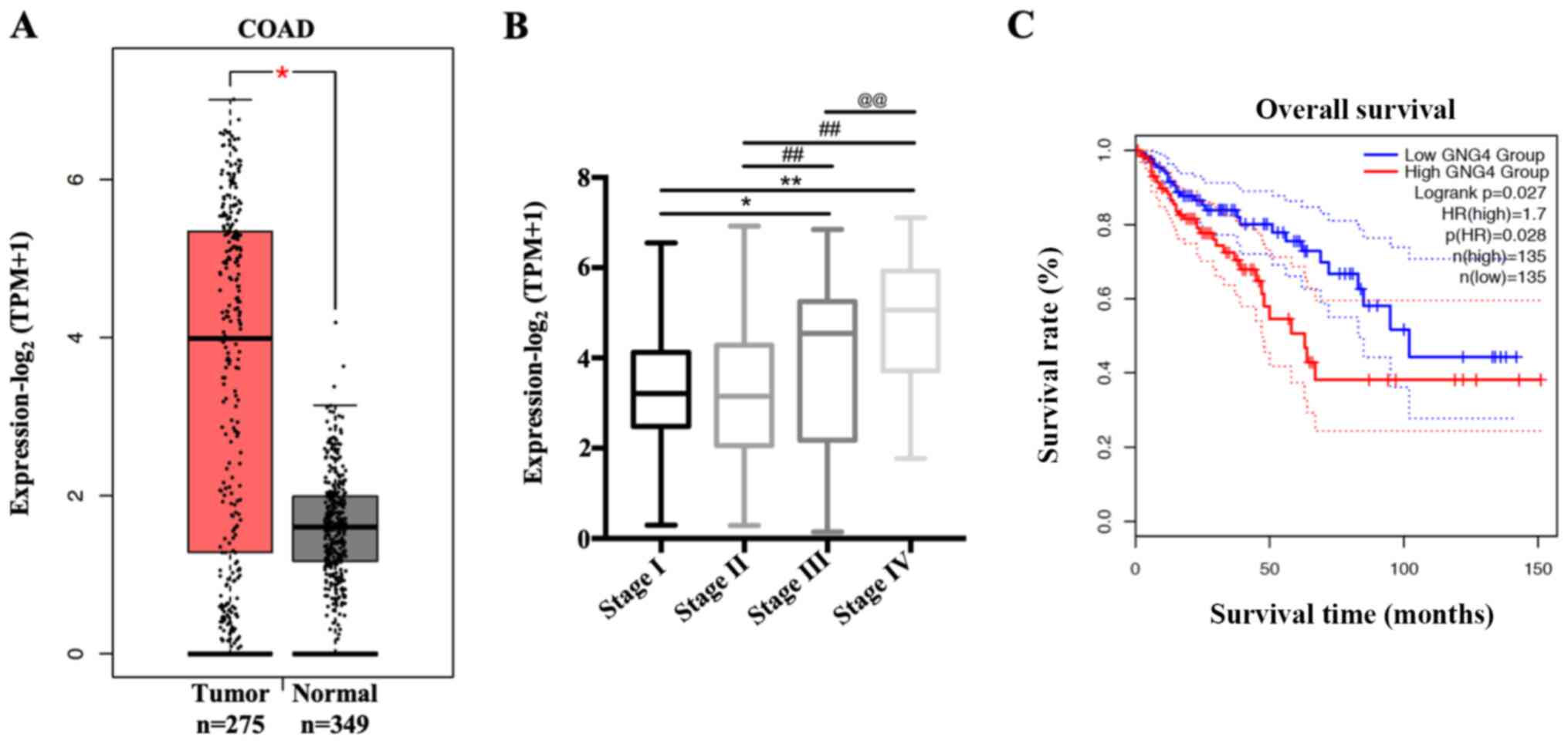 | Figure 1.Association of GNG4 status with
disease stage and overall survival in patients with COAD. (A)
Different transcriptional expression levels of GNG4 in COAD samples
(n=275) and normal controls (n=349). Gene expression data were
collected from the GEPIA 2.0 database and analyzed with GraphPad
Prism using two-tailed t-tests (unpaired). (B) Patients with COAD
were divided into four groups based on disease stage, including
stage I, II, III and IV. Subsequently, the association between GNG4
expression and disease stage was analyzed by one-way ANOVA followed
by Tukeys post hoc test. (C) Survival analysis was performed using
GEPIA 2.0 in patients with COAD with low and high GNG4 expression.
*P<0.05; **, ##, @@P<0.01. COAD, colon
adenocarcinoma; GEPIA, Gene Expression Profiling Interactive
Analysis; GNG4, guanine nucleotide binding-protein γ subunit 4;
TPM, transcripts per million. |
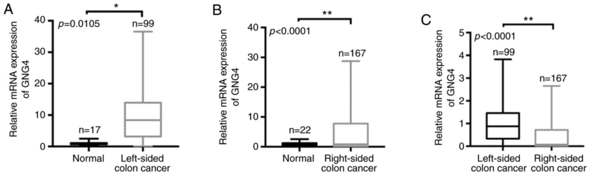
Molecular heterogeneity of GNG4 is
observed in White patients with LCC and RCC
Given that GNG4 is an important hub gene during the
tumorigenesis of COAD, the present study aimed to investigate the
differences in GNG4 expression between White patients with LCC and
those with RCC. Firstly, the raw data of patients with COAD was
collected from TCGA and screened according to grouping criteria.
Subsequently, all samples (ethnicity, White) were divided into two
groups according to the tumor site in the colon, i.e. LCC and RCC.
Based on the sample information, only 15 normal samples in LCC and
22 normal samples in RCC were included. In LCC cohorts, there were
99 tumor samples and the corresponding normal samples (n=15), and
there were 167 RCC tumor samples and corresponding normal tissues
(n=22). When comparing normal control tissues (n=15) with LCC
tissues (n=99), it was revealed that GNG4 expression was
significantly elevated in the LCC tissues (P=0.0105; Fig. 2A). Consistent with this, there was a
significant increase in GNG4 expression in RCC tissues (n=167)
compared with the corresponding normal tissues (n=22) (P<0.0001;
Fig. 2B). However, compared with the
level in patients with RCC (n=167), the GNG4 expression level was
increased in patients with LCC (n=99) (P<0.0001; Fig. 2C). Therefore, molecular heterogeneity
of GNG4 was present in LCC and RCC.
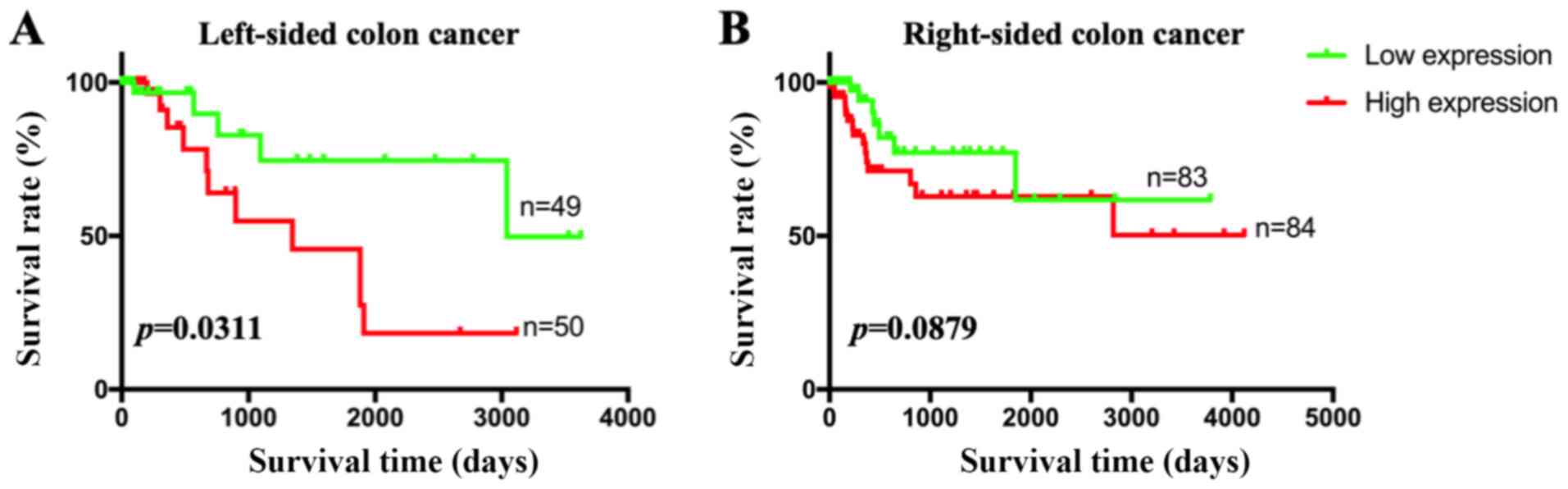
GNG4 is associated with disease stage
and prognosis in White patients with LCC
Since genetic heterogeneity of GNG4 was observed
between LCC and RCC, it was speculated that there may be a
difference in the clinical diagnostic value of GNG4 in the two
cancer types. First, patients were divided into two groups based on
GNG4 expression (cut-off-high, 50%; cut-off-low, 50%). High
GNG4 expression was associated with high disease stage in patients
with LCC (P=0.0356; Table I).
However, no association between GNG4 status and disease stage was
identified in patients with RCC (P=0.6260; Table I). Additionally, patients with LCC in
the high GNG4 expression group had a lower survival rate than those
in the low GNG4 expression group (P=0.0311; Fig. 3A). In patients with RCC, no
association between GNG4 expression and OS was observed (P=0.0879;
Fig. 3B). Therefore, GNG4 status was
associated with pathological stage and OS in White patients with
LCC, but not in those with RCC.
 | Table I.Association between GNG4
expression and pathological stage in LCC and RCC. |
Table I.
Association between GNG4
expression and pathological stage in LCC and RCC.
|
| LCC | RCC |
|---|
|
|
|
|
|---|
| Stage | Low GNG4, n | High GNG4, n | Low GNG4, n | High GNG4, n |
|---|
| I | 8 | 5 | 18 | 15 |
| II | 24 | 18 | 35 | 29 |
| III | 9 | 18 | 22 | 28 |
| IV | 4 | 13 | 9 | 11 |
| P-value |
| 0.0356a |
| 0.6260 |
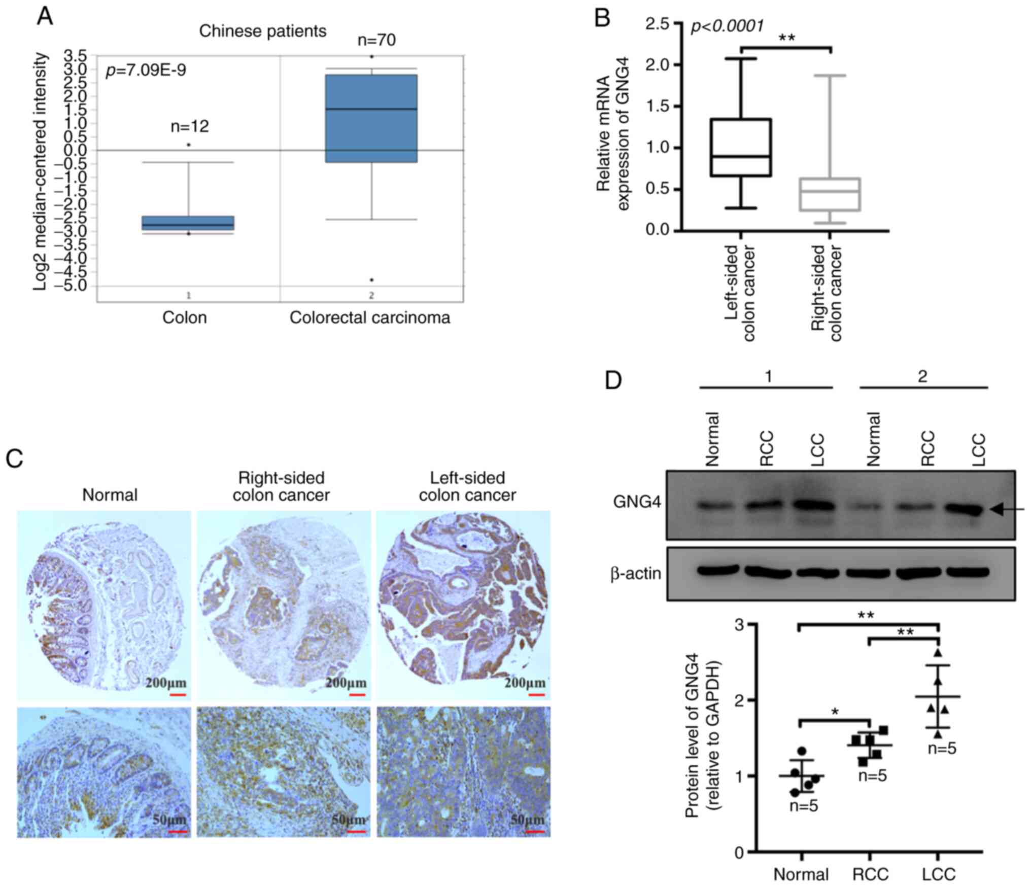
GNG4 is highly expressed in Chinese
patients with LCC
To further ascertain the expression pattern of GNG4
in Chinese patients with colorectal cancer, the present study
collected raw data, including that of 70 cases of colorectal cancer
and 12 normal colon samples, from the Oncomine database. All 82
samples were from a Chinese population. The results revealed that
GNG4 expression was significantly increased in patients with
colorectal cancer compared with that in normal controls
(P<0.0001; Fig. 4A).
Subsequently, tissue and cDNA microarrays containing 78 pairs of
COAD samples and paired adjacent colon tissues were used to
evaluate the expression levels of GNG4 in human specimens by IHC
staining and RT-qPCR. The microarray comprised 32 cases of LCC and
46 cases of RCC. The data indicated that the relative GNG4
mRNA expression in patients with LCC was markedly increased
compared with that in patients with RCC (P<0.0001; Fig. 4B). Additionally, the IHC results
demonstrated that GNG4 expression was notably increased in both LCC
and RCC samples compared with that in the normal colonic mucosal
tissues, with higher GNG4 expression observed in LCC compared with
that in RCC (Fig. 4C). Furthermore,
the increase in GNG4 protein expression was also observed in the
fresh clinical samples. GNG4 protein expression was markedly
increased in LCC (P=0.0002) and RCC tissues (P=0.0491) compared
with that in normal colon tissues. Consistently, higher GNG4
expression was observed in LCC compared with that in RCC (P=0.0099;
Fig. 4D). Based on the IHC results,
high GNG4 expression (++ and +++) was identified in 16 LCC tumor
tissues, with a higher percentage (14/32) in the strong positive
(+++) category. However, only 7/46 RCC samples exhibited strong
positive staining (Table II). Based
on multiple factor analysis, it was observed that there was a
significant difference in GNG4 status between LCC and RCC in the
Chinese patients (P=0.0019; Table
II). These data implied that GNG4 expression was increased in
Chinese patients with colorectal cancer, particularly in patients
with LCC, and that it may be involved in the progression of
COAD.
| Table II.Immunohistochemical staining of
guanine nucleotide binding-protein γ subunit 4 in samples from
Chinese patients with LCC and RCC. |
Table II.
Immunohistochemical staining of
guanine nucleotide binding-protein γ subunit 4 in samples from
Chinese patients with LCC and RCC.
|
| High
expression | Low expression |
|
|---|
|
|
|
|
|
|---|
| Type | Strong (+++),
n | Elevated (++),
n | Moderate (+),
n | Absent (−), n | P-value |
|---|
| LCC | 14 | 2 | 13 | 3 | 0.0019a |
| RCC | 7 | 16 | 13 | 10 |
|
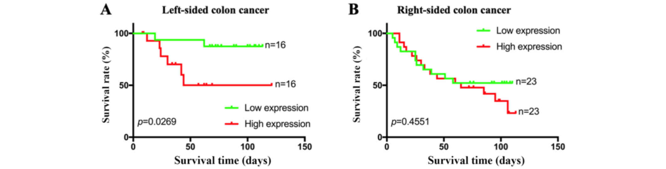
GNG4 is negatively associated with OS
in Chinese patients with LCC
When combining the mRNA expression levels of
GNG4 and the clinical data of patients in the microarray, it
was observed that both high GNG4 expression and low GNG4 expression
were more frequently present in patients with stage II colorectal
cancer, in both LCC and RCC (Table
III). Additionally, no associations between GNG4 status and
disease stage were identified in patients with LCC (P=0.4324) or in
patients with RCC (P=0.2717; Table
III). Survival analysis of the 32 patients with LCC indicated
that low expression levels of GNG4 were associated with a good
prognosis and high survival rate compared with high expression
levels of GNG4 (P=0.0269; Fig. 5A).
However, GNG4 expression status did not affect the prognosis of
patients with colorectal cancer with tumors located in the
right-sided colon (P=0.4551; Fig.
5B). Overall, GNG4 expression was only associated with the
prognosis of Chinese patients with LCC.
| Table III.Association between GNG4 expression
and pathological stage of LCC and RCC in Chinese patients. |
Table III.
Association between GNG4 expression
and pathological stage of LCC and RCC in Chinese patients.
|
| LCC | RCC |
|---|
|
|
|
|
|---|
| Stage | Low GNG4, n | High GNG4, n | Low GNG4, n | High GNG4, n |
|---|
| I | 1 | 1 | 2 | 3 |
| II | 13 | 10 | 18 | 13 |
| III | 2 | 5 | 3 | 7 |
| P-value |
| 0.4324 |
| 0.2717 |
Discussion
Genetic and immunological differences between the
proximal (right-sided) colon and the distal (left-sided) colorectum
have a strong impact on surgical and oncological outcomes, which
increases the difficulty of the management of patients with COAD
(21,22). Investigation of the diagnostic
markers for different anatomical sites (left or right) facilitates
personalized medicine and the treatment of colon cancer (23). The present study revealed the
potential diagnostic and prognostic values of GNG4 in LCC. To the
best of our knowledge, the present study is the first to illuminate
the genetic heterogeneity of GNG4 between LCC and RCC.
Based on sequencing and the Search Tool for the
Retrieval of Interacting Genes/Proteins database, GNG4 has been
demonstrated to be involved in the development of gastric cancer in
patients with H. pylori infection (24). In comparison with normal samples,
GNG4 is highly expressed in rectal adenocarcinoma samples; however,
there is no association between GNG4 expression and the survival
rate of patients with rectal cancer (25). In the present study, GNG4 was
upregulated in COAD samples, including both LCC and RCC samples.
The data indicated that the increase in GNG4 expression in COAD
samples was positively associated with tumors on both sides of the
colon. However, GNG4 expression was higher in patients with LCC
compared with that in patients with RCC. To the best of our
knowledge, the present study was the first to reveal the genetic
heterogeneity of GNG4 in colorectal cancer. Hypermethylated GNG4
has been identified in thymic carcinoma in relation to thymoma and
thymus, and is positively associated with poor relapse-free
survival rate in patients with all types of thymic epithelial tumor
(26). Aberrant DNA methylation of
GNG4, which is involved in cellular regulatory events, including
cell adhesion and signal transduction, can serve as a diagnostic
and therapeutic biomarker for bladder cancer (27). In the present study, GNG4 status was
only associated with disease stage and prognosis in patients with
LCC, but not in patients with RCC. These data suggested that GNG4
may be a biomarker for diagnosis and prognosis in patients with
LCC, which is partly consistent with the results from a previous
study (17). Due to the limited
sample size, this result should be further confirmed in large-scale
cohorts. In summary, GNG4 might be a good indicator for the
treatment of patients with LCC.
There are clear differences in genetic heterogeneity
and prognosis of colon cancer among different ethnicities. African
American (AA) patients with COAD have a poor prognosis compared
with Caucasian (CA) patients with COAD (28). Compared with that in Iranian patients
(a White population), hypermethylation of the glycoprotein nmb,
intercellular adhesion molecule 5 and chromodomain helicase DNA
binding protein 5 genes is identified in AA patients, and these are
the candidate cancer genes specifically involved in the progression
of COAD in the AA population (29).
The present study revealed that GNG4 was notably upregulated in
Chinese patients with COAD, and there was genetic heterogeneity of
GNG4 between LCC and RCC. In this study, most of the cohorts
downloaded from datasets from the TCGA dataset were of Caucasian
ethnicity. This finding indicated that abnormal GNG4 expression did
not differ between the Chinese study population and other CA
patients. Despite the limited sample size, the present study also
demonstrated that GNG4 was associated with the OS of patients with
LCC, whereas there was no association between disease stage and
GNG4 status in Chinese patients with COAD, including LCC and RCC.
The data in the microarray results were different from the database
results of Caucasian patients, implying that the clinical
application value of GNG4 differs depending on geographical
location. More subjects of different ethnicities are required to
investigate the diagnostic and prognostic role of GNG4 in COAD,
particularly in patients with LCC.
GNG4, c-Myc, DNA polymerase α1, catalytic subunit
and ribonucleotide reductase catalytic subunit M1 could serve as
prognostic factors for the response to treatment in patients with
locally advanced rectal cancer (30). Whether there is a association between
GNG4 and drug-sensitivity during the treatment of patients with LCC
remains unclear and requires more comprehensive analysis.
Mechanistically, forced expression of GNG4 inhibits
GBM cell migration by decreasing the activation of ERK and JNK via
stromal cell-derived factor 1α/C-X-C motif chemokine receptor
4-dependent chemokine signaling (31). Depletion of PSMC3-interacting protein
represses cell viability and xenograft tumorigenesis of
hepatocellular carcinoma (HCC) cells via upregulation of GNG4
(32). Exogenous GNG4 decreases cell
proliferation in renal cell carcinoma by affecting hypoxic response
signaling pathways (33). These
studies suggest the inhibitory effect of GNG4 on tumorigenesis in
GBM, HCC and renal cell carcinoma. Protein-protein interaction
analysis of different genes in LCC and RCC has demonstrated that
GNG4 may be a hub gene at the core of the interaction network
(34). This implies that GNG4 is an
important molecular switch in the development of LCC.
However, further in vitro and in vivo
experiments are required to investigate the biological function of
GNG4 in colorectal cancer. Additionally, the sample volume of
Chinese patients in the present study was small and limited the
analysis on the clinical significance of GNG4 in patients with LCC
and RCC. Future studies with larger cohorts are needed for
verification of the findings in the present study.
In summary, high expression levels of GNG4 and
genetic heterogeneity of GNG4 between LCC and RCC were present in
both White and Chinese patients. However, GNG4 was only associated
with the OS of Chinese patients with LCC, while GNG4 status was
associated with the disease stage and prognosis of LCC in White
patients. GNG4 may be a good prognostic factor for patients with
LCC worldwide.
Supplementary Material
Supporting Data
Acknowledgements
Not applicable.
Funding
The present study was supported by a grant from the
Startup Fund for Scientific Research, Fujian Medical University
(no. 2017XQ1211).
Availability of data and materials
The datasets generated and/or analyzed during the
current study are available in the [GEPIA2] repository, [http://gepia2.cancer-pku.cn/#general].
Authors contributions
YC and JS conceived the idea and drafted the
manuscript. JS and JY analyzed the expression pattern of GNG4 in
LCC and RCC. RL, XC and LZ performed the statistical analysis. All
authors discussed the results, edited this manuscript, read and
approved the final version.
Ethics approval and consent to
participate
All experiments were approved by the Ethics
Committee of the Affiliated Cancer Hospital of Fujian Medical
University (Fuzhou, China).
Patient consent for publication
Not applicable.
Competing interests
The authors declare that they have no competing
interests.
Glossary
Abbreviations
Abbreviations:
|
LCC
|
left-sided colon cancer
|
|
RCC
|
right-sided colon cancer
|
|
GNG4
|
guanine nucleotide binding-protein γ
subunit 4
|
|
GEPIA
|
Gene Expression Profiling Interactive
Analysis
|
|
TCGA
|
The Cancer Genome Atlas
|
|
COAD
|
colon adenocarcinoma
|
|
GBM
|
glioblastoma
|
|
HCC
|
hepatocellular carcinoma
|
References
|
1
|
Siegel RL, Miller KD, Fedewa SA, Ahnen DJ,
Meester RGS, Barzi A and Jemal A: Colorectal cancer statistics,
2017. CA Cancer J Clin. 67:177–193. 2017. View Article : Google Scholar : PubMed/NCBI
|
|
2
|
Siegel RL, Miller KD and Jemal A: Cancer
statistics, 2020. CA Cancer J Clin. 70:7–30. 2020. View Article : Google Scholar : PubMed/NCBI
|
|
3
|
Dallas NA, Xia L, Fan F, Gray MJ, Gaur P,
van Buren G II, Samuel S, Kim MP, Lim SJ and Ellis LM:
Chemoresistant colorectal cancer cells, the cancer stem cell
phenotype, and increased sensitivity to insulin-like growth
factor-I receptor inhibition. Cancer Res. 69:1951–1957. 2009.
View Article : Google Scholar : PubMed/NCBI
|
|
4
|
Aoyagi T, Terracina KP, Raza A and Takabe
K: Current treatment options for colon cancer peritoneal
carcinomatosis. World J Gastroentero. 20:12493–12500. 2014.
View Article : Google Scholar
|
|
5
|
Brulé SY, Jonker DJ, Karapetis CS,
OCallaghan CJ, Moore MJ, Wong R, Tebbutt NC, Underhill C, Yip D,
Zalcberg JR, et al: Location of colon cancer (right-sided versus
left-sided) as a prognostic factor and a predictor of benefit from
cetuximab in NCIC CO.17. Eur J Cancer. 51:1405–1414. 2015.
View Article : Google Scholar : PubMed/NCBI
|
|
6
|
Nitsche U, Stögbauer F, Späth C, Haller B,
Wilhelm D, Friess H and Bader FG: Right sided colon cancer as a
distinct histopathological subtype with reduced prognosis. Digest
Surg. 33:157–163. 2016. View Article : Google Scholar
|
|
7
|
Xiang L, Zhan Q, Zhao XH, Wang YD, An SL,
Xu YZ, Li AM, Gong W, Bai Y, Zhi FC and Liu SD: Risk factors
associated with missed colorectal flat adenoma: A multicenter
retrospective tandem colonoscopy study. World J Gastroentero.
20:10927–10937. 2014. View Article : Google Scholar
|
|
8
|
Weiss JM, Pfau PR, OConnor ES, King J,
LoConte N, Kennedy G and Smith MA: Mortality by stage for right-
versus left-sided colon cancer: Analysis of surveillance,
epidemiology, and end results-medicare data. J Clin Oncol.
29:4401–4409. 2011. View Article : Google Scholar : PubMed/NCBI
|
|
9
|
Yahagi M, Okabayashi K, Hasegawa H,
Tsuruta M and Kitagawa Y: The worse prognosis of right-sided
compared with left-sided colon cancers: A systematic review and
meta-analysis. J Gastrointest Surg. 20:648–655. 2016. View Article : Google Scholar : PubMed/NCBI
|
|
10
|
Petrelli F, Tomasello G, Borgonovo K,
Ghidini M, Turati L, Dallera P, Passalacqua R, Sgroi G and Barni S:
Prognostic survival associated with left-sided vs. right-sided
colon cancer: A systematic review and meta-analysis. JAMA Oncol.
3:211–219. 2017. View Article : Google Scholar : PubMed/NCBI
|
|
11
|
Lee GH, Malietzis G, Askari A, Bernardo D,
Al-Hassi HO and Clark SK: Is right-sided colon cancer different to
left-sided colorectal cancer?-A systematic review. Eur J Surg
Oncol. 41:300–308. 2015. View Article : Google Scholar : PubMed/NCBI
|
|
12
|
Slattery ML, Pellatt DF, Mullany LE, Wolff
RK and Herrick JS: Gene expression in colon cancer: A focus on
tumor site and molecular phenotype. Genes Chromosomes Cancer.
54:527–541. 2015. View Article : Google Scholar : PubMed/NCBI
|
|
13
|
Kalyanaraman S, Copeland NG, Gilbert DG,
Jenkins NA and Gautam N: Structure and chromosomal localization of
mouse G protein subunit gamma 4 gene. Genomics. 49:147–151. 1998.
View Article : Google Scholar : PubMed/NCBI
|
|
14
|
Bonham LW, Evans DS, Liu Y, Cummings SR,
Yaffe K and Yokoyama JS: Neurotransmitter pathway genes in
cognitive decline during aging: Evidence for GNG4 and KCNQ2 Genes.
Am J Alzheimers Dis Other Demen. 33:153–165. 2018. View Article : Google Scholar : PubMed/NCBI
|
|
15
|
Shukla S, Pia Patric IR, Thinagararjan S,
Srinivasan S, Mondal B, Hegde AS, Chandramouli BA, Santosh V,
Arivazhagan A and Somasundaram K: A DNA methylation prognostic
signature of glioblastoma: Identification of NPTX2-PTEN-NF-κB
nexus. Cancer Res. 73:6563–6573. 2013. View Article : Google Scholar : PubMed/NCBI
|
|
16
|
Liao JQ, Chen Z, He QH, Liu YM and Wang J:
Differential gene expression analysis and network construction of
recurrent cardiovascular events. Mol Med Rep. 13:1746–1764. 2016.
View Article : Google Scholar : PubMed/NCBI
|
|
17
|
Yang W, Ma J, Zhou W, Li Z, Zhou X, Cao B,
Zhang Y, Liu J, Yang Z, Zhang H, et al: Identification of hub genes
and outcome in colon cancer based on bioinformatics analysis.
Cancer Manag Res. 11:323–338. 2019. View Article : Google Scholar : PubMed/NCBI
|
|
18
|
Chen L, Lu D, Sun K, Xu Y, Hu P, Li X and
Xu F: Identification of biomarkers associated with diagnosis and
prognosis of colorectal cancer patients based on integrated
bioinformatics analysis. Gene. 692:119–125. 2019. View Article : Google Scholar : PubMed/NCBI
|
|
19
|
Livak KJ and Schmittgen TD: Analysis of
relative gene expression data using real-time quantitative PCR and
the 2(-Delta Delta C(T)) method. Methods. 25:402–408. 2001.
View Article : Google Scholar : PubMed/NCBI
|
|
20
|
Tang ZF, Li CW, Kang BX, Gao G, Li C and
Zhang ZM: GEPIA: A web server for cancer and normal gene expression
profiling and interactive analyses. Nucleic Acids Res. 45:W98–W102.
2017. View Article : Google Scholar : PubMed/NCBI
|
|
21
|
Bufill JA: Colorectal cancer: Evidence for
distinct genetic categories based on proximal or distal tumor
location. Ann Intern Med. 113:779–788. 1990. View Article : Google Scholar : PubMed/NCBI
|
|
22
|
Hu W, Yang Y, Li X, Huang M, Xu F, Ge W,
Zhang S and Zheng S: Multi-omics approach reveals distinct
differences in left-and right-sided colon cancer. Mol Cancer Res.
16:476–485. 2018. View Article : Google Scholar : PubMed/NCBI
|
|
23
|
Gervaz P, Bucher P and Morel P: Two
colons-two cancers: Paradigm shift and clinical implications. J
Surg Oncol. 88:261–266. 2004. View Article : Google Scholar : PubMed/NCBI
|
|
24
|
Chu AN, Liu JW, Yuan Y and Gong YH:
Comprehensive Analysis of Aberrantly Expressed ceRNA network in
gastric cancer with and without H. pylori infection. J
Cancer. 10:853–863. 2019. View Article : Google Scholar : PubMed/NCBI
|
|
25
|
Liu BX, Huang GJ and Cheng HB:
Comprehensive analysis of core genes and potential mechanisms in
rectal cancer. J Comput Biol. 26:1262–1277. 2019. View Article : Google Scholar : PubMed/NCBI
|
|
26
|
Kishibuchi R, Kondo K, Soejima S, Tsuboi
M, Kajiura K, Kawakami Y, Kawakita N, Sawada T, Toba H, Yoshida M,
et al: DNA methylation of GHSR, GNG4, HOXD9 and SALL3 is a common
epigenetic alteration in thymic carcinoma. Int J Oncol. 56:315–326.
2020.PubMed/NCBI
|
|
27
|
Zhang Y, Fang L, Zang Y and Xu Z:
Identification of core genes and key pathways via integrated
analysis of gene expression and DNA methylation profiles in bladder
cancer. Med Sci Monit. 24:3024–3033. 2018. View Article : Google Scholar : PubMed/NCBI
|
|
28
|
Govindarajan R, Posey J, Chao CY, Lu R,
Jadhav T, Javed AY, Javed A, Mahmoud FA, Osarogiagbon RU and Manne
U: A comparison of 12-gene colon cancer assay gene expression in
African American and Caucasian patients with stage II colon cancer.
BMC Cancer. 16:3682016. View Article : Google Scholar : PubMed/NCBI
|
|
29
|
Mokarram P, Kumar K, Brim H,
Naghibalhossaini F, Saberi-firoozi M, Nouraie M, Green R, Lee E,
Smoot DT and Ashktorab H: Distinct high-profile methylated genes in
colorectal cancer. PLoS One. 4:e70122009. View Article : Google Scholar : PubMed/NCBI
|
|
30
|
Palma P, Cano C, Conde-Muiño R, Comino A,
Bueno P, Ferrón JA and Cuadros M: Expression profiling of rectal
tumors defines response to neoadjuvant treatment related genes.
PLoS One. 9:e1121892014. View Article : Google Scholar : PubMed/NCBI
|
|
31
|
Pal J, Patil V, Mondal B, Shukla S, Hegde
AS, Arivazhagan A, Santosh V and Somasundaram K: Epigenetically
silenced GNG4 inhibits SDF1α/CXCR4 signaling in mesenchymal
glioblastoma. Genes Cancer. 7:136–147. 2016. View Article : Google Scholar : PubMed/NCBI
|
|
32
|
Ding JL, Li Y, Fan HX, Xu W, Gao R, Bai S,
Zhu Z, Yang W, Gong Y, Yang J and Zhou J: Knockdown of PSMC3IP
suppresses the proliferation and xenografted tumorigenesis of
hepatocellular carcinoma cell. J Cell Biochem. 120:5449–5458. 2019.
View Article : Google Scholar : PubMed/NCBI
|
|
33
|
Maina EN, Morris MR, Zatyka M, Raval RR,
Banks RE, Richards FM, Johnson CM and Maher ER: Identification of
novel VHL target genes and relationship to hypoxic response
pathways. Oncogene. 24:4549–4558. 2005. View Article : Google Scholar : PubMed/NCBI
|
|
34
|
Liang L, Zeng JH, Qin XG, Chen JQ, Luo DZ
and Chen G: Distinguishable prognostic signatures of left- and
right-sided colon cancer: A study based on sequencing data. Cell
Physiol Biochem. 48:475–490. 2018. View Article : Google Scholar : PubMed/NCBI
|