Introduction
Lung cancer is one of the leading causes of
cancer-related deaths globally, 1.8 million deaths occurred in
2018, representing close to 1 in 5 (18.4%) cancer deaths (1). Non-small-cell lung cancer (NSCLC)
accounts for 80–90% of lung cancer cases (2). Surgical treatment is recommended for
patients with early-stage lung cancer, whereas chemotherapy is
preferred in patients with advanced or recurrent disease (3,4).
Although multimodal therapy for NSCLC has exhibited progress in
recent years, the overall 5-year survival rate for advanced NSCLC
is only 4% (1,5). Previous studies have demonstrated the
treatment efficacy of an immune checkpoint inhibitor (ICI),
nivolumab, developed as an anti-programmed death-1 (PD-1) antibody,
for patients previously treated with one platinum-containing
regimen for NSCLC (6,7). Although ICIs such as nivolumab or
pembrolizumab serve a key role in systemic therapy for advanced
NSCLC, almost all non-responder patients receive no therapeutic
benefits from expensive ICIs, accompanied by immune-related adverse
events (8). Kurman and Murgu
(9) have reported accelerating
disease progression following the administration of ICIs in
patients with advanced NSCLC, referred to as hyper-progressive
disease. Therefore, established biomarkers to estimate the
sensitivity to ICIs are needed. A number of studies have reported
on useful biomarkers for predicting sensitivity to ICIs, including
programmed death ligand-1 (PD-L1) expression in tumor tissues,
tumor mutation burden and interferon-γ (IFN-γ) signature (10–12).
However, evaluating these factors requires sufficient tumor tissues
obtained through invasive procedures. Therefore, novel, less
invasive diagnostic tools that can predict the sensitivity to ICIs
are necessary.
The mechanisms of resistance to ICIs have attracted
attention from oncologists to improve treatment efficacy. One of
the most validated and characterized mechanisms is the
downregulation of antigen presentation. Genetic alterations in
β-2-microglobulin, an essential component of the major
histocompatibility complex (MHC) class I antigen presentation, have
been identified in progressing lesions in patients who exhibited an
initial response to PD-1 blockade (13). Loss of human leukocyte antigen genes
has been observed in patients with NSCLC, resulting in a loss of
MHC class I presentation, which is associated with resistance to
ICI (14). Recruitment and
activation of immune-suppressive cells, including myeloid-derived
suppressor cells, and regulatory T cells also induce resistance to
ICIs (15,16). Epithelial-mesenchymal transition
(EMT) is another mechanism of resistance to ICIs and is an
important regulatory mechanism of cancer invasion, metastasis,
therapeutic resistance and poor prognosis (17). EMT directly induces the expression of
PD-L1 and other checkpoint molecules that inhibit T cell-mediated
cytotoxicity against cancer cells (18,19).
Tumor cells undergoing EMT also express immune-suppressive
cytokines, such as transforming growth factor (TGF)-β, and enhanced
recruitment of immune-suppressive cells (20). Therefore, EMT is considered to be a
resistance factor to ICIs and a target mechanism that may improve
the efficacy of ICIs.
Plastin-3 (PLS3) encodes an actin-bundling protein
that facilitates cofilin-mediated polymerization of actin fibers
(21,22). PLS3 is associated with
EMT-induced aggressive phenotypes and is expressed in whole solid
cancers, but is not present in circulation, including whole blood
cells (23). However, in the
aforementioned study, PLS3 expression was identified as a
novel marker for circulating tumor cells (CTCs) with EMT phenotypes
in colorectal cancer, and the results demonstrated the usability of
PLS3 detection in peripheral blood as a marker for
EMT-induced CTCs and poor prognosis using PCR-based analyses
(23). By contrast, the clinical
significance of PLS3 protein concentration in the peripheral blood
measured using ELISA, which is one of the most popular diagnostic
tools in the clinic, has not been determined. In addition, whether
PLS3 detection in blood samples may be associated with sensitivity
to nivolumab treatment and prognosis in patients with NSCLC remains
unknown.
The aim of the present study was to determine the
usability of plasma PLS3 protein levels as a tumor marker of NSCLC
and a predictive marker of sensitivity to nivolumab treatment.
Materials and methods
Patients
For ELISA analysis, a total of 33 patients with
recurrent or advanced NSCLC who were treated with nivolumab at
Gunma University Hospital (Maebashi, Japan) and Hidaka Hospital
(Takasaki, Japan) between February 2016 and February 2017 were
included in this retrospective study. The mean age was 66.4 years
(age range, 47–82 years); 25 patients were male and 8 were female.
The inclusion criteria were as follows: i) Pathologically diagnosed
NSCLC; ii) recurrent or advanced NSCLC; iii) candidates for
nivolumab treatment after initial chemotherapy; and iv) performance
status based on the Eastern Cooperative Oncology Group guidelines
corresponding to 0–2. Blood samples and medical records of patients
included in a previous study (24)
were evaluated. Response Evaluation Criteria In Solid Tumors
(RECIST) version 1.1 was used to discriminate between partial
response (PR) and stable disease (SD) or progressive disease (PD)
(25). The follow-up duration for
censored cases ranged between 2.9 and 50.7 months (median, 15.8
months). The patients provided written informed consent. The
present study conformed to the Declaration of Helsinki and was
approved by the Institutional Review Board for Clinical Research at
the Gunma University Hospital (approval no. 1404) and Hidaka
Hospital (approval no. 124). For PLS3 immunohistostaining, a total
of 34 patients including five patients in the aforementioned cohort
and another 29 patients with NSCLC who underwent surgical resection
at Gunma University Hospital between January 2001 and December 2007
were included. The study of these 29 patients was also approved by
the Institutional Review Board for Clinical Research at the Gunma
University Hospital (approval no. 150044), and patient consent was
obtained using the opt-out method. The mean age was 66.4 years (age
range, 45–84 years); 20 patients were male and 14 were female.
ELISA
Peripheral blood was collected from each patient
with NSCLC before treatment with nivolumab. Plasma was obtained by
centrifugation at 1,689 × g for 10 min at room temperature and
stored at −80°C. Plasma concentrations of PLS3 were determined
using a Human Plastin 3 (PLS3) ELISA kit (cat. no. MBS4503796;
MyBioSource, Inc.) ELISA according to the manufacturer's
instructions. In addition, 10 anonymized plasma samples collected
from healthy volunteers were used. All healthy volunteers provided
informed consent, and the use of blood samples obtained from
healthy volunteers was approved by the Institutional Review Board
for Clinical Research at the Gunma University Hospital (approval
no. HS2019-271). All samples were examined in duplicate. The
dose-response curves for standards were obtained by 4-parameter
curve fitting using SkanIt Software 4.1 for Microplate Readers
(Thermo Fisher Scientific, Inc.).
Immunohistochemistry
Immunohistostaining of PLS3 was performed using
surgically resected lung cancer specimens to evaluate the
expression levels of PLS3 in lung cancer tissues. In total, tissues
from 34 patients (27 with adenocarcinoma and 7 with squamous cell
carcinoma) were evaluated for PLS3 expression using
immunohistochemistry. Subsequently, immunohistostaining of PD-L1,
CD8, and forkhead box protein P3 (Foxp3) was performed in the ELISA
cohort. Among 33 patients in the ELISA cohort, serial sections
consisting of the resected specimens (n=14) and needle biopsies
(n=8) from patients with NSCLC were assessed for the expression of
PD-L1 and CD8, only for those cases in whom we could obtain the
clinical samples. For Foxp3 staining, 21 available samples in the
ELISA cohort were evaluated. For immunohistochemistry, 4-µm
sections were cut from the paraffin blocks of each sample. Each
section was mounted on a silane-coated glass slide, deparaffinized
in xylene, rehydrated in 100% ethanol and incubated for 30 min at
room temperature in 0.3% hydrogen peroxide to block endogenous
peroxidase activity. After rehydration through a graded series of
the ethanol treatments (90% for 1 min, 80% for 1 min, and 60% for 1
min), the sections for PLS3 staining were subjected to antigen
retrieval by heating in citrate buffer (pH 6.0) at 120°C for 20 min
in an autoclave. The sections for CD8 and Foxp3 staining were
heated in boiling water using Immunosaver (Nisshin EM Co., Ltd.)
for 45 min at 98–100°C for antigen retrieval. For PD-L1 staining,
Universal HIER antigen retrieval reagent (cat. no. ab208572; Abcam)
was used at 120°C for 20 min in an autoclave. Non-specific binding
sites were blocked by incubating the sections with Protein Block
Serum-Free (Dako; Agilent Technologies, Inc.) for 30 min at room
temperature. The samples were incubated overnight at 4°C with the
following primary antibodies: PLS3 (cat. no. sc-166208; 1:200;
Santa Cruz Biotechnology, Inc.), PD-L1 (E1L3N Rabbit mAb 1:200;
cat. no. 13684; Cell signaling Technology, Inc.), CD8 (cat. no.
ab4055; 1:1,000; Abcam) and Foxp3 (cat. no. ab20034; 1:200; Abcam).
The primary antibody was visualized by the Histofine Simple Stain
MAX-PO (Multi) Kit (Nichirei Biosciences, Inc.). Chromogen
3,3-diaminobenzidine tetrahydrochloride was used as a 0.02%
solution in 50 mM ammonium acetate-citrate acid buffer (pH 6.0)
containing 0.005% hydrogen peroxide. The sections were lightly
counterstained with hematoxylin and mounted.
The tissue sections were examined by two independent
evaluators who were blinded to the patient data. For evaluation of
the PLS3 expression, the staining intensity was scored as follows:
0, none; 1, weak; 2, moderate; or 3, strong. Scores 1–3 were
defined as positive PLS3 expression. The expression of PD-L1 was
evaluated using a semiquantitative scoring method according to the
percentage of stained cells: 1, ≤1; 2, 1–5; 3, 5–10; 4, 10–50; and
5, ≥50%. Tumors with a score >3 were graded as positive. CD8 and
Foxp3 were semi-quantitatively evaluated based on the extent of
positive lymphocytes infiltrating the tumor specimens, and patients
with >5% positive lymphocytes were defined as positive for CD8
and Foxp3 based on a previous study (24,26).
Statistical analysis
The data were presented as the mean ± standard
deviation or median and the interquartile range (IQR). Statistical
analyses were performed using the Mann-Whitney U test for
continuous variables and the χ2 test for categorical
variables. Kaplan-Meier curves were generated for overall survival,
and statistical significance was examined using the log-rank test.
Univariate analyses were performed using logistic regression
analysis or Cox's proportional hazards model. Receiver operating
characteristic (ROC) curve analyses were used to evaluate the
potential of plasma PLS3 concentration as a cancer diagnostic
marker, predictor of sensitivity to nivolumab and a marker of poor
prognosis. All statistical analyses were performed using JMP
software (SAS Institute, Inc.). A two-tailed P<0.05 was
considered to indicate a statistically significant difference.
Results
Immunohistochemistry of PLS3
expression
Prior to the ELISA analyses, immunohistochemical
analysis of PLS3 expression was performed using 34 surgically
resected NSCLC specimens to evaluate the expression levels of PLS3
in lung cancer tissues. Fig. S1
demonstrates the representative immunohistochemical staining of
PLS3 in negative (Fig. S1A) and
positive (Fig. S1B) cases. The
expression of the PLS3 protein was predominantly observed in the
cytoplasm. The rate of positive PLS3 expression was 88.2% (31/34
patients).
Plasma PLS3 levels as a diagnostic
marker of cancer
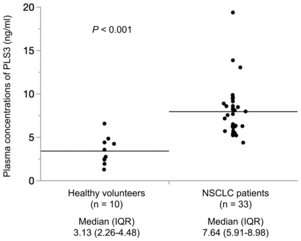
In total, 10 healthy volunteers (8 male and 2
female) and 33 patients with recurrent or advanced NSCLC were
enrolled in the present study. The mean age of the healthy
volunteers and patients with NSCLC was 33.7±3.6 and 66.4±9.1 years,
respectively (P<0.001). Plasma PLS3 concentrations in healthy
volunteers and patients with NSCLC are presented in Fig. 1. The median plasma PLS3 concentration
was significantly higher in patients with NSCLC compared with that
in healthy volunteers (median 7.64 ng/ml vs. 3.13 ng/ml,
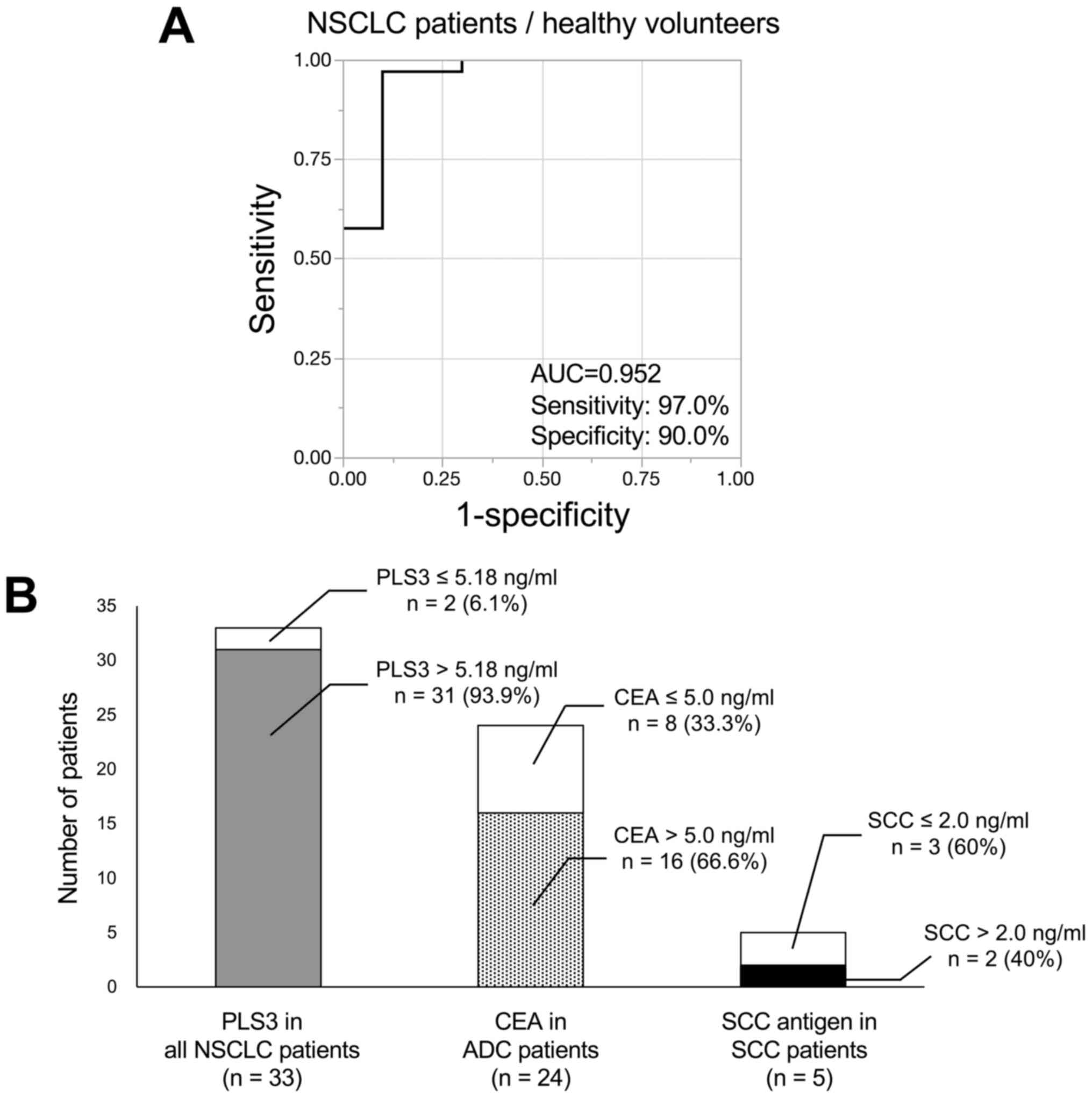
P<0.001). The diagnostic value of plasma PLS3 concentration to
discriminate between patients with NSCLC and healthy volunteers was
next determined. ROC analysis for plasma PLS3 concentration
revealed that the optimal cut-off value for patients with NSCLC was
5.18 ng/ml [area under the curve (AUC), 0.952; sensitivity, 97.0%;
specificity, 90.0%; Fig. 2A). Using
this cut-off value, 31 patients with NSCLC (93.9%) had PLS3
>5.18 ng/ml (Fig. 2B). The
positivity rate of PLS3 in all patients with NSCLC was higher
compared with that of CEA in patients with adenocarcinoma (66.6%)
or SCC antigen in patients with squamous cell carcinoma (40%).
Plasma PLS3 levels and sensitivity to
nivolumab treatment
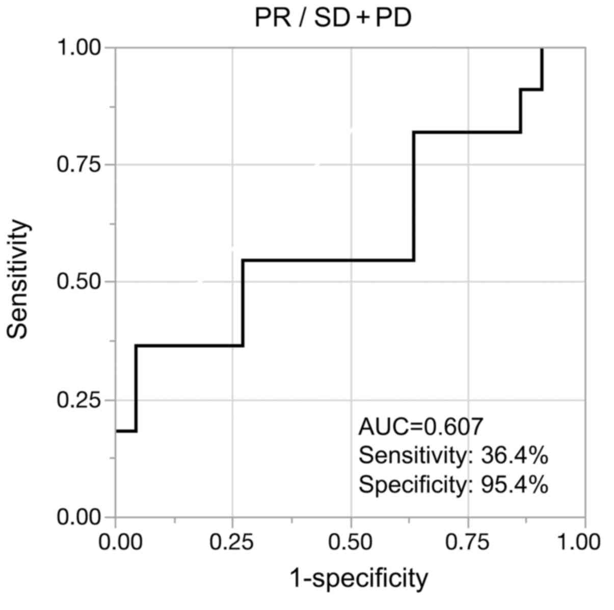
The association between plasma PLS3 concentration
and the sensitivity to nivolumab treatment was analyzed in patients
with NSCLC. Fig. 3 demonstrates the
results of the ROC analysis of plasma PLS3 concentration to
discriminate between PR and SD or PD according to the RECIST. The
optimal cut-off value for PR was 5.43 ng/ml (AUC, 0.607;
sensitivity, 36.4%; specificity, 95.4%). Using this cut-off value,
the rate of PR in the PLS3 ≤5.43 ng/ml group was significantly
higher compared with that in the PLS3 >5.43 ng/ml group
(P=0.016; Table I). In addition,
seven variables, including tumoral PD-L1, CD8 and PLS3
concentration, were evaluated as potential predictive factors of
PR; however, univariate analysis indicated that PLS3 ≤5.43 ng/ml
was the sole predictor of PR (odds ratio, 0.08; 95% CI, 0.01–0.68;
P=0.019; Table II).
 | Table I.Association between plasma PLS3
concentration and sensitivity to nivolumab treatment. |
Table I.
Association between plasma PLS3
concentration and sensitivity to nivolumab treatment.
| Group | PR (n=11) | SD + PD (n=22) | P-value |
|---|
| PLS3 ≤5.43
ng/ml | 4 | 1 | 0.016a |
| PLS3 >5.43
ng/ml | 7 | 21 |
|
| Table II.Univariate analysis of the predictors
associated with nivolumab treatment efficacy. |
Table II.
Univariate analysis of the predictors
associated with nivolumab treatment efficacy.
|
| Univariate
analysis |
|---|
|
|
|
|---|
| Clinicopathological
characteristic | OR | 95% CI | P-value |
|---|
| Age, years (≤65 vs.
>65) | 1.46 | 0.34–6.95 | 0.617 |
| Sex (male vs.
female) | 0.21 | 0.01–1.48 | 0.127 |
| Histology (ADC vs.
SCC) | 0.76 | 0.09–4.34 | 0.761 |
| PD-L1 expression
(negative vs. positive) | 1.25 | 0.21–8.13 | 0.805 |
| CD8 expression
(negative vs. positive) | 2.55 | 0.29–55.41 | 0.417 |
| Recurrent disease
(no vs. yes) | 0.69 | 0.15–3.06 | 0.616 |
| Plasma PLS3
concentration, ng/ml (≤5.43 vs. >5.43) | 0.08 | 0.01–0.68 | 0.019a |
Plasma PLS3 levels and overall
survival
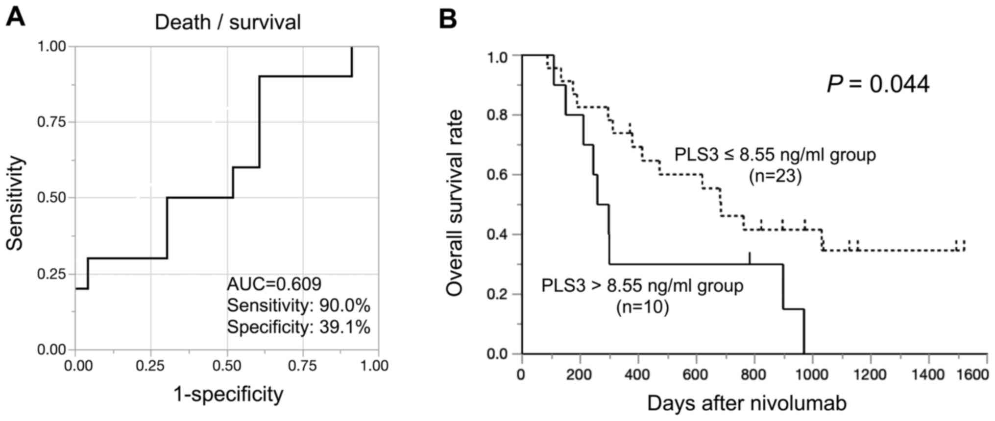
The prognostic value of the plasma PLS3
concentration was evaluated in patients with NSCLC treated with
nivolumab. ROC analysis of plasma PLS3 concentration revealed that
the optimal cut-off value for mortality was 8.55 ng/ml (AUC,0.609;
sensitivity, 90.0%; specificity, 39.1%; Fig. 4A). The patients were divided into
PLS3 ≤8.55 and >8.55 ng/ml groups according to this cut-off
point. The associations between clinicopathological characteristics
and PLS3 concentration in patients with NSCLC are presented in
Table III; no significant
differences in clinicopathological characteristics were identified
between the PLS3 ≤8.55 and >8.55 ng/ml groups. The prognostic
analysis of patients with NSCLC according to PLS3 concentration
demonstrated that the patients in the PLS3 >8.55 ng/ml group
exhibited a poorer prognosis compared with those in the PLS3 ≤8.55
ng/ml group (Fig. 4B). Univariate
analysis revealed that the high plasma PLS3 concentration was the
sole predictor of poor survival (hazard ratio, 2.40; 95% CI,
1.01–5.67; P=0.047; Table IV). The
prognostic values of the other two cut-off point (5.18 ng/ml used
for diagnosis and 5.43 ng/ml used for treatment sensitivity) are
presented in Fig. S2. Using the
cut-off values of 5.18 and 5.43 ng/ml, no significant differences
were observed in patient overall survival rates according to the
PLS3 concentration (P=0.214 and 0.306, respectively).
| Table III.Associations between
clinicopathological characteristics and plasma PLS3 concentrations
in patients with non-small cell lung cancer treated with
nivolumab. |
Table III.
Associations between
clinicopathological characteristics and plasma PLS3 concentrations
in patients with non-small cell lung cancer treated with
nivolumab.
| Clinicopathological
characteristic | PLS3 ≤8.55 ng/ml
(n=23) | PLS3 >8.55 ng/ml
(n=10) | P-value |
|---|
| Age, years, mean ±
SD | 67.3±8.6 | 64.2±10.3 | 0.377 |
| Sex |
|
|
|
|
Male | 18 | 7 | 0.612 |
|
Female | 5 | 3 |
|
| Histology |
|
|
|
|
SCC | 6 | 1 | 0.299 |
|
ADC | 17 | 9 |
|
| PD-L1 |
|
|
|
|
Negative | 5 | 4 | 0.512 |
|
Positive | 9 | 4 |
|
| CD8 |
|
|
|
|
Negative | 2 | 3 | 0.211 |
|
Positive | 12 | 5 |
|
| Foxp3 |
|
|
|
|
Negative | 2 | 4 | 0.163 |
|
Positive | 10 | 5 |
|
| Driver
mutation |
|
|
|
|
EGFR | 2 | 1 | 0.364 |
|
ALK | 0 | 1 |
|
|
Wild-type | 15 | 7 |
|
|
Unknown | 6 | 1 |
|
| Recurrent
disease |
|
|
|
| No | 8 | 5 | 0.411 |
|
Yes | 15 | 5 |
|
| Number of previous
regimens, mean ± SD | 2.7±2.3 | 3.2±1.9 | 0.419 |
| Nivolumab
administrations, mean ± SD | 17.5±20.4 | 9.2±10.1 | 0.215 |
| irAE |
|
|
|
|
Negative | 19 | 7 | 0.416 |
|
Positive | 4 | 3 |
|
| Therapeutic
effect |
|
|
|
| PR | 9 | 2 | 0.284 |
| SD +
PD | 14 | 8 |
|
| Reason for
treatment discontinuation |
|
|
|
| Disease
progression | 15 | 7 | 0.448 |
|
irAE | 3 | 0 |
|
|
Decreased PS | 3 | 0 |
|
| Death
due to AMI | 0 | 1 |
|
| Long
stable disease | 1 | 0 |
|
|
Treatment continuation | 1 | 0 |
|
| Table IV.Univariate analysis of
clinicopathological factors affecting overall survival. |
Table IV.
Univariate analysis of
clinicopathological factors affecting overall survival.
|
| Univariate
analysis |
|---|
|
|
|
|---|
| Clinicopathological
characteristics | HR | 95% CI | P-value |
|---|
| Age, years (≤65 vs.
>65) | 0.60 | 0.26–1.37 | 0.224 |
| Sex (male vs.
female) | 2.07 | 0.81–5.27 | 0.128 |
| Histology (ADC vs.
SCC) | 1.58 | 0.57–4.38 | 0.380 |
| PD-L1 expression
(negative vs. positive) | 0.72 | 0.26–2.02 | 0.535 |
| CD8 expression
(negative vs. positive) | 0.74 | 0.24–2.30 | 0.602 |
| Recurrent disease
(no vs. yes) | 0.77 | 0.34–1.77 | 0.539 |
| Plasma PLS3
concentration, ng/ml (≤8.55 vs. >8.55) | 2.40 | 1.01–5.67 | 0.047a |
Discussion
The clinical usability of plasma PLS3 in patients
with NSCLC remains unknown. In addition, the identification of
novel, less invasive diagnostic tools that can predict the
sensitivity to ICIs are necessary. The present study first
demonstrated that the plasma PLS3 concentration in patients with
NSCLC was significantly higher compared with that in healthy
volunteers. In addition, low PLS3 concentration was associated with
PR according to the RECIST in patients with NSCLC treated with
nivolumab, with a cut-off value of 5.43 ng/ml. PLS3 concentration
≥8.55 ng/ml was the sole predictor of poor overall survival. To the
best of our knowledge, the present study is the first to indicate
the clinical significance of plasma PLS3 concentration in the
prediction of the diagnosis, prognosis and sensitivity to nivolumab
treatment in patients with NSCLC. Furthermore, the present study is
unique due to the use of ELISA to measure peripheral PLS3 protein
concentration in the plasma, which may enhance its clinical
utility.
Tumor markers are broadly used in screening,
diagnosis, monitoring and predicting therapeutic response and
prognosis in various types of cancer, such as CEA or SCC antigen in
NSCLC (27–30). Ideally, it is desirable that tumor
markers are detected only in patients with cancer and not in
healthy individuals. The present study focused on EMT-related
characteristics and unique expression profiles of PLS3 to examine
its potential as a novel tumor marker in the blood. Our previous
study validated the PLS3 expression profiles in several
cancer cell lines, including hematopoietic malignancies; as a
result, the PLS3 expression was detected in solid cancer
cell lines including esophageal cancer, gastric cancer, liver
tumor, pancreatic cancer, breast cancer, lung cancer, prostate
cancer and melanoma, but not in hematopoietic malignancies or blood
samples from healthy individuals (23). The results of the present study
demonstrated that plasma PLS3 protein concentration in patients
with NSCLC was significantly higher compared with that in healthy
volunteers. Further studies are warranted to clarify the clinical
significance of plasma PLS3 as a diagnostic marker in a broad range
of solid tumors.
Various studies have reported the predictive value
of ICI sensitivity markers, such as tumoral PD-L1 expression
(10), tumor mutation burden
(11) and IFN-γ gene signature
(12); however, for predicting the
sensitivity to nivolumab, established biomarkers that are obtained
via less invasive techniques are urgently needed. Recent studies
have reported that cancer cells undergoing EMT were resistant to
immunotherapy (20,31). In addition, Dodagatta-Marri et
al (32) have demonstrated that
the combination of anti-PD-1 and anti-TGF-β antibodies inhibit the
EMT signaling pathways in murine tumor models. Furthermore, PLS3
induces the EMT via the TGF-β signaling pathway, and PLS3
expression is also detectable in the EMT-induced CTCs (33,34). In
the present study, plasma PLS3, but not tumoral PD-L1, expression
was associated with sensitivity to nivolumab treatment, although
the origin of plasma PLS3 (EMT-induced CTCs, tumor mass or other
tissue) was unclear. A previous study has reported that EMT causes
therapeutic resistance to not only nivolumab, but also other
immunotherapies (20). Therefore,
the evaluation of plasma PLS3 may also be a useful predictive
marker of sensitivity to treatment with a range of ICIs, including
anti-cytotoxic T lymphocyte-associated protein 4 and anti-PD-L1
antibodies.
As aforementioned, EMT promotes tumor invasion,
metastasis, therapeutic resistance and poor prognosis, and PLS3 has
been reported as one of the regulators of EMT (17,23). Our
previous study demonstrated by PCR detection that the mRNA levels
of PLS3 in the tumor tissue were associated with cancer
progression and a poor prognosis in patients with colorectal cancer
(23). In addition, Ueo et al
(34) have demonstrated that
PLS3 evaluation in the peripheral blood by PCR-based
analysis is associated with a poor prognosis in patients with
breast cancer. However, whether the protein expression level of
PLS3 in blood samples using ELISA may predict therapeutic response
and cancer prognosis remained unknown. The results of the present
study demonstrated that the high plasma PLS3 concentration was
associated with nivolumab resistance and poor survival. PCR-based
gene expression analysis in the peripheral blood may require
complicated methods to extract the unstable total RNA from blood
samples. By contrast, ELISA-based analysis requires serum or plasma
samples obtained by standard collection tubes frequently used in
clinical practice. The evaluation of plasma PLS3 by ELISA may be
easier to use in clinical application compared with gene
expression-based analysis.
The present study had several limitations. First, a
retrospective design was used, and a small sample size was
included. Second, the mean age of healthy volunteers was different
compared with that of patients with NSCLC, which may have affected
the plasma PLS3 concentration among individuals. Third, there is
the possibility of false-positive cases, as plasma PLS3 may
originate from not only CTCs or primary tumors, but also healthy
solid tissues of the whole body. Fourth, only patients with
recurrent or advanced NSCLC who possessed large numbers of cancer
cells were enrolled; therefore, further studies in patients with
early-stage cancer who may possess relatively small numbers of
cancer cells are warranted to determine whether plasma PLS3 may be
useful as a diagnostic marker for cancer. Fifth, all patients in
the present study had history of previous chemotherapy treatment.
In Japan, nivolumab has been covered by the medical insurance
system for NSCLC as second-line therapy since December 2015, and at
present, a number of ICIs are available as first-line therapy
(4). The present study did not
include untreated patients with NSCLC; therefore, it remains
unclear whether plasma PLS3 may predict the sensitivity to
nivolumab treatment or prognosis as first-line therapy. Sixth, the
present study did not include functional experiments on the
relationship between plasma PLS3 expression and nivolumab using
in vitro or in vivo analyses.
In conclusion, the results of the present study
demonstrated the diagnostic value of plasma PLS3 concentration in
distinguishing patients with NSCLC from healthy volunteers. In
addition, the low pretreatment plasma PLS3 concentration was
associated with high responsiveness to nivolumab treatment, whereas
high plasma PLS3 concentration predicted poor overall survival in
patients with NSCLC treated with nivolumab. These observations
suggest that the evaluation of PLS3 concentration in plasma samples
may be used as a cancer diagnostic marker, predictor of sensitivity
to nivolumab and a marker of poor prognosis for patients with
NSCLC. Future prospective studies are required to confirm these
results and explore the use of PLS3 as a marker in other types of
cancer.
Supplementary Material
Supporting Data
Acknowledgements
The authors would like to thank Ms. Mariko Nakamura,
Ms. Yukie Saito, Ms. Sayaka Okada, Ms. Kayoko Takahashi, Ms. Mizuho
Murata, Ms. Harumi Kanai, Ms. Fumie Takada, Ms. Yukiko Suto and Ms.
Sawa Nagayama (Department of General Surgical Science, Graduate
School of Medicine, Gunma University, Gunma, Japan) for their
assistance.
Funding
This study was supported by Grants-in-Aid for
Scientific Research from the Japanese Society for the Promotion of
Science (grant nos. 17K19893 and 18K07665). The work was also
supported in part by the Research Grant of the Princess Takamatsu
Cancer Research Fund (grant no. 18-25034) and Suzuken Memorial
Foundation.
Availability of data and materials
The datasets used and or/analyzed during the current
study are available from the corresponding author on reasonable
request.
Authors' contributions
TYo designed the study and wrote the initial draft
of the manuscript. TYo, KS and HS contributed to data analysis and
interpretation and assisted in the preparation of the manuscript.
KKu, MS, NN, TYa, IN, HK and KKa contributed to data collection and
interpretation, critically reviewed the manuscript. All authors
read and approved the final manuscript.
Ethics approval and consent to
participate
This study conformed to the Declaration of Helsinki
and was approved by the Institutional Review Board for Clinical
Research at the Gunma University Hospital (Maebashi, Japan;
approval nos. 1404, 150044 and HS2019-271). Informed consent was
obtained from the participants included in the study.
Patient consent for publication
Not applicable.
Competing interests
Kyoichi Kaira has received research grants and a
speaker honorarium from Ono Pharmaceutical Company, Boehringer
Ingelheim, Chugai Pharmaceutical, Taiho Pharmaceutical, Eli Lilly
Japan and AstraZeneca. Department of Innovative Cancer
Immunotherapy, Gunma University is an endowment department,
supported with an unrestricted grant from Ono Pharmaceutical
Company, Chugai Pharmaceutical Company and Memolead Company. The
other authors declare that they have no competing interests.
Glossary
Abbreviations
Abbreviations:
|
PLS3
|
plastin-3
|
|
NSCLC
|
non-small-cell lung cancer
|
|
ICI
|
immune checkpoint inhibitor
|
|
PD-L1
|
programmed death ligand-1
|
|
MHC
|
major histocompatibility complex
|
|
EMT
|
epithelial-mesenchymal transition
|
|
CTCs
|
circulating tumor cells
|
|
PR
|
partial response
|
|
SD
|
stable disease
|
|
PD
|
progressive disease
|
|
Foxp3
|
forkhead box protein P3
|
|
ROC
|
receiver operating characteristic
|
References
|
1
|
Bray F, Ferlay J, Soerjomataram I, Siegel
RL, Torre LA and Jemal A: Global cancer statistics 2018: GLOBOCAN
estimates of incidence and mortality worldwide for 36 cancers in
185 countries. CA Cancer J Clin. 68:394–424. 2018. View Article : Google Scholar : PubMed/NCBI
|
|
2
|
Planchard D, Popat S, Kerr K, Novello S,
Smit EF, Faivre-Finn C, Mok TS, Reck M, Van Schil P, Hellmann MD,
et al: Metastatic non-small cell lung cancer: ESMO clinical
practice guidelines for diagnosis, treatment and follow-up. Ann
Oncol. 29 (Suppl 4):iv192–iv237. 2018. View Article : Google Scholar
|
|
3
|
Sawabata N, Miyaoka E, Asamura H,
Nakanishi Y, Eguchi K, Mori M, Nomori H, Fujii Y, Okumura M and
Yokoi K; Japanese Joint Committee for Lung Cancer Registration, :
Japanese lung cancer registry study of 11,663 surgical cases in
2004: Demographic and prognosis changes over decade. J Thorac
Oncol. 6:1229–1235. 2011. View Article : Google Scholar : PubMed/NCBI
|
|
4
|
Akamatsu H, Ninomiya K, Kenmotsu H, Morise
M, Daga H, Goto Y, Kozuki T, Miura S, Sasaki T, Tamiya A, et al:
The Japanese lung cancer society guideline for non-small cell lung
cancer, stage IV. Int J Clin Oncol. 24:731–770. 2019. View Article : Google Scholar : PubMed/NCBI
|
|
5
|
Siegel RL, Miller KD and Jemal A: Cancer
statistics, 2017. CA Cancer J Clin. 67:7–30. 2017. View Article : Google Scholar : PubMed/NCBI
|
|
6
|
Brahmer J, Reckamp KL, Baas P, Crinò L,
Eberhardt WEE, Poddubskaya E, Antonia S, Pluzanski A, Vokes EE,
Holgado E, et al: Nivolumab versus docetaxel in advanced
squamous-cell non-small-cell lung cancer. N Engl J Med.
373:123–135. 2015. View Article : Google Scholar : PubMed/NCBI
|
|
7
|
Borghaei H, Paz-Ares L, Horn L, Spigel DR,
Steins M, Ready NE, Chow LQ, Vokes EE, Felip E, Holgado E, et al:
Nivolumab versus docetaxel in advanced nonsquamous non-small-cell
lung cancer. N Engl J Med. 373:1627–1639. 2015. View Article : Google Scholar : PubMed/NCBI
|
|
8
|
Shields BD, Mahmoud F, Taylor EM, Byrum
SD, Sengupta D, Koss B, Baldini G, Ransom S, Cline K, Mackintosh
SG, et al: Indicators of responsiveness to immune checkpoint
inhibitors. Sci Rep. 7:8072017. View Article : Google Scholar : PubMed/NCBI
|
|
9
|
Kurman JS and Murgu SD: Hyperprogressive
disease in patients with non-small cell lung cancer on
immunotherapy. J Thorac Dis. 10:1124–1128. 2018. View Article : Google Scholar : PubMed/NCBI
|
|
10
|
Garon EB, Rizvi NA, Hui R, Leighl N,
Balmanoukian AS, Eder JP, Patnaik A, Aggarwal C, Gubens M, Horn L,
et al: Pembrolizumab for the treatment of non-small-cell lung
cancer. N Engl J Med. 372:2018–2028. 2015. View Article : Google Scholar : PubMed/NCBI
|
|
11
|
Hellmann MD, Ciuleanu TE, Pluzanski A, Lee
JS, Otterson GA, Audigier-Valette C, Minenza E, Linardou H, Burgers
S, Salman P, et al: Nivolumab plus ipilimumab in lung cancer with a
high tumor mutational burden. N Engl J Med. 378:2093–2104. 2018.
View Article : Google Scholar : PubMed/NCBI
|
|
12
|
Fehrenbacher L, Spira A, Ballinger M,
Kowanetz M, Vansteenkiste J, Mazieres J, Park K, Smith D,
Artal-Cortes A, Lewanski C, et al: Atezolizumab versus docetaxel
for patients with previously treated non-small-cell lung cancer
(POPLAR): A multicentre, open-label, phase 2 randomised controlled
trial. Lancet. 387:1837–1846. 2016. View Article : Google Scholar : PubMed/NCBI
|
|
13
|
Sade-Feldman M, Jiao YJ, Chen JH, Rooney
MS, Barzily-Rokni M, Eliane JP, Bjorgaard SL, Hammond MR, Vitzthum
H, Blackmon SM, et al: Resistance to checkpoint blockade therapy
through inactivation of antigen presentation. Nat Commun.
8:11362017. View Article : Google Scholar : PubMed/NCBI
|
|
14
|
McGranahan N, Rosenthal R, Hiley CT, Rowan
AJ, Watkins TBK, Wilson GA, Birkbak NJ, Veeriah S, Ven Loo P,
Herrero J, et al: Allele-specific HLA loss and immune escape in
lung cancer evolution. Cell. 171:1259–1271.e11. 2017. View Article : Google Scholar : PubMed/NCBI
|
|
15
|
Pitt JM, Vetizou M, Daillere R, Roberti
MP, Yamazaki T, Routy B, Lepage P, Boneca IG, Chamaillard M,
Kroemer G and Zitvogel L: Resistance mechanisms to
immune-checkpoint blockade in cancer: Tumor-intrinsic and
-extrinsic factors. Immunity. 44:1255–1269. 2016. View Article : Google Scholar : PubMed/NCBI
|
|
16
|
O'Donnell JS, Long GV, Scolyer RA, Teng MW
and Smyth MJ: Resistance to PD1/PDL1 checkpoint inhibition. Cancer
Treat Rev. 52:71–81. 2017. View Article : Google Scholar : PubMed/NCBI
|
|
17
|
Polyak K and Weinberg RA: Transitions
between epithelial and mesenchymal states: Acquisition of malignant
and stem cell traits. Nat Rev Cancer. 9:265–273. 2009. View Article : Google Scholar : PubMed/NCBI
|
|
18
|
Chen L, Gibbons DL, Goswami S, Cortez MA,
Ahn YH, Byers LA, Zhang X, Yi X, Dwyer D, Lin W, et al: Metastasis
is regulated via microRNA-200/ZEB1 axis control of tumour cell
PD-L1 expression and intratumoral immunosuppression. Nat Commun.
5:52412014. View Article : Google Scholar : PubMed/NCBI
|
|
19
|
Loi S, Dushyanthen S, Beavis PA, Salgado
R, Denkert C, Savas P, Combs S, Rimm DL, Giltnane JM, Estrada MV,
et al: RAS/MAPK Activation is associated with reduced
tumor-infiltrating lymphocytes in triple-negative breast cancer:
Therapeutic cooperation between MEK and PD-1/PD-L1 immune
checkpoint inhibitors. Clin Cancer Res. 22:1499–1509. 2016.
View Article : Google Scholar : PubMed/NCBI
|
|
20
|
Soundararajan R, Fradette JJ, Konen JM,
Moulder S, Zhang X, Gibbons DL, Varadarajan N, Wistuba II, Tripathy
D, Bernatchez C, et al: Targeting the interplay between
epithelial-to-mesenchymal-transition and the immune system for
effective immunotherapy. Cancers (Basel). 11:7142019. View Article : Google Scholar
|
|
21
|
Su MW, Dorocicz I, Dragowska WH, Ho V, Li
G, Voss N, Gascoyne R and Zhou Y: Aberrant expression of T-plastin
in sezary cells. Cancer Res. 63:7122–7127. 2003.PubMed/NCBI
|
|
22
|
Giganti A, Plastino J, Janji B, Van Troys
M, Lentz D, Ampe C, Sykes C and Friederichet E: Actin-filament
cross-linking protein T-plastin increases Arp2/3-mediated
actin-based movement. J Cell Sci. 118:1255–1265. 2005. View Article : Google Scholar : PubMed/NCBI
|
|
23
|
Yokobori T, Iinuma H, Shimamura T, Imoto
S, Sugimachi K, Ishii H, Iwatsuki M, Ota D, Ohkuma M, Iwaya T, et
al: Plastin3 is a novel marker for circulating tumor cells
undergoing the epithelial-mesenchymal transition and is associated
with colorectal cancer prognosis. Cancer Res. 73:2059–2069. 2013.
View Article : Google Scholar : PubMed/NCBI
|
|
24
|
Kaira K, Higuchi T, Naruse I, Arisaka Y,
Tokue A, Altan B, Suda S, Mogi A, Shimizu K, Sunaga N, et al:
Metabolic activity by 18F-FDG-PET/CT is predictive of
early response after nivolumab in previously treated NSCLC. Eur J
Nucl Med Mol Imaging. 45:56–66. 2018. View Article : Google Scholar : PubMed/NCBI
|
|
25
|
Eisenhauer EA, Therasse P, Bogaerts J,
Schwartz LH, Sargent D, Ford R, Dancey J, Arbuck S, Gwyther S,
Mooney M, et al: New response evaluation criteria in solid tumors:
Revised RECIST guideline (version 1.1). Eur J Cancer. 45:228–247.
2009. View Article : Google Scholar : PubMed/NCBI
|
|
26
|
Kurashige J, Yokobori T, Mima K, Sawada G,
Takahashi Y, Ueo H, Takano Y, Matsumura T, Uchi R, Eguchi H, et al:
Plastin3 is associated with epithelial-mesenchymal transition and
poor prognosis in gastric cancer. Oncol Lett. 17:2393–2399.
2019.PubMed/NCBI
|
|
27
|
Vargas AJ and Harris CC: Biomarker
development in the precision medicine era: Lung cancer as a case
study. Nat Rev Cancer. 16:525–537. 2016. View Article : Google Scholar : PubMed/NCBI
|
|
28
|
Grunnet M and Sorensen JB:
Carcinoembryonic antigen (CEA) as tumor marker in lung cancer. Lung
Cancer. 76:138–143. 2012. View Article : Google Scholar : PubMed/NCBI
|
|
29
|
De Stefano F, Chacon E, Turcios L, Marti F
and Gedaly R: Novel biomarkers in hepatocellular carcinoma. Dig
Liver Dis. 50:1115–1123. 2018. View Article : Google Scholar : PubMed/NCBI
|
|
30
|
Shimada H, Noie T, Ohashi M, Oba K and
Takahashi Y: Clinical significance of serum tumor markers for
gastric cancer: A systematic review of literature by the task force
of the Japanese gastric cancer association. Gastric Cancer.
17:26–33. 2014. View Article : Google Scholar : PubMed/NCBI
|
|
31
|
Shrestha R, Prithviraj P, Anaka M, Bridle
KR, Crawford DHG, Dhungel B, Steel JC and Jayachandran A:
Monitoring immune checkpoint regulators as predictive biomarkers in
hepatocellular carcinoma. Front Oncol. 8:2692018. View Article : Google Scholar : PubMed/NCBI
|
|
32
|
Dodagatta-Marri E, Meyer DS, Reeves MQ,
Paniagua R, To MD, Binnewies M, Broz ML, Mori H, Wu D, Adoumie M,
et al: α-PD-1 therapy elevates Treg/Th balance and increases tumor
cell pSmad3 that are both targeted by α-TGFβ antibody to promote
durable rejection and immunity in squamous cell carcinomas. J
Immunother Cancer. 7:622019. View Article : Google Scholar : PubMed/NCBI
|
|
33
|
Sugimachi K, Yokobori T, Iinuma H, Ueda M,
Ueo H, Shinden Y, Eguchi H, Sudo T, Suzuki A, Maehara Y, et al:
Aberrant expression of plastin-3 via copy number gain induces the
epithelial-mesenchymal transition in circulating colorectal cancer
cells. Ann Surg Oncol. 21:3680–3690. 2014. View Article : Google Scholar : PubMed/NCBI
|
|
34
|
Ueo H, Sugimachi K, Gorges TM, Bartkowiak
K, Yokobori T, Müller V, Shinden Y, Ueda M, Ueo H, Mori M, et al:
Circulating tumour cell-derived plastin3 is a novel marker for
predicting long-term prognosis in patients with breast cancer. Br J
Cancer. 112:1519–1526. 2015. View Article : Google Scholar : PubMed/NCBI
|