Introduction
Colorectal cancer (CRC) is one of the most common
malignancies worldwide. Although recent advances in surgical
techniques, chemotherapy, and molecular targeted drugs have
improved the prognosis of CRC patients, it ranks fourth among all
cancer-related deaths worldwide (1).
Liver metastasis is the most frequently observed metastasis site
and the strongest determinant for prognosis in CRC patients. It
occurs in 50% of patients during follow-up for CRC after surgery
(2) and is responsible for
two-thirds of CRC patient deaths (3). Therefore, it is extremely important to
develop new treatments for liver metastasis to improve the
prognosis of CRC patients. To this end, it is indispensable to
understand the detailed mechanism by which CRC cells metastasize to
the liver.
The adhesion molecule with immunoglobulin like
domain (AMIGO) family of molecules was identified as novel
transmembrane proteins that are involved in neuronal processes by a
homophilic binding mechanism (4). It
was reported that AMIGO2 inhibits apoptosis and promotes the
survival of cerebellar granule neurons (5). Recently, Kanda et al (6) revealed that AMIGO2 functions as a
driver gene for liver metastasis in mouse models. In fact, they
demonstrated that knockdown of AMIGO2 expression in highly liver
metastatic mouse fibrosarcoma cells in vitro and in
vivo resulted in the suppression of liver metastasis via
attenuation of tumor cell adhesion to hepatic vascular endothelial
cells. Conversely, forced expression of AMIGO2 in non-metastatic
parental fibrosarcoma cells induced an increase in hepatic vascular
endothelial cell adhesion and hepatic metastasis of tumor cells
(6). Furthermore, Huo et al
(7) recently reported that
upregulated AMIGO2 expression became evident as CRC advanced, as
detected by transcriptome analysis using The Cancer Genome Atlas.
These results encouraged us to determine the role of AMIGO2 in
terms of liver metastasis from CRC. Therefore, the aim of this
study was to clarify the role of AMIGO2 in liver metastasis in
CRC.
Materials and methods
Cell lines
Caco-2 was purchased from DS Pharma Biomedical
(Osaka, Japan) and HCT116 was purchased from the American Type
Culture Collection. Caco-2 cells were maintained in EMEM
(051-07615; Wako) supplemented with 10% fetal bovine serum (FBS;
P30-3306; PAN-Biotech GmbH), 5% non-essential amino acids
(139-15651; Wako), penicillin, and streptomycin (168-23191; Wako).
HCT116 cells were maintained in McCoy's 5A (16600-082; Gibco;
Thermo Fisher Scientific, Inc.) supplemented with 10% FBS,
penicillin, and streptomycin. Human hepatic sinusoidal endothelial
cells (HHSECs) were purchased from ScienCell Research Laboratories,
Inc. (cat. no. 5000) and maintained with Endothelial Cell Medium
(cat. no. 1001; ScienCell Research Laboratories, Inc.) supplemented
with 5% FBS and Endothelial Cell Growth Supplement (cat. no. 1052;
ScienCell Research Laboratories, Inc.). Cultures were maintained at
37°C in an atmosphere of 95% air and 5% CO2.
Western blot analysis
Cells were lysed in protein extraction buffer
(28941279; GE Healthcare) containing protease inhibitor (80650123;
GE Healthcare) to obtain whole cell lysates. Protein concentrations
were determined using a Bradford protein assay. Proteins were
separated by 10% SDS-PAGE, and then transferred onto 0.2 µm PVFD
membranes (1704156; Bio-Rad). Following incubation in 5% skimmed
milk, the membranes were reacted with mouse monoclonal anti-AMIGO2
antibody (1:500, clone G-7, sc-373699; Santa Cruz Biotechnology,
Inc.) or with mouse monoclonal anti-β-actin antibody (1:5,000,
clone AC-15, A5441; Sigma-Aldrich; Merck KGaA), and then with
peroxidase-conjugated sheep monoclonal anti-mouse IgG antibody
(1:3,000, NA931; GE Healthcare) in 5% skimmed milk.
Transfection
The pEZ-M02-AMIGO2 expression vector
(EX-Mm13004-M02) was purchased from GeneCopoeia. An empty vector
used as a control was generated by removing the Amigo2
insert by restriction digest as follows. The pEZ-M02-AMIGO2 plasmid
was digested with EcoRI and NotI (R0101S and R0189L,
respectively; New England Biolabs). Then 5′overhangs were filled
using KOD DNA polymerase (KOD-101; Toyobo) to generate blunt ends,
which were ligated together using Ligation high (LGK-101; Toyobo).
Caco-2 cells were transfected with the AMIGO2 expression vector
(OE-AMIGO2) or the empty vector (OE-Empty) using
Lipofectamine® 2000 (12566014; Invitrogen; Thermo Fisher
Scientific, Inc.) and two stable clones for OE-AMIGO2
(OE-AMIGO2-Caco-2-1 and OE-AMIGO2-Caco-2-2) and OE-Empty
(OE-Empty-Caco-2-1 and OE-Empty-Caco-2-2) were selected in the
presence of 750 µg/ml G-418 sulfate (074-05963; Wako). HCT116 cells
were transfected with 0.67 µM siRNA targeting AMIGO2 (4392420;
Ambion) or with negative control siRNA (4390843; Ambion) using
Lipofectamine RNAiMAX (13778100; Invitrogen; Thermo Fisher
Scientific, Inc.).
Proliferation assay
The proliferation of CRC cells was evaluated using
the CCK-8 assay. Briefly, CRC cells in 200 µl culture medium were
seeded in three wells of a 96-well plate at a density of 5,000
cells/well. After 72 h, 10 µl CCK-8 assay solution (347-07621;
Dojindo Molecular Technologies, Inc.) was added to each well and
the cells were incubated for 60 min at 37°C in an atmosphere of 95%
air and 5% CO2. The absorbance was determined at 450 nm
against a reference wavelength of 620 nm using a microplate reader
(Infinite F50R; Tecan). The mean value of three wells was used for
statistical analysis.
Invasion assay
Cell invasion assays were performed using BioCoat
Matrigel invasion chambers (BD Biosciences) in accordance with the
manufacturer's protocol. Briefly, CRC cells (1×10⁵) were
seeded in the inserts of Matrigel-coated invasion chambers (24
wells, 8-µm pore size) filled with serum-free DMEM medium. Then,
the cells were incubated with DMEM medium containing 20% FBS in the
lower chamber at 37°C in an atmosphere of 95% air and 5%
CO2. After 24 h, non-migrating cells were removed from
the top of the filter with a cotton swab. The invading cells at the
bottom of the filter were fixed with methanol for 10 min and
stained with 0.2% crystal violet and then counted using a
microscope (ECLIPSE Ts2; Nikon) in three different visual fields
(magnification, ×100). The mean value of three different fields was
used for statistical analysis.
Adhesion assay
Tumor cell adhesion assays were performed in
accordance with previous reports (6,8).
Briefly, a 96-well plate (165305; Thermo Fisher Scientific, Inc.)
was coated with 1% gelatin (071-06291; Wako) for 16 h. A total of
8×10³ HHSECs were seeded in each well after removal of
the gelatin solution. Tumor cells (2×10⁵) labeled with
the PKH67 green fluorescent dye (PKH67GL-1KT; Sigma-Aldrich; Merck
KGaA) were then placed onto HHSEC monolayers in the wells and
incubated for 30 min. The non-adherent cells were removed by
washing with PBS, and the adherent cells were quantified with a
fluorescent plate reader (Infinite M200 PRO; Tecan) at an
excitation of 485 nm and an emission of 535 nm. The percentage of
adherence was calculated as the fluorescence ratio (post-wash
fluorescence/pre-wash fluorescence ×100).
Patient samples
Immunohistochemical analysis was performed using
paraffin-embedded CRC samples from 267 patients with CRC who
underwent proctocolectomies at our institution between January 2007
and December 2015. Normal colorectal tissues were available in 119
patients out of 267 patients in which immunohistochemical analysis
was performed. Clinicopathological findings were determined by the
Japanese Classification of Colorectal Carcinoma (9). None of the patients had received
radiotherapy, chemotherapy, or other medical interventions before
surgery.
Immunohistochemistry
Tissue samples were fixed in formalin and embedded
in paraffin. Serial sections were cut at 4 µm, deparaffinized in
xylene, and rehydrated through a graded alcohol series. For
retrieval of AMIGO2, the sections were boiled for 20 min in a
microwave oven in 10 mM citrate buffer (pH 6.0). The samples were
incubated in 3% hydrogen peroxidase for 30 min to block endogenous
peroxidases and in Block Ace (UK-B80; DS Pharma Biomedical) for 30
min to prevent non-specific antigen binding. The slides were
subsequently incubated with primary antibodies (mouse anti-AMIGO2,
1:400; Santa Cruz Biotechnology, Inc.) overnight at 4°C and then
incubated with Envision+ Dual Link (K4063; Dako) for 30 min.
Staining was visualized with diaminobenzidine (SK-4105; Vector
Laboratories) and the sections were counterstained with
hematoxylin. The expression of AMIGO2 in CRC cells was evaluated in
a blinded manner. In brief, five fields were chosen at random and
examined at ×400 magnification. The staining intensity on the cell
surfaces of CRC cells was scored as 0 (negative), 1 (weak), or 2
(moderate to strong) as previously reported (6). This evaluation was based on the
comparison of AMIGO2 expression of lymphocytes in the tissue of CRC
since AMIGO2 is expressed in T-cells (10).
Collection of data of AMIGO2 mRNA
expression in CRC patients
The data of AMIGO2 mRNA expression in CRC patients
were obtained from The Cancer Genome Atlas (TCGA) Research Network
(http://cancergenome.nih.gov/) through
The Human Protein Atlas (https://www.proteinatlas.org) on April 20, 2020. Five
hundred and seventy seven patients, in whom both survival data and
stage of disease are available, were used for survival
analysis.
Statistical analysis
The data of proliferation, invasion, and adhesion
were checked for normality with Shapiro Wilk test. Differences in
proliferation, invasion, and adhesion were evaluated using the
Kruskal-Wallis test and Dunn's test. Differences between
categorical variables were determined using the χ2 test.
Differences in AMIGO2 expression between normal tissues and cancer
tissue and between the tissues obtained from liver metastasis of
CRC and their primary lesions were determined with the Wilcoxon
test. Univariate and multivariate analyses to identify the risk
factors of liver metastasis were performed by logistic regression
analysis and a stepwise procedure. Survival curves were calculated
using the Kaplan-Meier method and differences between survival
curves were examined using the log-rank test. P<0.05 was
considered significant. GraphPad Prism 6 (GraphPad Software, Inc.)
and SPSS statistics version 24.0 (IBM Corp.) software were used for
the statistical analyses.
Results
AMIGO2 expression in CRC cell
lines
We first assessed the expression of AMIGO2 in CRC
cell lines by western blotting and found that it was low in Caco-2
and high in HCT116 cells (Fig. 1A).
We then determined the efficacy of transfection of EX-Mm13004-M02
and siRNA targeting AMIGO2 in CRC cells. EX-Mm13004-M02 was able to
increase AMIGO2 expression in Caco-2 cells (Fig. 1B). Furthermore, siRNA targeting
AMIGO2 could efficiently decrease AMIGO2 expression in HCT116 cells
(Fig. 1C).
AMIGO2 regulates CRC cell
proliferation and invasion
We next determined the effect of AMIGO2 on the
proliferation of CRC cell lines by CCK-8 assay. The proliferation
of OE-AMIGO2-Caco-2 cells was significantly higher than that of
OE-Empty-Caco-2 cells (P=0.002, Fig.
2A). The proliferation of si-AMIGO2-HCT116 cells was
significantly less than that of si-NTC-HCT116 cells (P=0.007,
Fig. 2B). Regarding the invasion of
CRC cells, the invasive ability of OE-AMIGO2-Caco-2 cells was
significantly more than that of OE-Empty-Caco-2 cells (P<0.001,
Fig. 2C and D). Furthermore, the
invasive ability of si-AMIGO2-HCT116 cells was significantly less
than that of si-NTC-HCT116 cells (P<0.001, Fig. 2E and F). These results indicated that
AMIGO2 regulated the proliferation and invasive ability of CRC
cells.
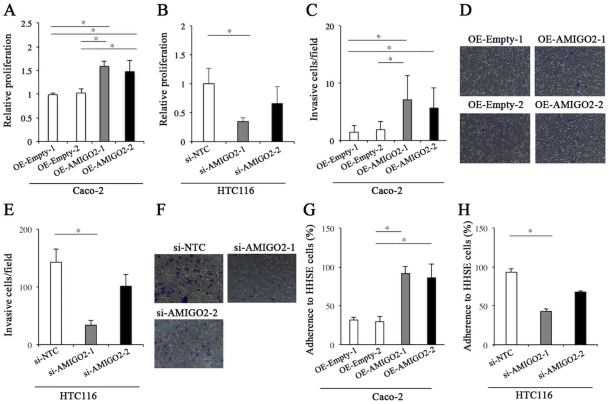 | Figure 2.Proliferation, invasion, and adhesion
assays. (A) The proliferation of Caco-2 cells transfected with
EX-Mm13004-M02 was significantly higher than that of Caco-2 cells
transfected with empty vector. (B) The proliferation of HCT-116
cells transfected with siRNA targeting AMIGO2 was significantly
lower than that of HCT-116 transfected with negative control siRNA.
(C) The invasive ability of Caco-2 cells transfected with
EX-Mm13004-M02 was significantly greater than that of Caco-2 cells
transfected with empty vector. (D) The representative images for
the invasion assay in each condition of (C) Magnification, ×100.
(E) The invasive ability of HCT116 cells transfected with siRNA
targeting AMIGO2 was significantly less than that of HCT116 cells
transfected with negative control siRNA. (F) The representative
images for the invasion assay in each condition of (E)
Magnification, ×100. (G) Caco-2 cells transfected with
EX-Mm13004-M02 demonstrated significantly increased adhesion to
human hepatic sinusoidal endothelial cells (HHSECs), when compared
with controls. (H) HCT116 cells transfected with AMIGO2 siRNA
demonstrated significantly reduced adhesion to HHSECs, when
compared with controls. The data were checked for normality with
Shapiro Wilk test. Differences in proliferation, invasion, and
adhesion were evaluated using the Kruskal-Wallis test and Dunn's
test. *significant difference between 2 groups by Dunn's test
(P<0.05). AMIGO2, adhesion molecule with Ig like domain 2;
siRNA, small interfering RNA; OE, overexpression; NTC,
non-targeting control; HHSE, human hepatic sinusoidal
endothelial. |
AMIGO2 mediates the adhesion of CRC
cells to human hepatic sinusoidal endothelial cells
We then determined the propensity for CRC cells to
adhere to HHSECs. OE-AMIGO2-Caco-2 cells demonstrated significantly
increased adhesion to HHSECs, when compared with OE-Empty-Caco-2
cells (P=0.001, Fig. 2G).
Furthermore, si-AMIGO2-HCT116 cells demonstrated significantly
decreased adhesion to HHSECs, when compared with si-NTC-HCT116
cells (P<0.001, Fig. 2H). These
results indicate that AMIGO2 regulates the adhesion of CRC cells to
HHSECs.
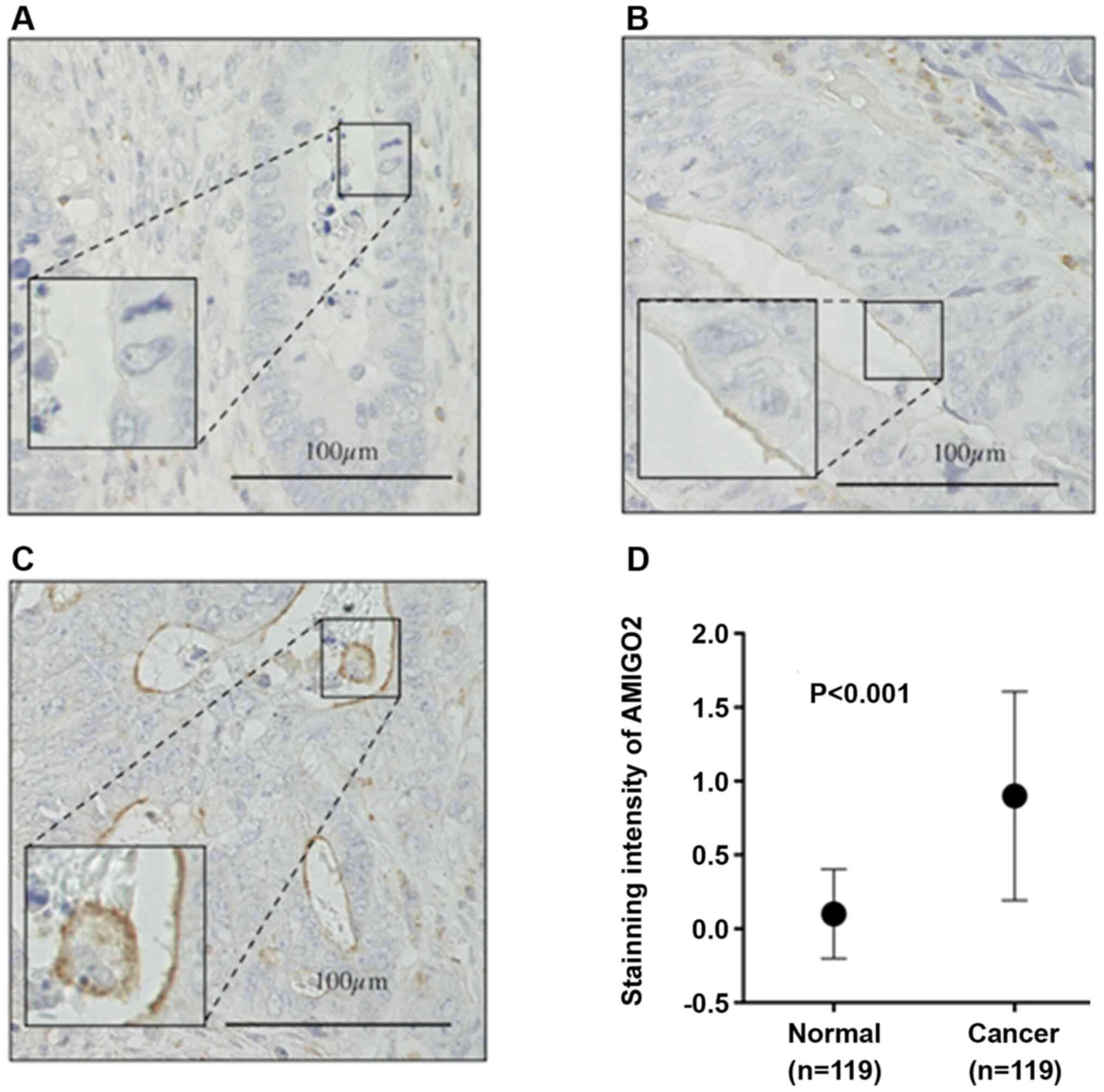
AMIGO2 expression in CRC tissue
We then determined AMIGO2 expression in CRC tissue
(Fig. 3). The staining intensity of
AMIGO2 on the surfaces of CRC cells was scored as 0 (negative), 1
(weak), or 2 (moderate to strong) as previously reported (6) (Fig.
3A-C). The intensity of AMIGO2 staining was significantly
stronger in cancer tissue, when compared with normal tissue
(P<0.001, Fig. 3D).
Table I shows the
association between AMIGO2 expression in CRC tissue and
clinicopathological characteristics. AMIGO2 expression was observed
more frequently in differentiated tumor than in undifferentiated
tumor (P=0.001).
 | Table I.Association between AMIGO2 expression
and clinicopathological features. |
Table I.
Association between AMIGO2 expression
and clinicopathological features.
|
| AMIGO2
expression |
|
|---|
|
|
|
|
|---|
| Variables | 0 (n=82) (%) | 1 (n=126) (%) | 2 (n=59) (%) | P-value |
|---|
| Age, years |
|
|
|
|
| <70
(n=127) | 35 (27.6) | 68 (53.5) | 24 (18.9) | 0.137 |
| ≥70
(n=140) | 47 (33.6) | 58 (41.4) | 35 (25.0) |
|
| Sex |
|
|
|
|
| Male
(n=146) | 48 (32.9) | 64 (43.8) | 34 (23.3) | 0.480 |
| Female
(n=121) | 34 (28.1) | 62 (51.2) | 25 (20.7) |
|
| Tumor location |
|
|
|
|
| Colon
(n=190) | 62 (32.6) | 84 (44.2) | 44 (23.2) | 0.306 |
| Rectum
(n=77) | 20 (26.0) | 42 (54.5) | 15 (19.5) |
|
| Tumor size, cm |
|
|
|
|
| <4.0
(n=103) | 34 (33.0) | 45 (43.7) | 24 (23.3) | 0.659 |
| ≥4.0
(n=164) | 48 (29.3) | 81 (49.4) | 35 (21.3) |
|
|
Histologya |
|
|
|
|
|
Differentiated (n=235) | 63 (26.8) | 115 (48.9) | 57 (42.3) | 0.001 |
|
Undifferentiated (n=32) | 19 (59.4) | 11 (34.4) | 2 (6.2) |
|
| Depth of
invasionb |
|
|
|
|
| T1/T2
(n=19) | 5 (26.3) | 9 (47.4) | 5 (26.3) | 0.863 |
| T3/T4
(n=248) | 77 (31.0) | 117 (47.2) | 54 (21.8) |
|
| Lymph node
metastasis |
|
|
|
|
| Absent
(n=139) | 46 (33.1) | 67 (48.2) | 26 (18.7) | 0.348 |
| Present
(n=128) | 36 (28.1) | 59 (46.1) | 33 (25.8) |
|
| Lymphatic
invasionc |
|
|
|
|
| ly0/1
(n=105) | 32 (30.5) | 52 (49.5) | 21 (20.0) | 0.761 |
| ly2/3
(n=162) | 50 (30.9) | 74 (45.7) | 38 (23.4) |
|
| Vascular
invasiond |
|
|
|
|
| v0/1
(n=159) | 45 (28.3) | 81 (50.9) | 33 (20.8) | 0.327 |
| v2/3
(n=108) | 37 (34.2) | 45 (41.7) | 26 (24.1) |
|
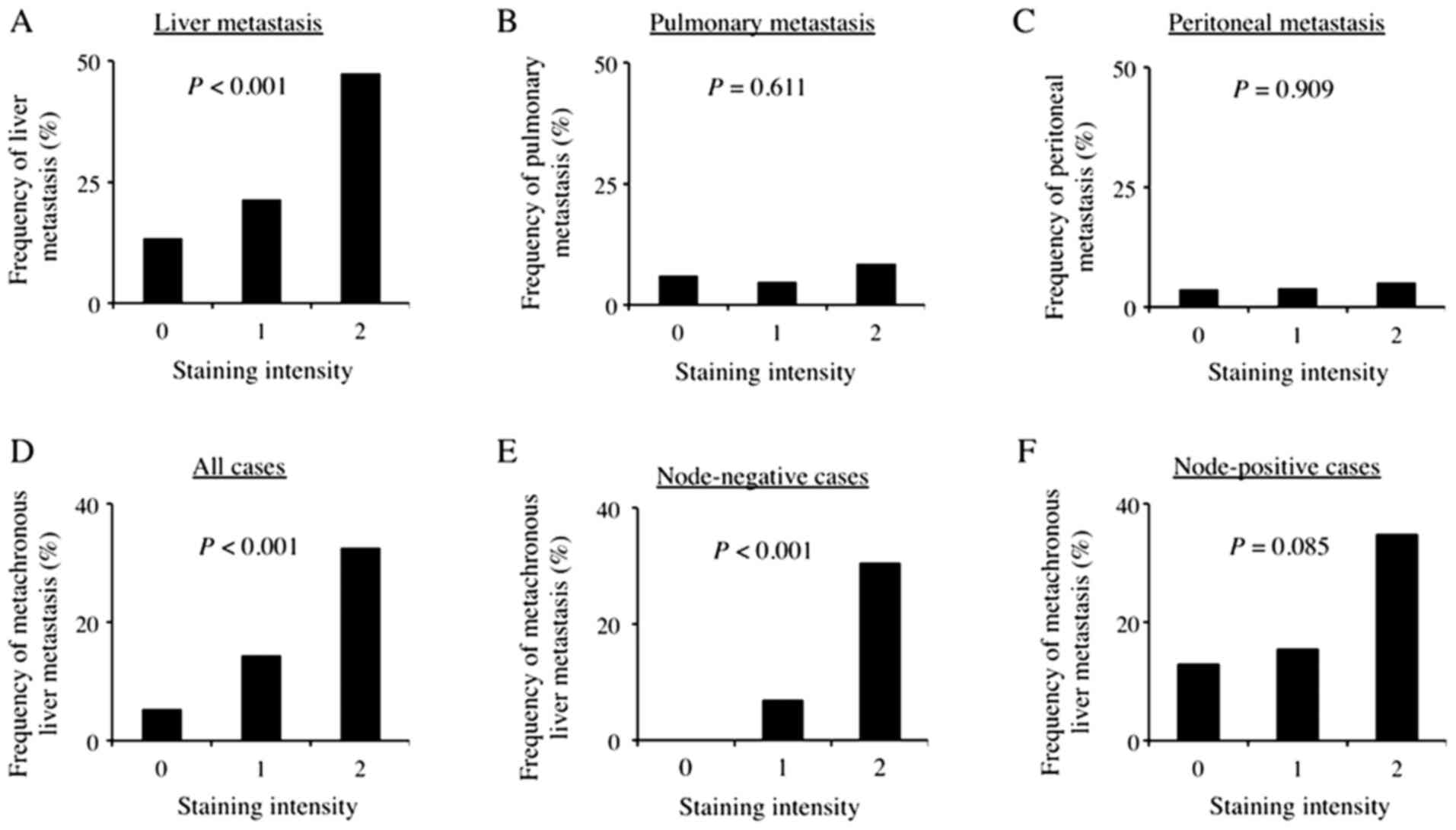
Regarding the association between AMIGO2 expression
in CRC tissue and the site of metastasis, including both
synchronous and metachronous metastasis, AMIGO2 expression in CRC
tissue was significantly related to liver metastasis (P<0.001,
Fig. 4A), but not to pulmonary
metastasis (P=0.61, Fig. 4B) and
peritoneal metastasis (P=0.91, Fig.
4C). Univariate analysis indicated that sex, lymph node
metastasis, vascular invasion, and AMIGO2 expression in CRC tissue
were factors associated with liver metastasis. Multivariate
analysis also revealed that AMIGO2 expression in CRC tissue was an
independent predictor of liver metastasis, along with sex, lymph
node metastasis, and vascular invasion (Table II). Furthermore, we determined the
association between AMIGO2 expression in CRC tissue and
metachronous liver metastasis and found that AMIGO2 expression in
CRC tissue was also significantly related to metachronous liver
metastasis (P<0.001, Fig. 4D).
Univariate analysis indicated that lymph node metastasis and AMIGO2
expression in CRC tissue were factors associated with metachronous
liver metastasis (Table III).
Multivariate analysis also revealed that AMIGO2 expression in CRC
tissue was an independent predictor of metachronous liver
metastasis, along with lymph node metastasis (Table III). Furthermore, AMIGO2 expression
in CRC tissue was also significantly related to metachronous liver
metastasis in node-negative patients (P<0.001, Fig. 4E), but not node-positive patients
(P=0.085, Fig. 4F).
| Table II.Univariate and multivariate analyses
of risk factors for liver metastases in patients with colorectal
cancer. |
Table II.
Univariate and multivariate analyses
of risk factors for liver metastases in patients with colorectal
cancer.
|
| Univariate
analysis | Multivariate
analysis |
|---|
|
|
|
|
|---|
| Variables | P-value | HR | 95% CI | P-value | HR | 95% CI |
|---|
| Age (<70 vs. ≥70
years) | 0.459 | 1.234 | 0.707–2.153 |
|
|
|
| Sex (female vs.
male) | 0.025 | 1.941 | 1.085–3.473 | 0.012 | 2.260 | 1.200–4.254 |
| Tumor location
(colon vs. rectum) | 0.992 | 1.003 | 0.543–1.854 |
|
|
|
| Tumor size (<4.0
vs. ≥4.0 cm) | 0.113 | 1.618 | 0.892–2.934 |
|
|
|
| Histology
(differentiated vs. undifferentiated) | 0.210 | 0.528 | 0.195–1.433 |
|
|
|
| Depth of invasion
(pT1/2 vs. pT3/4) | 0.155 | 2.957 | 0.665–13.151 |
|
|
|
| Lymph node
metastasis (absent vs. present) | 0.004 | 2.340 | 1.318–4.155 | 0.014 | 2.165 | 1.170–4.006 |
| Lymphatic invasion
(ly0/1 vs. ly2/3) | 0.252 | 1.407 | 0.785–2.522 |
|
|
|
| Vascular invasion
(v0/1 vs. v2/3) | 0.008 | 2.150 | 1.223–3.778 | 0.010 | 2.220 | 1.206–4.088 |
| AMIGO2 (0 vs. 1 vs.
2) | <0.001 | 2.511 | 1.651–3.817 | <0.001 | 2.585 | 1.677–3.985 |
| Table III.Univariate and multivariate analyses
of risk factors for metachronous liver metastases in patients with
colorectal cancer. |
Table III.
Univariate and multivariate analyses
of risk factors for metachronous liver metastases in patients with
colorectal cancer.
|
| Univariate
analysis | Multivariate
analysis |
|---|
|
|
|
|
|---|
| Variables | P-value | HR | 95% CI | P-value | HR | 95% CI |
|---|
| Age (<70 vs. ≥70
years) | 0.432 | 1.363 | 0.629–2.957 |
|
|
|
| Sex (female vs.
male) | 0.449 | 1.344 | 0.625–2.888 |
|
|
|
| Tumor location
(colon vs. rectum) | 0.984 | 1.009 | 0.438–2.321 |
|
|
|
| Tumor size (<4.0
vs. ≥4.0 cm) | 0.341 | 1.477 | 0.661–3.300 |
|
|
|
| Histology
(differentiated vs. undifferentiated) | 0.564 | 0.690 | 0.196–2.429 |
|
|
|
| Depth of invasion
(pT1/2 vs. pT3/4) | 0.311 | 2.772 | 0.356–21.603 |
|
|
|
| Lymph node
metastasis (absent vs. present) | 0.027 | 2.431 | 1.107–5.341 | 0.040 | 2.357 | 1.042–5.332 |
| Lymphatic invasion
(ly0/1 vs. ly2/3) | 0.105 | 2.022 | 0.863–4.741 |
|
|
|
| Vascular invasion
(v0/1 vs. v2/3) | 0.318 | 1.475 | 0.687–3.167 |
|
|
|
| AMIGO2 (0 vs. 1 vs.
2) | <0.001 | 3.175 | 1.785–5.733 | <0.001 | 3.151 | 1.729–5.742 |
Finally, we determined AMIGO2 expression in the
tissues obtained from liver metastasis of CRC. We compared the
intensity of AMIGO2 between a primary CRC lesion (Fig. 5A) and a matched liver metastatic
lesion (Fig. 5B). The intensity of
AMIGO2 staining was significantly stronger in the tissue obtained
from the liver metastasis of CRC, when compared with the primary
lesion (n=21, P=0.012, Fig. 5C).
With regard to the association between AMIGO2 expression in CRC
tissue and prognosis of CRC patients, the prognosis of patients
with high AMIGO2 mRNA expression in CRC tissue was significantly
worse than that of patients with low AMIGO2 mRNA expression in CRC
tissue (P=0.013, Fig. 5D).
Discussion
Metastasis is a multi-stage process that is
collectively termed the invasion-metastasis cascade, in which
cancer cells: i) Proliferate and locally invade through the
surrounding extracellular matrix and stromal cell layers; ii)
intravasate into the lumina of blood vessels; iii) survive the
rigors of transport through the vasculature; iv) arrest at distant
organ sites; v) extravasate into the parenchyma of distant tissues;
vi) initially survive in these foreign microenvironments to form
micrometastases; and vii) re-initiate their proliferative programs
at metastatic sites, thereby generating macroscopic, clinically
detectable neoplastic growths. These processes are orchestrated by
molecular pathways operating within carcinoma cells. Among these
seven processes, we first demonstrated that AMIGO2 was closely
related to the proliferation and invasion of CRC cells in the
current study. With regards to the function of AMIGO2, it was
reported to be involved in cell survival and angiogenesis via AKT
signaling (11). However, it remains
unclear whether AMIGO2 is involved in cell survival and
angiogenesis via AKT signaling in CRC. It has recently been
reported that AMIGO2 is upregulated in malignant melanoma, and that
AMIGO2 and its interactor tyrosine-protein kinase-like 7 (PTK7)
regulate the proliferation and survival of tumor cells in this
disease (12). PTK7 is also known as
colon carcinoma kinase 4. This gene is thought to be expressed in
CRC but not in normal colon, and therefore may be involved in CRC
progression (13–15). Although there is no report regarding
the correlation between AMIGO2 and PTK7 in CRC thus far, it is
likely that AMIGO2 regulates the proliferation of CRC cells through
PTK7. With regards to invasion, Sonzogni et al (16) recently reported AMIGO2 as a new
mediator of invasion in breast cancer. In the current study, we
demonstrated that AMIGO2 knockdown in HCT116 cells suppressed
invasive behavior. Furthermore, its overexpression in Caco-2 cells
was sufficient to induce invasion, demonstrating that AMIGO2 also
has important roles in the invasiveness of CRC cells.
Our study also demonstrated that AMIGO2 was closely
associated with the adhesion of CRC cells to HHSECs. Furthermore,
AMIGO2 expression on cancer cells in CRC tissue was related to
increased frequency of liver metastasis. Of importance is that
AMIGO2 expression on cancer cells in CRC tissue was not related to
lung and peritoneal metastasis. In this regard, Kanda et al
(6) demonstrated that the adhesion
of LV12 cells, an isolated subline of QRsP-11 fibrosarcoma cells
with high liver-metastatic properties, to liver endothelial cells
was significantly higher than that of QRsP-11 cells, whereas
adhesion to lung endothelial cells (LE-1) was similar in both cell
lines. They also showed that the incidence of liver metastasis was
higher following intravenous injection of LV12 cells than following
injection of QRsP-11 cells. However, there was no difference in the
incidence of lung metastasis between the two cell lines, suggesting
that LV12 cells have a higher capacity to colonize the liver.
Finally, they revealed that AMIGO2, which was overexpressed in LV12
cells, functioned as a driver gene for liver metastasis. We also
found that AMIGO2 was more strongly expressed on the surfaces of
cells in liver metastasis lesions compared with its expression in
the primary lesions of human CRC in this study. These results may
verify the involvement of AMIGO2 expression in the formation of
liver metastases in clinical samples. However, the precise
mechanisms by which selective and firm adhesion to liver
endothelial cells, but not lung endothelial cells, can be brought
about by AMIGO2 expression remain unclear. One possible mechanism
to explain the AMIGO2-mediated cell adhesion is homophilic or
heterophilic binding among the three homologous proteins that
comprise the AMIGO family, i.e., AMIGO1, AMIGO2, and AMIGO3
(4). Regarding this, Kanda et
al (6) showed that all these
AMIGO family molecules are expressed on liver endothelial cells;
however, none of these AMIGO molecules are expressed on LE-1 lung
endothelial cells. Further experiments are required to resolve
these mechanisms.
Of critical importance is the question of whether
our results will prove useful to the diagnosis and treatment of CRC
patients. With respect to the clinical usefulness of AMIGO2 in CRC,
this study revealed that AMIGO2 was an independent predictive
factor of metachronous liver metastasis. The resection of liver
metastasis improves the prognosis of CRC patients. A recent study
demonstrated more than 60 months of median overall survival in CRC
patients with up to four liver metastases who underwent surgery
combined with perioperative FOLFOX4. It is likely that early
detection of metachronous liver metastasis increases the resection
rates of liver metastasis, improving the prognosis of CRC patients.
Our results also demonstrated that lymph node metastasis was a
useful predictive indicator of metachronous liver metastasis. Of
importance is that AMIGO2 expression was a useful predictive
indicator of metachronous liver metastasis even in node-negative
CRC patients. Therefore, evaluation of AMIGO2 in resected specimens
by immunohistochemistry might be useful in detecting node-negative
CRC patients who have a high possibility of metachronous liver
metastasis and need intensive follow-up and adjuvant chemotherapy
after surgery, which may improve the prognosis of CRC patients.
Regarding treatment, a recent study demonstrated
that AMIGO2 was upregulated in melanoma cells and tissues compared
with human melanocytes and nevi, and AMIGO2 silencing in melanoma
cells induces G1/S arrest followed by apoptosis. Furthermore, a BET
inhibitor was shown to be able to silence AMIGO2 expression
(12). Considering the
multifunctional aspects of AMIGO2 shown in the current study, it is
likely that BET inhibition may be useful in the treatment of CRC
patients with high AMIGO2 expression. Considering the close
association between AMIGO2 expression on CRC cells and liver
metastasis observed in the current study, treatment directly
targeting AMIGO2 might be effective in preventing liver metastasis
in CRC, eventually improving the prognosis of CRC patients.
In conclusion, AMIGO2 contributes to the formation
of liver metastasis by regulating CRC cell adhesion to human
hepatic sinusoidal endothelial cells, as well as the proliferation
and invasion of CRC cells. Treatments that target AMIGO2 could
therefore provide a novel form of CRC treatment.
Acknowledgements
The authors would like to thank Dr H. Nikki March
for editing a draft of this manuscript.
Funding
This work was supported by grants from Takeda
Pharmaceutical Company Limited.
Availability of data and materials
The datasets used and/or analyzed during the current
study are available from the corresponding author upon reasonable
request.
Authors' contributions
HS, AT, MA, KN, MO, FO and YF designed the study and
wrote the paper. AT and RS carried out experiments. AT, KH, KS, CU,
YT, KK and MY analyzed clinical data of patients with colorectal
cancer. All authors have read and approved the final
manuscript.
Ethics approval and consent to
participate
The study protocol was approved by the Institutional
Review Board at Tottori University Hospital (approval number:
1705A039). All procedures were in accordance with the ethical
standards of the responsible committee on human experimentation
(institutional and national) and with the Helsinki Declaration of
1964 and later versions. Written informed consent to be included in
the study was obtained from all patients.
Patient consent for publication
Not applicable.
Competing interests
The authors declare that they have no competing
interests.
Glossary
Abbreviations
Abbreviations:
|
AMIGO2
|
adhesion molecule with Ig like domain
2
|
|
BET
|
bromodomain and extra terminal
domain
|
|
CRC
|
colorectal cancer
|
|
HHSECs
|
human hepatic sinusoidal endothelial
cells
|
|
PTK7
|
tyrosine-protein kinase-like 7
|
|
siRNA
|
small interfering RNA
|
References
|
1
|
Torre LA, Bray F, Siegel RL, Ferlay J,
Lortet-Tieulent J and Jemal A: Global cancer statistics, 2012. CA
Cancer J Clin. 65:87–108. 2015. View Article : Google Scholar : PubMed/NCBI
|
|
2
|
Matsuoka H, Morise Z, Tanaka C, Hayashi T,
Ikeda Y, Maeda K, Masumori K, Koide Y, Katsuno H, Tanahashi Y, et
al: Repeat hepatectomy with systemic chemotherapy might improve
survival of recurrent liver metastasis from colorectal cancer-a
retrospective observational study. World J Surg Oncol. 17:332019.
View Article : Google Scholar : PubMed/NCBI
|
|
3
|
Abdalla EK, Adam R, Bilchik AJ, Jaeck D,
Vauthey JN and Mahvi D: Improving resectability of hepatic
colorectal metastases: Expert consensus statement. Ann Surg Oncol.
13:1271–1280. 2006. View Article : Google Scholar : PubMed/NCBI
|
|
4
|
Kuja-Panula J, Kiiltomaki M, Yamashiro T,
Rouhiainen A and Rauvala H: AMIGO, a transmembrane protein
implicated in axon tract development, defines a novel protein
family with leucine-rich repeats. J Cell Biol. 160:963–973. 2003.
View Article : Google Scholar : PubMed/NCBI
|
|
5
|
Ono T, Sekino-Suzuki N, Kikkawa Y,
Yonekawa H and Kawashima S: Alivin 1, a novel neuronal
activity-dependent gene, inhibits apoptosis and promotes survival
of cerebellar granule neurons. J Neurosci. 23:5887–5896. 2003.
View Article : Google Scholar : PubMed/NCBI
|
|
6
|
Kanda Y, Osaki M, Onuma K, Sonoda A,
Kobayashi M, Hamada J, Nicolson GL, Ochiya T and Okada F:
Amigo2-upregulation in tumour cells facilitates their attachment to
liver endothelial cells resulting in liver metastases. Sci Rep.
7:435672017. View Article : Google Scholar : PubMed/NCBI
|
|
7
|
Huo T, Canepa R, Sura A, Modave F and Gong
Y: Colorectal cancer stages transcriptome analysis. PLoS One.
12:e01886972017. View Article : Google Scholar : PubMed/NCBI
|
|
8
|
Onuma K, Suenaga Y, Sakaki R, Yoshitome S,
Sato Y, Ogawara S, Suzuki S, Kuramitsu Y, Yokoyama H, Murakami A,
et al: Development of a quantitative bioassay to assess preventive
compounds against inflammation-based carcinogenesis. Nitric Oxide.
25:183–194. 2011. View Article : Google Scholar : PubMed/NCBI
|
|
9
|
Japanese Society for Cancer of the Colon
and Rectum: Japanese classification of colorectal carcinoma, 2nd
English edition. Kanehara & Co., Ltd; 2009
|
|
10
|
Li Z, Khan MM, Kuja-Panula J, Wang H, Chen
Y, Guo D, Chen ZJ, Lahesmaa R, Rauvala H and Tian L: AMIGO2
modulates T cell functions and its deficiency in mice ameliorates
experimental autoimmune encephalomyelitis. Brain Behav Immun.
62:110–123. 2017. View Article : Google Scholar : PubMed/NCBI
|
|
11
|
Park H, Lee S, Shrestha P, Kim J, Park JA,
Ko Y, Ban YH, Park DY, Ha SJ, Koh GY, et al: AMIGO2, a novel
membrane anchor of PDK1, controls cell survival and angiogenesis
via Akt activation. J Cell Biol. 211:619–637. 2015. View Article : Google Scholar : PubMed/NCBI
|
|
12
|
Fontanals-Cirera B, Hasson D, Vardabasso
C, Di Micco R, Agrawal P, Chowdhury A, Gantz M, de
Pablos-Aragoneses A, Morgenstern A, Wu P, et al: Harnessing BET
inhibitor sensitivity reveals AMIGO2 as a melanoma survival gene.
Mol Cell. 68:731–744.e9. 2017. View Article : Google Scholar : PubMed/NCBI
|
|
13
|
Saha S, Sparks AB, Rago C, Akmaev V, Wang
CJ, Vogelstein B, Kinzler KW and Velculescu VE: Using the
transcriptome to annotate the genome. Nat Biotechnol. 20:508–512.
2002. View Article : Google Scholar : PubMed/NCBI
|
|
14
|
Tian X, Yan L, Zhang D, Guan X, Dong B,
Zhao M and Hao C: PTK7 overexpression in colorectal tumors:
Clinicopathological correlation and prognosis relevance. Oncol Rep.
36:1829–1836. 2016. View Article : Google Scholar : PubMed/NCBI
|
|
15
|
Lhoumeau AC, Martinez S, Boher JM, Monges
G, Castellano R, Goubard A, Doremus M, Poizat F, Lelong B, de
Chaisemartin C, et al: Overexpression of the promigratory and
prometastatic PTK7 receptor is associated with an adverse clinical
outcome in colorectal cancer. PLoS One. 10:e01237682015. View Article : Google Scholar : PubMed/NCBI
|
|
16
|
Sonzogni O, Haynes J, Seifried LA, Kamel
YM, Huang K, Be Gora MD, Yeung FA, Robert-Tissot C, Heng YJ, Yuan
X, et al: Reporters to mark and eliminate basal or luminal
epithelial cells in culture and in vivo. PLoS Biol.
16:e20040492018. View Article : Google Scholar : PubMed/NCBI
|