Introduction
Hepatocellular carcinoma (HCC) is one of the most
common cancer worldwide, with similar rates of incidence and
mortality (1,2). As the third most common cause of
cancer-related death, HCC leads to >60,000 deaths globally each
year (3,4). The majority of patients with HCC are
already in advanced stages when they are diagnosed, and the most
effective treatment for early-stage patients is surgical resection
(5). However, the long-term survival
rate of patients with HCC is low, and the prognosis is poor due to
the high rate of HCC recurrence and metastasis so resection is not
available for most patients (6). Due
to changes in lifestyle and environment, HCC is currently
demonstrating a trend of continuously increasing global incidence
that seriously affects quality of life (7). Numerous types of antitumor therapy,
such as radiotherapy, chemotherapy and adjuvant therapy have been
used for HCC, however, there are few specific markers for the
diagnosis and prognosis of HCC to date (8). It is important to identify effective
targets for early detection, treatment, and prognostic monitoring
of patients with HCC; hence, it is necessary to identify new
biomarkers (9).
MicroRNAs (miRNAs) are a major class of small
non-coding RNAs consisting of ~18–25 nucleotides that were
originally identified in Caenorhabditis elegans and
negatively regulate gene expression at the mRNA level (10–12).
Numerous studies have revealed the important role of miRNAs in
biological processes, such as cell proliferation and
differentiation, embryogenesis, metabolism, organogenesis and
apoptosis (12–14). Growing evidence indicates that miRNAs
are altered in a number of diseases, including cardiovascular
diseases, hepatitis and various types of cancer including prostate
cancer and osteosarcoma due to genomic events, such as gene
mutations or downregulation caused by enzymes that involved in
miRNAs biogenesis (15–17). Previous studies have demonstrated
that miR-29a, miR-21 and miR-221 and 222 are abnormally expressed
in HCC (18–21). Shi et al (22) recently found that 29 miRNAs,
including miR-1266, were upregulated in the tissues of patients
with HCC. It can be concluded from previous studies that miR-1266
may serve an important role in the occurrence and development of
HCC. However, the expression pattern, biological function and
clinical significance of miR-1266 in the pathogenesis of HCC remain
unclear.
The present study aimed to investigate the
difference in miR-1266 expression between HCC and normal liver
tissues and evaluated the clinical prognostic significance of
miR-1266 in patients with HCC. In addition, the effects of miR-1266
on the proliferation, migration and invasion of HCC cell lines were
also investigated. The present study aimed to provide a new
biomarker for HCC.
Materials and methods
Patients and specimens
A total of 119 patients (median age, 60 years; age
range, 26–77 years) with HCC admitted to Mengchao Hepatobiliary
Hospital of Fujian Medical University (Fuzhou, China) between
January 2012 and December 2014 were included in this study. HCC
tissue and normal adjacent tissues [all were confirmed by at least
two pathologists of Mengchao Hepatobiliary Hospital of Fujian
Medical University (Fuzhou, China)] 5 cm away from the cancer
lesion were collected from each patient. The tissue samples were
immediately frozen in liquid nitrogen at −80°C until subsequent
total RNA extraction experiments were performed. All patients
underwent resection surgery and did not receive any treatment, such
as chemotherapy and radiotherapy before surgery. The exclusion
criteria were as follows: i) Patients with no complete follow-up
data; ii) patients suffering from other types of cancer; and iii)
patients with systemic intolerance to surgery. In order to collect
the overall survival outcome information of patients with HCC, all
of them were enrolled in the 5-year follow up investigation by
telephone. The follow-up was performed at intervals of 3 months
during the first 2 years, 6 months in years 2–4 and annually in the
last year. The results of the follow up are listed in Table I. Each patient signed a written
consent approving the use of their tissues for research purposes
after the operation. The present study was approved by the Research
Ethics Committee of Mengchao Hepatobiliary Hospital of Fujian
Medical University (approval no. 2012-032-01; Fuzhou, China).
 | Table I.Association between miR-1266
expression and clinical characteristics of patients with
hepatocellular carcinoma (n=119). |
Table I.
Association between miR-1266
expression and clinical characteristics of patients with
hepatocellular carcinoma (n=119).
|
|
| miR-1266
expression |
|
|---|
|
|
|
|
|
|---|
|
Characteristics | No. of
patients | Low (n=57) | High (n=62) | P-value |
|---|
| Age, years |
|
|
| 0.655 |
|
<60 | 58 | 29 | 29 |
|
|
≥60 | 61 | 28 | 33 |
|
| Sex |
|
|
| 0.584 |
|
Female | 45 | 23 | 22 |
|
|
Male | 74 | 34 | 40 |
|
| AFP, ng/ml |
|
|
| 0.060 |
|
<20 | 50 | 29 | 21 |
|
|
≥20 | 69 | 28 | 41 |
|
| Tumor size, cm |
|
|
| 0.066 |
|
<5 | 69 | 38 | 31 |
|
| ≥5 | 50 | 19 | 31 |
|
| HBV infection |
|
|
| 0.087 |
|
Absent | 55 | 31 | 24 |
|
|
Present | 64 | 26 | 38 |
|
| Cirrhosis |
|
|
| 0.105 |
|
Absent | 66 | 36 | 30 |
|
|
Present | 53 | 21 | 32 |
|
| TNM stage |
|
|
| 0.014 |
|
I–II | 88 | 48 | 40 |
|
|
III–IV | 31 | 9 | 22 |
|
Cell lines and transfection
In total 4 types of HCC cell lines (Hep3B, Huh-7,
MHCC97 and SNU-387) and one normal human liver cell line MIHA were
purchased from Shanghai Cell Bank of the Chinese Academy of
Sciences. In Hep3B cells there are obvious granular endoplasmic
reticulum pools and mitochondria; ring-shaped lamellar accumulation
and glycogen aggregates are found in the cytoplasm, and
α-fetoprotein (AFP) is positive (23). HuH-7 cells are flat polygonal in the
early stage with fine particles in the cytoplasm, while later cells
are elongated in the form of coarse particles (24). MHCC97 cells grow into dense colonies
or single-layer flakes with a doubling time of about 31 h
presenting a typical malignant epithelial morphology, and AFP is
positive (25). The SNU-387 cell
line demonstrates adherent cell growth and obvious nuclear
convolution (26). The Hep3B, Huh-7
and MHCC97 cells were cultured in DMEM medium (Gibco; Thermo Fisher
Scientific, Inc.) supplemented with 10% FBS (Gibco; Thermo Fisher
Scientific, Inc.), while the MIHA and SNU-387 cells were cultured
in RPMI 1640 medium (Gibco; Thermo Fisher Scientific, Inc.)
supplemented with 10% FBS. All cells were cultured at 37°C in a
humidified incubator with 5% CO2.
Cells (2×10⁵) were seeded into a 6-well
plate before transfection. According to the manufacturer's
instructions, miR-1266 mimic (5′-CCUCAGGGCUGUAGAACAGGGCU-3′),
scrambled mimic negative control (mimic NC;
5′-UUCUCCGAACGUGUCACGUTT-3′), miR-1266 inhibitor
(5′-AGCCCUGUUCUACAGCCCUGAGG-3′) and scrambled inhibitor NC
(5′-CAGUACUUUUGUGUAGUACAA-3′) (Shanghai GenePharma Co., Ltd.) all
at a final concentration of 50 nM were transfected into cells by
using Lipofectamine® 2000 Reagent (Invitrogen; Thermo
Fisher Scientific, Inc.) in serum-free condition for 6 h at 37°C
and then complete DMEM medium was added. Transfection efficiency
was measured after 24 h of transfection at 37°C and then the
transfected cells were used in subsequent experimentation.
Untreated cells served as the blank control group.
RNA extraction and reverse
transcription-quantitative (RT-q) PCR analysis
Total RNA of tissues or cell lines was extracted
using TRIzol® reagent (Takara Bio Inc.) according to the
manufacturer's instructions. The DNA was removed from the extracted
total RNA by trypsin and then RNA concentration was detected by
NanoDrop. Complementary DNA (cDNA) was obtained by reverse
transcription from the extracted RNA using the cDNA Reverse
Transcription kit (Applied Biosystems; Thermo Fisher Scientific
Inc.) with incubation at 16°C for 30 min, then 42°C for 30 min and
terminated at 85°C for 5 min. Relative expression of miR-1266 was
assessed in cell lines and tissues using the SYBR Green I Real-Time
PCR kit (Shanghai GenePharma Co. Ltd.) on an Applied Biosystems
7900 Real-Time PCR system (Applied Biosystems; Thermo Fisher
Scientific Inc.) using the following thermocycling conditions:
Preliminary denaturation at 95°C for 2 min, then 35 cycles of 94°C
for 30 sec, 60°C for 30 sec, followed by 72°C for 45 sec. The
sequences used in RT-qPCR were as follows: miR-1266 forward,
5′-GCCGAGCCTCAGGGCTGTAGA-3′, reverse, 5′-CTCAACTGGTGTCGTGGA-3′; U6
forward, 5′-GCTTCGGCAGCACATATACTAAAAT-3′ and reverse
5′-CGCTTCACGAATTTGCGTGTCAT-3′. U6 small nuclear RNA was used for
normalization and relative quantification of miR-1266 expression
was calculated by using the 2−ΔΔCq method (27). Each sample measurement was performed
in triplicate.
Cell proliferation assay
Cell proliferation was assessed by using the Cell
Counting Kit-8 assay (CCK-8; Dojindo Molecular Technologies, Inc.)
according to the manufacturer's instructions. HCC Huh-7 and MHCC97H
cells (1×10³/well) were seeded into a 96-well culture
plate at 7°C for 24 h after transfection. Then, the CCK-8 solution
was added to each well for 0, 24, 48, and 72 h and incubated for 2
h. The absorbance was analyzed at 450 nm using a microplate reader
(Thermo Fisher Scientific, Inc.). Each sample measurement was
performed in triplicate at each time point.
Cell migration and invasion assay
Migration and cell invasion assays were performed
using the Transwell chamber (BD Biosciences) with 8-µm pores. The
cells were inoculated in serum-free DMEM medium after transfection.
In the invasion assay, Matrigel (Becton-Dickinson and Company) was
pre-coated on the bottom layer of the upper chamber at 37°C for 4
h, but Matrigel was not added in the upper chamber of the migration
assay. Huh-7 and MHCC97H cell suspension (5×10⁴
cells/well) was added into the upper chamber and 600–800 µl of
medium containing 10% FBS as a chemokine was added into the lower
chamber. After 24 h of incubation at 37°C, the bottom membrane of
the upper chamber was fixed in methanol for 30 min at room
temperature, and then stained with 0.1% crystal violet for 15 min
at room temperature. After air-drying, the stained cells were
mounted with neutral resin and counted under a light
microscope.
Luciferase reporter assay
First, it was predicted that DAB2 Interacting
Protein (DAB2IP) may be the target gene of miR-1266 through
TargetScan (http://www.targetscan.org/vert_71/). The wild-type
(WT) and mutant (MUT) promoters of DAB2IP were constructed by
Shanghai GenePharma Co. Ltd. and inserted into the pGL3 basic
vector (Promega Corporation). The DAB2IP 3′-untranslated region
(3′-UTR) sequence was amplified from normal human genomic DNA and
subcloned into the pmirGLO luciferase reporter vector (Promega
Corporation). MHCC97 cells were cultured in a 24-well plate, and
after 24 h of culture, they were co-transfected with WT or MUT
3′-UTR vector and miR-1266 mimic, 5′-CCUCAGGGCUGUAGAACAGGGCU-3′;
mimic NC, 5′-UUCUCCGAACGUGUCACGUTT-3′; miR-1266 inhibitor,
5′-AGCCCUGUUCUACAGCCCUGAGG-3′ or inhibitor NC,
5′-CAGUACUUUUGUGUAGUACAA-3′ using Lipofectamine® 2000
reagent (Invitrogen; Thermo Fisher Scientific, Inc.). Following 24
h of transfection, luciferase activity was measured by the dual
luciferase reporter gene assay system (Promega Corporation)
according to the manufacturer's instructions. The relative
luciferase activity was estimated by normalizing the luciferase
activity to the Renilla luciferase activity. All assays were
repeated three times.
Statistical analysis
All biological experiments were repeated at least 3
times and statistical analyses were performed using SPSS 19.0
software (IBM Corp.) and GraphPad Prism 7.0 software (GraphPad
Software Inc.). By using mean value of miR-1266 expression (0.8732)
as the cut-off value, patients were divided into high miR-1266
expression group and low miR-1266 expression group. χ2
test was used for assessing the association between miR-1266
expression and clinical features of patients. The difference
between two groups were analyzed using paired Student's t-test.
Multiple comparisons were performed using one-way ANOVA followed by
the post hoc Tukey's test. Kaplan-Meier analysis and log-rank test
were used to test the overall survival rate of patients, and the
prognostic significance of miR-1266 was evaluated by Cox regression
analysis. Data are presented as the mean ± standard deviation.
P<0.05 was considered to indicate a statistically significant
difference.
Results
Expression of miR-1266 in HCC tissues
and cell lines
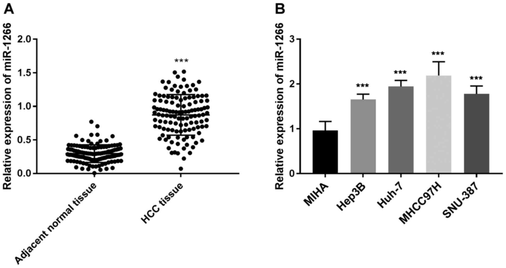
Firstly, the expression of miR-1266 in 119 pairs of
HCC tissues and adjacent normal liver tissues was assessed. The
results indicated that the miR-1266 expression level was
significantly higher in the HCC tissues compared with the adjacent
normal tissues (P<0.001; Fig.
1A). In addition, further analysis of miR-1266 expression in
HCC cell lines (Hep3B, Huh-7, MHCC97 and SNU-387) was performed.
The expression of miR-1266 in the HCC cell lines was significantly
higher compared with that of the normal cell line MHA (P<0.001;
Fig. 1B). In addition, the highest
expression of miR-1266 in the HCC cell lines was in the MHCC97 cell
line followed by the Huh-7 cell line (Fig. 1B). Hence, these 2 cell lines were
selected for subsequent experimentation due to their high miR-1266
expression.
miR-1266 expression is associated with
the clinicopathological features of patients with HCC
As shown in Table I,
the association between miR-1266 expression and the
clinicopathological characteristics of patients with HCC was
assessed. The patients were divided into the miR-1266 low
expression group (n=57) and the miR-1266 high expression group
(n=62) with the mean value of miR-1266 expression as the cut-off
value. The results demonstrated that high mRNA expression of
miR-1266 was significantly associated with Tumor-Node-Metastasis
(TNM) stage (28) (P=0.014; Table I). There was no significant
association observed for the other characteristics, such as age,
sex, hepatitis B virus infection, tumor size, AFP and cirrhosis
(P>0.05; Table I).
High expression of miR-1266 is
associated with poor prognosis in patients with HCC
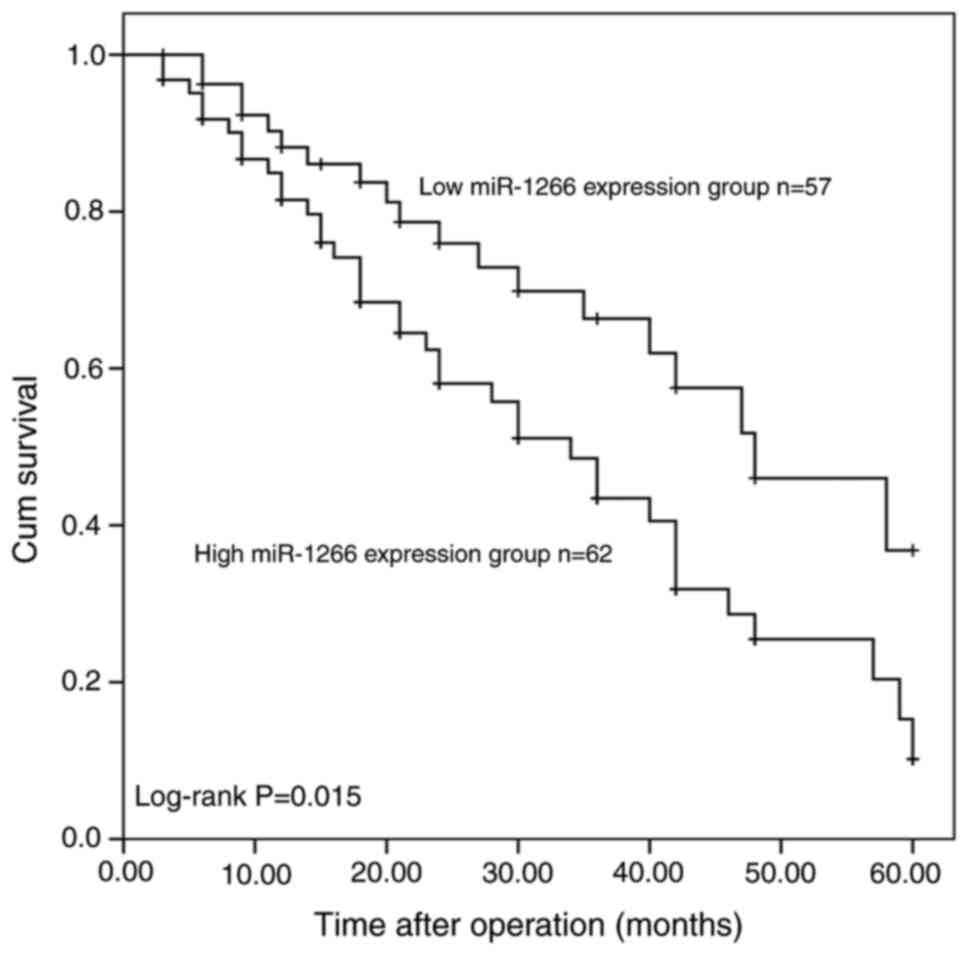
To assess the potential prognostic value of using
miR-1266 as a biomarker in HCC tissues, a cumulative survival curve
was generated using the Kaplan-Meier method according to the
patients' miR-1266 expression and overall survival information. The
results demonstrated that, when compared with patients with lower
miR-1266 expression levels, the 5-year survival rate of patients
with HCC with higher miR-1266 expression levels was significantly
lower (log-rank test P=0.015; Fig.
2). In the multivariate Cox hazard regression model, further
multivariate analysis of the association between miR-1266
expression levels and overall survival of patients with HCC was
performed. The results demonstrated that the miR-1266 expression
level [hazard ratio (HR)=2.048; 95% confidence interval
(CI)=1.151–3.664; P=0.015; Table
II) and TNM stage (HR=1.900; 95% CI=1.074–3.364; P=0.028;
Table II) were independent
prognostic factors for the 5-year overall survival of patients with
HCC.
| Table II.Multivariate Cox regression analysis
for risk prognostic factors to the overall survival of patients
with hepatocellular carcinoma. |
Table II.
Multivariate Cox regression analysis
for risk prognostic factors to the overall survival of patients
with hepatocellular carcinoma.
|
| Multivariate
analysis |
|---|
|
|
|
|---|
| Parameters | HR | 95% CI | P-value |
|---|
| miR-1266
expression | 2.048 | 1.151–3.644 | 0.015 |
| Age | 1.149 | 0.641–2.058 | 0.641 |
| Sex | 1.423 | 0.803–2.523 | 0.227 |
| AFP | 1.175 | 0.674–2.048 | 0.570 |
| Tumor size | 1.457 | 0.791–2.685 | 0.227 |
| HBV infection | 1.583 | 0.888–2.822 | 0.119 |
| Cirrhosis | 2.011 | 0.960–4.212 | 0.064 |
| TNM stage | 1.900 | 1.074–3.364 | 0.028 |
Overexpression of miR-1266 promotes
HCC cell proliferation, migration, and invasion
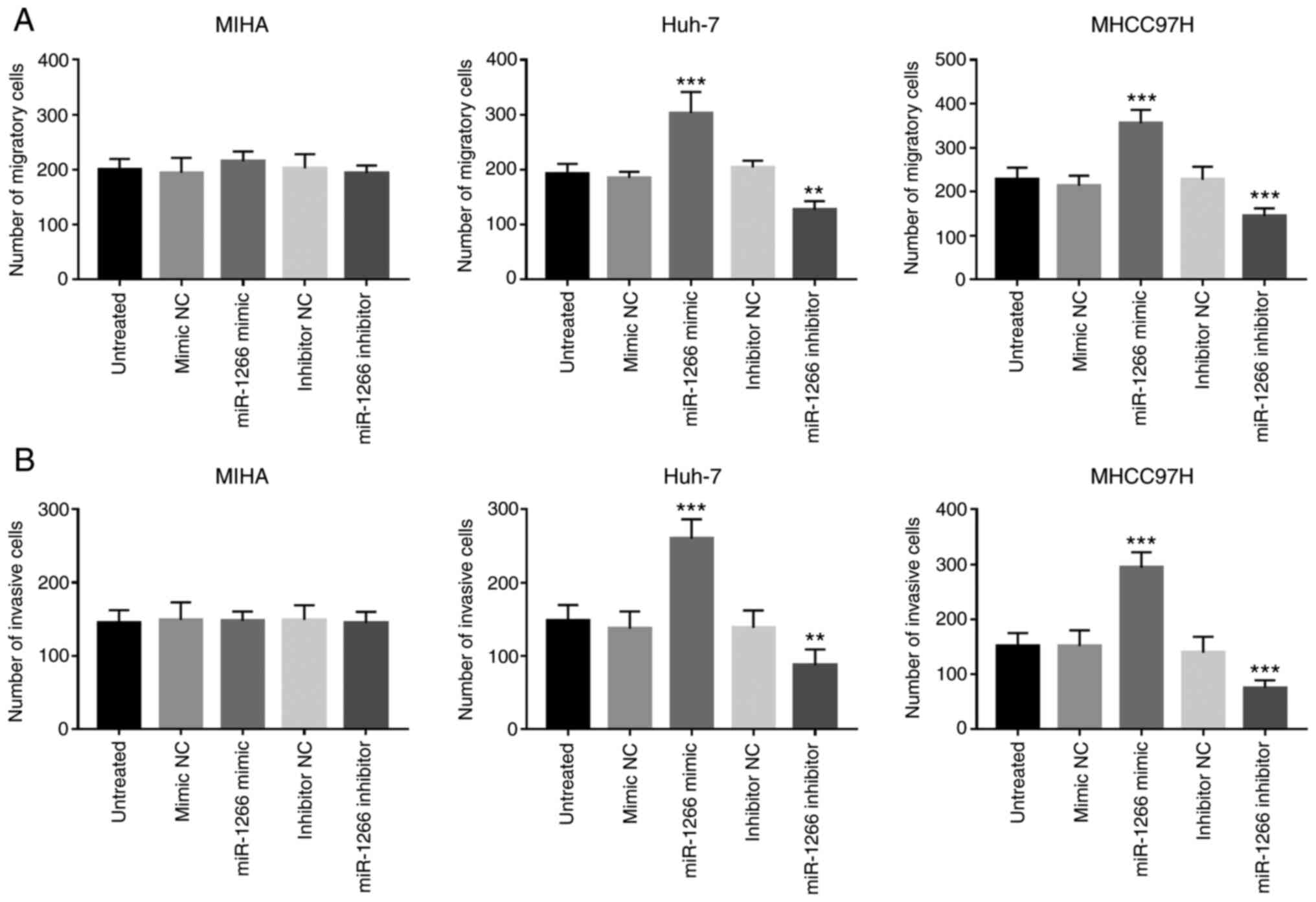
To investigate whether miR-1266 regulates HCC cell
proliferation, migration and invasion, in vitro HCC cell
experiments were performed by transfecting miR-1266 mimics or
miR-1266 inhibitor into MHCC97 and Huh7 cells. The effect of
miR-1266 on the aforementioned cellular behavior was also measured
in MIHA cells, which acted as a control for comparison with HCC
cell lines. MHCC97 and Huh7 cells with significantly increased
expression of miR-1266 (P<0.001) and MHCC97 and Huh7 cells with
inhibited expression of miR-1266 were obtained through cell
transfection, compared with untreated cells (P<0.01; Fig. 3A). The CCK-8 cell proliferation assay
demonstrated that overexpression of miR-1266 by miR-1266 mimics in
cells increased Transwell assays indicated that the migration and
invasion of MHCC97 and Huh7 cells were significantly increased due
to the overexpression of miR-1266 (P<0.001), while the
downregulation of miR-1266 led to a significant decrease in the
migration and invasion of MHCC97 (P<0.001) and Huh7 (P<0.01)
cells (Fig. 4A and B), however, no
effect was observed in normal MIHA cell line (Fig. 4A and B). It can be seen from the
images of MIHA, MHCC97 and Huh7 cells in Fig. S1A and B that the invasion and
migration trends were consistent with those in Fig. 4A and B.
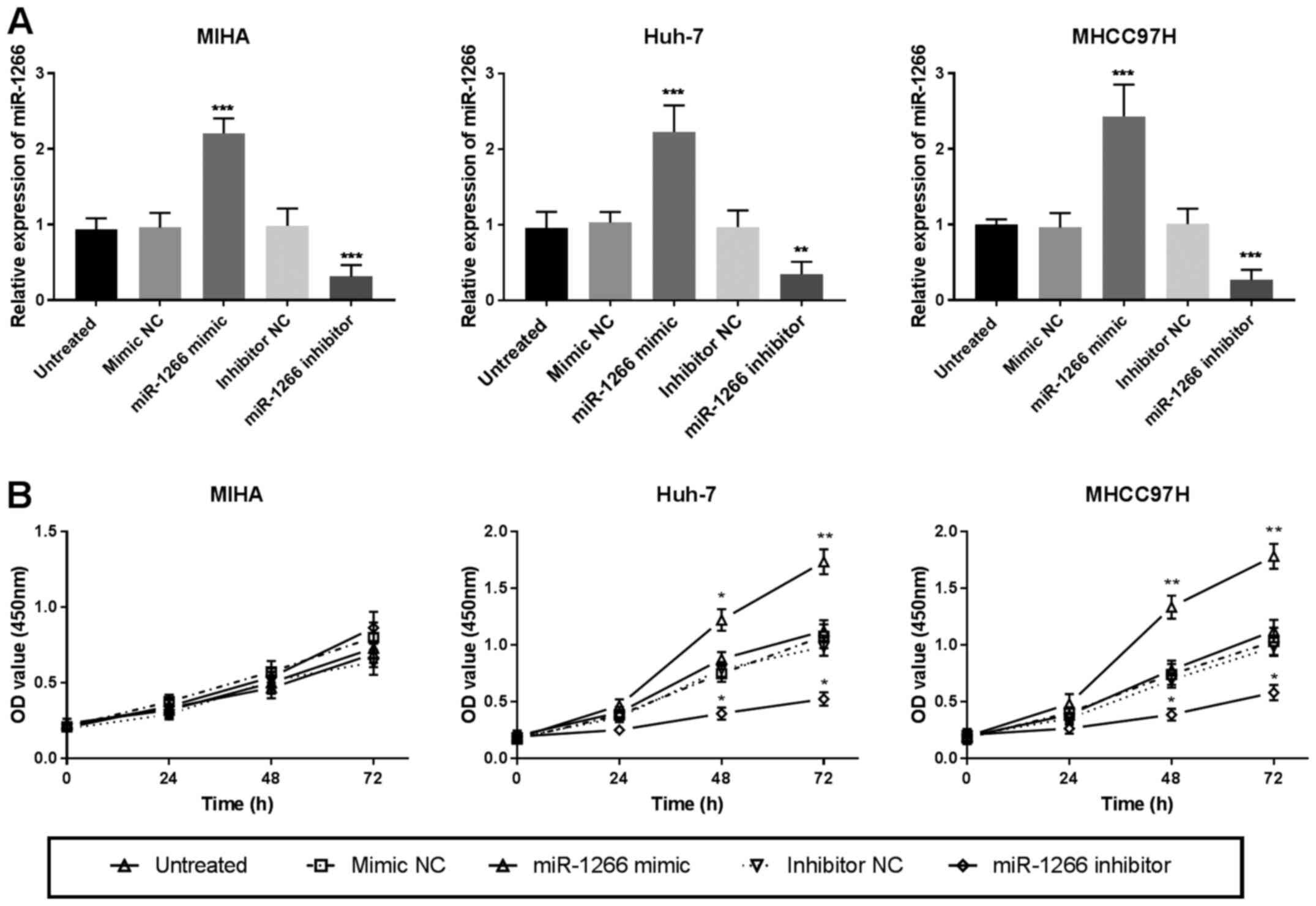 | Figure 3.Effects of miR-1266 on proliferation
of HCC cell lines. (A) Changes in the proliferation of MIHA, MHCC97
and Huh-7 cells following the transfection of miR-1266 mimics NC,
miR-1266 mimic, inhibitor NC or miR-1266 inhibitor (**P<0.01,
***P<0.001) vs. untreated cells. (B) Effects of miR-1266 on the
proliferation of MIHA, MHCC97 and Huh-7 cells (*P<0.05,
**P<0.01, ***P<0.001) vs. untreated cells. HCC,
hepatocellular carcinoma; miR, microRNA; NC, negative control; OD,
optical density. |
DAB2IP may be a direct target of
miR-1266
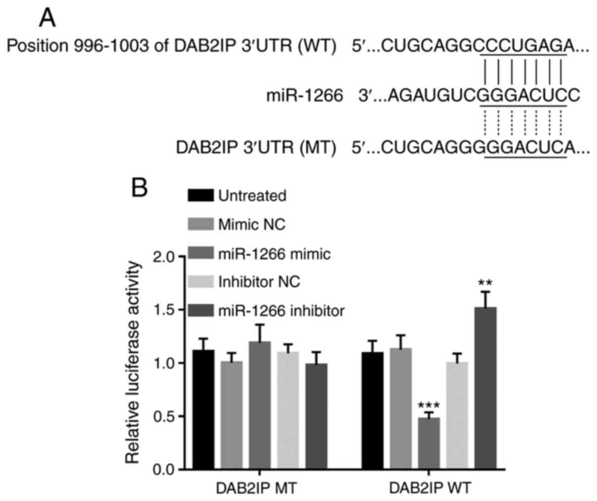
Luciferase reporter assay was performed using
MHCCH97 cells due to its superior cellular activities
(proliferative, migratory, and invasive abilities) compared with
Huh-7 cells. Based on the TargetScan database, position 996–1003 of
DAB2IP mRNA 3′UTR was discovered to have binding sites of miR-1266
(Fig. 5A). Luciferase reporter gene
detection was used to verify the effect of miR-1266 on DAB2IP in
MHCCH97 cell because of its better cellular activities. miR-1266
mimics significantly reduced the relative luciferase activities of
cells with wild miR-1266-binding site (P<0.001; Fig. 5B) and miR-1266 inhibitors
significantly increased its relative luciferase activities
(P<0.01; Fig. 5B). However,
neither miR-1266 mimics nor inhibitors caused significant changes
in cells with mutated vectors (Fig.
5B). The aforementioned results demonstrated that miR-1266 can
directly target DAB2IP expression.
Discussion
HCC is a disease that is common worldwide (29). The majority of patients find that the
disease is already advanced upon diagnosis, and its prognosis is
poor (30,31). It is necessary to discover new and
effective biomarkers for the diagnosis and prognosis of HCC, which
will help to improve the clinical efficacy of treatments in
patients (32,33). As a new type of biomarker, miRNAs
serve an important role in the occurrence, development and
metastasis of numerous types of cancer, such as HCC and prostate
cancer (34,35). Accumulating studies have investigated
the association between cancer and miRNAs, and it has been proven
that various cancers exhibit abnormal expression of miRNAs
(36–38). For example, the low expression of
miR-129 is significantly associated with the poor prognosis of
prostate cancer, and can be regarded as a new molecular target for
the diagnosis and treatment of prostate cancer (39). Wang et al (40) discovered that the expression of
miR-339-5p is significantly reduced in HCC patients tissues
compared with non-cancerous liver tissues, and is closely related
to the invasion of HCC cells indicating that the expression of
miR-339-5p is an independent prognostic factor in patients with
HCC. Several studies have proven that the abnormal expression of
miR-1266 serves an important role in various diseases and cancers,
such as papillary thyroid carcinoma and prostate cancer (41–44).
miRNAs have been proven to be oncogenes in various
cancers (45). Wang et al
(44) confirmed that miR-1266
demonstrated a highly upregulated trend in the tissues and serum of
patients with cervical cancer compared with the controls. The
aforementioned study is consistent with the findings of the present
study. In the present study, the expression of miR-1266 in HCC
tissues and cell lines was examined, and its potential role in HCC
was investigated in relation to the pathological characteristics of
patients. The results demonstrated that the expression of miR-1266
was significantly increased in HCC tissues and cell lines compared
with non-cancerous tissue and a normal cell line. In addition, in
the present study high expression of miR-1266 demonstrated a
significant association with TNM stage, which was consistent with
other studies (44,46). Lu et al (47) analyzed the miRNA expression profile
of patients in The Cancer Genome Atlas database and concluded that
miR-1266 is upregulated in HCC. The aforementioned results indicate
that miR-1266 is an oncogene of HCC and is involved in the
occurrence, development and metastasis of HCC.
In addition, the present study examined the
relationship between miR-1266 expression and the survival of
patients with HCC using Kaplan-Meier curves and Cox regression
analysis. The present study found that high expression levels of
miR-1266 reduced the 5-year overall survival rate of patients with
HCC. In addition, it was found that the expression level of
miR-1266 was an independent prognostic factor for 5-year overall
survival. These results indicated that miR-1266 may be an important
prognostic biomarker for HCC. Sevinc et al (42) proved that miR-1266 is associated with
the recurrence and metastasis of estrogen receptor positive
patients with breast cancer. Wang et al (44) revealed that the overall survival rate
of patients with cervical cancer with high expression of miR-1266
was significantly lower compared with patients with low expression
of miR-1266. These studies indicated that miR-1266 may be used as a
new molecular target for the diagnosis and prognosis of cancer.
However, a previous study did not find a significant association
between miR-1266 expression and the clinical prognosis of HCC
(47). This may be because the
previous study (47) selected
patients from TCGA database and the patients came from different
hospitals in various cities and the time span was large, which may
lead to inconsistent criteria for determining patient
information.
In addition, the present study investigated the
effects of miR-1266 on the proliferation, migration and invasion of
HCC cells using CCK-8 assays and Transwell assays, respectively.
The results demonstrated that miR-1266 significantly increased the
cell proliferation, migration and invasion ability of HCC cell
lines, while inhibitors of miR-1266 inhibited their proliferation,
migration and invasion. These results indicate that miR-1266 is
involved in the molecular biological changes of HCC cells. Fu et
al demonstrated that the upregulated expression of miR-1266
significantly inhibited the proliferation, migration and invasion
of papillary thyroid carcinoma cells (41). Sun et al (43) revealed that overexpression of
miR-1266 significantly attenuated the proliferation, migration and
invasion of prostate cancer cell lines. Chen et al (48) confirmed that miR-1266 affected the
proliferation and invasion of gastric cancer cells by targeting
telomerase reverse transcriptase. These studies indicated that
miR-1266 may participate in the occurrence and development of
different diseases by affecting proliferation, migration and
invasion, which is consistent with the findings of the present
study.
Wang et al (44) proved that miR-1266 promoted
proliferation, migration and invasion by targeting DAB2IP in HeLa
and SiHa cells. Hence, the present study sought to investigate the
interaction between miR-1266 and DAB2IP. The results revealed that
the overexpression of miR-1266 directly targeted downregulation of
DAB2IP. From the results of the present study it can be concluded
that DAB2IP is a target gene of miR-1266 expressed in HCC tissues
and cells. Previous studies have found that DAB2IP can be used as a
target gene for regulating HCC. Liu et al (49) found that miR-328-5p decreased its
expression by targeting DAB2IPmRNA and promoted the development of
HCC. Chen et al (50) proved
that there was a direct interaction between miR-1307-3p and the
3′UTR of DAB2IP, and miR-1307-3p served a driving role in the
process of HCC by targeting DAB2IP. The results of the present
study indicate that DAB2IP may be a target gene for miR-1266
serving a role in HCC cells. This speculation needs further
investigation in future studies.
The lack of in vivo experiments was a
limitation in the present study. The current study is relatively
microscopic, and the lack of in vivo assays makes it
impossible to observe the effects of miR-1266 expression on tumor
growth and invasion from a macroscopic perspective. In future
studies, in vivo assays should be performed in order to
verify the findings of the present study and further investigate
the role and mechanism of miR-1266 expression in vivo.
In conclusion, the results of the present study
indicated that the expression of miR-1266 was significantly
increased in patients with HCC and is associated with poor
prognosis in patients, and promotes the proliferation, migration
and invasion of HCC cells. The findings of the present study
indicated that miR-1266 may be used as a biomarker for the
prognosis of HCC.
Supplementary Material
Supporting Data
Acknowledgements
Not applicable.
Funding
This study was supported by the Science Foundation
of Fujian province (grant no. 2017J05144).
Availability of data and materials
The datasets used and/or analyzed during the current
study are available from the corresponding author on reasonable
request.
Authors' contributions
XH, YL, NY, YC, XY, ZC, YT, YH and JZ designed the
study and performed the experiments. XH, YL and NY carried out the
literature search, data acquisition, and data analysis. YC, XY, ZC
and YT provided assistance for data acquisition, data analysis and
manuscript editing. XH, YL, NY, YC, and XY drafted the manuscript.
YH and JZ revised the manuscript for important intellectual
content. All authors read and approved the final manuscript.
Ethics approval and consent to
participate
The present study was approved by the Research
Ethics Committee of Mengchao Hepatobiliary Hospital of Fujian
Medical University (approval no. 2012-032-01; Fuzhou, China). All
patients signed a written consent approving the use of their
tissues for research purposes after the operation.
Patient consent for publication
Not applicable.
Competing interests
The authors declare that they have no competing
interests.
References
|
1
|
Forner A, Llovet JM and Bruix J:
Hepatocellular carcinoma. Curr Opin Gastroenterol. 10:339–351.
2006.
|
|
2
|
Heinrich B, Czauderna C and Marquardt JU:
Immunotherapy of hepatocellular carcinoma. Oncol Res Treat.
41:292–297. 2018. View Article : Google Scholar : PubMed/NCBI
|
|
3
|
Etik DO, Suna N and Boyacioglu AS:
Management of hepatocellular carcinoma: Prevention, surveillance,
diagnosis, and staging. Exp Clin Transplant. 31–35. 2017.
|
|
4
|
Intaraprasong P, Siramolpiwat S and
Vilaichone RK: Advances in management of hepatocellular carcinoma.
Asian Pac J Cancer Prev. 17:3697–3703. 2016.PubMed/NCBI
|
|
5
|
Sherman M: Recurrence of hepatocellular
carcinoma. N Engl J Med. 359:2045–2047. 2008. View Article : Google Scholar : PubMed/NCBI
|
|
6
|
Poon RTP: Differentiating early and late
recurrences after resection of HCC in Cirrhotic Patients:
Implications on Surveillance, prevention, and treatment strategies.
Ann Surg Oncol. 16:792–794. 2009. View Article : Google Scholar : PubMed/NCBI
|
|
7
|
Cabrera R and Nelson DR: Review article:
The management of hepatocellular carcinoma. Aliment Pharmacol Ther.
31:461–476. 2010. View Article : Google Scholar : PubMed/NCBI
|
|
8
|
Forner A, Da Fonseca LG, Diaz-Gonzalez A,
Sanduzzi-Zamparelli M, Reig M and Bruix J: Controversies in the
management of hepatocellular carcinoma. JHEP Rep. 1:17–29. 2019.
View Article : Google Scholar : PubMed/NCBI
|
|
9
|
De Stefano F, Chacon E, Turcios L, Marti F
and Gedaly R: Novel biomarkers in hepatocellular carcinoma. Dig
Liver Dis. 50:1115–1123. 2018. View Article : Google Scholar : PubMed/NCBI
|
|
10
|
Awasthi R, Rathbone MJ, Hansbro PM, Bebawy
M and Dua K: Therapeutic prospects of microRNAs in cancer treatment
through nanotechnology. Drug Deliv Transl Res. 8:97–100. 2018.
View Article : Google Scholar : PubMed/NCBI
|
|
11
|
Mohr AM and Mott JL: Overview of microRNA
biology. Semin Liver Dis. 35:3–11. 2015. View Article : Google Scholar : PubMed/NCBI
|
|
12
|
Saliminejad K, Khorram Khorshid HR,
Soleymani Fard S and Ghaffari SH: An overview of microRNAs:
Biology, functions, therapeutics, and analysis methods. J Cell
Physiol. 234:5451–5465. 2019. View Article : Google Scholar : PubMed/NCBI
|
|
13
|
Vienberg S, Geiger J, Madsen S and
Dalgaard LT: MicroRNAs in metabolism. Acta Physiol (Oxf).
219:346–361. 2017. View Article : Google Scholar : PubMed/NCBI
|
|
14
|
Cai Z, Zhang F, Chen W, Zhang J and Li H:
miRNAs: A promising target in the chemoresistance of bladder
cancer. Onco Targets Ther. 12:11805–11816. 2019. View Article : Google Scholar : PubMed/NCBI
|
|
15
|
Aghdam SG, Ebrazeh M, Hemmatzadeh M,
Seyfizadeh N, Shabgah AG, Azizi G, Ebrahimi N, Babaie F and
Mohammadi H: The role of microRNAs in prostate cancer migration,
invasion, and metastasis. J Cell Physiol. 234:9927–9942. 2019.
View Article : Google Scholar : PubMed/NCBI
|
|
16
|
Schulte C, Karakas M and Zeller T:
microRNAs in cardiovascular disease-clinical application. Clin Chem
Lab Med. 55:687–704. 2017. View Article : Google Scholar : PubMed/NCBI
|
|
17
|
Li C, Xu B, Miu X, Deng Z, Liao H and Hao
L: Inhibition of miRNA-21 attenuates the proliferation and
metastasis of human osteosarcoma by upregulating PTEN. Exp Ther
Med. 15:1036–1040. 2018.PubMed/NCBI
|
|
18
|
Garofalo M, Di Leva G, Romano G, Nuovo G,
Suh SS, Ngankeu A, Taccioli C, Pichiorri F, Alder H, Secchiero P,
et al: miR-221&222 regulate TRAIL resistance and enhance
tumorigenicity through PTEN and TIMP3 downregulation. Cancer Cell.
16:498–509. 2009. View Article : Google Scholar : PubMed/NCBI
|
|
19
|
Kong G, Zhang J, Zhang S, Shan C, Ye L and
Zhang X: Upregulated microRNA-29a by Hepatitis B Virus X protein
enhances hepatoma cell migration by targeting PTEN in cell culture
model. PLoS One. 6:e195182011. View Article : Google Scholar : PubMed/NCBI
|
|
20
|
Meng F, Henson R, Wehbe-Janek H, Ghoshal
K, Jacob ST and Patel T: MicroRNA-21 regulates expression of the
PTEN tumor suppressor gene in human hepatocellular cancer.
Gastroenterology. 133:647–658. 2007. View Article : Google Scholar : PubMed/NCBI
|
|
21
|
Chen F, Li XF, Fu DS, Huang JG and Yang
SE: Clinical potential of miRNA-221 as a novel prognostic biomarker
for hepatocellular carcinoma. Cancer Biomark. 18:209–214. 2017.
View Article : Google Scholar : PubMed/NCBI
|
|
22
|
Shi B, Zhang X, Chao L, Zheng Y, Tan Y,
Wang L and Zhang W: Comprehensive analysis of key genes, microRNAs
and long non-coding RNAs in hepatocellular carcinoma. FEBS Open
Bio. 8:1424–1436. 2018. View Article : Google Scholar : PubMed/NCBI
|
|
23
|
Aden DP, Fogel A, Plotkin S, Damjanov I
and Knowles BB: Controlled synthesis of HBsAg in a differentiated
human liver carcinoma-derived cell line. Nature. 282:615–616. 1979.
View Article : Google Scholar : PubMed/NCBI
|
|
24
|
Nakabayashi H, Taketa K, Yamane T,
Miyazaki M, Miyano K and Sato J: Phenotypical stability of a human
hepatoma cell line, HuH-7, in long-term culture with chemically
defined medium. Gan. 75:151–158. 1984.PubMed/NCBI
|
|
25
|
Tian J, Tang ZY, Ye SL, Liu YK, Lin ZY,
Chen J and Xue Q: New human hepatocellular carcinoma (HCC) cell
line with highly metastatic potential (MHCC97) and its expressions
of the factors associated with metastasis. Br J Cancer. 81:814–821.
1999. View Article : Google Scholar : PubMed/NCBI
|
|
26
|
Park JG, Lee JH, Kang MS, Park KJ, Jeon
YM, Lee HJ, Kwon HS, Park HS, Yeo KS, Lee KU, et al:
Characterization of cell lines established from human
hepatocellular carcinoma. Int J Cancer. 62:276–282. 1995.
View Article : Google Scholar : PubMed/NCBI
|
|
27
|
Livak KJ and Schmittgen TD: Analysis of
relative gene expression data using real-time quantitative PCR and
the 2(-Delta Delta C(T)) method. Methods. 25:402–408. 2001.
View Article : Google Scholar : PubMed/NCBI
|
|
28
|
Cheng CH, Lee CF, Wu TH, Chan KM, Chou HS,
Wu TJ, Yu MC, Chen TC, Lee WC and Chen MF: Evaluation of the new
AJCC staging system for resectable hepatocellular carcinoma. World
J Surg Oncol. 9:1142011. View Article : Google Scholar : PubMed/NCBI
|
|
29
|
Zachau L, Zeckey C, Schlue J, Sander J,
Meyer-Heithuis C, Winkler M, Klempnauer J and Schrem H:
Haematogenous abdominal wall metastasis of differentiated,
alpha-fetoprotein-negative hepatocellular carcinoma after previous
antiandrogen therapy within a site of lipoma manifestation since
childhood. World J Surg Oncol. 10:982012. View Article : Google Scholar : PubMed/NCBI
|
|
30
|
Poon RT and Fan ST: Hepatectomy for
hepatocellular carcinoma: Patient selection and postoperative
outcome. Liver Transpl. 10 (2 Suppl 1):S39–S45. 2004. View Article : Google Scholar : PubMed/NCBI
|
|
31
|
Bray F, Ferlay J, Soerjomataram I, Siegel
RL, Torre LA and Jemal A: Global cancer statistics 2018: GLOBOCAN
estimates of incidence and mortality worldwide for 36 cancers in
185 countries. CA Cancer J Clin. 68:394–424. 2018. View Article : Google Scholar : PubMed/NCBI
|
|
32
|
Su CW, Lei HJ, Chau GY, Hung HH, Wu JC,
Hsia CY, Lui WY, Su YH, Wu CW and Lee SD: The effect of age on the
long-term prognosis of patients with hepatocellular carcinoma after
resection surgery: A propensity score matching analysis. Arch Surg.
147:137–144. 2012. View Article : Google Scholar : PubMed/NCBI
|
|
33
|
Xu RH, Wei W, Krawczyk M, Wang W, Luo H,
Flagg K, Yi S, Shi W, Quan Q, Li K, et al: Circulating tumour DNA
methylation markers for diagnosis and prognosis of hepatocellular
carcinoma. Nat Mater. 16:1155–1161. 2017. View Article : Google Scholar : PubMed/NCBI
|
|
34
|
Sohn W, Kim J, Kang SH, Yang SR, Cho JY,
Cho HC, Shim SG and Paik YH: Serum exosomal microRNAs as novel
biomarkers for hepatocellular carcinoma. Exp Mol Med. 47:e1842015.
View Article : Google Scholar : PubMed/NCBI
|
|
35
|
Ostadrahimi S, Fayaz S, Parvizhamidi M,
Abedi-Valugerdi M, Hassan M, Kadivar M, Teimoori-Toolabi L, Asgari
M, Shahrokh H, Abolhasani M, et al: Downregulation of miR-1266-5P,
miR-185-5P and miR-30c-2 in prostatic cancer tissue and cell lines.
Oncol Lett. 15:8157–8164. 2018.PubMed/NCBI
|
|
36
|
Cañueto J, Cardeñoso-Álvarez E,
García-Hernández JL, Galindo-Villardón P, Vicente-Galindo P,
Vicente-Villardón JL, Alonso-López D, De Las Rivas J, Valero J,
Moyano-Sanz E, et al: MicroRNA (miR)-203 and miR-205 expression
patterns identify subgroups of prognosis in cutaneous squamous cell
carcinoma. Br J Dermatol. 177:168–178. 2017. View Article : Google Scholar : PubMed/NCBI
|
|
37
|
Zhang Y, Zhao Y, Sun S, Liu Z, Zhang Y and
Jiao S: Overexpression of MicroRNA-221 is associated with poor
prognosis in non-small cell lung cancer patients. Tumour Biol.
37:10155–10160. 2016. View Article : Google Scholar : PubMed/NCBI
|
|
38
|
Galardi S, Mercatelli N, Giorda E,
Massalini S, Frajese GV, Ciafrè SA and Farace MG: miR-221 and
miR-222 expression affects the proliferation potential of human
prostate carcinoma cell lines by targeting p27Kip1. J Biol Chem.
282:23716–23724. 2007. View Article : Google Scholar : PubMed/NCBI
|
|
39
|
Xu S, Yi XM, Zhang ZY, Ge JP and Zhou WQ:
miR-129 predicts prognosis and inhibits cell growth in human
prostate carcinoma. Mol Med Rep. 14:5025–5032. 2016. View Article : Google Scholar : PubMed/NCBI
|
|
40
|
Wang YL, Chen CM, Wang XM and Wang L:
Effects of miR-339-5p on invasion and prognosis of hepatocellular
carcinoma. Clin Res Hepatol Gastroenterol. 40:51–56. 2016.
View Article : Google Scholar : PubMed/NCBI
|
|
41
|
Fu YT, Zheng HB, Zhang DQ, Zhou L and Sun
H: MicroRNA-1266 suppresses papillary thyroid carcinoma cell
metastasis and growth via targeting FGFR2. Eur Rev Med Pharmacol
Sci. 22:3430–3438. 2018.PubMed/NCBI
|
|
42
|
Sevinc ED, Egeli U, Cecener G, Tezcan G,
Tunca B, Gokgoz S, Tasdelen I, Tolunay S and Evrensel T:
Association of miR-1266 with recurrence/metastasis potential in
estrogen receptor positive breast cancer patients. Asian Pac J
Cancer Prev. 16:291–297. 2015. View Article : Google Scholar : PubMed/NCBI
|
|
43
|
Sun CM, Zhang GM, Qian HN, Cheng SJ, Wang
M, Liu M and Li D: MiR-1266 suppresses the growth and metastasis of
prostate cancer via targeting PRMT5. Eur Rev Med Pharmacol Sci.
23:6436–6444. 2019.PubMed/NCBI
|
|
44
|
Wang J, Liu Y, Wang X, Li J, Wei J, Wang
Y, Song W and Zhang Z: MiR-1266 promotes cell proliferation,
migration and invasion in cervical cancer by targeting DAB2IP.
Biochim Biophys Acta Mol Basis Dis. 1864:3623–3630. 2018.
View Article : Google Scholar : PubMed/NCBI
|
|
45
|
Tutar Y: miRNA and cancer; computational
and experimental approaches. Curr Pharm Biotechnol. 15:4292014.
View Article : Google Scholar : PubMed/NCBI
|
|
46
|
Zhang X, Ren D, Wu X, Lin X, Ye L, Lin C,
Wu S, Zhu J, Peng X and Song L: miR-1266 contributes to pancreatic
cancer progression and chemoresistance by the STAT3 and NF-κB
signaling pathways. Mol Ther Nucleic Acids. 11:142–158. 2018.
View Article : Google Scholar : PubMed/NCBI
|
|
47
|
Lu M, Kong X, Wang H, Huang G, Ye C and He
Z: A novel microRNAs expression signature for hepatocellular
carcinoma diagnosis and prognosis. Oncotarget. 8:8775–8784. 2017.
View Article : Google Scholar : PubMed/NCBI
|
|
48
|
Chen L, Lü MH, Zhang D, Hao NB, Fan YH, Wu
YY, Wang SM, Xie R, Fang DC, Zhang H, et al: miR-1207-5p and
miR-1266 suppress gastric cancer growth and invasion by targeting
telomerase reverse transcriptase. Cell Death Dis. 5:e10342014.
View Article : Google Scholar : PubMed/NCBI
|
|
49
|
Liu Z, Yu Y, Huang Z, Kong Y, Hu X, Xiao
W, Quan J and Fan X: CircRNA-5692 inhibits the progression of
hepatocellular carcinoma by sponging miR-328-5p to enhance DAB2IP
expression. Cell Death Dis. 10:9002019. View Article : Google Scholar : PubMed/NCBI
|
|
50
|
Chen S, Wang L, Yao B, Liu Q and Guo C:
miR-1307-3p promotes tumor growth and metastasis of hepatocellular
carcinoma by repressing DAB2 interacting protein. Biomed
Pharmacother. 117:1090552019. View Article : Google Scholar : PubMed/NCBI
|