Introduction
Globally in 2018, there were an estimated 18.1
million new cancer cases and 9.6 million cancer-related deaths.
Globally, ~20% of people aged ≤75 years are at risk of developing
cancer and the mortality for these people is 10% (1,2). Cancer
morbidity and mortality are rapidly increasing worldwide (3). It is estimated that by 2035, the annual
number of new cancer cases and cancer-related deaths globally will
be 24 million and 14.5 million, respectively (3). Since patients with cancer can display
atypical symptoms at an early clinical stage for example, early
gastric cancer is asymptomatic or only manifests as nausea,
vomiting and other symptoms, and it is difficult to distinguish
early gastric cancer from benign dyspepsia (4). If the patient doesn't pay enough
attention the disease gradually deteriorates (5). The disease develops to the late stage
showing obvious abdominal pain, anorexia, dyspepsia, weight loss,
gastrointestinal bleeding and other symptoms before diagnosis
(6). Metastasis is the major cause
of cancer-related mortality, accounting for ~90% of treatment
failure and cancer-related deaths (7). The development of reliable technologies
to detect tumorigenesis and tumor metastasis at an earlier stage
could enable the timely excision of tumors, which would undoubtedly
reduce cancer-related mortality (5).
Metastasis is a multi-stage process, and cancer
cells are initially released into the bloodstream from a primary
tumor (8). Cancer cells in
circulation are termed circulating tumor cells (CTCs) (9). Some CTCs from the primary tumor or
metastatic cells survive in circulation and extravasate into the
surrounding tissues and grow at the new site as single or cluster
cells (10). The immune system and
tumor microenvironment are important in tumor progression (11). Thousands of tumor cells are released
into the blood from the primary tumor each day (11). However, on average, only a very small
number of CTCs can be detected (12). Most tumor cells die during
transmission to the blood (12).
Biological and physical factors, such as shear stress and immune
surveillance cause death of CTCs (12). Only a minor subset of CTCs (0.01%)
survive (12), and their survival
time is short, with a half-life from 1.0–2.4 h (13). It is currently estimated that there
are typically 0–100 individual CTCs and ~0-5 CTCs clusters in 10 ml
of blood sampled from the peripheral circulation of a patient with
metastatic cancer, while red blood cells (RBCs), white blood cells
(WBCs) and platelets in these blood samples are ~50×109,
80×109, and 3×10⁹, respectively (14). These estimates can vary greatly
according to cancer type, blood collection site and treatment stage
(14).
Tumors have been traditionally diagnosed primarily
based on tumor tissue biopsy (15).
However, this means of diagnosis is limited by some inherent
factors, such as the patient being unsuitable for surgery,
inconvenient location of the tumor, clinical risks involved in
tissue biopsy, and tumor heterogeneity (15). Compared with traditional tissue
biopsy, liquid biopsy has notable advantages (15). Liquid biopsy was originally used in
the analysis of CTCs and then extended to the analysis of cell-free
DNA, cell-free RNA, micro RNA, extracellular vesicles, and
tumor-derived metabolites found in blood, urine, cerebrospinal
fluid, and other fluids, such as plueral fluid and ascites in
patients with cancer (16). Liquid
biopsy is non-invasive, hence it can be used to indicate the
real-time status of disease, including information on how the tumor
evolves and tumor heterogeneity (17). CTCs are integral cells that break
away from the tumor and undergo metastasis (10). In contrast to other liquid biopsies,
CTCs can be used to analyze the levels of DNA, RNA and protein
(15,18). The functional cellular
characteristics of CTCs can also be analyzed, which can provide
information on cancer biology (19).
The existence of CTCs has been confirmed in many cancer types,
including lung (20), prostate
(21), liver (22), breast (23), pancreas (24), colon (25), ovarian (26) cancers and so on. Its development
includes early cancer diagnosis, prognosis monitoring, treatment
efficacy, drug resistance analysis, and therapeutic targeting of
CTCs to prevent metastasis, hence enabling personalized therapy
(27,28). The potential value of harnessing CTCs
as a non-invasive means of understanding the biology of tumor cells
has been recognized by the global oncology research community
(29).
CTCs can be distinguished from whole blood based on
physical properties, such as size, deformability, density,
adhesion, and dielectric properties (8,15).
However, because of the low numbers, CTCs are very difficult to
detect and capture at a satisfactorily high efficiency and purity
(15). From a technical point of
view, the heterogeneity of tumor cells has become another challenge
in technology (8). Different tumor
types can display histologic differences, which can manifest as the
expression of different proteins on the cell surface, resulting in
tumor cell heterogeneity (15). In
addition, the same type of tumor occurring in different patients
may also display different patterns and levels of protein
expression on the surface of the cancer cell (30). Furthermore, cancer cells must be
released from the primary tumor to disseminate. This goal
necessitates the transition of epithelial tumor cells in the form
of epithelial-to-mesenchymal transition (EMT) (31). During EMT, surface markers of
epithelial cells, such as epithelial cell adhesion molecule (EpCAM)
are downregulated, while markers of mesothelial cells are
upregulated, such as vimentin (30).
These alterations allow tumor cells to sever their intercellular
adhesions, hence achieving their variability and invasiveness. The
tumor cells invade the bloodstream and become CTCs (30). After extravasation, CTCs can invade a
new organ and undergo mesenchymal-to-epithelial transition (MET)
(31). MET enables CTCs that have
undergone EMT to reverse and restore the epithelial phenotype,
regain the ability to adhesion and metastasize (31). As tumor cells are heterogeneous, CTCs
can be detected and captured depending on the targeting molecule,
although this is also technically challenging (10).
There is an urgency to effectively and reliably
detect and isolate CTCs (32).
During recent decades, various methods have been developed to solve
these technological problems (32).
The aim of the present review is to summarize the latest advances
in the methodology for improved enrichment and detection of
CTCs.
Novel materials for enrichment and detection
of CTCs
A number of novel materials and devices have been
recently developed to enrich and detect CTCs (33,34).
These include the most widely used nanomaterials and microfluidic
devices (33,34).
Application of nanomaterials for
isolation and detection of CTCs
Development of nanotechnology has included the
adaptation of numerous state-of-the-art nanoscale materials, such
as metals, metal oxides, semiconductors, liposomes, graphene and
graphene oxide, into nanoparticles, nanowires, nanofibers,
nanopillars, and nanotubes (18,35–40).
These nanomaterials have been successfully incorporated into
platforms capable of detecting CTCs. The numerous advantages of
nanoparticles include their small size, ease of modification with
different ligands and high surface-to-volume ratios (41). The size, shape and surface
characteristics of nanoparticles enable their use in detecting
biomarkers that are present in low levels and indirectly amplify
the signal to enhance the detection rate, even in the early stage
of cancer (42). Nanomaterials can
be combined with a number of reactive functional groups to further
interact with antibodies, polypeptides, aptamers, and other
molecules to produce multifunctional hybrid nanomaterials for
biological targeted cancer therapies, multiple detectors,
biosensors, and other applications (33,41).
A recent emerging novel engineering strategy is the
fabrication of biomimetic nanoparticles that combine synthetic
nanomaterials with natural biomaterials, such as the membranes of
leukocytes (43), RBCs (44) and platelets (45). A fluidic and multivalent engineered
nanointerface decorating a microfluidic chip with
aptamer-functionalized leukocyte membrane nanovesicles has been
described (46). Platelet-leukocyte
hybrid membrane-coated magnetic nanoparticles were designed with
cell membranes modified with anti-EpCAM for the high-performance
isolation of CTCs (45). The
combination of nanoparticles and cell membranes significantly
decreases the nonspecific adsorption and scavenging probability of
nanoparticles (45). In addition,
placing a layer of soft yet flexible nanovesicles between the cell
and capture substrate, which serves as a cushion, can minimize cell
damage resulting from interfacial collision (46).
Application of microfluidics
technology for isolation and detection of CTCs
Conventional chemical and biological approaches can
be integrated onto a microfluidic chip to achieve a number of basic
operations, such as reaction, separation, detection, cell culture,
and others (47). This variety of
applications has been recognized by the description of the
microfluidic chip as a lab-on-a-chip (47,48). The
chip can consist of different kinds of materials, including glass,
silicon, polymethylmethacrylate and polydimethylsiloxane (49–52). By
manipulating the fluids at the microscale, microfluidics can be
used to minimize sample consumption, enhance sensitivity and
purity, and allow high-speed and high-throughput analysis (47). In addition, microfluidic platforms
can be integrated into other technologies, such as nanotechnology,
to enhance the efficacy of the assay in detecting CTCs (53). CTCs detection can be automated to a
greater degree using microfluidic technology, as can cell culture
and molecular analysis of CTCs (54). Thus, the microfluidics approach
provides versatile platforms for liquid biopsy (47). For these reasons, this technology has
become important in enriching and detecting CTCs (47). Numerous microfluidic devices have
been developed for the separation and analysis of CTCs, such as
ApoStream™ (ApoCell), system CellSearch® system
(Veridex; Menarini Silicon Biosystems) and
CellCollector® (Gilupi).
Novel technologies for isolation and
detection of CTCs
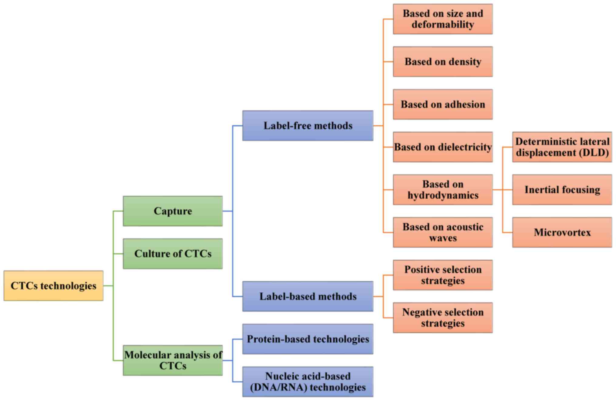
CTCs technologies mainly consist of two steps
(15,55). They can be used to identify and
enumerate these rare cells (8). They
can also be used to better understand their molecular features
(56). The first stage is detection
or capture. In this stage, some methods is used to increase the
concentration of CTCs and/or deplete surrounding normal blood cells
(15,55). In the second stage, the retrieved
CTCs are characterized by various molecular techniques (15,55). In
this new age of cancer treatment, the ex vivo culture of
captured CTCs also provides exceptional reagents to study cancer
metastasis, as well as perform individualized preclinical testing
for drug susceptibility (57)
(Fig. 1).
Isolation technologies for CTCs
CTCs are mainly detected and separated using
label-free and label-based methods (34). Label-free methods mainly depend on
physical properties of cells, which include size, deformability,
density, adhesion, and dielectric properties (15,34).
Label-based methods are mainly based on the binding affinity
between unique surface proteins expressed on CTCs, such as EpCAM
and synthetic molecular probes (15,34).
Aptamers are synthetic oligonucleotide ligands with high affinity
and specificity for targets compared to an antibody/antigen
interaction (18). They can be
selected using systematic evolution of ligands by exponential
enrichment technology. Aptamers can be used specifically to
recognize numerous kinds of targets, including small metal ions,
proteins, organic molecules, and whole cells (58). A number of aptamers have been
developed over the past decades to target cancer cell biomarkers,
such as prostate-specific membrane antigen (59), mucin 1 (60), cell surface vimentin (61), and EpCAM (41). Compared to antibodies, aptamers can
easily be synthesized in large quantities and modified with
different chemical groups (62). If
captured, cells can be released gently by using nucleases or a
complementary strand of aptamers (62). Peptides can serve as surrogates for
antibodies because short peptides at the contacting interfaces
participate in the molecular recognition between the antigens and
the antibodies (63). Since the
peptides are small and stable, they can be synthesized easily
(64). They are promising probes for
biological detection (63). Numerous
peptides with high affinity have been designed for the detection of
CTCs, and high capture efficiency has been achieved compared to
antibodies (63). For instance, a
microfluidic chip was developed to lithographically pattern silicon
nanowires functionalized with the specific CKAAKN peptide to
capture CTCs in patients with pancreatic cancer. The recovery rate
exceeded 95.6%, and after enzymatic release, the purity and cell
viability of the obtained CTCs was 28.5 and 93.5%, respectively
(63). Capture of CTCs may be
impeded because the tumor cells are heterogeneous (65). To solve this problem, two or more
probes could be made in response to cancer cells for wide use in
cancer diagnosis (65).
Indicators for CTCs isolation
technologies
A total of six technical indicators are typically
used to compare the performance of isolation technologies: i)
Recovery rate; ii) purity; iii) throughput; iv) sensitivity; v)
specificity; and vi) biocompatibility (32). Recovery rate, also known as capture
efficiency or capture rate, refers to the proportion of captured
CTCs from the total number of CTCs in the blood sample, which is
used to indicate the assay efficiency in isolating CTCs. However,
the recovery rate is almost always measured by recovering the cells
from the estimated cells when the total number of cancer cells is
already known. It is imperative to translate the aforementioned
measurements into clinical samples. Purity is the percentage of
isolated CTCs in the total isolated cells. The purity may be
heavily biased because of different experimental designs or because
of clinical samples at different disease stages. Throughput is used
to demonstrate how much blood is required for CTCs analysis. While
5–10 ml volume of blood was once required for detection of CTCs,
only 1–2 ml blood is now required owing to the development in CTCs
isolation technology. Clinical sensitivity and specificity are used
to indicate whether the technology can be used to correctly
identify patients with cancer and avoid false positives.
Biocompatibility refers to cells that maintain their integrity even
after sequential processing (34,66).
Since the actual number of CTCs in the clinical samples from
patients is unknown, blood samples from healthy donors spiked with
known numbers of tumor cells from cancer cell lines are used for
evaluating a system's performance for these parameters (67). An optimal technology should have high
recovery and purity of CTCs, high-throughput for sample processing,
and the ability to retain heterogeneous CTCs populations for
further downstream analysis to aid in providing options for
clinical management.
Label-free methods
Methods based on size and
deformability of CTCs
Size and deformability of cells are the foundation
of the physical separation approaches for CTCs enrichment in
peripheral blood (8). In some
studies, CTCs have been described as being larger and stiffer
compared with hematopoietic cells (68). Numerous technologies have been
developed based on these physical differences to increase recovery
rates in CTCs enrichment (34).
Size-based filtration is the most common enrichment strategy for
CTCs separation (69). The
commercially available ISET system was one of the earliest
technologies (70). In this system,
blood is diluted and then filtered with a 8-µm pore size filter
(70). CTCs are retained by the
filter as they are larger in size, while RBCs and leukocytes can
pass through the randomly distributed pores as their sizes are
smaller than the pore diameter (70). The variety of CTCs separation methods
depends solely on the size difference (69). Since the sizes of CTCs and leukocytes
overlap, the efficiency of the size-dependent filtration system to
capture target cells is limited (68). Therefore, a strategy to increase cell
size by utilizing modified microbeads that specifically bind to
CTCs before cell filtration was proposed to improve capture
efficiency (71). Up to 91% of
target cells were isolated from whole blood samples using this
microfluidic capture system at a flow rate of 1 ml/min (71).
Another possible solution to this limitation is to
harness the features of CTCs (72).
Cells that are more deformable or smaller can more easily pass
through the channel, while higher critical pressure is required to
help rigid and larger cells pass through the channel; the channel
will be blocked if the pressure provided is insufficient (72). A proposed mechanical low-pass
filtering technique based on microfluidic constriction has a
microfluidic device with consecutive constriction channels of
different sizes and an electrical current-sensing detector for
detection of novel CTCs in whole blood without any specific
antigen-antibody interaction or biochemical modification of the
cell surface (73). The ionic
current is reduced when cells pass through the constriction
channel. The intensity of the ionic current at the wide
constriction reflects the volume of the migrating cell, while the
residence time of the migrating cell at the narrow constriction
depends on the deformability of the cell (73). This consecutive constriction device
has been applied to distinguish CTCs from blood cells with an
accuracy rate of almost 95% (73).
Cell clogging reduces the efficiency of filtration
(66). To resolve this problem, an
electromagnetic vibration-based filtration (eVBF) device was
developed (74). The eVBF device can
be used to generate an electromagnetic force with a periodic
vibration that prevents cells from clogging (74). High-throughput (1 ml/min) is achieved
without the need for antibodies or chemical reagents in the
continuous isolation of CTCs or pretreatment processes (74). In addition, the eVBF device has been
optimized with a high-amplitude vibration, and its recovery rate
has increased to 80–90% for whole blood sampled with 100 or 1,000
gastric cancer cells/ml (74).
Caution and precision are required in the
determination of pore size and in measurement based on cell
deformability (75). In clinical
cases, CTCs from patients can be small and similar in size to
leukocytes (69). CTCs that have
undergone EMT and stem cell-like CTCs are capable of considerable
deformation and can be missed with deformability-based separation
technologies (69). Therefore,
physical isolation of CTCs using rational design and performance
analyses based on cell size and deformability may be unreliable
(75).
Methods based on density of CTCs
Density gradient centrifugation can be used to
separate CTCs from other blood cells based on the different
densities of different cell types (19,68). As
erythrocytes and granulocytes have a higher density compared with
the separation medium, they precipitate at the bottom of the pipe
when centrifugation is completed (19,68).
Monocytes and lymphocytes have lower densities compared with the
separation medium (19,68). Hence, they remain at the surface of
the medium or suspended in the medium. CTCs are mainly deposited in
the monocyte enrichment layer, allowing for their convenient
collection and analysis (19,68).
Ficoll-Hypaque® (Cytiva) and OncoQuick®
(Greiner Bio-One) density gradient centrifugation technologies are
the most popular in preclinical and clinical research (8). Ficoll-Hypaque® (Cytiva) is
based on a co-polymer of sucrose and epichlorohydrin (8). It is mainly used to recover mononuclear
cells from peripheral blood (8).
Ficoll-Hypaque® (Cytiva) has been used in laboratories
for a long time (67). Nonetheless,
the use of Ficoll-Hypaque® (Cytiva) remains hampered by
limitations (67). For instance, the
tumor cells may migrate to the plasma fraction or to the bottom of
the gradient owing to the formation of aggregates (8). This can result in the loss of CTCs
(8). Unlike the simple
Ficoll-Hypaque separation, OncoQuick® (Greiner Bio-One)
incorporates a porous barrier during the separation process to
prevent separated cells from mixing (76). This approach also has disadvantages.
For example, large CTCs and clusters of CTCs fall to the bottom,
making it impossible to eliminate leukocytes and difficult to
obtain a pure preparation of CTCs (77). To overcome these limitations, it is
necessary to combine centrifugation with another enrichment method
(8). The RosetteSep™ CTCs Enrichment
Cocktail (StemCell Technologies Inc.) in combination with
Ficoll-Hypaque® (Cytiva) separation improves the
enrichment and purity of CTCs (77).
The system utilizes tetrameric antibody complexes that crosslink
CD45-expressing leukocytes to RBCs (77). This artificially increases the
density of the labeled leukocytes, which gather at the bottom of
the density gradient (78). The
aforementioned system has successfully detected CTCs in 77% of
patients with prostate cancer and 90% of metastatic epithelial
ovarian cancer (79).
The sedimentation rate of a particle in suspension
is determined by the size of the particle and the difference in
density between the particle and the surrounding solution (68). The sedimentation rate increases
dramatically as the size and density of the particles increase
(68). Selective density
amplification of CTCs using antibody-coated microbeads is another
strategy to separate CTCs based on density (34). The microbeads specifically bind to
CTCs to maximize the difference in sedimentation rates between CTCs
and normal blood cells, such as leukocytes and erythrocytes in
blood samples (34). Huang et
al (68) proposed a novel
density gradient centrifugation method that used biodegradable
gelatin nanoparticle-coated silicon microbeads (SiO2@Gel MBs)
coated with anti-EpCAM and anti-CD146 antibodies to enhance the
size and density of mesenchymal CTCs. The authors reported high
rates of recovery (>80%) and purity (>85%). Degradation of
the gelatin coating by matrix metalloproteinase-9 enzyme enabled
high rates of CTCs release (94%) and viability (92.5%) (68). The authors also demonstrated the
unparalleled robustness of this method in downstream CTCs analyses,
such as the detection of phosphatidylinositol-4,5-bisphosphate
3-kinase catalytic subunit α mutations (68). The efficiency and versatility of the
multifunctional density microbeads approach provides new
opportunities for personalized cancer diagnostics and
treatments.
Methods based on adhesion of CTCs
Compared to normal blood cells, a simple and
effective strategy was reported by Chen et al (80). The authors used the preferential
adhesion of cancer cells to surfaces that were rough on the
nanoscale to capture CTCs regardless of their physical size and
without using capture antibody. The reactive ion etching method was
used to treat bare glass surfaces to produce different degrees of
roughness at nanoscale resolution (80). On average, the recovery rates were
88.7±3.0% and 93.3±1.5% for MCF-7 cells mixed with peripheral blood
mononuclear cells and spiked in lysed blood, respectively, while
for MD-MB-231 cells, the recovery rates were 94.9±2.4% and
95.4±2.2% for the peripheral blood mononuclear cells and lysed
blood samples, respectively (80).
The aforementioned results confirmed the efficiency of nanorough
substrates to capture cancer cells (80).
Methods based on dielectricity of
CTCs
In addition to size, density, and deformability, the
dielectricity of CTCs can also be harnessed to distinguish CTCs
from other blood cells including RBCs, platelets and WBCs (81). Dielectrophoresis-based enrichment
technologies rely on the different levels of polarization between
cells generated using a non-uniform alternative electric field
(82). The cells are also isolated
when they move toward electric fields that have different
intensities (82). The
polarizability of a cell depends on its composition, morphology,
and frequency of the applied electric field (82). Different cell populations can be
isolated when they move in opposite directions if the electrical
excitation frequency is set between the cross-over frequencies for
different cells (81).
Dielectrophoretic field-flow fractionation has been utilized in the
commercial ApoStream™ (ApoCell) system that enables the label-free
isolation of viable cancer cells (83). The system utilizes a 45–85 kHz AC
signal that is in the range of the cross-over frequencies of cancer
cells (~30-40 kHz) and peripheral blood cells (90–140 kHz)
(84). Cancer cells move toward
electrodes and force blood cells to the center of the channel
(85,86). The cancer cells are enriched and
collected at the product collection port, while blood cells exit
from the chamber via the waste outlet (85,86). The
system was used to obtain recovery rates of 75.4±3.1% and 71.2±1.6%
for SKOV3 ovarian cancer cells and MDA-MB-231 breast cancer cells,
respectively. In addition, the viability of these enriched cancer
cells exceeded 97.1% (84). The
continuous-flow design of the ApoStream™ allows the processing of a
7.5-ml sample within 1 h, which is faster compared with most
dielectrophoresis-based CTCs enrichment technologies (84).
Methods based on hydrodynamics
A novel approach used to enrich CTCs relies on
unique hydrodynamics and is used in microfluidic channels (87–89). The
hydrodynamics method is primarily based on the different movements
of cells with different sizes in response to the hydrodynamic force
(90). There are three main types,
which are separately detailed below: Deterministic lateral
displacement (DLD), inertial focusing and micro-vortex (90).
Methods based on DLD
DLD is a technology in which the microcolumns in a
chip are designed and controlled to have spacings smaller or larger
compared with the critical particle diameter (69). This microcolumn array can influence
the displacement of the suspended particles according to
hydrodynamic forces created by the flow conditions (69). A parameter termed the critical
particle diameter defined in these devices predicts the
displacement of suspended particles as they pass through the array
of microcolumns (69,88). If CTCs are larger compared with the
critical diameter of the device, they are laterally displaced when
they collide with the microcolumn and move to one side (following
the bumped mode) (69,88). In contrast, blood cells that are
smaller than the critical diameter maintain their original path
without being laterally displaced after collision with the
microcolumn (following the zigzag mode) (69,88). The
array of microcolumns in DLD devices is arranged so that the
trajectory of the particles is precisely controlled and particles
larger or smaller compared with the critical diameter are more
rapidly separated (69,88). A DLD array device was designed by
Loutherback et al (91) to
separate CTCs at a flow rate of 10 ml/min. The capture efficiency
of this device exceeded 85% without affecting cell viability
(91). The DLD device can become
clogged with cells if the detected clinical samples are too sticky
or contain large amounts of debris (91).
Methods based on inertial
focusing
The inertial focusing method used in microfluidic
devices exploits the advantage of the hydraulic phenomenon, in
which particles/cells of different sizes occupy different
equilibrium positions in the cross-section of the microfluidic
channel because of the balance between the inertial lift force and
the Dean drag force (87). As a
result, when flowing in a spiral channel, larger cells move toward
the inner wall, while smaller cells move toward the outer wall
under the influences of these forces (69). CTCs separate from blood cells
(87). The device can be operated at
a wide range of flow rates from 5 µl/min-8 ml/min (87). Hydraulic-based separation has been
further exploited by constructing a cascaded microfluidic device
consisting of two spiral channels and one zigzag channel, to
simultaneously isolate different types of CTCs from human blood
(87). Recovery rates of 80.75% for
A549 human lung cancer cells and 73.75% for MCF-7 human breast
cancer cells have been reported (87).
Methods based on microvortex
Microvortex trapping is another hydrodynamics-based
CTCs enrichment technology that enriches CTCs in the microvortex
formed in a microfluidic device. The method is based on the high
flow rate that produces microvortices when cells flow through
regions of expanded and contracted volume. Smaller cells flow
smoothly through the main channel, while larger cells swirl in the
vortex and remain in the chamber. Finally, the flow rate is reduced
to release the captured cells from the vortex (89,92,93). The
microfluidic vortex chip was used by Rennier et al (94) to isolate CTCs from patients with
advanced prostate cancer. CTCs were isolated with a purity of
1.74–37.59% (94). A disadvantage of
the method is that some microfluidic vortex chips may produce
bubbles inside the channel (94).
Methods based on acoustic waves
In 2014, Ding et al (95) proposed the use of acoustic waves to
separate pathological cells from normal blood cells. Other studies
have also addressed the use of acoustic waves to separate CTCs
(96). Cells that are sustained by
acoustic force with different magnitudes depending on the cell
sizes, density, and deformation are translated by the acoustic
force to the nodes with zero periodic pressure variations or
maximum pressure to reach a balance point (95). An acoustic-based microfluidic device
was developed by Wu et al (96) using tilted-angle surface acoustic
waves to form multiple regions of slanted nodes or antinodes inside
the microfluidic channel. Cells transiting these regions experience
different levels of acoustic forces, which may change their
positions inside the channel (90).
A constructed high-throughput platform integrating acoustics and
microfluidics has enabled the separation of CTCs from peripheral
blood (96). The separated cells
remain structurally, biologically, and functionally intact
(96). The system can separate
cancer cells from leukocytes at a throughput of up to 7.5 ml
h−1 with an 86% recovery rate and no effect on cell
proliferation (96). Using acoustic
waves to separate cells is a relatively mild method that can
maintain the original state, phenotype, and genotype of CTCs to the
greatest extent (96). However, this
method is greatly influenced by environmental factors (96).
Label-based methods
Specific cancer biomarkers are indispensable in
most biological detection methods (47). Cancer biomarkers refer to the
molecular changes that are measurable and occur between normal and
cancerous tissues of patients (47).
Specific molecular characteristics and pathological changes occur
in each cancer during the transition from normal to cancerous cells
(47). It is important to identify
these biomarkers so that they can be further applied in capturing
and isolating CTCs that are commonly described to express EpCAM and
cytokeratins (CKs), and to be nucleated (they are identified by
staining with a nuclear dye like 4′,6-diamidino-2-phenylindole)
(67). These biomarkers are not used
to express the leukocyte surface marker CD45 (67). Other biomarkers commonly used to
specifically detect CTCs include human epidermal growth factor
receptor 2, epidermal growth factor receptor, vimentin, and CD44
(47). In addition, ‘negative
markers’ may be used to identify and eliminate questionable cells
(47). These markers include the
leukocyte markers CD45, CD66, and CD15, platelet marker CD61, and
apoptosis marker M30 (97).
Label-based methods can be further divided into two sub-categories
according to the target cells: Negative selection and positive
selection (97).
Positive selection strategies
CTCs are regarded as target cells in positive
sorting methods (98). EpCAM is used
as a specific biomarker in most of these methods (19). Positive selection to isolate CTCs is
performed based on ligand-labeled beads, columns, or other devices
(8). The CellSearch®
system (Veridex; Menarini Silicon Biosystems) is a commonly
accepted method that automatically detects EpCAM- and
keratin-positive CTCs (32). This is
followed by immunostaining of the captured cells and semi-automated
fluorescence microscopy to evaluate the immunofluorescence
(19). The aforementioned system has
been approved by the United States Food and Drug Administration for
analysis of blood samples from patients with metastatic breast,
prostate, and colorectal cancer (99). The capture step is characterized by
high sensitivity, but may lack specificity. In another innovative
method marketed as MagSweeper by Illumina Inc., blood samples are
diluted and prelabeled with EpCAM-coated magnetic particles
(8). The cells are subsequently
captured with a magnetic rod that is swept through the sample
(56). The magnetic rod is then
placed in a washing well, and the labeled cells are released by an
external magnetic field in a buffer solution (56). The released cells remain viable and
can be successfully transferred and cultured (56).
All the aforementioned methods have limitations
because they rely on the enrichment of EpCAM-positive cells. Hence,
they can only detect the EpCAM-positive subpopulation of CTCs.
However, once CTCs undergo EMT, these EpCAM-based methods may be
ineffective because EMT reduces EpCAM expression. CTCs with low or
no EpCAM expression can remain undetected and their capture rate is
greatly decreased (30).
Negative selection strategies
Leukocytes are regarded as target cells in negative
sorting (34). Anti-CD45 is the
biomarker used most often, reflecting its binding affinity with
leukocytes (19). The
EasySep® leukocyte depletion kit marketed by Stem Cells
Technologies Inc. allows the enrichment of CTCs through leukocyte
depletion using CD45-labeled magnetic beads (100). The CTCs remain viable for use in
further experiments after leukocyte depletion (100). Leukocyte depletion methods
reportedly reduce purity compared to preparations obtained by
positive CTCs selection. Hence, negative selection strategies are
often used in combination with other enrichment technologies
(34).
Combination of label-free and
label-based methods
Label-free isolation does not vary with the level
of expression of the selected marker on the cell surface (90). Instead, the approach is used
according to the physical differences between CTCs and blood cells
(90). In addition, a label-free
isolation method can be realized simply, rapidly, and at low cost
compared with label-based isolation (90). However, label-free isolation has low
specificity due to the overlap in size, density, rigidity, and
other parameters between CTCs and leukocytes (68). Numerous researchers have sought to
combine label-free and label-based methods to increase the
efficiency of the isolation process, as both the label-free methods
and label-based methods have limitations (101,102).
The novel Herringbone (HB)-Chip is designed such that the
microvortex increases the number of interactions between target
CTCs and the antibody-coated chip surface (101). This system has been used to
successfully isolate CTCs in patients with metastatic disease for
different cancers, such as metastatic prostate and breast cancer
(103), as well as to isolate CTCs
clusters (104).
In vivo isolation of CTCs
Ex vivo CTCs isolation technologies have
relatively low sensitivity as limited volume of blood is sampled
from patients (47). A total of 1–10
ml blood is required for CTCs detection (90). However, the amount of CTCs in blood
is very small with 1–10 CTCs/ml (90). The detection systems used also have
limitations. CellCollector® marketed by Gilupi GmbH
isolates CTCs based on EpCAM recognition. It is the first in
vivo CTCs isolation product with CE approval (CE abbreviation
of French phrase ‘Conformité Européene’ which literally means
‘European Conformity’) (67).
Specifically, CellCollector® (Gilupi) is a medical wire
coated with anti-EpCAM antibodies. The wire is directly placed in
the bloodstream of the patient through a permanent catheter. The
wire remains in the vein of the arm for 30 min, where it contacts a
larger volume of blood and captures CTCs in vivo during this
period (105,106). CTCs that have not been detected in
healthy volunteers could be isolated in early-stage cancer patients
not yet diagnosed with distant metastases (75). However, it is not simple to implement
this technique in the clinic as manual selection is required to
detect CTCs (67).
Culture of CTCs
Culture of viable CTCs is an attractive option for
the study of captured CTCs (107).
Once a CTCs line is established in cell culture, it can be used for
direct testing of drug sensitivity or can be implanted into
immunosuppressed mice to create xenograft models, permitting
further drug testing and genetic profiling studies (57,107).
Additionally, the metastatic mechanism can be further understood by
using experimental platforms that mimic the tumor microenvironment
in vitro (108). A
three-dimensional porous poly(ε-caprolactone) scaffold-based method
was established by De et al (108) to mimic more closely the native
cellular in vivo environment and allow the deposition of
extracellular matrix. Culture of CTCs from RBC-depleted nucleated
cell pellets of patients with advanced breast cancer under hypoxic
conditions without any prior enrichment allowed the detection of
CK-positive and CD45-negative CTCs in 12 of 16 patient samples
(108). However, as the captured
CTCs were removed from their host environment and the immune
system, they may be altered, precluding meaningful conclusions
regarding disease evolution or treatment resistance (108). Hence, it is necessary to establish
robust and reliable culture conditions for CTCs.
Molecular analysis of CTCs
It has been suggested in some studies that the
enumeration of CTCs is not sufficient to guide therapeutic
decisions (109). Rather, molecular
analysis of CTCs should be performed to determine the design of the
target therapy (110). Two main
approaches have been adopted according to current technologies for
molecular characterization of CTCs: Protein-based technologies and
nucleic acid-based (DNA/RNA) technologies (10). These are reviewed in detail elsewhere
(10,15,98,110)
and not discussed in the present review.
Discussion
CTCs detection has numerous advantages compared to
conventional invasive biopsy (41).
For example, with approval of the patients, blood samples can be
easily collected. CTCs harbor considerable information on
tumorigenesis, tumor progression, metastasis, and drug resistance
(41). This information increases
the knowledge and treatment of tumor diseases (15). The new approach of employing CTCs has
paved the way for next-generation liquid biopsy diagnostics,
especially in tumors that are difficult to biopsy and in metastatic
lesions (109).
There is great anticipation for label-free and
label-based methods for CTCs isolation (90). Each method has value, and each aims
to overcome the pitfalls involving sensitivity, specificity,
throughput, and/or purity (90).
Numerous reliable CTCs isolation platforms are available (90). However, the methodology that is
superior and that would be the most suitable for the end user
requirements remains unclear (82).
These questions can be answered based on accrued biological
knowledge (82).
The only two types of CTCs assays that are
presently used are CellSearch® system (Veridex; Menarini
Silicon Biosystems) and CellCollector® (Gilupi)
(67). Both technologies have been
limited in their clinical application, largely due to their low
sensitivity (33). The technologies
need to satisfy the following demands to meet the clinical
requirements of CTCs detection (90). First, the assays must fully consider
cell heterogeneity to distinguish different subpopulations of CTCs,
and to achieve more efficient isolation of the target cells
(111). This goal may require new
technologies or a combination of several technologies (10). Second, high-throughput is conducive
for the rapid recovery of a sufficient amount of cancer cells
collected from a large volume. A high-throughput system that is
more suited to clinical applications is required (34). Third, a shorter separation time would
help preserve cell freshness, viability, and integrity and would
maximize the potential to establish in vitro cultured CTCs
lines (108). These advancements
would make it possible to perform tasks such as investigation of
CTCs phenotypic and biomarker heterogeneity or isolation of CTCs
clusters that are not possible with slower processing methods due
to the degradation of CTCs (108).
Fourth, specific verification strategies to prove that the isolated
targeted cells are real CTCs are important for clinical
applications (8). Fifth, applying
automated devices as much as possible is desirable to minimize
intervention by human operators (98). Sixth, standardization is crucial to
yield convincing and reproducible results and ensure the
generalized use of the approach (42). Finally, clinical demonstration of the
prowess of this technology in clinical trials is important
(112). Most of the detection
technologies have not yet been fully applied in clinical practice,
and there is a demand for large-scale clinical validation (32). Moving the aforementioned techniques
from the bench to the clinic is becoming a hot topic of research in
the CTCs field.
It is hypothesized that there are two main
directions for the development of CTCs in the future. One is the
development of CTCs detection technology with higher sensitivity
and specificity. The other is functional assessments of CTCs,
especially the decoding of molecular pathological characteristics
such as genes, proteins, and methylation expression profiles.
Although great progress has been made, there is a long way to go
before CTCs-based liquid biopsy is widely used as a routine test in
clinical applications.
Acknowledgements
Not applicable.
Funding
The present study was funded by the Maternal and
Child Health Research Project of Jiangsu Province in 2019 (grant
no. F201952) and Huai'an Health and Scientific Research Project in
2019 (grant no. 20).
Availability of data and materials
Data sharing is not applicable to this article, as
no datasets were generated or analyzed during the current
study.
Authors' contributions
XH, XZ and YL all contributed toward data analysis,
drafting and critically revising the paper, gave final approval of
the version to be published, and agree to be accountable for all
aspects of the work.
Ethics approval and consent to
participate
Not applicable.
Patient consent for publication
Not applicable.
Competing interests
The authors declare that they have no competing
interests.
Glossary
Abbreviations
Abbreviations:
|
CTCs
|
circulating tumor cells
|
|
RBCs
|
red blood cells
|
|
WBCs
|
white blood cells
|
|
EMT
|
epithelial-to-mesenchymal
transition
|
|
EpCAM
|
epithelial cell adhesion molecule
|
|
MET
|
mesenchymal-to-epithelial
transition
|
|
eVBF
|
electromagnetic vibration-based
filtration
|
|
DLD
|
deterministic lateral
displacement
|
References
|
1
|
Bray F, Ferlay J, Soerjomataram I, Siegel
RL, Torre LA and Jemal A: Global cancer statistics 2018: GLOBOCAN
estimates of incidence and mortality worldwide for 36 cancers in
185 countries. CA Cancer J Clin. 68:394–424. 2018. View Article : Google Scholar : PubMed/NCBI
|
|
2
|
Ferlay J, Colombet M, Soerjomataram I,
Mathers C, Parkin DM, Piñeros M, Znaor A and Bray F: Estimating the
global cancer incidence and mortality in 2018: GLOBOCAN sources and
methods. Int J Cancer. 144:1941–1953. 2019. View Article : Google Scholar : PubMed/NCBI
|
|
3
|
Cheng SJ, Hsieh KY, Chen SL, Chen CY,
Huang CY, Tsou HI, Kumar PV, Hsieh JC and Chen GY: Microfluidics
and nanomaterial-based technologies for circulating tumor cell
isolation and detection. Sensors (Basel). 20:18752020. View Article : Google Scholar
|
|
4
|
Maconi G, Manes G and Porro GB: Role of
symptoms in diagnosis and outcome of gastric cancer. World J
Gastroenterol. 14:1149–1155. 2008. View Article : Google Scholar : PubMed/NCBI
|
|
5
|
Necula L, Matei L, Dragu D, Neagu AI,
Mambet C, Nedeianu S, Bleotu C, Diaconu CC and Chivu-Economescu M:
Recent advances in gastric cancer early diagnosis. World J
Gastroenterol. 25:2029–2044. 2019. View Article : Google Scholar : PubMed/NCBI
|
|
6
|
Smyth EC, Nilsson M, Grabsch HI, van
Grieken NC and Lordick F: Gastric cancer. Lancet. 396:635–648.
2020. View Article : Google Scholar : PubMed/NCBI
|
|
7
|
Chaffer CL and Weinberg RA: A perspective
on cancer cell metastasis. Science. 331:1559–1564. 2011. View Article : Google Scholar : PubMed/NCBI
|
|
8
|
Gabriel MT, Calleja LR, Chalopin A, Ory B
and Heymann D: Circulating tumor cells: A review of non-EpCAM-based
approaches for cell enrichment and isolation. Clin Chem.
62:571–581. 2016. View Article : Google Scholar : PubMed/NCBI
|
|
9
|
Prasanna BK, Balakrishnan A and Kumar P:
Circulating tumor cell clusters and circulating tumor cell-derived
explant models as a tool for treatment response. Biotechniques.
69:362–363. 2020. View Article : Google Scholar : PubMed/NCBI
|
|
10
|
Tellez-Gabriel M, Heymann MF and Heymann
D: Circulating tumor cells as a tool for assessing tumor
heterogeneity. Theranostics. 9:4580–4594. 2019. View Article : Google Scholar : PubMed/NCBI
|
|
11
|
Leone K, Poggiana C and Zamarchi R: The
interplay between circulating tumor cells and the immune system:
From immune escape to cancer immunotherapy. Diagnostics (Basel).
8:592018. View Article : Google Scholar
|
|
12
|
Tayoun T, Faugeroux V, Oulhen M, Aberlenc
A, Pawlikowska P and Farace F: CTC-derived models: A window into
the seeding capacity of circulating tumor cells (CTCs). Cells.
8:11452019. View Article : Google Scholar
|
|
13
|
Meng S, Tripathy D, Frenkel EP, Shete S,
Naftalis EZ, Huth JF, Beitsch PD, Leitch M, Hoover S, Euhus D, et
al: Circulating tumor cells in patients with breast cancer
dormancy. Clin Cancer Res. 10:8152–8162. 2004. View Article : Google Scholar : PubMed/NCBI
|
|
14
|
Au SH, Edd J, Haber DA, Maheswaran S,
Stott SL and Toner M: Clusters of circulating tumor cells: A
biophysical and technological perspective. Curr Opin Biomed Eng.
3:13–19. 2017. View Article : Google Scholar : PubMed/NCBI
|
|
15
|
Thiele JA, Bethel K, Králíčková M and Kuhn
P: Circulating tumor cells: Fluid surrogates of solid tumors. Annu
Rev Pathol. 12:419–447. 2017. View Article : Google Scholar : PubMed/NCBI
|
|
16
|
Mocan T, Simão AL, Castro RE, Rodrigues
CMP, Słomka A, Wang B, Strassburg C, Wöhler A, Willms AG and Kornek
M: Liquid biopsies in hepatocellular carcinoma: Are we winning? J
Clin Med. 9:15412020. View Article : Google Scholar
|
|
17
|
Wu C, Zhang J, Li H, Xu W and Zhang X: The
potential of liquid biopsies in gastrointestinal cancer. Clin
Biochem. 84:1–12. 2020. View Article : Google Scholar : PubMed/NCBI
|
|
18
|
Li X, Zhang P, Dou L, Wang Y, Sun K, Zhang
X, Song G, Zhao C, Li K, Bai Y, et al: Detection of circulating
tumor cells in breast cancer patients by nanopore sensing with
aptamer-mediated amplification. ACS Sens. 5:2359–2366. 2020.
View Article : Google Scholar : PubMed/NCBI
|
|
19
|
Yap K, Cohen EN, Reuben JM and Khoury JD:
Circulating tumor cells: State-of-the-art update on technologies
and clinical applications. Curr Hematol Malig Rep. 14:353–357.
2019. View Article : Google Scholar : PubMed/NCBI
|
|
20
|
Yang D, Yang X, Li Y, Zhao P, Fu R, Ren T,
Hu P, Wu Y, Yang H and Guo N: Clinical significance of circulating
tumor cells and metabolic signatures in lung cancer after surgical
removal. J Transl Med. 18:2432020. View Article : Google Scholar : PubMed/NCBI
|
|
21
|
Zapatero A, Gómez-Caamaño A, Cabeza
Rodriguez MÁ, Muinelo-Romay L, Martin de Vidales C, Abalo A, Calvo
Crespo P, Leon Mateos L, Olivier C and Vega Piris LV: Detection and
dynamics of circulating tumor cells in patients with high-risk
prostate cancer treated with radiotherapy and hormones: A
prospective phase II study. Radiat Oncol. 15:1372020. View Article : Google Scholar : PubMed/NCBI
|
|
22
|
Sun YF, Wang PX, Cheng JW, Gong ZJ, Huang
A, Zhou KQ, Hu B, Gao PT, Cao Y, Qiu SJ, et al: Postoperative
circulating tumor cells: An early predictor of extrahepatic
metastases in patients with hepatocellular carcinoma undergoing
curative surgical resection. Cancer Cytopathol. 128:733–745. 2020.
View Article : Google Scholar : PubMed/NCBI
|
|
23
|
Deutsch TM, Stefanovic S, Feisst M,
Fischer C, Riedel F, Fremd C, Domschke C, Pantel K, Hartkopf AD,
Sutterlin M, et al: Cut-off analysis of CTC change under systemic
therapy for defining early therapy response in metastatic breast
cancer. Cancers (Basel). 12:10552020. View Article : Google Scholar
|
|
24
|
Dimitrov-Markov S, Perales-Patón J,
Bockorny B, Dopazo A, Muñoz M, Baños N, Bonilla V, Menendez C,
Duran Y, Huang L, et al: Discovery of new targets to control
metastasis in pancreatic cancer by single-cell transcriptomics
analysis of circulating tumor cells. Mol Cancer Ther. 19:1751–1760.
2020. View Article : Google Scholar : PubMed/NCBI
|
|
25
|
Pan X and Zhang X: Utility of circulating
tumor cells and DNA in the management of advanced colorectal
cancer. Future Oncol. 16:1289–1299. 2020. View Article : Google Scholar : PubMed/NCBI
|
|
26
|
Kim H, Lim M, Kim JY, Shin SJ, Cho YK and
Cho CH: Circulating tumor cells enumerated by a centrifugal
microfluidic device as a predictive marker for monitoring ovarian
cancer treatment: A pilot study. Diagnostics (Basel). 10:2492020.
View Article : Google Scholar
|
|
27
|
Hong Y, Fang F and Zhang Q: Circulating
tumor cell clusters: What we know and what we expect (Review). Int
J Oncol. 49:2206–2216. 2016. View Article : Google Scholar : PubMed/NCBI
|
|
28
|
Cabel L, Proudhon C, Gortais H, Loirat D,
Coussy F, Pierga JY and Bidard FC: Circulating tumor cells:
Clinical validity and utility. Int J Clin Oncol. 22:421–430. 2017.
View Article : Google Scholar : PubMed/NCBI
|
|
29
|
Aoki M, Shoji H, Kashiro A, Takeuchi K,
Shimizu Y and Honda K: Prospects for comprehensive analyses of
circulating tumor cells in tumor biology. Cancers (Basel).
12:11352020. View Article : Google Scholar
|
|
30
|
Brown HK, Tellez-Gabriel M, Cartron PF,
Vallette FM, Heymann MF and Heymann D: Characterization of
circulating tumor cells as a reflection of the tumor heterogeneity:
Myth or reality? Drug Discov Today. 24:763–772. 2019. View Article : Google Scholar : PubMed/NCBI
|
|
31
|
Lowes LE and Allan AL: Circulating tumor
cells and implications of the epithelial-to-mesenchymal transition.
Adv Clin Chem. 83:121–181. 2018. View Article : Google Scholar : PubMed/NCBI
|
|
32
|
Lin E, Cao T, Nagrath S and King MR:
Circulating tumor cells: Diagnostic and therapeutic applications.
Annu Rev Biomed Eng. 20:329–352. 2018. View Article : Google Scholar : PubMed/NCBI
|
|
33
|
Hong S and Wang AZ: Nanotechnology
enabling the use of circulating tumor cells (CTCs) as reliable
cancer biomarkers. Adv Drug Deliv Rev. 125:1–2. 2018. View Article : Google Scholar : PubMed/NCBI
|
|
34
|
Zou D and Cui D: Advances in isolation and
detection of circulating tumor cells based on microfluidics. Cancer
Biol Med. 15:335–353. 2018. View Article : Google Scholar : PubMed/NCBI
|
|
35
|
Peng Y, Peng Y, Tang S, Shen H, Sheng S,
Wang Y, Wang T, Cai J, Xie G and Feng W: PdIrBP mesoporous
nanospheres combined with superconductive carbon black for the
electrochemical determination and collection of circulating tumor
cells. Mikrochim Acta. 187:2162020. View Article : Google Scholar : PubMed/NCBI
|
|
36
|
Chen Y, Peng J, Lai Y, Wu B, Sun L and
Weng J: Ultrasensitive label-free detection of circulating tumor
cells using conductivity matching of two-dimensional semiconductor
with cancer cell. Biosens Bioelectron. 142:1115202019. View Article : Google Scholar : PubMed/NCBI
|
|
37
|
Chen J, Chen L, Du S, Wu J, Quan M, Yin H,
Wu Y, Ye X, Liang X and Jiang H: High sensitive detection of
circulating tumor cell by multimarker lipid magnetic nanoparticles
and clinical verifications. J Nanobiotechnology. 17:1162019.
View Article : Google Scholar : PubMed/NCBI
|
|
38
|
Dou B, Xu L, Jiang B, Yuan R and Xiang Y:
Aptamer-functionalized and gold nanoparticle array-decorated
magnetic graphene nanosheets enable multiplexed and sensitive
electrochemical detection of rare circulating tumor cells in whole
blood. Anal Chem. 91:10792–10799. 2019. View Article : Google Scholar : PubMed/NCBI
|
|
39
|
Chen SL, Chen CY, Hsieh JC, Yu ZY, Cheng
SJ, Hsieh KY, Yang JW, Kumar PV, Lin SF and Chen GY: Graphene
oxide-based biosensors for liquid biopsies in cancer diagnosis.
Nanomaterials (Basel). 9:17252019. View Article : Google Scholar
|
|
40
|
Zhu Y, Zou C, Zhang J, Jiang W, Guan F,
Tang K, Li S, Li G, Wang J and Ke Z: dynamically monitoring the
clonal evolution of lung cancer based on the molecular
characterization of circulating tumor cells using aptamer
cocktail-modified nanosubstrates. ACS Appl Mater Interfaces.
12:5671–5679. 2020. View Article : Google Scholar : PubMed/NCBI
|
|
41
|
Ding P, Wang Z, Wu Z, Zhou Y, Sun N and
Pei R: Natural biointerface based on cancer cell membranes for
specific capture and release of circulating tumor cells. ACS Appl
Mater Interfaces. 12:20263–20270. 2020. View Article : Google Scholar : PubMed/NCBI
|
|
42
|
Li W, Wang H, Zhao Z, Gao H, Liu C, Zhu L,
Wang C and Yang Y: Emerging nanotechnologies for liquid biopsy: The
detection of circulating tumor cells and extracellular vesicles.
Adv Mater. 31:e18053442019. View Article : Google Scholar : PubMed/NCBI
|
|
43
|
Chen M, Liu A, Chen B, Zhu DM, Xie W, Deng
FF, Ji LW, Chen LB, Huang HM, Fu YR, et al: Erythrocyte-derived
vesicles for circulating tumor cell capture and specific tumor
imaging. Nanoscale. 11:12388–12396. 2019. View Article : Google Scholar : PubMed/NCBI
|
|
44
|
Xiong K, Wei W, Jin Y, Wang S, Zhao D,
Wang S, Gao X, Qiao C, Yue H, Ma G and Xie HY: Biomimetic
immuno-magnetosomes for high-performance enrichment of circulating
tumor cells. Adv Mater. 28:7929–7935. 2016. View Article : Google Scholar : PubMed/NCBI
|
|
45
|
Rao L, Meng QF, Huang Q, Wang Z, Yu GT, Li
A, Ma W, Zhang N, Guo SS, Zhao XZ, et al: Platelet-leukocyte hybrid
membrane-coated immunomagnetic beads for highly efficient and
highly specific isolation of circulating tumor cells. Adv Funct
Mater. 28:18035312018. View Article : Google Scholar
|
|
46
|
Wu L, Ding H, Qu X, Shi X, Yang J, Huang
M, Zhang J, Zhang H, Song J, Zhu L, et al: Fluidic multivalent
membrane nanointerface enables synergetic enrichment of circulating
tumor cells with high efficiency and viability. J Am Chem Soc.
142:4800–4806. 2020. View Article : Google Scholar : PubMed/NCBI
|
|
47
|
Gribko A, Künzel J, Wünsch D, Lu Q, Nagel
SM, Knauer SK, Stauber RH and Ding GB: Is small smarter?
Nanomaterial-based detection and elimination of circulating tumor
cells: Current knowledge and perspectives. Int J Nanomedicine.
14:4187–4209. 2019. View Article : Google Scholar : PubMed/NCBI
|
|
48
|
Salmanogli A and Gokcen D: Identification
of circulating tumor cells using plasmonic resonance effect:
Lab-on-a-chip analysis and modelling. J Nanosci Nanotechnol.
20:1341–1350. 2020. View Article : Google Scholar : PubMed/NCBI
|
|
49
|
Al-Halhouli A, Al-Faqheri W, Alhamarneh B,
Hecht L and Dietzel A: Spiral microchannels with trapezoidal cross
section fabricated by femtosecond laser ablation in glass for the
inertial separation of microparticles. Micromachines (Basel).
9:1712018. View Article : Google Scholar
|
|
50
|
Hao N, Nie Y, Tadimety A, Shen T and Zhang
JXJ: Microfluidics-enabled rapid manufacturing of hierarchical
silica-magnetic microflower toward enhanced circulating tumor cell
screening. Biomater Sci. 6:3121–3125. 2018. View Article : Google Scholar : PubMed/NCBI
|
|
51
|
Jackson JM, Witek MA, Hupert ML, Brady C,
Pullagurla S, Kamande J, Aufforth RD, Tignanelli CJ, Torphy RJ, Yeh
JJ and Soper SA: UV activation of polymeric high aspect ratio
microstructures: Ramifications in antibody surface loading for
circulating tumor cell selection. Lab Chip. 14:106–117. 2014.
View Article : Google Scholar : PubMed/NCBI
|
|
52
|
Fan X, Jia C, Yang J, Li G, Mao H, Jin Q
and Zhao J: A microfluidic chip integrated with a high-density
PDMS-based microfiltration membrane for rapid isolation and
detection of circulating tumor cells. Biosens Bioelectron.
71:380–386. 2015. View Article : Google Scholar : PubMed/NCBI
|
|
53
|
Wu Z, Zhao D, Zhang Y, Huang L, Huang H,
Guo Q, Zhang W, Hou C, Wang H, Zhang Q, et al: Facile synthesis of
3D hierarchical micro-/nanostructures in capillaries for efficient
capture of circulating tumor cells. J Colloid Interface Sci.
575:108–118. 2020. View Article : Google Scholar : PubMed/NCBI
|
|
54
|
Khoo BL, Grenci G, Lim YB, Lee SC, Han J
and Lim CT: Expansion of patient-derived circulating tumor cells
from liquid biopsies using a CTC microfluidic culture device. Nat
Protoc. 13:34–58. 2018. View Article : Google Scholar : PubMed/NCBI
|
|
55
|
Harigopal M, Kowalski D and Vosoughi A:
Enumeration and molecular characterization of circulating tumor
cells as an innovative tool for companion diagnostics in breast
cancer. Expert Rev Mol Diagn. 20:815–828. 2020. View Article : Google Scholar : PubMed/NCBI
|
|
56
|
Deng G, Krishnakumar S, Powell AA, Zhang
H, Mindrinos MN, Telli ML, Davis RW and Jeffrey SS: Single cell
mutational analysis of PIK3CA in circulating tumor cells and
metastases in breast cancer reveals heterogeneity, discordance, and
mutation persistence in cultured disseminated tumor cells from bone
marrow. BMC Cancer. 14:4562014. View Article : Google Scholar : PubMed/NCBI
|
|
57
|
Maheswaran S and Haber DA: Ex vivo culture
of CTCs: An emerging resource to guide cancer therapy. Cancer Res.
75:2411–2415. 2015. View Article : Google Scholar : PubMed/NCBI
|
|
58
|
Sun D, Lu J, Zhang L and Chen Z:
Aptamer-based electrochemical cytosensors for tumor cell detection
in cancer diagnosis: A review. Anal Chim Acta. 1082:1–17. 2019.
View Article : Google Scholar : PubMed/NCBI
|
|
59
|
Fan X, Guo Y, Wang L, Xiong X, Zhu L and
Fang K: Diagnosis of prostate cancer using anti-PSMA aptamer
A10-3.2-oriented lipid nanobubbles. Int J Nanomedicine.
11:3939–3950. 2016. View Article : Google Scholar : PubMed/NCBI
|
|
60
|
Yin X, Chen B, He M and Hu B: A
multifunctional platform for the capture, release, and enumeration
of circulating tumor cells based on aptamer binding, nicking
endonuclease-assisted amplification, and inductively coupled plasma
mass spectrometry detection. Anal Chem. 92:10308–10315. 2020.
View Article : Google Scholar : PubMed/NCBI
|
|
61
|
Zheng Y, Zhang J, Huang M, Wang T, Qu X,
Wu L, Song J, Wang W, Song Y and Yang C: Selection of aptamers
against vimentin for isolation and release of circulating tumor
cells undergoing epithelial mesenchymal transition. Anal Chem.
92:5178–5184. 2020. View Article : Google Scholar : PubMed/NCBI
|
|
62
|
Ding P, Wang Z, Wu Z, Zhu W, Liu L, Sun N
and Pei R: Aptamer-based nanostructured interfaces for the
detection and release of circulating tumor cells. J Mater Chem B.
8:3408–3422. 2020. View Article : Google Scholar : PubMed/NCBI
|
|
63
|
Shen Q, Yang H, Peng C, Zhu H, Mei J,
Huang S, Chen B, Liu J, Wu W and Cao S: Capture and biological
release of circulating tumor cells in pancreatic cancer based on
peptide-functionalized silicon nanowire substrate. Int J
Nanomedicine. 14:205–214. 2018. View Article : Google Scholar : PubMed/NCBI
|
|
64
|
Wang SG, Zhang B, Li CG, Zhu JQ, Sun BS
and Wang CL: Sorting and gene mutation verification of circulating
tumor cells of lung cancer with epidermal growth factor receptor
peptide lipid magnetic spheres. Thorac Cancer. 11:2887–2895. 2020.
View Article : Google Scholar : PubMed/NCBI
|
|
65
|
Lin Y, Jiang L, Huang Y, Yang Y, He Y, Lu
C and Yang H: DNA-mediated reversible capture and release of
circulating tumor cells with a multivalent dual-specific aptamer
coating network. Chem Commun (Camb). 55:5387–5390. 2019. View Article : Google Scholar : PubMed/NCBI
|
|
66
|
Magbanua MJ, Das R, Polavarapu P and Park
JW: Approaches to isolation and molecular characterization of
disseminated tumor cells. Oncotarget. 6:30715–30729. 2015.
View Article : Google Scholar : PubMed/NCBI
|
|
67
|
Costa C and Dávila-Ibáñez AB: Methodology
for the isolation and analysis of CTCs. Adv Exp Med Biol.
1220:45–59. 2020. View Article : Google Scholar : PubMed/NCBI
|
|
68
|
Huang Q, Wang FB, Yuan CH, He Z, Rao L,
Cai B, Chen B, Jiang S, Li Z, Chen J, et al: Gelatin
nanoparticle-coated silicon beads for density-selective capture and
release of heterogeneous circulating tumor cells with high purity.
Theranostics. 8:1624–1635. 2018. View Article : Google Scholar : PubMed/NCBI
|
|
69
|
Hao SJ, Wan Y, Xia YQ, Zou X and Zheng SY:
Size-based separation methods of circulating tumor cells. Adv Drug
Deliv Rev. 125:3–20. 2018. View Article : Google Scholar : PubMed/NCBI
|
|
70
|
Magbanua MJ and Park JW: Isolation of
circulating tumor cells by immunomagnetic enrichment and
fluorescence-activated cell sorting (IE/FACS) for molecular
profiling. Methods. 64:114–118. 2013. View Article : Google Scholar : PubMed/NCBI
|
|
71
|
Sun N, Li X, Wang Z, Li Y and Pei R:
High-purity capture of CTCs based on micro-beads enhanced isolation
by size of epithelial tumor cells (ISET) method. Biosens
Bioelectron. 102:157–163. 2018. View Article : Google Scholar : PubMed/NCBI
|
|
72
|
Aghaamoo M, Zhang Z, Chen X and Xu J:
Deformability-based circulating tumor cell separation with
conical-shaped microfilters: Concept, optimization, and design
criteria. Biomicrofluidics. 9:0341062015. View Article : Google Scholar : PubMed/NCBI
|
|
73
|
Suzuki T, Kaji N, Yasaki H, Yasui T and
Baba Y: Mechanical low-pass filtering of cells for detection of
circulating tumor cells in whole blood. Anal Chem. 92:2483–2491.
2020. View Article : Google Scholar : PubMed/NCBI
|
|
74
|
Xiang A, Xue M, Ren F, Wang L, Ye Z, Li D,
Ji Q, Ji G and Lu Z: High-throughput and continuous flow isolation
of rare circulating tumor cells and clusters in gastric cancer from
human whole blood samples using electromagnetic vibration-based
filtration. Oncol Rep. 43:1975–1985. 2020.PubMed/NCBI
|
|
75
|
Lei KF: A review on microdevices for
isolating circulating tumor cells. Micromachines (Basel).
11:5312020. View Article : Google Scholar
|
|
76
|
Lin Z, Luo G, Du W, Kong T, Liu C and Liu
Z: Recent advances in microfluidic platforms applied in cancer
metastasis: Circulating tumor cells' (CTCs) isolation and
tumor-on-a-chip. Small. 16:e19038992020. View Article : Google Scholar : PubMed/NCBI
|
|
77
|
Drucker A, Teh EM, Kostyleva R, Rayson D,
Douglas S and Pinto DM: Comparative performance of different
methods for circulating tumor cell enrichment in metastatic breast
cancer patients. PLoS One. 15:e02373082020. View Article : Google Scholar : PubMed/NCBI
|
|
78
|
Soler A, Cayrefourcq L, Mazel M and
Alix-Panabières C: EpCAM-independent enrichment and detection of
viable circulating tumor cells using the EPISPOT assay. Methods Mol
Biol. 1634:263–276. 2017. View Article : Google Scholar : PubMed/NCBI
|
|
79
|
He W, Kularatne SA, Kalli KR, Prendergast
FG, Amato RJ, Klee GG, Hartmann LC and Low PS: Quantitation of
circulating tumor cells in blood samples from ovarian and prostate
cancer patients using tumor-specific fluorescent ligands. Int J
Cancer. 123:1968–1973. 2008. View Article : Google Scholar : PubMed/NCBI
|
|
80
|
Chen W, Weng S, Zhang F, Allen S, Li X,
Bao L, Lam RH, Macoska JA, Merajver SD and Fu J: Nanoroughened
surfaces for efficient capture of circulating tumor cells without
using capture antibodies. ACS Nano. 7:566–575. 2013. View Article : Google Scholar : PubMed/NCBI
|
|
81
|
Waheed W, Alazzam A, Mathew B,
Christoforou N and Abu-Nada E: Lateral fluid flow fractionation
using dielectrophoresis (LFFF-DEP) for size-independent, label-free
isolation of circulating tumor cells. J Chromatogr B Analyt Technol
Biomed Life Sci. 1087-1088:133–137. 2018. View Article : Google Scholar : PubMed/NCBI
|
|
82
|
Murlidhar V, Rivera-Báez L and Nagrath S:
Affinity versus label-free isolation of circulating tumor cells:
Who wins? Small. 12:4450–4463. 2016. View Article : Google Scholar : PubMed/NCBI
|
|
83
|
Nasiri R, Shamloo A, Ahadian S, Amirifar
L, Akbari J, Goudie MJ, Lee K, Ashammakhi N, Dokmeci MR, Di Carlo D
and Khademhosseini A: Microfluidic-based approaches in targeted
cell/particle separation based on physical properties: Fundamentals
and applications. Small. 16:e20001712020. View Article : Google Scholar : PubMed/NCBI
|
|
84
|
Gupta V, Jafferji I, Garza M, Melnikova
VO, Hasegawa DK, Pethig R and Davis DW: ApoStream(™), a
new dielectrophoretic device for antibody independent isolation and
recovery of viable cancer cells from blood. Biomicrofluidics.
6:241332012. View Article : Google Scholar : PubMed/NCBI
|
|
85
|
Gascoyne PR, Noshari J, Anderson TJ and
Becker FF: Isolation of rare cells from cell mixtures by
dielectrophoresis. Electrophoresis. 30:1388–1398. 2009. View Article : Google Scholar : PubMed/NCBI
|
|
86
|
Shim S, Gascoyne P, Noshari J and Hale KS:
Dynamic physical properties of dissociated tumor cells revealed by
dielectrophoretic field-flow fractionation. Integr Biol (Camb).
3:850–862. 2011. View Article : Google Scholar : PubMed/NCBI
|
|
87
|
Abdulla A, Liu W, Gholamipour-Shirazi A,
Sun J and Ding X: High-throughput isolation of circulating tumor
cells using cascaded inertial focusing microfluidic channel. Anal
Chem. 90:4397–4405. 2018. View Article : Google Scholar : PubMed/NCBI
|
|
88
|
Aghilinejad A, Aghaamoo M and Chen X: On
the transport of particles/cells in high-throughput deterministic
lateral displacement devices: Implications for circulating tumor
cell separation. Biomicrofluidics. 13:0341122019. View Article : Google Scholar : PubMed/NCBI
|
|
89
|
Lin MX, Hyun KA, Moon HS, Sim TS, Lee JG,
Park JC, Lee SS and Jung HI: Continuous labeling of circulating
tumor cells with microbeads using a vortex micromixer for highly
selective isolation. Biosens Bioelectron. 40:63–67. 2013.
View Article : Google Scholar : PubMed/NCBI
|
|
90
|
Xu X, Jiang Z, Wang J, Ren Y and Wu A:
Microfluidic applications on circulating tumor cell isolation and
biomimicking of cancer metastasis. Electrophoresis. 41:933–951.
2020. View Article : Google Scholar : PubMed/NCBI
|
|
91
|
Loutherback K, D'Silva J, Liu L, Wu A,
Austin RH and Sturm JC: Deterministic separation of cancer cells
from blood at 10 ml/min. AIP Adv. 2:421072012. View Article : Google Scholar : PubMed/NCBI
|
|
92
|
Khojah R, Stoutamore R and Di Carlo D:
Size-tunable microvortex capture of rare cells. Lab Chip.
17:2542–2549. 2017. View Article : Google Scholar : PubMed/NCBI
|
|
93
|
Sollier E, Go DE, Che J, Gossett DR,
O'Byrne S, Weaver WM, Kummer N, Rettig M, Goldman J, Nickols N, et
al: Size-selective collection of circulating tumor cells using
vortex technology. Lab Chip. 14:63–77. 2014. View Article : Google Scholar : PubMed/NCBI
|
|
94
|
Renier C, Pao E, Che J, Liu HE, Lemaire
CA, Matsumoto M, Triboulet M, Srivinas S, Jeffrey SS, Rettig M, et
al: Label-free isolation of prostate circulating tumor cells using
vortex microfluidic technology. NPJ Precis Oncol. 1:152017.
View Article : Google Scholar : PubMed/NCBI
|
|
95
|
Ding X, Peng Z, Lin SC, Geri M, Li S, Li
P, Chen Y, Dao M, Suresh S and Huang T: Cell separation using
tilted-angle standing surface acoustic waves. Proc Natl Acad Sci U
S A. 111:12992–12997. 2014. View Article : Google Scholar : PubMed/NCBI
|
|
96
|
Wu M, Huang PH, Zhang R, Mao Z, Chen C,
Kemeny G, Li P, Lee AV, Gyanchandani R, Armstrong AJ, et al:
Circulating tumor cell phenotyping via high-throughput acoustic
separation. Small. 14:e18011312018. View Article : Google Scholar : PubMed/NCBI
|
|
97
|
Castro-Giner F and Aceto N: Tracking
cancer progression: From circulating tumor cells to metastasis.
Genome Med. 12:312020. View Article : Google Scholar : PubMed/NCBI
|
|
98
|
Rawal S, Yang YP, Cote R and Agarwal A:
Identification and quantitation of circulating tumor cells. Annu
Rev Anal Chem (Palo Alto Calif). 10:321–343. 2017. View Article : Google Scholar : PubMed/NCBI
|
|
99
|
Riethdorf S, O'Flaherty L, Hille C and
Pantel K: Clinical applications of the CellSearch platform in
cancer patients. Adv Drug Deliv Rev. 125:102–121. 2018. View Article : Google Scholar : PubMed/NCBI
|
|
100
|
Awasthi NP, Kumari S, Neyaz A, Gupta S,
Agarwal A, Singhal A and Husain N: EpCAM-based flow cytometric
detection of circulating tumor cells in gallbladder carcinoma
cases. Asian Pac J Cancer Prev. 18:3429–3437. 2017.PubMed/NCBI
|
|
101
|
Stott SL, Hsu CH, Tsukrov DI, Yu M,
Miyamoto DT, Waltman BA, Rothenberg SM, Shah AM, Smas ME, Korir GK,
et al: Isolation of circulating tumor cells using a
microvortex-generating herringbone-chip. Proc Natl Acad Sci USA.
107:18392–18397. 2010. View Article : Google Scholar : PubMed/NCBI
|
|
102
|
Chen K, Dopico P, Varillas J, Zhang J,
George TJ and Fan ZH: Integration of lateral filter arrays with
immunoaffinity for circulating tumor cell isolation. Angew Chem Int
Ed Engl. 58:7606–7610. 2019. View Article : Google Scholar : PubMed/NCBI
|
|
103
|
Aceto N, Bardia A, Miyamoto DT, Donaldson
MC, Wittner BS, Spencer JA, Yu M, Pely A, Engstrom A, Zhu H, et al:
Circulating tumor cell clusters are oligoclonal precursors of
breast cancer metastasis. Cell. 158:1110–1122. 2014. View Article : Google Scholar : PubMed/NCBI
|
|
104
|
Wang S, Thomas A, Lee E, Yang S, Cheng X
and Liu Y: Highly efficient and selective isolation of rare tumor
cells using a microfluidic chip with wavy-herringbone
micro-patterned surfaces. Analyst. 141:2228–2237. 2016. View Article : Google Scholar : PubMed/NCBI
|
|
105
|
Dizdar L, Fluegen G, van Dalum G, Honisch
E, Neves RP, Niederacher D, Neubauer H, Fehm T, Rehders A, Krieg A,
et al: Detection of circulating tumor cells in colorectal cancer
patients using the GILUPI CellCollector: Results from a
prospective, single-center study. Mol Oncol. 13:1548–1558. 2019.
View Article : Google Scholar : PubMed/NCBI
|
|
106
|
He Y, Shi J, Shi G, Xu X, Liu Q, Liu C,
Gao Z, Bai J and Shan B: Using the new CellCollector to capture
circulating tumor cells from blood in different groups of pulmonary
disease: A cohort study. Sci Rep. 7:95422017. View Article : Google Scholar : PubMed/NCBI
|
|
107
|
Que Z, Luo B, Zhou Z, Dong C, Jiang Y,
Wang L, Shi Q and Tian J: Establishment and characterization of a
patient-derived circulating lung tumor cell line in vitro and in
vivo. Cancer Cell Int. 19:212019. View Article : Google Scholar : PubMed/NCBI
|
|
108
|
De T, Goyal S, Balachander G, Chatterjee
K, Kumar P, Babu KG and Rangarajan A: A novel ex vivo system using
3d polymer scaffold to culture circulating tumor cells from breast
cancer patients exhibits dynamic E-M phenotypes. J Clin Med.
8:14732019. View Article : Google Scholar
|
|
109
|
Hwang WL, Pleskow HM and Miyamoto DT:
Molecular analysis of circulating tumors cells: Biomarkers beyond
enumeration. Adv Drug Deliv Rev. 125:122–131. 2018. View Article : Google Scholar : PubMed/NCBI
|
|
110
|
Rossi E and Zamarchi R: Single-cell
analysis of circulating tumor cells: How far have we come in
the-omics era? Front Genet. 10:9582019. View Article : Google Scholar : PubMed/NCBI
|
|
111
|
Pramani KA, Jones S, Gao Y, Sweet C,
Vangara A, Begum S and Ray PC: Multifunctional hybrid graphene
oxide for circulating tumor cell isolation and analysis. Adv Drug
Deliv Rev. 125:21–35. 2018. View Article : Google Scholar : PubMed/NCBI
|
|
112
|
Bankó P, Lee SY, Nagygyörgy V, Zrínyi M,
Chae CH, Cho DH and Telekes A: Technologies for circulating tumor
cell separation from whole blood. J Hematol Oncol. 12:482019.
View Article : Google Scholar : PubMed/NCBI
|