Introduction
An estimated 572,034 new esophageal cancer cases and
508,585 associated deaths are expected annually according to the
Global Cancer Statistics 2018 (1).
Esophageal cancer is one of the least studied and most fatal cancer
types worldwide due to its extremely aggressive nature and poor
survival rate (2). Esophageal
squamous cell carcinoma (ESCC) is the most common histological
subtype of esophageal cancer in Asia, while adenocarcinoma is
dominant in Western countries. Despite the global incidence of ESCC
decreasing slightly in recent years, ESCC is still a major cause of
cancer-related morbidity and mortality worldwide (3). The majority of patients with ESCC die
due to local recurrence and distant metastasis, even after curative
surgery; however, no prognostic biomarkers are currently used for
the treatment decision in the clinical setting (4,5).
The tumor microenvironment (TME) is known to play an
important role in esophageal cancer development and progression
(6). The TME is composed not only of
cellular components, such as cancer-associated fibroblasts (CAFs),
endothelial cells and immune cells, but also of the extracellular
matrix (ECM), a network of macromolecules that provide mechanical
and biochemical support for surrounding cells (7). CAFs are commonly described as having a
myofibroblastic phenotype; i.e., a secretory and contractile cell
that expresses α-smooth muscle actin (αSMA)(6). CAFs regulate a number of
tumor-promoting functions, including invasion and angiogenesis, and
may also affect tumor cell function by remodeling and generating
tissue tension (8). The increased
expression of these CAF proteins is induced by growth factors and
microRNAs secreted by cancer cells. CAFs can modulate tumor
progression in several pathways, such as via the alteration of ECM
protein structure and stiffness (6).
In the TME, CAFs can produce ECM proteins, growth factors and
cytokines to promote tumor progression and metastasis (9). In a previous study, genome-wide
expression profiling of ESCC demonstrated that CAF-related
molecules, including versican, periostin and lumican, were highly
expressed in ESCC (6). Versican,
periostin and lumican are all TGF-β-related molecules in CAFs
(6,10,11).
Although the expression of all three molecules in cancer stroma was
found to be associated with poor survival outcomes in several types
of cancer and their tumor-promoting functions in the TME have also
been reported (10,12–15), to
the best of our knowledge, it remains to be determined whether the
expression levels of these stromal proteins have a prognostic
impact in ESCC. The present study aimed to address the prognostic
role of CAF-related molecules, including versican, periostin and
lumican, via immunohistochemical analysis of 106 specimens obtained
from patients with stage I–IV ESCC treated by curative surgery
without preoperative therapy.
Materials and methods
Patients and specimens
From 303 consecutive patients with esophageal cancer
who underwent curative esophagectomy between July 2004 and July
2019 at the Department of Gastrointestinal Tract Surgery (Fukushima
Medical University, Fukushima, Japan), 106 patients with ESCC who
did not receive preoperative chemotherapy or radiotherapy were
enrolled in the present study (Fig.
S1). The clinical and pathological data were retrospectively
collected from medical records, with the date of last follow-up
being July 2019. These data included age, sex, tumor location,
tumor depth, presence of lymph node metastasis, lymphatic and
venous invasion, and Tumor-Node-Metastasis (TNM) classification
defined by The TNM Classification of Malignant Tumors, 8th edition
(16). This study was retrospective
and the tissue samples for the patients were obtained from the
Department of Gastrointestinal Tract Surgery and the Department of
Diagnostic Pathology, Fukushima Medical University. This study was
conducted in accordance with the Declaration of Helsinki and was
approved by the Institutional Review Board of Fukushima Medical
University.
Immunostaining and scoring
For immunostaining, the EnVision system (Dako;
Agilent Technologies, Inc.) was used to observe the expression of
versican, periostin and lumican. Primary rabbit polyclonal
antibodies, anti-versican (cat. no. HPA004726) and anti-lumican
(cat. no. HPA001522), were purchased from Sigma-Aldrich; Merck
KGaA, and rabbit polyclonal anti-periostin (RD181045050) was
purchased from BioVendor R&D (10,12).
Rabbit monoclonal anti-αSMA (cat. no. 19245) was purchased from
Cell Signaling Technology, Inc. For immunostaining, the tissue
samples were fixed in 10% formalin at room temperature for 48 h.
and embedded in paraffin. The specimens were cut into 4-µm
sections, which were deparaffinized in xylene and treated with a
series of ethanol (100, 100 and 95% for 5 min each). Endogenous
peroxidase activity was blocked with 0.3%
H2O2 in methanol. For versican stainging,
antigens were retrieved by autoclaving with 10 mM citrate buffer
solution (pH 6.0) at 100°C for 10 min. For the staining of
periostin, lumican and αSMA, antigens were not retrieved. Next, the
slides were incubated with the following primary antibodies:
Versican (1:500), periostin (1:1,000), lumican (1:500), and αSMA
(1:400). All primary antibody incubations were performed at 4°C
overnight. The sections were subsequently incubated with
anti-rabbit secondary antibodies (DAKO Envisison+ System; cat. no.
K4003; Agilent Technologies, Inc.). Peroxidase was visualized with
diaminobenzidine (Dojindo), and the nuclei were counterstained with
Mayer's Hematoxylin solution. Immunostaining score was composed of
two factors: Staining intensity and percentage of positivity in the
stromal area. Staining intensity was scored as follows: 0,
negative; 1, weak; and 2, strong. The percentage of positivity in
the stromal area was scored as follows: 0, 0–5%; 1, 5–25%; and 2,
≥25%. Scores were combined to generate each immunohistochemistry
(IHC) score (min, 0; max, 4) (14).
The individuals with a total score of 4 were defined as the high
expression group, while individuals with a score of 0–3 were
defined as the low expression group for each molecule. Evaluation
of staining for αSMA was assessed as the percentage of positivity
and the staining intensity in the stromal area; staining intensity
was scored as follows: 0, negative; 1, weak; and 2, strong. The
percentage of positive staining in the stromal area was scored as
follows: 1, 0–50%; 2, >51%. The scores were combined to generate
each IHC score (min, 0; max, 4). The individuals with a total score
of 2–4 were defined as the high expression group, whereas
individuals with total score of 0–1 were defined as the low
expression group. Microscopic analysis was conducted using
NanoZoomer-SQ (Hamamatsu Photonics K.K.) by three independent
investigators, including two pathologists, who had been blinded to
the clinical data, The scoring was determined through
discussion.
Statistical analysis
All statistical analyses were performed with R
software (Ver. 3.6.1.) (17) in the
present study. The χ2 test was used to evaluate age,
presence of lymph node metastasis, lymphatic invasion and venous
invasion, and tumor location. Fisher's exact test was applied to
analyze differences in sex, postoperative additional therapy and
tumor differentiation, and the Mann-Whitney U test was applied for
tumor depth and the TNM classification for comparisons between each
high and low group. Relapse-free survival (RFS) time was defined as
the time from the date of surgery to the date of tumor relapse at
any site, and overall survival (OS) time was defined as the time
from the date of surgery to the date of death. RFS and OS were
analyzed using the Kaplan-Meier method and log-rank and Wilcoxon
tests. The associations between stromal versican, periostin,
lumican and αSMA were calculated by the χ2 test, and the
association between stroma periostin and lumican calculated by
Fisher's exact test. The Cox hazard regression model was used for
univariate and multivariate survival analysis. The results were
presented as hazard ratios (HRs) and their 95% confidence intervals
(CIs). P<0.05 was considered to indicate a statistically
significant difference.
Results
Expression of versican, periostin and
lumican in ESCC as determined via immunohistochemistry
The clinicopathological characteristics of the 106
patients with ESCC are summarized in Table SI. According to the expression of
versican, periostin and lumican, as assessed by
immunohistochemistry, the clinicopathological factors are shown in
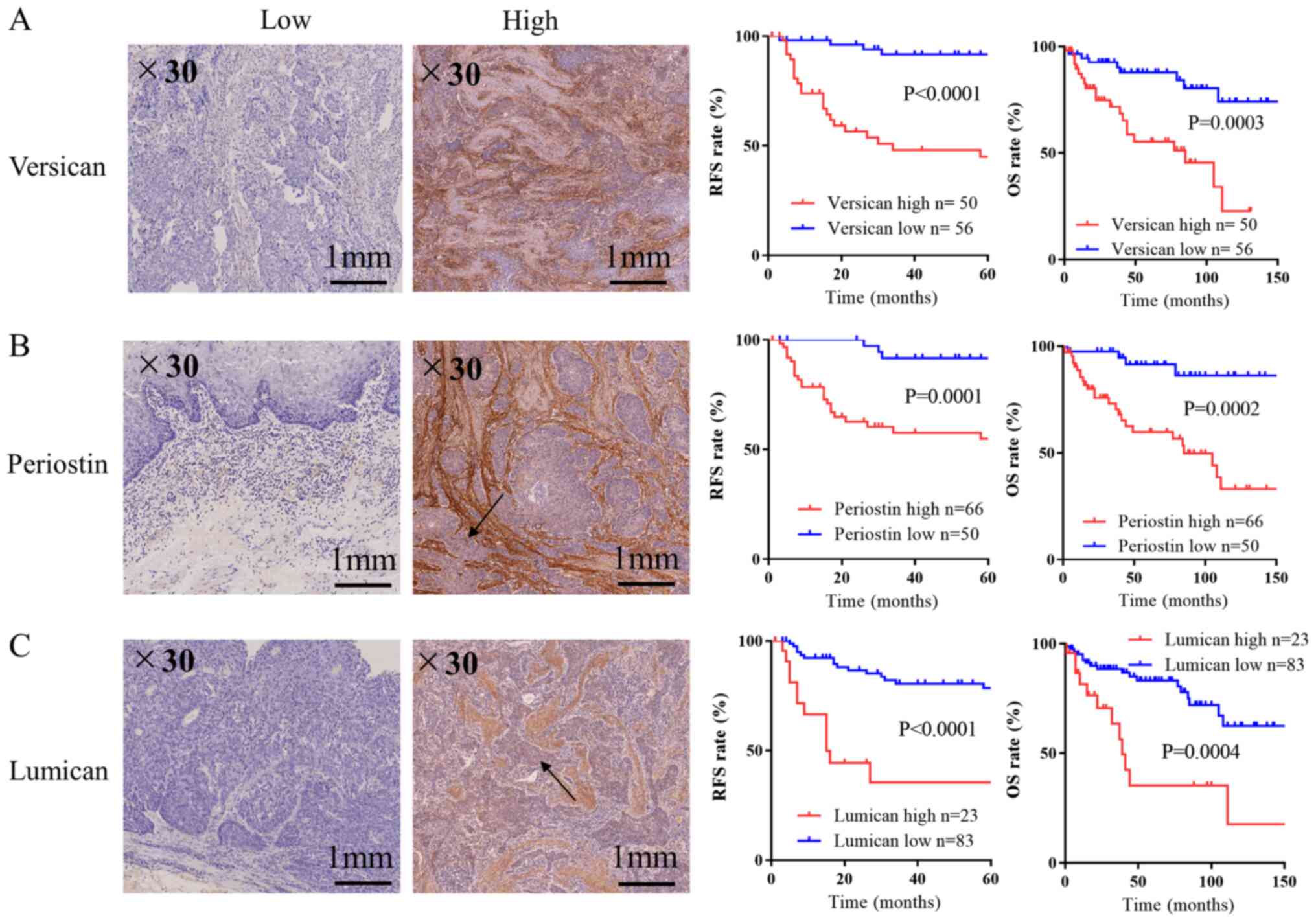
Table I. As demonstrated in Fig. 1A, versican staining was found
specifically in the cancer stroma, and 50 out of 106 patients (47%)
were determined to have high stromal versican expression. Likewise,
the expression of periostin and lumican was also observed in the
cancer stroma, and 66 (62%) and 23 (22%) tumors were considered to
exhibit high stromal periostin and lumican, respectively (Table I; Fig. 1B
and C). The present study investigated whether cancer cells
expressed the three CAF-related molecules, and the expression of
periostin and lumican was detected, whereas that of versican was
very low. The expression of periostin in cancer cells was
significantly associated with that in stromal cells, while the
expression of lumican in cancer cells did not exhibit any
significant association with that in stromal cells (data not
shown). By contrast, versican was not expressed in cancer cells,
but was expressed in stromal cells. High levels of versican and
periostin in the cancer stroma were significantly associated with
aggressive clinicopathological features, including a greater depth
of invasion, the presence of lymph node metastasis, positive
lymphatic and venous invasion, and a more advanced TNM stage
(P<0.05; Table I). In addition,
high level of versican was associated with postoperative additional
therapy, and high level of periostin was associated with location
(P<0.05; Table I). High
expression of stromal lumican was significantly associated with
depth of invasion, lymph node metastasis, venous invasion and
stage, but not with lymphatic invasion (Table I). As shown in Tables II–IV, a significant positive association was
also found between stromal versican and periostin (P<0.0001),
stromal periostin and lumican (P<0.0002), and stromal lumican
and versican (P=0.0034). Moreover, the expression of all three
molecules was positively associated with the expression of αSMA
(Fig. S2 and Table SII).
 | Table I.Patient characteristics. |
Table I.
Patient characteristics.
|
| Versican
expression | Periostin
expression | Lumican
expression |
|---|
|
|
|
|
|
|---|
| Characteristic | High (n=50) | Low (n=56) | P-value | High (n=66) | Low (n=40) | P-value | High (n=23) | Low (n=83) | P-value |
|---|
| Age, years |
|
|
|
|
|
|
|
|
|
| Range
(mean ± SD) | 42–81 | 37–79 | 0.8011 | 42–81 | 37–78 | 0.1350 | 42–81 | 37–79 | 0.4588 |
|
| (65.82±8.55) | (64.00±8.53) |
| (65.94±8.55) | (63.08±8.53) |
| (66.26±8.55) | (64.47±8.58) |
|
| ≤60,
n | 14 | 18 |
| 16 | 16 |
| 5 | 27 |
|
| ≥61,
n | 36 | 38 |
| 50 | 24 |
| 18 | 56 |
|
| Sex, n |
|
| 0.3755 |
|
| >0.999 |
|
| 0.4727 |
|
Male | 42 | 51 |
| 58 | 35 |
| 19 | 74 |
|
|
Female | 8 | 5 |
| 8 | 5 |
| 4 | 9 |
|
| Postoperative
additional therapy, n |
|
| 0.0436a |
|
| 0.0529 |
|
| 0.0569 |
| No | 36 | 50 |
| 49 | 37 |
| 17 | 69 |
|
|
Chemotherapy | 12 | 6 |
| 15 | 3 |
| 4 | 14 |
|
|
Radiotherapy | 2 | 0 |
| 2 | 0 |
| 2 | 0 |
|
|
Chemoradiotherapy | 0 | 0 |
| 0 | 0 |
| 0 | 0 |
|
| Location, n |
|
| 0.2137 |
|
| 0.0409a |
|
| 0.2884 |
|
Upper | 4 | 11 |
| 5 | 10 |
| 1 | 14 |
|
|
Middle | 30 | 31 |
| 40 | 21 |
| 14 | 47 |
|
|
Lower | 16 | 14 |
| 21 | 9 |
| 8 | 22 |
|
| Invasion depth,
n |
|
|
<0.0001a |
|
|
<0.0001a |
|
| 0.0005a |
|
pT1 | 25 | 49 |
| 34 | 40 |
| 9 | 65 |
|
|
pT2 | 10 | 3 |
| 13 | 0 |
| 6 | 7 |
|
|
pT3 | 14 | 4 |
| 18 | 0 |
| 8 | 10 |
|
|
pT4 | 1 | 0 |
| 1 | 0 |
| 0 | 1 |
|
| Lymph node
metastasis, n |
|
| 0.0022a |
|
| 0.0002a |
|
| 0.0633 |
|
Yes | 27 | 13 |
| 34 | 6 |
| 13 | 27 |
|
| No | 23 | 43 |
| 32 | 34 |
| 10 | 56 |
|
| Lymphatic invasion,
n |
|
| 0.0002a |
|
| 0.0049a |
|
| 0.3773 |
|
Yes | 33 | 16 |
| 38 | 11 |
| 13 | 36 |
|
| No | 17 | 40 |
| 28 | 29 |
| 10 | 47 |
|
| Venous invasion,
n |
|
|
<0.0001a |
|
| 0.0026a |
|
| 0.0078a |
|
Yes | 40 | 17 |
| 46 | 11 |
| 18 | 39 |
|
| No | 10 | 39 |
| 20 | 29 |
| 5 | 44 |
|
| Tumor
differentiation, n |
|
| 0.1365 |
|
| 0.8843 |
|
| 0.3300 |
|
Well | 12 | 14 |
| 15 | 11 |
| 9 | 17 |
|
|
Moderate | 27 | 26 |
| 34 | 19 |
| 9 | 44 |
|
|
Poor | 10 | 8 |
| 12 | 6 |
| 3 | 15 |
|
|
Unknown | 1 | 8 |
| 5 | 4 |
| 2 | 7 |
|
| TNM stage, n |
|
| 0.0075a |
|
|
<0.0001a |
|
| 0.0091a |
| I | 25 | 42 |
| 30 | 36 |
| 8 | 59 |
|
| II | 10 | 6 |
| 16 | 0 |
| 8 | 8 |
|
|
III | 11 | 7 |
| 15 | 3 |
| 7 | 11 |
|
| IV | 4 | 1 |
| 4 | 1 |
| 0 | 5 |
|
| Table II.Association between stromal versican
and periostin expression levels. |
Table II.
Association between stromal versican
and periostin expression levels.
|
| Periostin |
|
|---|
|
|
|
|
|---|
|
| High (n=66) | Low (n=40) | P-value |
|---|
| Versican |
|
| <0.0001 |
| High
(n=50) | 43 | 7 |
|
| Low (n=56) | 23 | 33 |
|
| Table IV.Correlation between stromal lumican
and versican expression levels. |
Table IV.
Correlation between stromal lumican
and versican expression levels.
|
| Versican |
|
|---|
|
|
|
|
|---|
|
| High (n=50) | Low (n=56) | P-value |
|---|
| Lumican |
|
| 0.0076 |
| High
(n=23) | 17 | 6 |
|
| Low
(n=83) | 33 | 50 |
|
High expression of stromal versican,
periostin and lumican in ESCC tissues and the association with poor
prognosis
Kaplan-Meier survival analysis showed that patients
with stage I–IV ESCC exhibiting high stromal versican expression
experienced significantly shorter RFS and OS times, compared with
those patients exhibiting low expression (P<0.0001 and P=0.0003,
respectively; Fig. 1A). Similarly,
stromal periostin and lumican expression demonstrated a significant
impact resulting in poor prognosis for both RFS and OS (periostin,
P=0.0001 and P=0.0002, respectively; lumican: P<0.0001 and
P=0.0004, respectively; Fig. 1B and
C). Moreover, when patients were stratified according to TNM
stage, high expression of stromal versican and lumican each showed
a significant association with poorer RFS compared with low
expression only in stage I patients (Fig. S3). It was also found that the
prognostic performance of stromal versican had statistically
significant in the subgroup analyses of stage III patients.
Similarly, stromal periostin tended to be associated with RFS in
stage III patients, although this result was not significant
(P=0.0931; Fig. S3), while stromal
lumican was associated with RFS in stage I patients (Fig. S3).
Univariate and multivariate survival
analysis for patient prognosis
In the univariate survival analysis, RFS rate was
associated with invasion depth (HR, 4.47; 95% CI, 2.04–9.82;
P<0.001), lymph node metastasis (HR, 3.1; 95% CI, 1.44–6.71;
P=0.004), lymphatic invasion (HR, 3.59; 95% CI, 1.57–8.22;
P=0.002), venous invasion (HR, 9.26; 95% CI, 2.78–30.82;
P<0.001), TNM stage (HR, 1.98; 95% CI, 1.42–2.75; P<0.001),
stromal versican expression (HR, 9.11; 95% CI, 3.14–26.44;
P<0.0001), stromal periostin expression (HR, 7.46; 95% CI,
2.24–24.88; P=0.001) and stromal lumican expression (HR, 5.11; 95%
CI, 2.35–11.1; P<0.0001). Multivariate survival analysis by the
Cox hazard model showed that three factors, TNM stage (HR, 1.81;
95% CI, 1.03–3.16; P=0.039), stromal versican expression (HR, 3.96;
95% CI, 1.16–13.46; P=0.028) and stromal lumican expression (HR,
2.55; 95% CI, 1.06–6.17; P=0.037) were independent indicators for a
poor prognosis (Table V).
| Table V.Univariate and multivariate analysis
for relapse-free survival. |
Table V.
Univariate and multivariate analysis
for relapse-free survival.
|
| Univariate | Multivariate |
|---|
|
|
|
|
|---|
| Factor | HR | 95% CI | P-value | HR | 95% CI | P-value |
|---|
| Age (≤60 vs. ≥61
years) | 1.00 | 0.45–2.22 | 0.9940 |
|
|
|
| Sex (male vs.
female) | 1.58 | 0.54–4.57 | 0.4027 |
|
|
|
| Invasion depth
(pT1-2 vs. pT3-4) | 4.47 | 2.04–9.82 | 0.0002a | 0.80 | 0.30–2.15 | 0.6545 |
| Lymph node
metastasis (yes vs. no) | 3.10 | 1.44–6.71 | 0.0040a | 0.56 | 0.19–1.64 | 0.2870 |
| Lymphatic invasion
(yes vs. no) | 3.59 | 1.57–8.22 | 0.0025a | 1.01 | 0.37–2.79 | 0.9814 |
| Venous invasion
(yes vs. no) | 9.26 | 2.78–30.82 | 0.0003a | 3.07 | 0.78–12.09 | 0.1095 |
| TNM stage | 1.98 | 1.42–2.75 |
<0.0001a | 1.81 | 1.03–3.16 | 0.0390a |
| Versican (high vs.
low) | 9.11 | 3.14–26.44 |
<0.0001a | 3.96 | 1.16–13.46 | 0.0278a |
| Periostin (high vs.
low) | 7.46 | 2.24–24.88 | 0.0011a | 1.91 | 0.48–7.65 | 0.3582 |
| Lumican (high vs.
low) | 5.11 | 2.35–11.10 |
<0.0001a | 2.55 | 1.06–6.17 | 0.0371a |
Discussion
The present study investigated whether the
expression of versican, periostin and lumican has utility as a
prognostic biomarker for ESCC using 106 surgically resected
specimens assessed via immunohistochemistry. The staining of the
three CAF-related molecules in the cancer stroma was significantly
associated with worse RFS and OS times. Moreover, stromal versican
and lumican expression levels were independent prognostic factors
for ESCC.
A previous study revealed that CAFs can increase the
frequency of cancer stem cells, leading to a high tumor recurrence
rate and a poor prognosis, which is enhanced by TGF-β signaling,
while poor prognostic signatures share a stromal gene program that
is induced by TGF-β (18). Another
study showed that ovarian CAFs, which had much higher levels of
TGF-β receptors than other cell types, exhibited versican
upregulation by TGF-β. By contrast, TGF-β receptors were
downregulated in ovarian cancer cells, possibly conferring
resistance to inhibitory growth signals exerted by TGF-β (19). These results indicate that CAFs are
specifically responsive to elevated TGF-β levels, while cancer
cells can be the major source of TGF-β ligands (14). Furthermore, studies have shown that
TGF-β signaling plays a role in esophageal cancer progression. For
example, upregulation of TGF-β was associated with tumor size in
patients with ESCC (20).
Additionally, overexpression of TGF-β and decreased TGF-β receptor
expression were associated with depth of invasion and pathological
stage in patients with ESCC (21).
TGF-β/Smad signaling has been shown to promote
epithelial-mesenchymal transition in ESCC (22,23).
CAF-specific versican was upregulated by TGF-β in several cancer
types, such as colorectal and ovarian cancer, resulting in cancer
cell motility and invasion (14,19).
Versican is implicated in the regulation of cell proliferation,
differentiation, apoptosis, migration and adhesion in a variety of
cancer types, such as breast and ovarian cancer (24). Versican is a large chondroitin
sulfate proteoglycan that is a major component of the ECM (12,13) and
plays a role in the formation of the tumor-specific ECM, which can
support cancer cell growth and metastasis in certain solid cancer
types. Several clinical studies indicated that high versican
expression was a poor prognostic factor in a variety of cancer
types such as prostate, breast and gastric cancer (24). The present study showed that high
versican expression in the stroma was associated with poor RFS and
OS times in stage I–IV ESCC after resection, which was consistent
with earlier findings in other cancer types (25–28).
Furthermore, the expression of stromal versican was significantly
associated with poor RFS time in stratified analyses of stage I and
III patients. Correspondingly, stromal versican was found to be an
independent prognostic factor for RFS via multivariate analysis.
These results indicated that stromal versican may be used as a
prognostic biomarker for patients with ESCC after curative surgery,
and that immunohistochemical analysis for versican expression in
resected specimens may influence a decision on whether to complete
intensive postoperative treatment, including administration of
adjuvant chemotherapy, particularly for patients with stage I
ESCC.
Periostin is an extracellular matrix secreted
protein that is upregulated in tumor cells in several cancer types,
including pancreatic, colorectal, lung, ovarian, breast, head and
neck, thyroid, gastric, hepatic and esophageal cancer (5,15,29–36).
Periostin overexpression in tumor cells, not in stroma, has also
been associated with tumor invasion and metastasis in oral
carcinoma and esophageal cancer (37,38).
Periostin is regulated by TGF-β signaling, as well as versican
expression. An earlier study showed that periostin was expressed by
fibroblasts in the normal tissue and in the stroma of the primary
tumor (6). Infiltrating tumor cells
need to induce stromal periostin expression in the secondary target
organ to initiate colonization, and the induced periostin secreted
by CAFs in the stroma of the metastatic loci was required to allow
for the maintenance of cancer stem cells (24). Periostin is able to interact with
other ECM proteins, specific cell surface receptors and integrins
via multiple signal pathways affecting metastasis, invasion and
angiogenesis in cancer development (39). Periostin was reported to bind as a
ligand to ανβ3 and ανβ5 integrins, thus signaling via the PI3K-Akt
pathway within esophageal cancer. The study reported that
periostin-positive tumors exhibited higher levels of vascular
endothelial growth factor and greater microvessel density compared
with periostin-negative tumors (5).
These findings indicate that periostin serves a key role in ESCC
tumorigenesis through the induction and/or promotion of
angiogenesis. Periostin is an important mediator of tumor invasion
in ESCC (37). The present study
showed that high stromal periostin was significantly associated
with worse RFS and OS times.
Lumican is also known to be regulated by TGF-β
signaling. Lumican in stromal tissues, adjacent to cancer cells,
may modulate the characteristics of collagen fibers and induce the
invasion activity of pancreatic cancer cells (40). A previous study showed that in breast
cancer, a high expression level of stromal lumican was associated
with a high pathological tumor grade (40). By contrast, a high expression level
of lumican in breast cancer was reported to be associated with slow
progression and an improved prognosis (41). In pancreatic cancer, lumican
expression in cancer stroma is associated with a shorter survival
time (42). Another study reported
that the presence of lumican in the ECM surrounding pancreatic
ductal adenocarcinoma (PDAC) cells is associated with an improved
patient outcome. Secretion of lumican from activated pancreatic
stellate cells within PDAC is negatively regulated by TGF-β
(11). In ESCC, to the best of our
knowledge, there is still no report on lumican in association with
prognosis, and there are few studies describing the molecular
mechanisms of stromal lumican in malignant tumors (11). The present study showed that high
stromal lumican expression indicated a poor prognosis in patients
with ESCC, and a significant difference in RFS was found between
high and low stromal lumican expression groups in the analysis of
stage I patients. Furthermore, stromal lumican expression, as well
as versican expression, was found to be an independent prognostic
factor for RFS via multivariate analysis.
Overall, the present study examined the protein
levels of versican, periostin and lumican via IHC without exploring
the detailed molecular mechanisms. Further studies will be required
to clarify the general role, mechanisms and relationships of
versican, periostin and lumican in ESCC.
In summary, stromal periostin may have utility as a
prognostic biomarker, while stromal versican and, in particular,
could be independent prognostic factors for ESCC. The results of
the present study indicated that stromal versican and lumican
expression scoring may help to make a decision on whether to
administer adjuvant chemotherapy for patients with ESCC.
Supplementary Material
Supporting Data
Acknowledgements
Not applicable.
Funding
No funding was received.
Availability of data and materials
The datasets and materials used and analyzed during
the current study are available from the corresponding author on
reasonable request.
Authors' contributions
YK and KKo conceived and designed the present study.
SO and NY collected the data. YK, SO and NY confirm the
authenticity of all the raw data. KS performed sample
immunostaining. NY, KS, SY, YH, EE, KKa, LY, ZS, KM, TMa and YK
performed the majority of the current study and analyzed the data.
HO, HN, TMo, HH, YW, SH and MS participated in analyzing the data
and drafting the initial manuscript. NY, YK, SN, KS and KKo revised
the manuscript. All authors read and approved the manuscript.
Ethics approval and consent to
participate
The current study was approved by Fukushima Medical
University Certified Review Board (Fukushima, Japan; approval no.
2329). Informed consent was obtained from all patients.
Patient consent for publication
Not applicable.
Competing interests
The authors declare that they have no competing
interests.
References
|
1
|
Bray F, Ferlay J, Soerjomataram I, Siegel
RL, Torre LA and Jemal A: Global Cancer Statistics 2018: GLOBOCAN
Estimates of incidence and mortality worldwide for 36 cancers in
185 countries. CA Cancer J Clin. 68:394–424. 2018. View Article : Google Scholar : PubMed/NCBI
|
|
2
|
Zhang Y: Epidemiology of esophageal
cancer. World J Gastroenterol. 19:5598–5606. 2013. View Article : Google Scholar : PubMed/NCBI
|
|
3
|
Han-Ze Z, Guang-Fu J and Hong-Bing S:
Epidemiologic differences in esophageal cancer between Asian and
Western populations. Chin J Cancer. 31:281–286. 2012. View Article : Google Scholar
|
|
4
|
Lam AK: Introduction: Esophageal squamous
cell carcinoma-current status and future advances. Methods Mol
Biol. 2129:1–6. 2020. View Article : Google Scholar : PubMed/NCBI
|
|
5
|
Wang W, Sun QK, He YF, Ma DC, Xie MR, Ji
CS and Hu B: Overexpression of periostin is significantly
correlated to the tumor angiogenesis and poor prognosis in patients
with esophageal squamous cell carcinoma. Int J Clin Exp Pathol.
7:593–601. 2014.PubMed/NCBI
|
|
6
|
Underwood TJ, Hayden AL, Derouet M, Garcia
E, Noble F, White MJ, Thirdborough S, Mead A, Clemons N, Mellone M,
et al: Cancer-associated fibroblasts predict poor outcome and
promote periostin-dependent invasion in oesophageal adenocarcinoma.
J Pathol. 235:466–477. 2015. View Article : Google Scholar : PubMed/NCBI
|
|
7
|
Rui W, Si L, Shutai Z, Min L and Zhu S:
Cellular and extracellular components in tumor microenvironment and
their application in early diagnosis of cancer. Anal Cell Pathol
(Amst). 2020:62837962020.
|
|
8
|
R Core Team, . R: A language and
environment for statistical computing. R Foundation for Statistical
Computing; Vienna, Austria: 2014, http://www.R-project.org/February 10–2015
|
|
9
|
Palumbo A Jr, Meireles Da Costa N, Pontes
B, Leite de Oliveira F, Lohan Codeço M, Ribeiro Pinto LF and
Nasciutti LE: Esophageal cancer development: Crucial clues arising
from the extracellular matrix. Cells. 9:4552020. View Article : Google Scholar : PubMed/NCBI
|
|
10
|
Kashyap MK, Marimuthu A, Kishore CJ, Peri
S, Keerthikumar S, Prasad TS, Mahmood R, Rao S, Ranganathan P,
Sanjeeviah RC, et al: Genomewide mRNA profiling f esophageal
squamous cell carcinoma for identification of cancer biomarkers.
Cancer Biol Ther. 8:36–46. 2009. View Article : Google Scholar : PubMed/NCBI
|
|
11
|
Sotoodehnejadnematalahi F and Burke B:
Structure, function and regulation of versican: The most abundant
type of proteoglycan in the extracellular matrix. Acta Med Iran.
51:740–750. 2013.PubMed/NCBI
|
|
12
|
Kang Y, Roife D, Lee Y, Lv H, Suzuki R,
Ling J, Rios Perez MV, Li X, Dai B, Pratt M, et al: Transforming
growth factor-β limits secretion of lumican by activated stellate
cells within primary pancreatic adenocarcinoma tumors. Clin Cancer
Res. 22:4934–4946. 2016. View Article : Google Scholar : PubMed/NCBI
|
|
13
|
Ween MP, Oehler MK and Ricciardelli C:
Role of versican, hyaluronan and CD44 in ovarian cancer metastasis.
Int J Mol Sci. 12:1009–1029. 2011. View Article : Google Scholar : PubMed/NCBI
|
|
14
|
Du WW, Yang W and Yee AJ: Roles of
versican in cancer biology-tumorigenesis, progression and
metastasis. Histol Histopathol. 28:701–713. 2013.PubMed/NCBI
|
|
15
|
Chida S, Okayama H, Noda M, Saito K,
Nakajima T, Aoto K, Hayase S, Momma T, Ohki S, Kono K and
Takenoshita S: Stromal VCAN expression as a potential prognostic
biomarker for disease recurrence in stage II–III colon cancer.
Carcinogenesis. 37:878–887. 2016. View Article : Google Scholar : PubMed/NCBI
|
|
16
|
Jiang Q, Chen J, Zhang B, Niu J and He Y:
Prognostic significance of periostin and mammalian target of
rapamycin (mTOR) in locally advanced esophageal squamous cell
carcinoma. Med Sci Monit. 23:3200–3208. 2017. View Article : Google Scholar : PubMed/NCBI
|
|
17
|
Brierley JD, MK Gospodarowicz and
Wittekind C: The TNM Classification of Malignant Tumours. 8th
edition. Wiley-Blackwell; Hoboken, NJ, USA: pp. 2722016
|
|
18
|
Calon A, Lonardo E, Berenguer-Llergo A,
Espinet E, Hernando-Momblona X, Iglesias M, Sevillano M,
Palomo-Ponce S, Tauriello DV, Byrom D, et al: Stromal gene
expression defines poor-prognosis subtypes in colorectal cancer.
Nat Genet. 47:320–329. 2015. View
Article : Google Scholar : PubMed/NCBI
|
|
19
|
Yeung TL, Leung CS, Wong KK, Samimi G,
Thompson MS, Liu J, Zaid TM, Ghosh S, Birrer MJ and Mok SC: TGF-β
modulates ovarian cancer invasion by upregulating CAF-derived
versican in the tumor microenvironment. Cancer Res. 73:5016–5028.
2013. View Article : Google Scholar : PubMed/NCBI
|
|
20
|
Gholamin M, Moaven O, Memar B, Farshchian
M, Naseh H, Malekzadeh R, Sotoudeh M, Rajabi-Mashhadi MT, Forghani
MN, Farrokhi F and Abbaszadegan MR: Overexpression and interactions
of interleukin-10, transforming growth factor beta, and vascular
endothelial growth factor in esophageal squamous cell carcinoma.
World J Surg. 33:1439–1445. 2009. View Article : Google Scholar : PubMed/NCBI
|
|
21
|
Fukai Y, Fukuchi M, Masuda N, Osawa H,
Kato H, Nakajima T and Kuwano H: Reduced expression of transforming
growth factor-beta receptors is an unfavorable prognostic factor in
human esophageal squamous cell carcinoma. Int J Cancer.
104:161–166. 2003. View Article : Google Scholar : PubMed/NCBI
|
|
22
|
Pang L, Li Q, Wei C, Zou H, Li S, Cao W,
He J, Zhou Y, Ju X, Lan J, et al: TGF-β1/Smad signaling pathway
regulates epithelial-to-mesenchymal transition in esophageal
squamous cell carcinoma: In vitro and clinical analyses of cell
lines and nomadic Kazakh patients from northwest Xinjiang, China.
PLoS One. 9:e1123002014. View Article : Google Scholar : PubMed/NCBI
|
|
23
|
Luo J, Chen XQ and Li P: The role of TGF-β
and its receptors in gastrointestinal cancers. Transl Oncol.
12:475–484. 2019. View Article : Google Scholar : PubMed/NCBI
|
|
24
|
Asano K, Nelson CM, Nandadasa S,
Aramaki-Hattori N, Lindner DJ, Alban T, Inagaki J, Ohtsuki T,
Oohashi T, Apte SS and Hirohata S: Stromal versican regulates tumor
growth by promoting angiogenesis. Sci Rep. 7:172252017. View Article : Google Scholar : PubMed/NCBI
|
|
25
|
Ricciardelli C, Mayne K, Sykes PJ, Raymond
WA, McCaul K, Marshall VR and Horsfall DJ: Elevated levels of
versican but not decorin predict disease progression in early-stage
prostate cancer. Clin Cancer Res. 4:963–9371. 1998.PubMed/NCBI
|
|
26
|
Suwiwat S, Ricciardelli C, Tammi R, Tammi
M, Auvinen P, Kosma VM, LeBaron RG, Raymond WA, Tilley WD and
Horsfall DJ: Expression of extracellular matrix components
versican, chondroitin sulfate, tenascin, and hyaluronan, and their
association with disease outcome in node-negative breast cancer.
Clin Cancer Res. 10:2491–2498. 2004. View Article : Google Scholar : PubMed/NCBI
|
|
27
|
Kodama J, Hasengaowa, Kusumoto T, Seki N,
Matsuo T, Ojima Y, Nakamura K, Hongo A and Hiramatsu Y: Prognostic
significance of stromal versican expression in human endometrial
cancer. Ann Oncol. 18:269–274. 2007. View Article : Google Scholar : PubMed/NCBI
|
|
28
|
Shen XH, Lin WR, Xu MD, Qi P, Dong L,
Zhang QY, Ni SJ, Weng WW, Tan C, Huang D, et al: Prognostic
significance of Versican expression in gastric adenocarcinoma.
Oncogenesis. 4:e1782015. View Article : Google Scholar : PubMed/NCBI
|
|
29
|
Liu Y and Du L: Role of pancreatic
stellate cells and periostin in pancreatic cancer progression.
Tumour Biol. 36:3171–3177. 2015. View Article : Google Scholar : PubMed/NCBI
|
|
30
|
Li Z, Zhang X, Yang Y, Yang S, Dong Z, Du
L, Wang L and Wang C: Periostin expression and its prognostic value
for colorectal cancer. Int J Mol Sci. 16:12108–12118. 2015.
View Article : Google Scholar : PubMed/NCBI
|
|
31
|
Zhu M, Fejzo MS, Anderson L, Dering J,
Ginther C, Ramos L, Gasson JC, Karlan BY and Slamon DJ: Periostin
promotes ovarian cancer angiogenesis and metastasis. Gynecol Oncol.
119:337–344. 2010. View Article : Google Scholar : PubMed/NCBI
|
|
32
|
Wu SQ, Lv YE, Lin BH, Luo LM, Lv SL, Bi AH
and Jia YS: Silencing of periostin inhibits nicotine-mediated tumor
cell growth and epithelial-mesenchymal transition in lung cancer
cells. Mol Med Rep. 7:875–880. 2013. View Article : Google Scholar : PubMed/NCBI
|
|
33
|
Lee YJ, Kim IS, Park SA, Kim Y, Lee JE,
Noh DY, Kim KT, Ryu SH and Suh PG: Periostin-binding DNA aptamer
inhibits breast cancer growth and metastasis. Mol Ther.
21:1004–1013. 2013. View Article : Google Scholar : PubMed/NCBI
|
|
34
|
Kim CJ, Sakamoto K, Tambe Y and Inoue H:
Opposite regulation of epithelial-to-mesenchymal transition and
cell invasiveness by periostin between prostate and bladder cancer
cells. Int J Oncol. 38:1759–1766. 2011.PubMed/NCBI
|
|
35
|
Liu AY, Zheng H and Ouyang G: Periostin, a
multifunctional matricellular protein in inflammatory and tumor
microenvironments. Matrix Biol. 37:150–156. 2014. View Article : Google Scholar : PubMed/NCBI
|
|
36
|
Luo JH, Zhou J and Gao Y: Correlation
between periostin and SNCG and esophageal cancer invasion,
infiltration and apoptosis. Asian Pac J Trop Med. 6:516–519. 2013.
View Article : Google Scholar : PubMed/NCBI
|
|
37
|
Michaylira CZ, Wong GS, Miller CG,
Gutierrez CM, Nakagawa H, Hammond R, Klein-Szanto AJ, Lee JS, Kim
SB, Herlyn M, et al: Periostin, a cell adhesion molecule,
facilitates invasion in the tumor microenvironment and annotates a
novel tumor-invasive signature in esophageal cancer. Cancer Res.
70:5281–5292. 2010. View Article : Google Scholar : PubMed/NCBI
|
|
38
|
Qin X, Yan M, Zhang J, Wang X, Shen Z, Lv
Z, Li Z, Wei W and Chen W: TGFβ3-mediated induction of Periostin
facilitates head and neck cancer growth and is associated with
metastasis. Sci Rep. 6:205872016. View Article : Google Scholar : PubMed/NCBI
|
|
39
|
Lv YJ, Wang W, Ji CS, Jia, Xie MR and Hu
B: Association between periostin and epithelial-mesenchymal
transition in esophageal squamous cell carcinoma and its clinical
significance. Oncol Lett. 14:376–382. 2017. View Article : Google Scholar : PubMed/NCBI
|
|
40
|
Troup S, Njue C, Kliewer EV, Parisien M,
Roskelley C, Chakravarti S, Roughley PJ, Murphy LC and Watson PH:
Reduced expression of the small leucine-rich proteoglycans,
lumican, and decorin is associated with poor outcome in
node-negative invasive breast cancer. Clin Cancer Res. 9:207–214.
2003.PubMed/NCBI
|
|
41
|
Leygue E, Snell L, Dotzlaw H, Hole K,
Hiller-Hitchcock T, Roughley PJ, Watson PH and Murphy LC:
Expression of lumican in human breast carcinoma. Cancer Res.
58:1348–1352. 1998.PubMed/NCBI
|
|
42
|
Ishiwata T, Cho K, Kawahara K, Yamamoto T,
Fujiwara Y, Uchida E, Tajiri T and Naito Z: Role of lumican in
cancer cells and adjacent stromal tissues in human pancreatic
cancer. Oncol Rep. 18:537–543. 2007.PubMed/NCBI
|