Introduction
Gastric cancer (GC) is the fifth most common cancer
and the third leading cause of cancer-related death worldwide
(1). Although significant
advancements in therapeutic strategies have been achieved, and
several chemotherapeutic drugs, such as fluoropyrimidines
(5-fluorouracil, S-1, and capecitabine), cisplatin, oxaliplatin,
taxanes, and irinotecan, and molecular targeted drugs (trastuzumab
or ramucirumab), have improved the treatment of metastatic GC
patients (2–5), the overall prognosis of
advanced/metastatic GC remains dismal (6,7), and the
management of this disease is challenging.
In this context, complementary and alternative
medicine, especially dietary compounds, has gained economic and
sociological importance because of its cost-effectiveness and
reduced toxicity (8–10). Over the past three decades, nearly
100 natural products or direct derivatives from natural remedies
have been highlighted in the area of cancer therapy (11). Interestingly, most of these dietary
botanicals function by targeting multiple cancer-associated
pathways to exert anti-tumorigenic effects on cancer progression
(11). Andrographolide is a C20
diterpenoid lactone, which is an active ingredient derived from the
traditional Chinese herbal medicine Andrographis paniculate
(12–14). Because of its ability to circulate in
the bloodstream (15–17), Andrographis exhibits diverse
biological activities, such as anti-inflammatory, antiviral, and
immunomodulatory effects (18,19).
Furthermore, accumulating evidence has shown that Andrographis has
anti-tumorigenic properties in multiple malignancies, such as
melanoma, leukemia, glioblastoma, breast, lung, esophageal,
colorectal, bladder, pancreatic, and liver cancer (14,20–29). Its
various underlying mechanisms include the regulation of oxidative
stress, apoptosis, necrosis, autophagy, inhibition of cell
adhesion, proliferation, migration, invasion, and angiogenesis
(13,14,30,31).
Moreover, Andrographis influences several cancer-associated and
angiogenesis signaling pathways, such as PI3K/AKT/mTOR (20,24),
SRC/MAPKs/AP-1 (25),
TLR4/NF-κB/MMP-9 (26), and
VEGF/VEGFR2/AKT (29).
A previous study showed that Andrographis enhanced
tumor necrosis factor-related apoptosis-inducing ligand
(TRAIL)-mediated apoptosis in GC cells (32). However, other mechanistic pathways of
anti-tumorigenic events induced by Andrographis have not been fully
elucidated in GC. Therefore, in this study, we conducted a series
of experiments in GC cells and demonstrated that Andrographis
exerts its anti-tumorigenic effects via a novel mechanism. Our
study indicates that Andrographis may be a potential therapeutic or
adjunct option for GC patients.
Materials and methods
Cell culture and materials
The GC cell lines MKN74 and NUGC4 were provided by
the Cell Resource Center of Biomedical Research, Institute of
Development, Aging and Cancer (Tohoku University, Sendai, Japan).
All cell lines were authenticated using a panel of genetic and
epigenetic markers and tested for mycoplasma regularly. The cells
were cultured in RPMI-1640 medium (Nacalai Tesque) supplemented
with 10% fetal bovine serum (FBS; Biowest) and an
antibiotic-antimycotic mixed stock solution (Nacalai Tesque) and
maintained at 37°C in a humidified incubator at 5% CO2.
Andrographis (EP80 Andrographis extract standardized to 80%
andrographolide content, dissolved in DMSO) was purified by
EuroPharma-USA and kindly provided by Professor Ajay Goel at the
City of Hope Comprehensive Cancer Center. Andrographis was diluted
to appropriate experimental concentrations in culture medium.
Cell viability and proliferation
assays
For WST assays, cells were plated in 96-well tissue
culture plates (TPP Techno Plastic Products AG) at a density of
5,000 cells/well in RPMI-1640 medium supplemented with 10% FBS and
antibiotics and allowed to adhere overnight. First, we treated GC
cells with various doses of Andrographis (10, 20, 40, 60, 80 and
100 µg/ml) for 72 h to evaluate its cytotoxic effects and then
measured cell proliferation using WST-8 (Dojindo Laboratories) in
accordance with the manufacturer's instructions. Subsequently,
based on the IC50 concept (33), we evaluated cell proliferation after
treatment with 40 µg/ml Andrographis for 24, 48 and 72 h. The
absorbance in each well was measured at a wavelength of 450 nm
using SoftMax Pro (Molecular Devices).
Cell colony formation assays
Colony formation activity was measured according to
established procedures (34).
Briefly, 2×10³ MKN74 cells/well and 5×10³
NUGC4 cells/well were seeded in 6-well tissue culture plates (TPP
Techno Plastic Products AG) in the same culture medium as described
above and incubated for 24 h in Andrographis-free culture medium.
We then added 20 µg/ml Andrographis to the culture medium and
incubated the cells for 72 h. Subsequently, the medium was replaced
with Andrographis-free culture medium, and the cells were
maintained at 37°C/5% CO2 for 5 days in a humidified
atmosphere. The number of colonies was counted using Image J
software ver.1.52 (NIH) (35) and
compared between Control and Andrographis treatment groups.
Cell apoptosis assays
Apoptosis assays were conducted using PI/Annexin V
double staining and flow cytometry. Cells were plated in a 6-well
plate (MKN74: 1.2×10⁵/well; NUGC4:
1.5×10⁵/well) for 24 h, followed by treatment with 40
µg/ml Andrographis for 48 h. The apoptotic cells were harvested and
measured using a Muse® Annexin V and Dead Cell Assay
(Luminex) on a Muse™ Cell Analyzer (Millipore) in accordance with
the manufacturer's instructions.
Quantitative mRNA expression
analysis
For the quantification of mRNA expression, cells
were plated in 6-well dishes (MKN74: 2.5×10⁵/well;
NUGC4: 2.0×10⁵/well), incubated for 24 h, and then
treated with 40 µg/ml Andrographis or DMSO. Total RNA from cells in
the treatment and control groups was extracted using an RNA
extraction miRNeasy Mini kit (Qiagen). cDNA was synthesized from
5.0 ng total RNA using a Reverse Transcription kit (Toyobo), and
RT-qPCR was performed using the Power SYBR® Green PCR
Master Mix (Life Technology). Quantitative real-time reverse
transcription (RT)-PCR analysis was conducted using the StepOne™
Real-time PCR System (Applied BiosystemsA). The primer sequences
were as follows: heme oxygenase-1 (HMOX1): forward,
5′-AAGACTGCGTTCCTGCTCAAC-3′ and reverse,
5′-AAAGCCCTACAGCAACTGTCG-3′; glutamate-cysteine ligase catalytic
(GCLC): forward, 5′-AGGCCAACATGCGAAAAC-3′ and reverse,
5′-CGGATATTTCTTGTTAAGGTACTGG-3′; glutamate-cysteine ligase modifier
(GCLM): forward, 5′-GGGGAACCTGCTGAACTG-3′ and reverse,
5′-AGATACAGTGCATTCCAAGACATC-3′; and β-actin (ACTB): forward,
5′-CATGTACGTTGCTATCCAGGC-3′ and reverse,
5′-CTCCTTAATGTCACGCACGAT-3′. The relative expression of target
genes was calculated using the 2−ΔΔCq method (36) and normalized against the housekeeping
gene ACTB.
Western immunoblotting
For western immunoblotting experiments, cells
(MKN74: 3.5×10⁵/well; NUGC4: 2.5×10⁵/well)
were treated with 40 µg/ml Andrographis (treatment group) or DMSO
(control group) for 48 h, followed by cell lysis using RIPA buffer
(BioDynamics) supplemented with a proteinase inhibitor cocktail
(Sigma-Aldrich; Merck KGaA). The protein concentration of cells in
each group was measured using a BCA Protein Assay kit (Thermo
Fisher Scientific, Inc.). The proteins were mixed with loading
buffer and boiled for 5 min. Then, they were subjected to
electrophoresis on 5%-20% gradient e-PAGEL HRMINI gels (ATTO) for
85 min for protein separation and transferred to Clear Blot
Membrane-P plus (ATTO) using an EB RAPID for 10 min. The membranes
were blocked in 5% milk at room temperature and then incubated with
the indicated primary antibody at room temperature for 30 min. The
detailed information and dilutions of primary antibodies were as
follows: mouse monoclonal anti-HMOX1 (sc-136960; Santa Cruz
Biotechnology, Inc.; 1:500), mouse monoclonal anti-γ-GCLM
(sc-55586; Santa Cruz Biotechnology, Inc.; 1:1,000), and rabbit
polyclonal anti-GCLC (ab53179; Abcam; 1:2,000). The membranes were
then washed with cold PBS three times and incubated with anti-mouse
IgG (W4028; Promega; 1:5,000) and anti-rabbit IgG (W4018; Promega;
1:10,000) secondary antibodies at room temperature for 30 min. A
mouse monoclonal β-actin antibody (691001, 691002; MP Biomedicals)
was used as the loading control. Chemiluminescence detection was
performed using Immobilon® Western (Millipore), and
protein signals were detected using a chemiluminescent imaging
system (ATTO). Band intensity was quantified using Image J software
ver.1.52 (NIH) (35) and shown as a
ratio of the B-actin band intensity.
Statistical analysis
All experiments were repeated in triplicate. The
data were expressed as the mean ± SD. Statistical comparisons were
determined by a two-tailed unpaired Student's t-test. P-values less
than 0.05 were considered statistically significant. Statistical
analyses were performed using MedCalc Statistical Software version
19.1.2 (MedCalc Software bv) and GraphPad Prism Ver.7.0 (GraphPad
Software, Inc.).
Results
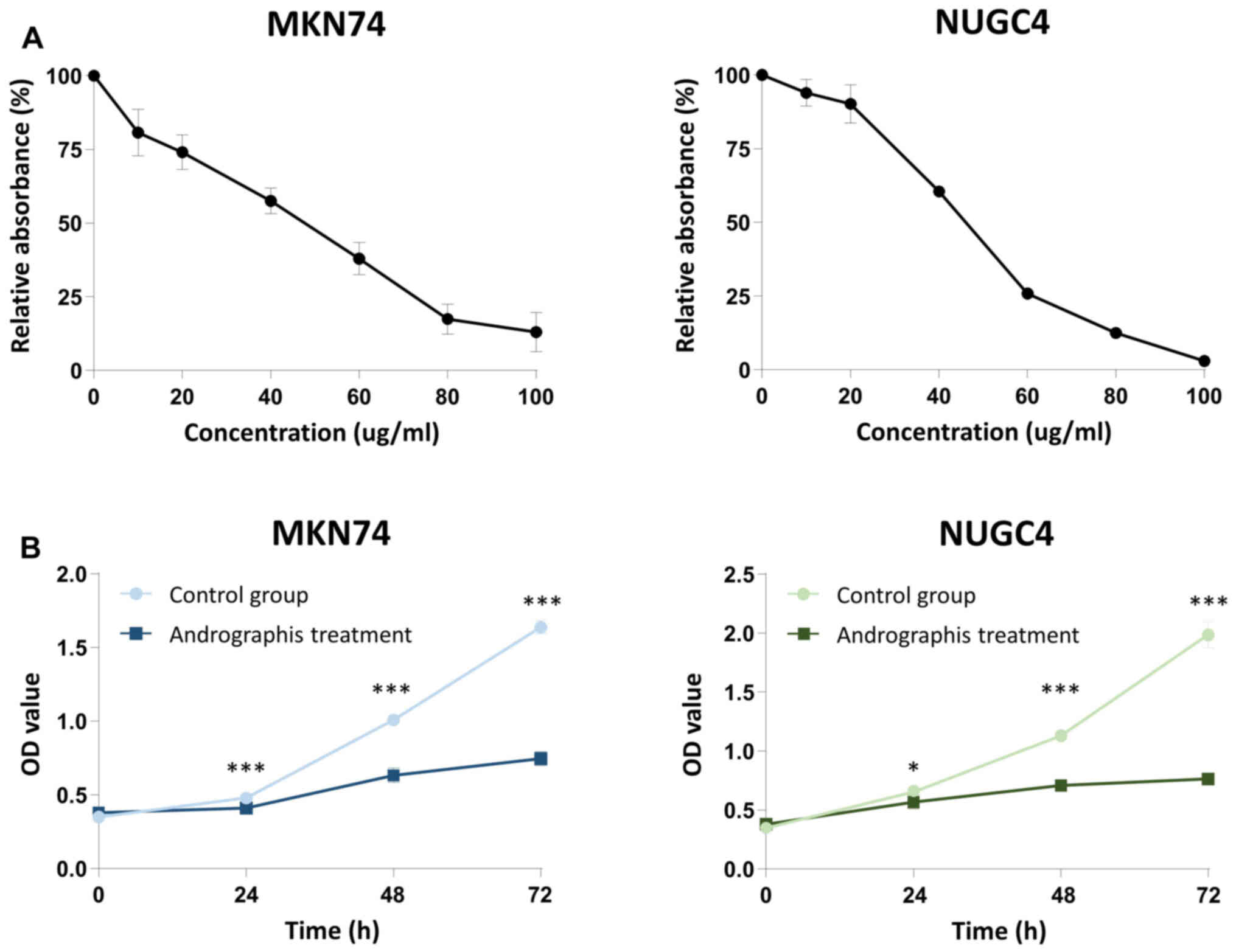
Andrographis exhibits
antiproliferative effects in GC cells
To evaluate the potential antiproliferative effects
of Andrographis in GC cells, we first treated two GC cell lines
(MKN74 and NUGC4) with Andrographis at concentrations of 10, 20,
40, 60, 80 and 100 µg/ml. As expected, Andrographis suppressed the
proliferation of MKN74 and NUGC4 cell lines in a dose-dependent
manner (Fig. 1A). Next, we treated
GC cells with 40 µg/ml Andrographis for 24, 48 and 72 h and
compared the proliferative ability of the Andrographis treatment
group with that of the control group. Intriguingly, the results
showed that Andrographis treatment significantly inhibited the
growth of both cell lines (MKN74: 24 h, P<0.0001; 48 h,
P<0.0001; 72 h, P<0.0001 and NUGC4: 24 h, P=0.03; 48 h,
P<0.0001; 72 h, P<0.0001) (Fig.
1B).
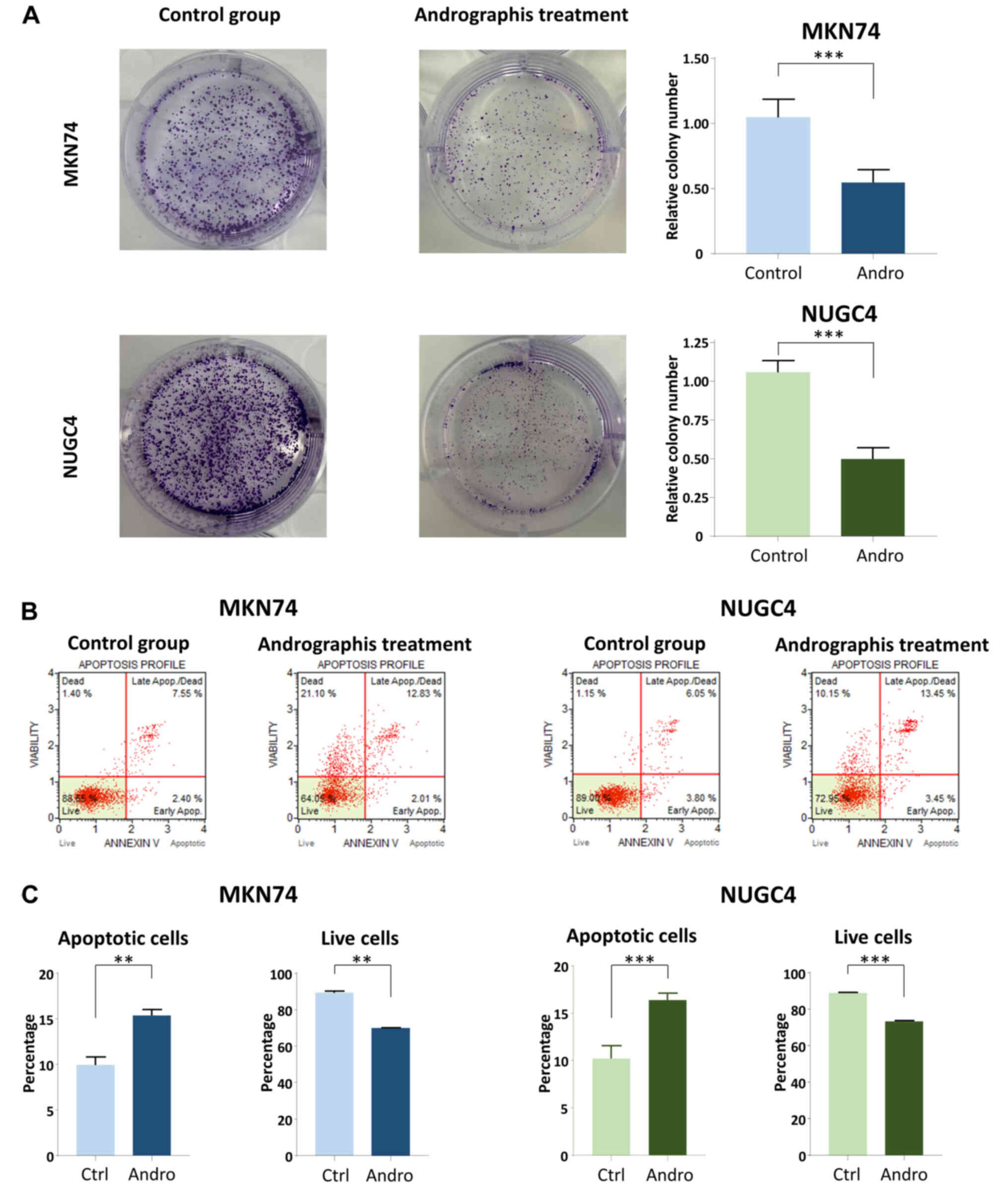
Andrographis inhibits the colony
formation activity of GC cells
We next investigated the colony forming ability of
two GC cell lines. After treatment of MKN74 and NUGC4 cells with
Andrographis, we observed a significant reduction in the size and
number of colonies compared with the corresponding controls
(Fig. 2A). These results also
indicated that Andrographis exhibits anti-tumorigenic effects on
the phenotype of GC cells.
Andrographis treatment enhances the
apoptosis of GC cells
To verify and strengthen the results of previous
studies showing the apoptosis-enhancing activity of Andrographis in
GC (32,37,38), we
next investigated whether Andrographis treatment influences
apoptosis in MKN74 and NUGC4 cells via an Annexin V binding assay.
Apoptosis was clearly enhanced in the Andrographis-treated group
compared with the control group in both cell lines (Fig. 2B). More specifically, compared with
the control group, the percentage of apoptotic cells in the
Andrographis-treated group significantly increased to 5.45±0.95%
for MKN74 cells (P=0.001) and 6.18±1.6% for NUGC4 cells (P=0.002),
and Andrographis treatment significantly reduced the percentage of
live cells to 19.43±1.13% for MKN74 cells (P=0.0001) and
15.42±0.65% for NUGC4 cells (P<0.0001) (Fig. 2C). Together, our results confirmed
the previous finding that Andrographis exhibits anti-cancer
potential through the enhancement of apoptosis using MKN74 and
NUGC4 cells.
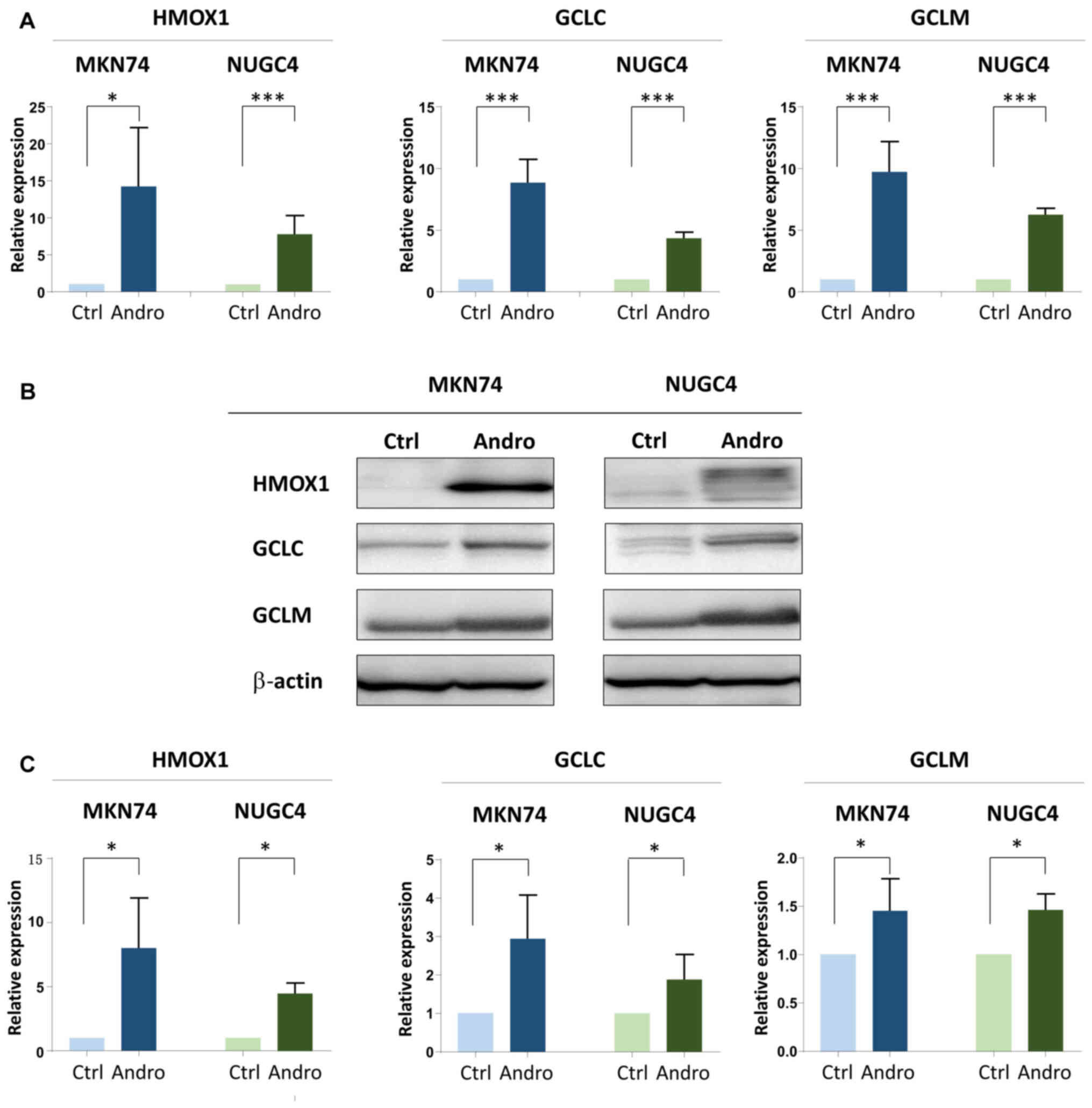
Andrographis mediates its anti-cancer
activity by activating ferroptosis-associated genes
Because previous evidence revealed that
Andrographolide induces the upregulation of ferroptosis-associated
genes, such as HMOX1, GCLC, and GCLM, in both
non-cancer (39–43) and cancer cells (44–46), we
investigated whether this finding applies to GC by performing
RT-qPCR and western blot assays. Intriguingly, the RT-qPCR results
demonstrated that all target genes were significantly upregulated
(P<0.0001) at the mRNA level following Andrographis treatment
compared with the corresponding control in both cell lines
(Fig. 3A). Furthermore, western blot
experiments confirmed a substantial increase in the expression of
HMOX-1 (P<0.05), GCLC (P<0.05), and GCLM (P<0.05) at the
protein level in both cell lines after Andrographis treatment
compared with the corresponding control (Fig. 3B and C). Collectively, these results
suggested that the alteration of ferroptosis-associated genes may
be one of the possible mechanisms by which Andrographis exerts its
anti-tumorigenic potential in GC cells.
Discussion
Accumulating evidence has shown that dietary
botanical compounds, such as herbal medicines, have increasingly
important roles in the field of cancer treatment, both as
therapeutic agents and adjunctive treatments to traditional
therapies (10,47). Andrographolide is a C20 labdane
diterpenoid derived from the traditional Chinese medicine
Andrographis paniculate (12–14),
which has been found to have cytotoxic/anti-tumorigenic potential
in various malignancies (14,20,22,24–26,28).
Intriguingly, Andrographolide was demonstrated to alter multiple
cancer-associated signaling pathways, such as BAX-dependent
apoptotic signaling in breast cancer (22), PI3K/AKT/mTOR-dependent signaling in
leukemia (20) and glioblastoma
(24), p38 signaling in melanoma
(14), STAT3/AKT activation in
pancreatic cancer (28), and the
Src/MAPKs/AP-1 axis (25) and
TLR4/NF-kB/MMP-9 pathway (32) in
colorectal cancer. Moreover, Andrographolide modulates
chemosensitization in multiple tumors (28,46,48). In
this study, we demonstrated that Andrographis exerts
anti-tumorigenic effects by suppressing cell proliferation and
colony formation and enhancing apoptotic activity in MKN74 and
NUGC4 GC cells. Intriguingly, we also revealed that Andrographis
treatment altered the expression of ferroptosis-associated genes,
including HMOX1, GCLC, and GCLM, which might offer
novel mechanistic insight into the pathogenesis of GC.
Collectively, our findings may provide additional evidence
supporting the anti-tumorigenic potential of Andrographis as an
adjunctive treatment in GC.
In our study, we showed that cell viability was
significantly reduced and apoptotic activity was enhanced in
Andrographis-treated GC cells compared with the control cells,
which is consistent with previous reports. Lim et al
(32) reported that Andrographolide
dose-dependently decreased the proliferation and viability of GC
cells, which was accompanied by increased apoptotic and
non-apoptotic cell death. They also demonstrated that
Andrographolide enhanced recombinant human TRAIL-induced apoptotic
cell death mediated by the TRAIL-RS (DR5) pathway (32). Furthermore, Li et al (37) verified that Andrographolide inhibited
cell proliferation and induced apoptosis by altering the expression
of BAX, caspase-3, and BCL-2. In a study by Dai et al
(38), Andrographolide inhibited
cell proliferation, invasion, and migration in a dose-dependent
manner and promoted apoptosis. They also showed that
Andrographolide influenced the expression of various targets,
including the upregulation of TIMP-1/2, cyclin B1, p-Cdc2, BAX, and
BIK and downregulation of MMP-2/9 and BCL-2 (38).
The most striking result of this study was that
Andrographis treatment remarkably upregulated the expression of the
ferroptosis-related genes HMOX1, GCLC, and GCLM.
Ferroptosis is a recently defined form of regulated cell death that
differs from apoptosis, necrosis, and necroptosis and is
characterized by iron-dependent reactive oxygen species (ROS)
generation, lipid peroxidation, and iron accumulation (49–51).
Because ferroptosis is often associated with resistance to
chemotherapeutic drugs, several types of malignancies, including
large B-cell lymphoma, leukemia, head and neck cancer, renal cell
carcinoma, osteosarcoma, prostate adenocarcinoma, hepatocellular
carcinoma, cholangiocarcinoma, ovarian cancer, pancreatic
carcinoma, and lung cancer (52–61),
exhibit sensitivity to ferroptosis inducers. For instance,
ferroptosis inducers (e.g., erastin and sorafenib) may be
considered a novel treatment regimen for non-small cell lung cancer
patients with cisplatin failure (62). Furthermore, sulfasalazine depletes
paclitaxel-resistant tumor cells by inducing ferroptosis in uterine
serous carcinoma, which may be an effective treatment for patients
with recurrent paclitaxel-resistant uterine serous carcinoma
(63).
In our study, significant upregulation of HMOX-1 was
observed after Andrographis treatment in GC cells. Heme oxygenase
is a rate-limiting enzyme that catalyzes the oxidative degradation
of cellular heme, which produces carbon monoxide, bilirubin, and
free iron (64). HMOX-1 is a subtype
of heme oxygenase that maintains cellular homeostasis and reduces
tissue oxidative damage and the inflammatory response (65,66).
Additionally, HMOX-1 regulates cellular iron and ROS levels during
ferroptosis (67–70). HMOX-1 was reported to play an
anticancer role in various types of human malignancies, such as
fibrosarcoma, breast cancer, and prostate cancer (45,71–73).
Moreover, some evidence indicates that Andrographolide induces the
upregulation of HMOX-1 in breast cancer, fibrosarcoma, and
colorectal cancer (44–46). In this study, we revealed that
Andrographis treatment induced significant upregulation of HMOX-1
in GC cells, which strengthens the idea that Andrographis may have
potential as an adjunctive treatment by enhancing ferroptotic
activity mediated by HMOX-1.
Our experimental findings also demonstrated that
Andrographis treatment upregulated the expression of GCLC
and GCLM. GCLC and GCLM are involved in the synthesis of GSH
in the oxidative stress response and metabolism of intracellular
labile iron, and GCLC and GCLM are critical genes in
the ferroptosis-associated pathway (74,75).
Several dietary botanical compounds, such as chrysin, apigenin, and
luteolin, can upregulate GCLC and GCLM in addition to
HMOX1 gene transcription via the ERK2/NRF2/ARE signaling pathway
(76). Consistent with this study,
our findings demonstrated that the altered expression of HMOX-1 was
accompanied by the upregulation of GCLC and GCLM following
treatment of GC cells with Andrographis.
There are several limitations to our current study.
First, although we showed the anti-tumorigenic potential of
Andrographis, we demonstrated this using just two GC cell lines. In
addition, although we revealed the upregulation of HMOX-1, GCLC,
and GCLM after Andrographis treatment, we did not perform detailed
mechanistic studies of ferroptosis pathways. In the future, we plan
to further identify the molecular mechanisms underlying the effects
of this dietary compound on ferroptosis.
Collectively, our study demonstrated the
anti-tumorigenic properties of Andrographis through the alteration
of the ferroptosis-associated genes HMOX1, GCLC, and
GCLM in GC cells. Although further mechanistic validation is
warranted, our study may provide substantial evidence for the use
of Andrographis as a potential adjunctive treatment in patients
with GC.
In conclusion, we demonstrated that Andrographis
exerts its anti-tumorigenic effects by altering the expression of
ferroptosis-associated genes, indicating that Andrographis could
serve as an adjunctive therapeutic option in patients with GC.
Acknowledgements
The authors would like to acknowledge Ms. Yuki Orito
(Department of Gastrointestinal and Pediatric Surgery, Institute of
Life Sciences, Mie University Graduate School of Medicine) and Ms.
Amphone Okada (Department of Gastrointestinal and Pediatric
Surgery, Institute of Life Sciences, Mie University Graduate School
of Medicine) for their experimental support, and Dr Priyanka Sharma
(Department of Molecular Diagnostics and Experimental Therapeutics,
Beckman Research Institute, City of Hope Comprehensive Cancer
Center) for advice on the discussion of this article. Finally, the
authors would like to thank Dr H. Nikki March and Dr Melissa
Crawford, for editing a draft of this manuscript.
Funding
The present study was supported by a Grant-in-Aid
for Scientific Research (grant nos. 18K08566 and 18K08700) from the
Ministry of Education, Culture, Sports, Science, and Technology,
Japan.
Availability of data and materials
All data generated or analyzed during this study are
included in this published article.
Authors' contributions
RM, TS, CY, YOku and YT conceived and designed the
study. RM, TS, CY, YOku, TK, YK, YT, AG, LY and XZ acquired,
analyzed and interpreted the data. RM, TS, CY, YOku, AG, LY, XZ and
YT drafted the manuscript. RM, TS, CY, YOki, MO and KU performed
statistical analysis. YT supervised the study. RM, TS, CY and YOku
confirm the authenticity of all the raw data. All authors read and
approved the final manuscript.
Ethics approval and consent to
participate
Not applicable.
Patient consent for publication
Not applicable.
Competing interests
The authors declare that they have no competing
interests.
Glossary
Abbreviations
Abbreviations:
|
GC
|
gastric cancer
|
|
FBS
|
fetal bovine serum
|
|
h
|
hours
|
|
ROS
|
reactive oxygen species
|
|
HMOX1
|
heme oxygenase-1
|
|
GCLC
|
glutamate-cysteine ligase
catalytic
|
|
GCLM
|
glutamate-cysteine ligase modifier
|
|
TRAIL
|
tumor necrosis factor-related
apoptosis-inducing ligand
|
References
|
1
|
Bray F, Ferlay J, Soerjomataram I, Siegel
RL, Torre LA and Jemal A: Global cancer statistics 2018: GLOBOCAN
estimates of incidence and mortality worldwide for 36 cancers in
185 countries. CA Cancer J Clin. 68:394–424. 2018.Erratum in: CA
Cancer J Clin 70: 313, 2020. View Article : Google Scholar : PubMed/NCBI
|
|
2
|
Digklia A and Wagner AD: Advanced gastric
cancer: Current treatment landscape and future perspectives. World
J Gastroenterol. 22:2403–2414. 2016. View Article : Google Scholar : PubMed/NCBI
|
|
3
|
Smyth EC, Nilsson M, Grabsch HI, van
Grieken NC and Lordick F: Gastric cancer. Lancet. 396:635–648.
2020. View Article : Google Scholar : PubMed/NCBI
|
|
4
|
Hironaka S, Sugimoto N, Yamaguchi K,
Moriwaki T, Komatsu Y, Nishina T, Tsuji A, Nakajima TE, Gotoh M,
Machida N, et al: S-1 plus leucovorin versus S-1 plus leucovorin
and oxaliplatin versus S-1 plus cisplatin in patients with advanced
gastric cancer: A randomised, multicentre, open-label, phase 2
trial. Lancet Oncol. 17:99–108. 2016. View Article : Google Scholar : PubMed/NCBI
|
|
5
|
Wagner AD, Syn NL, Moehler M, Grothe W,
Yong WP, Tai BC, Ho J and Unverzagt S: Chemotherapy for advanced
gastric cancer. Cochrane Database Syst Rev.
8:CD0040642017.PubMed/NCBI
|
|
6
|
Van Cutsem E, Sagaert X, Topal B,
Haustermans K and Prenen H: Gastric cancer. Lancet. 388:2654–2664.
2016. View Article : Google Scholar : PubMed/NCBI
|
|
7
|
Muro K, Van Cutsem E, Narita Y,
Pentheroudakis G, Baba E, Li J, Ryu MH, Zamaniah WIW, Yong WP, Yeh
KH, et al: Pan-Asian adapted ESMO Clinical Practice Guidelines for
the management of patients with metastatic gastric cancer: A
JSMO-ESMO initiative endorsed by CSCO, KSMO, MOS, SSO and TOS. Ann
Oncol. 30:19–33. 2019. View Article : Google Scholar : PubMed/NCBI
|
|
8
|
Hyodo I, Amano N, Eguchi K, Narabayashi M,
Imanishi J, Hirai M, Nakano T and Takashima S: Nationwide survey on
complementary and alternative medicine in cancer patients in Japan.
J Clin Oncol. 23:2645–2654. 2005. View Article : Google Scholar : PubMed/NCBI
|
|
9
|
Chan A, Tan HL, Ching TH and Tan HC:
Clinical outcomes for cancer patients using complementary and
alternative medicine. Altern Ther Health Med. 18:12–17.
2012.PubMed/NCBI
|
|
10
|
Liu TG, Xiong SQ, Yan Y, Zhu H and Yi C:
Use of chinese herb medicine in cancer patients: a survey in
southwestern china. Evid Based Complement Alternat Med.
2012:7690422012. View Article : Google Scholar : PubMed/NCBI
|
|
11
|
Newman DJ and Cragg GM: Natural products
as sources of new drugs from 1981 to 2014. J Nat Prod. 79:629–661.
2016. View Article : Google Scholar : PubMed/NCBI
|
|
12
|
Singh S, Pandey P, Ghosh S and Banerjee S:
Anti-cancer labdane diterpenoids from adventitious roots of
Andrographis paniculata: Augmentation of production prospect
endowed with pathway gene expression. Protoplasma. 255:1387–1400.
2018. View Article : Google Scholar : PubMed/NCBI
|
|
13
|
Islam MT, Ali ES, Uddin SJ, Islam MA, Shaw
S, Khan IN, Saravi SSS, Ahmad S, Rehman S, Gupta VK, et al:
Andrographolide, a diterpene lactone from Andrographis
paniculata and its therapeutic promises in cancer. Cancer Lett.
420:129–145. 2018. View Article : Google Scholar : PubMed/NCBI
|
|
14
|
Liu G and Chu H: Andrographolide inhibits
proliferation and induces cell cycle arrest and apoptosis in human
melanoma cells. Oncol Lett. 15:5301–5305. 2018.PubMed/NCBI
|
|
15
|
Suo XB, Zhang H and Wang YQ: HPLC
determination of andrographolide in rat whole blood: Study on the
pharmacokinetics of andrographolide incorporated in liposomes and
tablets. Biomed Chromatogr. 21:730–734. 2007. View Article : Google Scholar : PubMed/NCBI
|
|
16
|
Lu WJ, Lee JJ, Chou DS, Jayakumar T, Fong
TH, Hsiao G and Sheu JR: A novel role of andrographolide, an
NF-kappa B inhibitor, on inhibition of platelet activation: The
pivotal mechanisms of endothelial nitric oxide synthase/cyclic GMP.
J Mol Med (Berl). 89:1261–1273. 2011. View Article : Google Scholar : PubMed/NCBI
|
|
17
|
Jayakumar T, Hsieh CY, Lee JJ and Sheu JR:
Experimental and clinical pharmacology of Andrographis
paniculata and its major bioactive phytoconstituent
andrographolide. Evid Based Complement Alternat Med.
2013:8467402013. View Article : Google Scholar : PubMed/NCBI
|
|
18
|
Poolsup N, Suthisisang C, Prathanturarug
S, Asawamekin A and Chanchareon U: Andrographis paniculata
in the symptomatic treatment of uncomplicated upper respiratory
tract infection: Systematic review of randomized controlled trials.
J Clin Pharm Ther. 29:37–45. 2004. View Article : Google Scholar : PubMed/NCBI
|
|
19
|
Dai Y, Chen SR, Chai L, Zhao J and Wang Y
and Wang Y: Overview of pharmacological activities of
Andrographis paniculata and its major compound
andrographolide. Crit Rev Food Sci Nutr. 59 (Suppl 1):S17–S29.
2019. View Article : Google Scholar : PubMed/NCBI
|
|
20
|
Kumar D, Das B, Sen R, Kundu P, Manna A,
Sarkar A, Chowdhury C, Chatterjee M and Das P: Andrographolide
analogue induces apoptosis and autophagy mediated cell death in
U937 cells by inhibition of PI3K/Akt/mTOR pathway. PLoS One.
10:e01396572015. View Article : Google Scholar : PubMed/NCBI
|
|
21
|
Li L, Yue GG, Lee JK, Wong EC, Fung KP, Yu
J, Lau CB and Chiu PW: The adjuvant value of Andrographis
paniculata in metastatic esophageal cancer treatment - from
preclinical perspectives. Sci Rep. 7:8542017. View Article : Google Scholar : PubMed/NCBI
|
|
22
|
Banerjee M, Chattopadhyay S, Choudhuri T,
Bera R, Kumar S, Chakraborty B and Mukherjee SK: Cytotoxicity and
cell cycle arrest induced by andrographolide lead to programmed
cell death of MDA-MB-231 breast cancer cell line. J Biomed Sci.
23:402016. View Article : Google Scholar : PubMed/NCBI
|
|
23
|
Lai YH, Yu SL, Chen HY, Wang CC, Chen HW
and Chen JJ: The HLJ1-targeting drug screening identified Chinese
herb andrographolide that can suppress tumour growth and invasion
in non-small-cell lung cancer. Carcinogenesis. 34:1069–1080. 2013.
View Article : Google Scholar : PubMed/NCBI
|
|
24
|
Li Y, Zhang P, Qiu F, Chen L, Miao C, Li
J, Xiao W and Ma E: Inactivation of PI3K/Akt signaling mediates
proliferation inhibition and G2/M phase arrest induced by
andrographolide in human glioblastoma cells. Life Sci. 90:962–967.
2012. View Article : Google Scholar : PubMed/NCBI
|
|
25
|
Yuan M, Meng W, Liao W and Lian S:
Andrographolide antagonizes TNF-α-induced IL-8 via inhibition of
NADPH oxidase/ROS/NF-κB and Src/MAPKs/AP-1 axis in human colorectal
cancer HCT116 cells. J Agric Food Chem. 66:5139–5148. 2018.
View Article : Google Scholar : PubMed/NCBI
|
|
26
|
Zhang R, Zhao J, Xu J, Jiao DX, Wang J,
Gong ZQ and Jia JH: Andrographolide suppresses proliferation of
human colon cancer SW620 cells through the TLR4/NF-κB/MMP-9
signaling pathway. Oncol Lett. 14:4305–4310. 2017. View Article : Google Scholar : PubMed/NCBI
|
|
27
|
Deng Y, Bi R, Guo H, Yang J, Du Y, Wang C
and Wei W: Andrographolide enhances TRAIL-induced apoptosis via
p53-mediated death receptors up-regulation and suppression of the
NF-кB pathway in bladder cancer cells. Int J Biol Sci. 15:688–700.
2019. View Article : Google Scholar : PubMed/NCBI
|
|
28
|
Bao GQ, Shen BY, Pan CP, Zhang YJ, Shi MM
and Peng CH: Andrographolide causes apoptosis via inactivation of
STAT3 and Akt and potentiates antitumor activity of gemcitabine in
pancreatic cancer. Toxicol Lett. 222:23–35. 2013. View Article : Google Scholar : PubMed/NCBI
|
|
29
|
Yang W, Zhao J, Wang Y, Xu H, Wu Z, Hu Y,
Jiang K, Shen P, Ma C, Guan Z, et al: In vivo inhibitory activity
of andrographolide derivative ADN-9 against liver cancer and its
mechanisms involved in inhibition of tumor angiogenesis. Toxicol
Appl Pharmacol. 327:1–12. 2017. View Article : Google Scholar : PubMed/NCBI
|
|
30
|
Lim JC, Chan TK, Ng DS, Sagineedu SR,
Stanslas J and Wong WS: Andrographolide and its analogues:
Versatile bioactive molecules for combating inflammation and
cancer. Clin Exp Pharmacol Physiol. 39:300–310. 2012. View Article : Google Scholar : PubMed/NCBI
|
|
31
|
Yue GG, Li L, Lee JK, Kwok HF, Wong EC, Li
M, Fung KP, Yu J, Chan AW, Chiu PW and Lau CB: Multiple modulatory
activities of Andrographis paniculata on immune responses
and xenograft growth in esophageal cancer preclinical models.
Phytomedicine. 60:1528862019. View Article : Google Scholar : PubMed/NCBI
|
|
32
|
Lim SC, Jeon HJ, Kee KH, Lee MJ, Hong R
and Han SI: Andrographolide induces apoptotic and non-apoptotic
death and enhances tumor necrosis factor-related apoptosis-inducing
ligand-mediated apoptosis in gastric cancer cells. Oncol Lett.
13:3837–3844. 2017. View Article : Google Scholar : PubMed/NCBI
|
|
33
|
Stewart MJ and Watson ID: Standard units
for expressing drug concentrations in biological fluids. Br J Clin
Pharmacol. 16:3–7. 1983. View Article : Google Scholar : PubMed/NCBI
|
|
34
|
Takahashi M, Sung B, Shen Y, Hur K, Link
A, Boland CR, Aggarwal BB and Goel A: Boswellic acid exerts
antitumor effects in colorectal cancer cells by modulating
expression of the let-7 and miR-200 microRNA family.
Carcinogenesis. 33:2441–2449. 2012. View Article : Google Scholar : PubMed/NCBI
|
|
35
|
Schneider CA, Rasband WS and Eliceiri KW:
NIH Image to ImageJ: 25 years of image analysis. Nat Methods.
9:671–675. 2012. View Article : Google Scholar : PubMed/NCBI
|
|
36
|
Livak KJ and Schmittgen TD: Analysis of
relative gene expression data using real-time quantitative PCR and
the 2(-Delta Delta C(T)) Method. Methods. 25:402–408. 2001.
View Article : Google Scholar : PubMed/NCBI
|
|
37
|
Li SG, Wang YY, Ye ZY, Shao QS, Tao HQ,
Shu LS, Zhao YF, Yang YJ, Yang J, Peng T, et al: Proliferative and
apoptotic effects of andrographolide on the BGC-823 human gastric
cancer cell line. Chin Med J (Engl). 126:3739–3744. 2013.PubMed/NCBI
|
|
38
|
Dai L, Wang G and Pan W: Andrographolide
inhibits proliferation and metastasis of SGC7901 gastric cancer
cells. BioMed Res Int. 2017:62421032017. View Article : Google Scholar : PubMed/NCBI
|
|
39
|
Yu AL, Lu CY, Wang TS, Tsai CW, Liu KL,
Cheng YP, Chang HC, Lii CK and Chen HW: Induction of heme oxygenase
1 and inhibition of tumor necrosis factor alpha-induced
intercellular adhesion molecule expression by andrographolide in
EA.hy926 cells. J Agric Food Chem. 58:7641–7648. 2010. View Article : Google Scholar : PubMed/NCBI
|
|
40
|
Seo JY, Pyo E, An JP, Kim J, Sung SH and
Oh WK: Andrographolide activates Keap1/Nrf2/ARE/HO-1 pathway in
HT22 cells and suppresses microglial activation by Aβ42 through
Nrf2-related inflammatory response. Mediators Inflamm.
2017:59061892017. View Article : Google Scholar : PubMed/NCBI
|
|
41
|
Lu CY, Yang YC, Li CC, Liu KL, Lii CK and
Chen HW: Andrographolide inhibits TNFα-induced ICAM-1 expression
via suppression of NADPH oxidase activation and induction of HO-1
and GCLM expression through the PI3K/Akt/Nrf2 and PI3K/Akt/AP-1
pathways in human endothelial cells. Biochem Pharmacol. 91:40–50.
2014. View Article : Google Scholar : PubMed/NCBI
|
|
42
|
Lee JC, Tseng CK, Young KC, Sun HY, Wang
SW, Chen WC, Lin CK and Wu YH: Andrographolide exerts
anti-hepatitis C virus activity by up-regulating haeme oxygenase-1
via the p38 MAPK/Nrf2 pathway in human hepatoma cells. Br J
Pharmacol. 171:237–252. 2014. View Article : Google Scholar : PubMed/NCBI
|
|
43
|
Guan SP, Tee W, Ng DS, Chan TK, Peh HY, Ho
WE, Cheng C, Mak JC and Wong WS: Andrographolide protects against
cigarette smoke-induced oxidative lung injury via augmentation of
Nrf2 activity. Br J Pharmacol. 168:1707–1718. 2013. View Article : Google Scholar : PubMed/NCBI
|
|
44
|
Chao CY, Lii CK, Hsu YT, Lu CY, Liu KL, Li
CC and Chen HW: Induction of heme oxygenase-1 and inhibition of
TPA-induced matrix metalloproteinase-9 expression by
andrographolide in MCF-7 human breast cancer cells. Carcinogenesis.
34:1843–1851. 2013. View Article : Google Scholar : PubMed/NCBI
|
|
45
|
Kwon MY, Park E, Lee SJ and Chung SW: Heme
oxygenase-1 accelerates erastin-induced ferroptotic cell death.
Oncotarget. 6:24393–24403. 2015. View Article : Google Scholar : PubMed/NCBI
|
|
46
|
Sharma P, Shimura T, Banwait JK and Goel
A: Andrographis-mediated chemosensitization through activation of
ferroptosis and suppression of β-catenin/Wnt-signaling pathways in
colorectal cancer. Carcinogenesis. 41:1385–1394. 2020. View Article : Google Scholar : PubMed/NCBI
|
|
47
|
Yang G, Li X, Li X, Wang L, Li J, Song X,
Chen J, Guo Y, Sun X, Wang S, et al: Traditional chinese medicine
in cancer care: a review of case series published in the chinese
literature. Evid Based Complement Alternat Med. 2012:7510462012.
View Article : Google Scholar : PubMed/NCBI
|
|
48
|
Zhou J, Ong CN, Hur GM and Shen HM:
Inhibition of the JAK-STAT3 pathway by andrographolide enhances
chemosensitivity of cancer cells to doxorubicin. Biochem Pharmacol.
79:1242–1250. 2010. View Article : Google Scholar : PubMed/NCBI
|
|
49
|
Liang C, Zhang X, Yang M and Dong X:
Recent progress in ferroptosis inducers for cancer therapy. Adv
Mater. 31:e19041972019. View Article : Google Scholar : PubMed/NCBI
|
|
50
|
Ye Z, Liu W, Zhuo Q, Hu Q, Liu M, Sun Q,
Zhang Z, Fan G, Xu W, Ji S, et al: Ferroptosis: Final destination
for cancer? Cell Prolif. 53:e127612020. View Article : Google Scholar : PubMed/NCBI
|
|
51
|
Hassannia B, Vandenabeele P and Vanden
Berghe T: Targeting Ferroptosis to Iron Out Cancer. Cancer Cell.
35:830–849. 2019. View Article : Google Scholar : PubMed/NCBI
|
|
52
|
Yu Y, Xie Y, Cao L, Yang L, Yang M, Lotze
MT, Zeh HJ, Kang R and Tang D: The ferroptosis inducer erastin
enhances sensitivity of acute myeloid leukemia cells to
chemotherapeutic agents. Mol Cell Oncol. 2:e10545492015. View Article : Google Scholar : PubMed/NCBI
|
|
53
|
Yang S, Evens AM, Prachand S, Singh AT,
Bhalla S, David K and Gordon LI: Mitochondrial-mediated apoptosis
in lymphoma cells by the diterpenoid lactone andrographolide, the
active component of Andrographis paniculata. Clin Cancer
Res. 16:4755–4768. 2010. View Article : Google Scholar : PubMed/NCBI
|
|
54
|
Xia X, Fan X, Zhao M and Zhu P: The
relationship between ferroptosis and tumors: a novel landscape for
therapeutic approach. Curr Gene Ther. 19:117–124. 2019. View Article : Google Scholar : PubMed/NCBI
|
|
55
|
Chen Y, Fan Z, Yang Y and Gu C: Iron
metabolism and its contribution to cancer (Review). Int J Oncol.
54:1143–1154. 2019.PubMed/NCBI
|
|
56
|
Roh JL, Kim EH, Jang HJ, Park JY and Shin
D: Induction of ferroptotic cell death for overcoming cisplatin
resistance of head and neck cancer. Cancer Lett. 381:96–103. 2016.
View Article : Google Scholar : PubMed/NCBI
|
|
57
|
Louandre C, Ezzoukhry Z, Godin C, Barbare
JC, Mazière JC, Chauffert B and Galmiche A: Iron-dependent cell
death of hepatocellular carcinoma cells exposed to sorafenib. Int J
Cancer. 133:1732–1742. 2013. View Article : Google Scholar : PubMed/NCBI
|
|
58
|
Puntawee S, Theerasilp M, Reabroi S,
Saeeng R, Piyachaturawat P, Chairoungdua A and Nasongkla N:
Solubility enhancement and in vitro evaluation of PEG-b-PLA
micelles as nanocarrier of semi-synthetic andrographolide analogue
for cholangiocarcinoma chemotherapy. Pharm Dev Technol. 21:437–444.
2016.PubMed/NCBI
|
|
59
|
Basuli D, Tesfay L, Deng Z, Paul B,
Yamamoto Y, Ning G, Xian W, McKeon F, Lynch M, Crum CP, et al: Iron
addiction: A novel therapeutic target in ovarian cancer. Oncogene.
36:4089–4099. 2017. View Article : Google Scholar : PubMed/NCBI
|
|
60
|
Eling N, Reuter L, Hazin J, Hamacher-Brady
A and Brady NR: Identification of artesunate as a specific
activator of ferroptosis in pancreatic cancer cells. Oncoscience.
2:517–532. 2015. View Article : Google Scholar : PubMed/NCBI
|
|
61
|
Liu Q and Wang K: The induction of
ferroptosis by impairing STAT3/Nrf2/GPx4 signaling enhances the
sensitivity of osteosarcoma cells to cisplatin. Cell Biol Int.
43:1245–1256. 2019. View Article : Google Scholar : PubMed/NCBI
|
|
62
|
Li Y, Yan H, Xu X, Liu H, Wu C and Zhao L:
Erastin/sorafenib induces cisplatin-resistant non-small cell lung
cancer cell ferroptosis through inhibition of the Nrf2/xCT pathway.
Oncol Lett. 19:323–333. 2020.PubMed/NCBI
|
|
63
|
Sugiyama A, Ohta T, Obata M, Takahashi K,
Seino M and Nagase S: xCT inhibitor sulfasalazine depletes
paclitaxel-resistant tumor cells through ferroptosis in uterine
serous carcinoma. Oncol Lett. 20:2689–2700. 2020. View Article : Google Scholar : PubMed/NCBI
|
|
64
|
Bussolati B, Ahmed A, Pemberton H, Landis
RC, Di Carlo F, Haskard DO and Mason JC: Bifunctional role for
VEGF-induced heme oxygenase-1 in vivo: Induction of angiogenesis
and inhibition of leukocytic infiltration. Blood. 103:761–766.
2004. View Article : Google Scholar : PubMed/NCBI
|
|
65
|
Farombi EO and Surh YJ: Heme oxygenase-1
as a potential therapeutic target for hepatoprotection. J Biochem
Mol Biol. 39:479–491. 2006.PubMed/NCBI
|
|
66
|
Jozkowicz A, Was H and Dulak J: Heme
oxygenase-1 in tumors: Is it a false friend? Antioxid Redox Signal.
9:2099–2117. 2007. View Article : Google Scholar : PubMed/NCBI
|
|
67
|
Chiang SK, Chen SE and Chang LC: A Dual
Role of Heme Oxygenase-1 in Cancer Cells. Int J Mol Sci. 20:202018.
View Article : Google Scholar
|
|
68
|
Trachootham D, Alexandre J and Huang P:
Targeting cancer cells by ROS-mediated mechanisms: A radical
therapeutic approach? Nat Rev Drug Discov. 8:579–591. 2009.
View Article : Google Scholar : PubMed/NCBI
|
|
69
|
Suttner DM and Dennery PA: Reversal of
HO-1 related cytoprotection with increased expression is due to
reactive iron. FASEB J. 13:1800–1809. 1999. View Article : Google Scholar : PubMed/NCBI
|
|
70
|
Chang LC, Chiang SK, Chen SE, Yu YL, Chou
RH and Chang WC: Heme oxygenase-1 mediates BAY 11-7085 induced
ferroptosis. Cancer Lett. 416:124–137. 2018. View Article : Google Scholar : PubMed/NCBI
|
|
71
|
Chau LY: Heme oxygenase-1: Emerging target
of cancer therapy. J Biomed Sci. 22:222015. View Article : Google Scholar : PubMed/NCBI
|
|
72
|
Hill M, Pereira V, Chauveau C, Zagani R,
Remy S, Tesson L, Mazal D, Ubillos L, Brion R, Asghar K, et al:
Heme oxygenase-1 inhibits rat and human breast cancer cell
proliferation: Mutual cross inhibition with indoleamine
2,3-dioxygenase. FASEB J. 19:1957–1968. 2005. View Article : Google Scholar : PubMed/NCBI
|
|
73
|
Ferrando M, Gueron G, Elguero B, Giudice
J, Salles A, Leskow FC, Jares-Erijman EA, Colombo L, Meiss R,
Navone N, et al: Heme oxygenase 1 (HO-1) challenges the angiogenic
switch in prostate cancer. Angiogenesis. 14:467–479. 2011.
View Article : Google Scholar : PubMed/NCBI
|
|
74
|
Nishizawa H, Matsumoto M, Shindo T,
Saigusa D, Kato H, Suzuki K, Sato M, Ishii Y, Shimokawa H and
Igarashi K: Ferroptosis is controlled by the coordinated
transcriptional regulation of glutathione and labile iron
metabolism by the transcription factor BACH1. J Biol Chem.
295:69–82. 2020. View Article : Google Scholar : PubMed/NCBI
|
|
75
|
Lu SC: Glutathione synthesis. Biochim
Biophys Acta. 1830:3143–3153. 2013. View Article : Google Scholar : PubMed/NCBI
|
|
76
|
Huang CS, Lii CK, Lin AH, Yeh YW, Yao HT,
Li CC, Wang TS and Chen HW: Protection by chrysin, apigenin, and
luteolin against oxidative stress is mediated by the Nrf2-dependent
up-regulation of heme oxygenase 1 and glutamate cysteine ligase in
rat primary hepatocytes. Arch Toxicol. 87:167–178. 2013. View Article : Google Scholar : PubMed/NCBI
|