Introduction
Gastric carcinoma is the fifth most common type of
primary carcinoma and is the third most common cause of
cancer-associated death. Surgery is the primary mode of treatment
currently available for the management of gastric carcinoma
(1,2). At present, there is a significant
amount of interest in the factors influencing the prognosis of
stomach cancer, allowing for a more precise risk assessment of
preoperative patients. The four primary known potential risk
factors for gastric cancer are preoperative carcinoembronic antigen
(CEA) and cancer antigen 19-9 (CA19-9) levels, preoperative
systemic inflammation (3) and
perioperative blood transfusion (4). These factors have been shown to be
significantly correlated with prognosis in patients with stomach
malignancies. Apart from these four acknowledged factors, the
health condition of individuals with stomach cancer is a crucial
factor that influences their prognosis (5). The relationship between a patient's
body composition and their clinical prognosis for cancer has
garnered increasing interest in recent years. It is becoming more
widely acknowledged that skeletal muscle mass, a type of bodily
component that can be assessed by computed tomography (CT) scan,
affects the prognosis of patients who have undergone stomach cancer
surgery (6–8). A decreased skeletal muscle mass has
been found to be an independent risk factor for long-term survival
in patients with gastric cancer in prior clinical research
(9), yet there has been
disagreement on this finding due to variations in the diagnostic
criteria between cohorts (10,11).
The body mass index (BMI), sometimes referred to as
the weight index, provides insight into an individual's nutritional
state, amount of body fat and stage of human development (12). However, as individuals have
different fat distributions, BMI does not always accurately
represent the buildup of adipose tissue. Visceral adipose tissue is
a preferable option over BMI since it can more precisely indicate
the existence of fat cell dysfunction (13,14).
Previous studies have shown that metabolic diseases can have an
impact on changes in visceral adipose tissue gain or loss (15), and these changes further suggest
abnormalities in the intra-abdominal environment (16). As our knowledge of visceral adipose
tissue has expanded, excess adiposity, a physical characteristic
linked to malignant prognosis, has emerged as a topic of intense
interest (17,18). Adipose tissue accumulation worsens
prognosis in a number of cancer types and raises the probability of
additional cancer progression; this is partly due to the
interaction between adipocytes and cancer cells (19). High intraperitoneal fat thickness
has been linked to a reduced OS rate in patients with locally
advanced gastric cancer, according to clinical trials (20). However, these results will need to
be further confirmed in further studies, as there are few
investigations on whether pre-operative intraperitoneal fat impacts
the long-term survival of patients with stomach cancer following
surgical operations (21–23).
East Asia has a significantly higher incidence of
stomach cancer than the rest of the world (24). The majority of individuals in East
Asia are from China. However, there is very little research on the
impact of preoperative body composition on the postoperative
long-term survival of Chinese patients with gastric cancer, as the
majority of studies on visceral fat and skeletal muscle focus on
patients' postoperative condition (25,26).
In the present study, the visceral fat area and skeletal muscle
mass were measured using preoperative CT scans. Next, the collected
data were used to evaluate the predictive power of body composition
characteristics on overall survival (OS) time. To the best of our
knowledge, this is the first study assessing how preoperative body
composition affects postoperative patient survival following
gastric surgery in a Chinese cohort.
Patients and methods
Patients
The present study was a retrospective cohort study
including all patients with gastric cancer (stage I–III) who
underwent gastrectomy between January 2007 and December 2009 in the
First Affiliated Hospital of Bengbu Medical College (Bengbu,
China). The criteria for patient inclusion in the study were the
following: i) Histologically confirmed gastric adenocarcinoma and
planned to receive elective curative surgery for gastrointestinal
cancer; ii) age >18 years; iii) non-obese patients, defined as
body mass index <30 kg/m2; and iv) within 1 month
prior to gastric cancer surgery, the patient had abdominal CT image
data. Patients were excluded from the study cohort when CT image
quality was sufficient for an analysis to confirm that the patient
had metastatic cancer that could not be cured by surgical treatment
and when the patient had undergone a part gastrectomy for an
incomplete cure of stomach cancer. These reasons could also lead to
the exclusion of patients from the cohort if contact was lost with
them for some reason that prevented follow-up, or if a patient had
multiple malignancies at the same time. This study used patient
data from a digestive tumor clinical database that was established
with approval from the First Affiliated Hospital of Bengbu Medical
College (Bengbu, China; approval no. 2020KY022). The Clinical
Medical Research Ethics Committee of The First Affiliated Hospital
of Bengbu Medical College waived the requirement for informed
consent for the collection of retrospective data.
Data collection
Anthropometric data (height and weight closest to CT
time), tumor features, operative information, postoperative
treatment and comorbidity data were collected for patients in the
cohort from the institution's electronic medical records. Patients
were staged with gastric carcinoma according to the relevant
provisions in the American Joint Committee on Cancer staging manual
(eighth edition) (27). In
addition, to make the study more comprehensive and credible,
demographic information (including age and sex), laboratory data
and other relevant data for subsequent examination were included
for analysis. In addition, the systemic inflammation status of the
patients was quantified using the preoperative
neutrophil-to-lymphocyte ratio (NLR).
Body composition assessment
For the body composition assessments, a patient was
required to lie down in the supine position and then their skeletal
muscle area (SMA) and visceral fat area (VFA) were measured using
CT. At the time of measurement, the Hounsfield unit (HU) thresholds
ranged from-29 to 150 HU for skeletal muscle and 50 to 150 HU for
visceral adipose tissue (9,28). The tool used to calculate the total
cross-sectional SMA and VFA for the 3rd lumbral vertebra was ImageJ
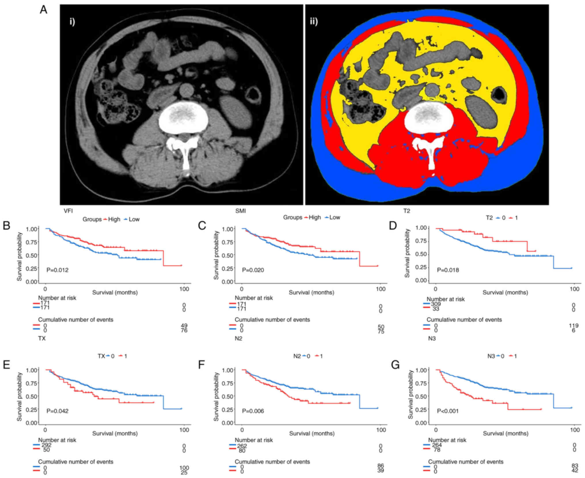
(National Institutes of Health) (Fig.
1A). For these data measurements, two experienced researchers
were selected. The Skeletal Muscle Index (SMI) was measured by
dividing the patient's skeletal muscle measurement by the square of
their height (m2), whereas the Visceral Fat Index (VFI)
was obtained by dividing the patient's area of visceral fat
(cm2) by the square of their height (m2). As
previous studies based on CT imaging have not given a valid
definition of low adiposity or low bone mass in patients with
cancer (29–31), the study cohort was divided into
high and low SMI and VFI groups based on the median value of the
data of the present cohort.
Outcomes
In this study, all individuals diagnosed with
gastric carcinoma were examined at standard outpatient clinics or
subsequently monitored. The outcome of this study was the overall
patient survival rate, which was the number of days the patient
being studied had survived from the initial date of diagnosis. A
sequence of study-related observations was performed on the
individual under study until their death, regardless of the cause,
or until the final follow-up visit. If the patient was still alive
at the final follow-up visit, they were removed from the study
cohort on the date of their last contact.
Statistical analysis
In the exploratory analysis, more structured
descriptive statistics were performed on the data collected,
including the means, standard deviations and percentages, which is
presented in a tabular form. Kaplan-Meier curves were also used to
further determine the role of each factor in survival outcomes. To
compare differences between survival curves, a Log-rank test was
used. A Cox proportional hazard model was used for the univariate
and multivariate survival analysis on the collected data. For
stratification of the SMI and VFI for Kaplan-Meier survival
analysis, the median value was used to separate the patients into
low and high VFI and SMI groups. For the calculation of overall
patient mortality, all patients included in the study who survived
gastric cancer from the time of surgery to the end of follow-up and
the total number of people who died midway or were lost to
follow-up for various reasons were used. Data were analyzed using
SPSS version 23.0 (IBM Corp.), and all calculated P-values were
two-tailed. P<0.05 was considered to indicate a statistically
significant difference.
Results
Baseline characteristics
Out of the total data gathered between 2007 and
2009, 342 patients fulfilled the inclusion criteria, with 271
(79.24%) of them being male. The median age of all participants in
the study was 64 years, with the range of 56–74. Following the
standard staging of the 342 patients, it was determined that 16
patients (4.68%) were diagnosed with stage I gastric carcinoma, 147
patients (42.98%) with stage II gastric carcinoma and 179 patients
(52.34%) with stage III gastric carcinoma. Out of all the patients
who were included, a total of 233 individuals (68.13%) underwent a
subtotal gastrectomy, while an additional 109 patients (31.87%)
underwent a total gastrectomy. Upon careful examination of the
follow-up records of all participants, it was determined that 125
individuals had died by the conclusion of the study period. The
median duration of follow-up for all patients included in the
present study was 29.54 months, ranging between 3 and 98 months.
Table I provides an overview of the
baseline characteristics of the included patients.
 | Table I.Clinicopathological characteristics
of the recruited cohort. |
Table I.
Clinicopathological characteristics
of the recruited cohort.
| Clinicopathological
factor | Value |
|---|
| Median age (range),
years | 64 (56–71) |
| Sex, n (%) |
|
|
Male | 271 (79.24) |
|
Female | 71 (20.76) |
| Preoperative
hemoglobin, g/la | 121.19±23.87 |
| Preoperative
albumin, g/la | 38.91±6.31 |
| Preoperative
neutrophil-lymphocyte ratiob | 2.03
(1.51–3.08) |
| Preoperative
carcinoembryonic antigenb | 2.80
(1.60–4.95) |
| Preoperative
CA19-9b | 9.60
(4.44–28.63) |
| Stage, n (%) |
|
| I | 16 (4.68) |
| II | 147 (42.98) |
|
III | 179 (52.34) |
| Body mass index,
mg/m2b | 21.53
(19.27–24.22) |
| SMI,
cm2/m2b | 47.73
(41.67–55.51) |
| VFI,
cm2/m2b | 41.28
(36.62–45.36) |
| Gastrectomy type, n
(%) |
|
| Partial
excision | 233 (68.13) |
| Total
excision | 109 (31.87) |
Body composition measurement
Two investigators examined CT images from the 342
participants. The median BMI of the cohort was 21.53 (IQR,
19.27–24.22), whereas the calculated median SMI and VFI were 47.73
(IQR, 41.67–55.51) and 41.28 (IQR, 36.62–45.36), respectively (data
not shown).
Association between body composition
and OS
The correlation between various body composition
parameters, such as SMI and VFI, and the subsequent data on OS were
assessed. The Kaplan-Meier curve revealed that patients with a low
VFI exhibited a lower OS time than patients with high VFI (P=0.012;
Fig. 1B). The Kaplan-Meier curves
also revealed a significant postoperative disparity in OS between
individuals with low SMI and those with high SMI (P=0.02; Fig. 1C).
Risk factors associated with a worse
prognosis
Next, other potential risk factors associated with a
worse prognosis in individuals with gastric carcinoma after surgery
were evaluated. Univariate Cox survival analysis was performed for
all clinical factors. Subgroups were set up for TNM clinical
staging (6th edition of the AJCC Cancer Staging Manual), and the
results are shown in Table II,
where T2, TX, N2 and N3 were independent factors affecting
prognosis. Kaplan-Meier curves were plotted based on the clinical
stages that independently influenced prognosis. T2 (P=0.018;
Fig. 1D) was a protective factor,
and TX (P=0.042; Fig. 1E), N2
(P=0.007; Fig. 1F) and N3
(P<0.0001 Fig. 1G) were risk
factors for a worse prognosis.
| Table II.Multivariate Cox regression
analysis. |
Table II.
Multivariate Cox regression
analysis.
|
| Univariate Cox
analysis | Multivariate Cox
analysis |
|---|
|
|
|
|
|---|
| Factor | P-value | HR (95% CI) | P-value | HR (95% CI) |
|---|
| Age | 0.403 | 1.008
(0.99–1.025) |
|
|
| Sex | 0.277 | 0.791
(0.519–1.206) |
|
|
| White blood cell
count | 0.705 | 0.989
(0.934–1.047) |
|
|
| Neutrophil
count | 0.652 | 0.986
(0.928–1.048) |
|
|
| Lymphocyte
count | 0.803 | 1.030
(0.816–1.301) |
|
|
|
Neutrophil-to-lymphocyte count | 0.069 | 1.389
(0.975–1.980) |
|
|
| Hemoglobin | 0.604 | 0.998
(0.991–1.005) |
|
|
| Albumin | 0.229 | 0.981
(0.952–1.012) |
|
|
| Skeletal muscle
index | 0.040a | 0.687
(0.480–0.983) | 0.349 | 0.806
(0.513–1.266) |
| Visceral fat
index | 0.046a | 0.989
(0.979–1.000) | 0.011 | 0.620
(0.428–0.898) |
| Triglyceride | 0.517 | 1.105
(0.817–1.469) |
|
|
| Low-density
lipoprotein | 0.948 | 0.991
(0.759–1.294) |
|
|
| High-density
lipoprotein | 0.122 | 1.468
(0.903–2.389) |
|
|
| Plasma glucose | 0.504 | 1.038
(0.931–1.157) |
|
|
| Carcinoembryonic
antigen | 0.388 | 0.997
(0.990–1.004) |
|
|
| CA19-9 | 0.002b | 1.001
(1.000–1.001) | <0.00001 | 1.001
(1.000–1.002) |
| Body mass
index | 0.311 | 0.977
(0.933–1.022) |
|
|
| Surgical
procedure | 0.443 | 1.159
(0.795–1.689) |
|
|
| T2 vs. T1 | 0.023a | 0.387
(0.170–0.879) | 0.186 | 0.566
(0.244–1.315) |
| T3 vs. T1 | 0.454 | 0.864
(0.590–1.26) |
|
|
| T4 vs. T1 | 0.159 | 1.290
(0.905–1.84) |
|
|
| TX vs. T1 | 0.045a | 1.567
(1.011–2.431) | 0.374 | 1.230
(0.779–1.941) |
| N1 vs. N0 | 0.036 | 0.592
(0.363–0.966) |
|
|
| N2 vs. N0 | 0.008b | 1.677
(1.147–2.451) | <0.00001 | 2.660
(1.719–4.118) |
| N3 vs. N0 |
<0.0001c | 2.631
(1.803–3.840) | <0.00001 | 3.313
(2.127–5.159) |
| M1 vs. M0 | 0.132 | 1.879
(0.827–4.270) |
|
|
Other clinical factors, including age, sex, NLR,
perioperative blood transfusion, preoperative CEA levels,
triglycerides, low-density lipoprotein, high-density lipoprotein,
plasma glucose and preoperative BMI, amongst other factors, were
not significantly associated with OS in the present cohort.
However, preoperative CA19-9 levels were an independent factor
affecting prognosis (P=0.001).
Predictors of adverse outcomes
To obtain more accurate results on independent risk
factors affecting the long-term survival of patients, multivariate
Cox analyses were performed for factors with P-values <0.1.
CA19-9, NLR, SMI, VFI and stages T2, TX, N2 and N3 cancer were
included in the Cox regression analysis. As shown in Table II, among the validated risk
factors, CA19-9 [hazard ratio (HR), 1.001; 95% confidence interval
(CI), 1.000–1.002; P<0.0001] and high VFI level (HR, 0.620; 95%
CI, 0.428–0.898; P=0.011) were associated with a long OS time,
while N2 (HR, 2.660; 95% CI: 1.719–4.118; P<0.0001) and N3 (HR
3.313; 95% CI: 2.127–5.159; P<0.0001) were associated with a
short OS time. NLR or SMI were not independent factors influencing
OS. Some of the serological indicators associated with VFI were
then analyzed to verify whether there were any differences in these
serological indicators in patients in the high and low VFI groups.
Among the serological indicators included (including triglycerides,
LDL, HDL and plasma glucose), only the expression level of
triglycerides was significantly different in the two groups of
patients (P<0.05).
Construction of a prognostic nomogram
including VFI
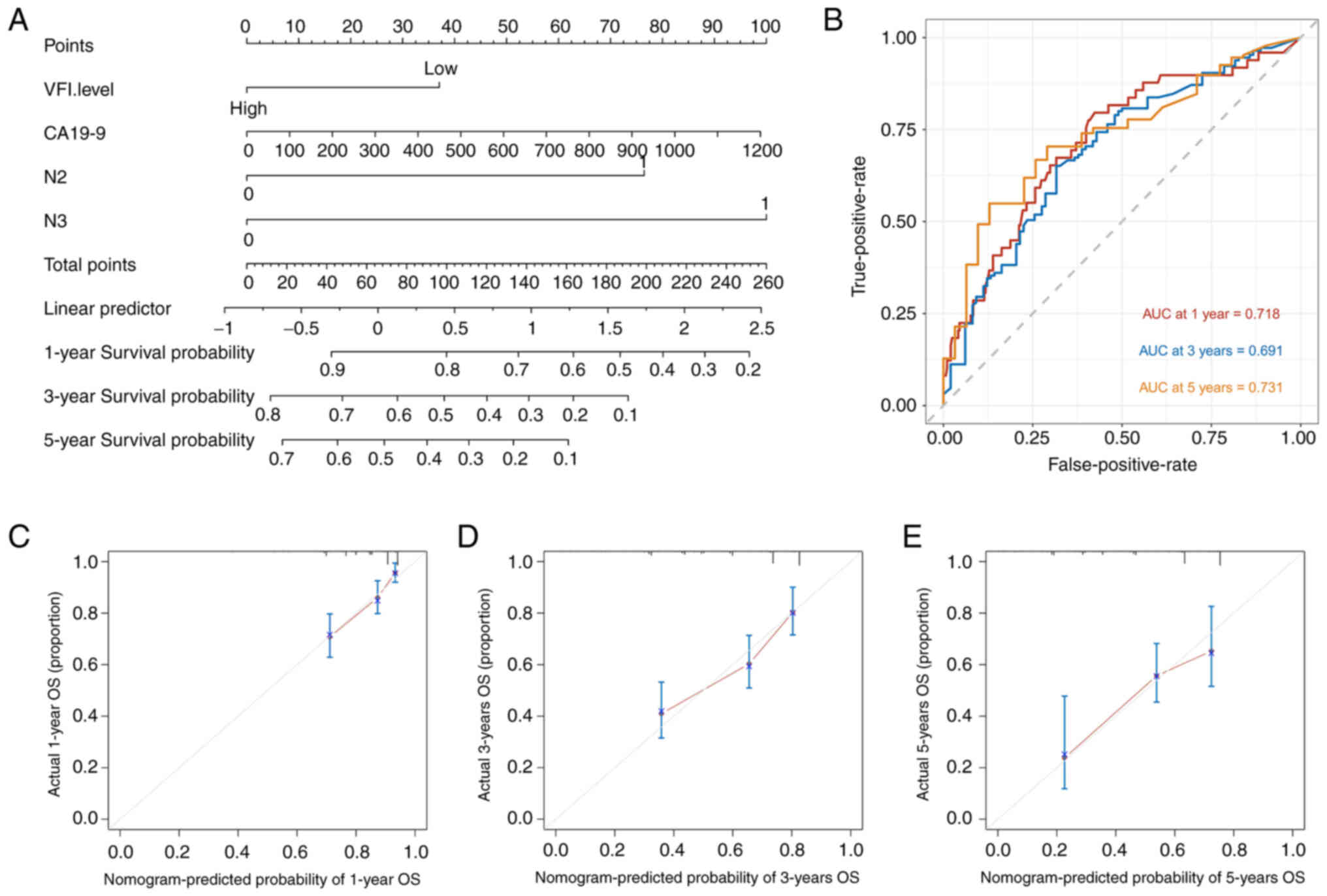
Next, a prognostic nomogram (Fig. 2A) was constructed using the
variables that were significant in the results of the multivariate
Cox survival analysis. To verify that the constructed nomogram
model had good clinical value, ROC curves were plotted (Fig. 2B) as well as 1-, 3- and 5-year
calibration curves (Fig. 2C-E). The
area under the ROC curve was 0.718, 0.691 and 0.731 for 1, 3 and 5
years, respectively. The results showed that the nomogram had good
clinical consistency and may thus be used for subsequent survival
prediction in patients after gastric cancer surgery.
Discussion
The present study examined the value of CT-measured
visceral adiposity and skeletal muscle mass in predicting
postoperative patient prognosis in patients with gastric cancer who
underwent gastrectomy. The results indicated an association between
a low VFI and a poor prognosis, further suggesting a potential role
for visceral fat status in identifying patients with gastric cancer
who have an unfavorable clinical outcome following gastrectomy.
Recent studies have found that visceral fat can
induce carcinogenesis via several pathways, including inflammation
associated with adipocytokines, reducing reactive oxygen levels and
inducing insulin resistance (15,16,32).
Adipocytes exert local and/or systemic effects through the
secretion of a wide range of signaling molecules, such as leptin,
adiponectin and resistin, and may have an impact on tumor growth
(19). It has been suggested that
these adipocytokines secreted by visceral fat can attract
aggregations of macrophages and T cells, which in turn produce
cytokines that promote carcinogenesis, such as tumor necrosis
factor-α and interleukin-6 (33,34).
Conversely, visceral fat lessens adiponectin discharge, and
clinical trials have shown that adiponectin exhibits anticancer
activity (35,36). Additionally, cancer-associated
adipocytes affect tumor biology via multiple mechanisms, including
certain indirect mechanisms such as promoting angiogenesis and
promoting inflammation via the secretion of inflammatory cytokines
(37,38); cancer-associated adipocytes also
provide direct metabolic benefits to cancer cells (39,40).
Therefore, theoretically, there may be a correlation between
visceral adipose tissue accumulation and a worse prognosis in
patients with gastric cancer. In fact, dysregulated deposition of
excess fat is associated with a worse prognosis in lethal cancer
types (41–43). More importantly, trials have
highlighted high intraperitoneal fat thickness as an independent
correlate of shorter OS time in patients with advanced gastric
cancer (20). A decrease in adipose
tissue, and skeletal muscle amyotrophy are common in the
development and treatment of people who suffer from cancer. In
clinical practice, tissue atrophy is a multifactorial disease, and
almost all common methods used to reverse this syndrome by
targeting specific circulatory factors have failed (44,45).
Likewise, due to the urgent need to treat the disease, a large
quantity of research is focused on muscle atrophy in patients with
malignancies and its impact on survival rates (46). The notion that a low VFI may reflect
cancer prognosis is supported by a previous study, which
established that low visceral fat content in patients with upper
gastrointestinal cancer was associated with a worse prognosis
(22). The data from the present
study also confirmed the notion that a low VFI was associated with
a worse prognosis in patients with gastric cancer. In addition, VFI
has been demonstrated to have a protective effect on cancer
prognosis in other types of cancer, such as colorectal cancer
(47). The present study provides
evidence that VFI measured by CT preoperatively may be a useful
measure of prognostic prediction in patients with gastric cancer
undergoing radical gastrectomy.
Skeletal muscle, of all the body's protein pools, is
the largest (48). Naturally,
measuring the volume of whole-body skeletal muscle is an important
parameter for assessing whole-body protein nutrition; however, this
is independent of serum albumin levels (49). Nevertheless, in individuals who
undergo surgery for gastric or colorectal cancer, skeletal muscle
volume, relative to other influencing factors (such as weight or
BMI), is a more accurate predictor of patient survival.
Preoperative skeletal muscle volume is an important factor that can
notably influence the prognosis of patients with gastric or
colorectal carcinoma (50).
Sarcopenia, a pathological syndrome of the skeletal muscle, is
characterized by low muscle mass and reduced muscle function
(strength or performance). In patients with a good status of
nutrition, regarding the development of complications following
gastrectomy, sarcopenia is an independent risk factor that has a
notable impact on prognosis (51).
Similarly, there is experimental evidence that low muscle mass is
related to a worse prognosis following colon or gastric surgery
(8,52). In one study, muscle volume was found
to be significantly better than BMI for predicting survival in
patients undergoing radical gastric cancer surgery under uniform
diagnostic criteria (53). A
systematic review and meta-analysis showed that low muscle mass was
indicative of a worse prognosis in numerous clinical conditions
(54). Notably, one study noted
that preoperative lean body mass was not a significant risk factor
for the death rate (10). Although
the predictive effect of low SMI was not significant in the
multivariate Cox regression analysis in the present study, in the
univariate analysis, the results supported the conclusion that a
low SMI was associated with a poorer prognosis. However, in the
present cohort, there was no distinct evidence that SMI was an
independent factor for a worse prognosis in patients with gastric
cancer in the presence of other factors such as cancer stage and
VFI.
In the present study, other potential risk factors
including preoperative systemic inflammation and perioperative
blood transfusion were evaluated. Systemic inflammation has been
reported to be reflected by certain hematological inflammatory
biomarkers, for example, NLR, lymphocyte-monocyte ratio and
platelet-lymphocyte ratio. Conversely, systemic or local
inflammation in association with the degree of cancer progression
and relevant prognosis can also be reflected by the aforementioned
biomarkers (55,56). It has been shown that systemic
inflammation has an undesirable influence on cancer prognosis
(3,57,58).
Whether perioperative blood transfusion affects recurrence,
survival and prognosis in patients who have had surgery for gastric
carcinoma remains contested (59–61).
By contrast, perioperative allogeneic blood transfusions have been
shown to be associated with no further recurrence and reduced
overall patient survival after gastrectomy for gastric cancer, but
not with clinicopathological factors (4). However, the results of the present
study do not demonstrate an association between preoperative
systemic inflammation and overall survival after gastrectomy. It
was not possible to determine the influence of perioperative blood
transfusion on postoperative survival.
In addition, visceral fat and skeletal muscle play
an important role in human homeostasis, thus the present study
analyzed certain neuroendocrine-related indices that affect
homeostasis (triglycerides, low-density lipoprotein, high-density
lipoprotein and plasma glucose), but unfortunately, thyroid
stimulating hormone could not be included in the analysis, as it is
not a common screening index in patients with gastric cancer. The
results showed that only triglyceride levels were significantly
different between the two groups when patients were divided into
high and low groups based on the median VFI, which is in line with
a previous study that concluded that visceral fat and triglycerides
are significantly correlated (13,62).
Based on an opinion piece by Slominski et al (63), neuroendocrine factors have a
significant impact on body homeostasis, of which visceral fat is
included, so a focus should be placed on TSH and other
neuroendocrine indices in patients with gastric cancer for
predicting patient prognosis.
There are several limitations to the present study.
First, the results of this study, which will be limited by its
retrospective nature, may not avoid the adverse effects of
selection bias and inevitably lack some relevant information, such
as complete information on chemotherapy, progression-free survival
and pro-inflammatory phenotype-related factors (TNF-α and IL-6),
which is why these factors were excluded from any analyses
performed. Second, there may be other confounding factors in the
study that could not be controlled for, such as the socioeconomic
status and dietary habits of the patients in the cohort, which may
have some influence on body composition following gastrectomy.
Third, the patients' muscular function was not validated, for
example, handgrip strength and walking speed, both of which are
important factors reflecting muscular function. Finally, this was a
study based on a population in a rural area, with no relevant data
from other areas/institutes to verify whether it applies to an
urban population. Although it was not possible to determine whether
these patients were affected by factors such as postoperative
socioeconomic status and diet or postoperative muscle and fat
changes, as the study was retrospective, the data derived from the
present cohort suggest that a high preoperative VFI may be a
predictor of long-term survival.
In conclusion, patient pre-surgical VFI, which is a
CT-based measure for the analysis of body composition, was
positively associated with OS in patients with gastric carcinoma.
More importantly, the present study can be applied to preoperative
patient VFI assessment in the future, and the results can be used
as a tool for risk stratification to assist in clinical
decision-making for patients with gastric cancer. VFI may be
recommended as a means to predict the perioperative risk for
gastric cancer patients, with more attention required for low VFI
patients prior to surgery, as these patients may benefit from
nutritional support.
Acknowledgements
Not applicable.
Funding
The present study was partly supported by funding from the
Jiangsu Province Hospital of Chinese Medicine (Nanjing, China;
grant no. kgr0255).
Availability of data and materials
The datasets used and/or analyzed during the present
study are available from the corresponding author on reasonable
request.
Authors' contributions
YT, LZ and JL contributed to study conception and
design, the acquisition, analysis, and interpretation of the data,
and drafting of the manuscript. JL contributed to revising the
manuscript. SG, RW, BL and YC contributed to study conception and
design, interpretation of data, and critical revision of the
manuscript for important intellectual content and the final article
corrections. All authors have read and approved the final
manuscript. YT, LZ and JL confirm the authenticity of all the raw
data.
Ethics approval and consent to
participate
The present study used patient data from a digestive
tumor clinical database that was established with approval from the
First Affiliated Hospital of Bengbu Medical College (Bengbu, China;
approval no. 2020KY022). The Ethics Committee of The First
Affiliated Hospital of Bengbu Medical College (Bengbu, China)
waived the requirement for informed consent for the collection of
retrospective data.
Patient consent for publication
Not applicable.
Competing interests
The authors declare that they have no competing
interests.
Glossary
Abbreviations
Abbreviations:
|
BMI
|
body mass index
|
|
CI
|
confidence interval
|
|
CT
|
computed tomography
|
|
HR
|
hazard ratio
|
|
HU
|
Hounsfield unit
|
|
IQR
|
interquartile range
|
|
NLR
|
neutrophil-to-lymphocyte ratio
|
|
SMI
|
skeletal muscle index
|
|
SMA
|
skeletal muscle area
|
|
VFA
|
visceral fat area
|
|
VFI
|
visceral fat index
|
References
|
1
|
Bray F, Ferlay J, Soerjomataram I, Siegel
RL, Torre LA and Jemal A: Global cancer statistics 2018: GLOBOCAN
estimates of incidence and mortality worldwide for 36 cancers in
185 countries. CA Cancer J Clin. 68:394–424. 2018. View Article : Google Scholar : PubMed/NCBI
|
|
2
|
Lepage C, Sant M, Verdecchia A, Forman D,
Esteve J and Faivre J; EUROCARE working group, : Operative
mortality after gastric cancer resection and long-term survival
differences across Europe. Br J Surg. 97:235–239. 2010. View Article : Google Scholar : PubMed/NCBI
|
|
3
|
Lin JX, Lin JP, Xie JW, Wang JB, Lu J,
Chen QY, Cao LL, Lin M, Tu R, Zheng CH, et al: Prognostic value and
association of sarcopenia and systemic inflammation for patients
with gastric cancer following radical gastrectomy. Oncologist.
24:e1091–e1101. 2019. View Article : Google Scholar : PubMed/NCBI
|
|
4
|
Squires MH III, Kooby DA, Poultsides GA,
Weber SM, Bloomston M, Fields RC, Pawlik TM, Votanopoulos KI,
Schmidt CR, Ejaz A, et al: Effect of perioperative transfusion on
recurrence and survival after gastric cancer resection: A
7-institution analysis of 765 patients from the US gastric cancer
collaborative. J Am Coll Surg. 221:767–777. 2015. View Article : Google Scholar : PubMed/NCBI
|
|
5
|
Piazuelo MB and Correa P: Gastric cáncer:
Overview. Colomb Med (Cali). 44:192–201. 2013.PubMed/NCBI
|
|
6
|
Sakurai K, Kubo N, Tamura T, Toyokawa T,
Amano R, Tanaka H, Muguruma K, Yashiro M, Maeda K, Hirakawa K and
Ohira M: Adverse effects of low preoperative skeletal muscle mass
in patients undergoing gastrectomy for gastric cancer. Ann Surg
Oncol. 24:2712–2719. 2017. View Article : Google Scholar : PubMed/NCBI
|
|
7
|
Kudou K, Saeki H, Nakashima Y, Edahiro K,
Korehisa S, Taniguchi D, Tsutsumi R, Nishimura S, Nakaji Y, Akiyama
S, et al: Prognostic significance of sarcopenia in patients with
esophagogastric junction cancer or upper gastric cancer. Ann Surg
Oncol. 24:1804–1810. 2017. View Article : Google Scholar : PubMed/NCBI
|
|
8
|
Yamamoto K, Hirao M, Nishikawa K, Omori T,
Yanagimoto Y, Shinno N, Sugimura K, Miyata H, Wada H, Takahashi H,
et al: Sarcopenia is associated with impaired overall survival
after gastrectomy for elderly gastric cancer. Anticancer Res.
39:4297–4303. 2019. View Article : Google Scholar : PubMed/NCBI
|
|
9
|
Nishigori T, Tsunoda S, Obama K, Hisamori
S, Hashimoto K, Itatani Y, Okada K and Sakai Y: Optimal cutoff
values of skeletal muscle index to define sarcopenia for prediction
of survival in patients with advanced gastric cancer. Ann Surg
Oncol. 25:3596–3603. 2018. View Article : Google Scholar : PubMed/NCBI
|
|
10
|
Sato T, Aoyama T, Hayashi T, Segami K,
Kawabe T, Fujikawa H, Yamada T, Yamamoto N, Oshima T, Rino Y, et
al: Impact of preoperative hand grip strength on morbidity
following gastric cancer surgery. Gastric Cancer. 19:1008–1015.
2016. View Article : Google Scholar : PubMed/NCBI
|
|
11
|
Tegels JJ, van Vugt JL, Reisinger KW,
Hulsewe KW, Hoofwijk AG, Derikx JP and Stoot JH: Sarcopenia is
highly prevalent in patients undergoing surgery for gastric cancer
but not associated with worse outcomes. J Surg Oncol. 112:403–407.
2015. View Article : Google Scholar : PubMed/NCBI
|
|
12
|
Renehan AG, Tyson M, Egger M, Heller RF
and Zwahlen M: Body-mass index and incidence of cancer: A
systematic review and meta-analysis of prospective observational
studies. Lancet. 371:569–578. 2008. View Article : Google Scholar : PubMed/NCBI
|
|
13
|
Tchernof A and Després JP: Pathophysiology
of human visceral obesity: An update. Physiol Rev. 93:359–404.
2013. View Article : Google Scholar : PubMed/NCBI
|
|
14
|
Amato MC, Giordano C, Galia M, Criscimanna
A, Vitabile S, Midiri M and Galluzzo A; AlkaMeSy Study Group, :
Visceral adiposity index: A reliable indicator of visceral fat
function associated with cardiometabolic risk. Diabetes Care.
33:920–922. 2010. View Article : Google Scholar : PubMed/NCBI
|
|
15
|
Després JP and Lemieux I: Abdominal
obesity and metabolic syndrome. Nature. 444:881–887. 2006.
View Article : Google Scholar : PubMed/NCBI
|
|
16
|
Pou KM, Massaro JM, Hoffmann U, Vasan RS,
Maurovich-Horvat P, Larson MG, Keaney JF Jr, Meigs JB, Lipinska I,
Kathiresan S, et al: Visceral and subcutaneous adipose tissue
volumes are cross-sectionally related to markers of inflammation
and oxidative stress: The framingham heart study. Circulation.
116:1234–1241. 2007. View Article : Google Scholar : PubMed/NCBI
|
|
17
|
Alemán JO, Eusebi LH, Ricciardiello L,
Patidar K, Sanyal AJ and Holt PR: Mechanisms of obesity-induced
gastrointestinal neoplasia. Gastroenterology. 146:357–373. 2014.
View Article : Google Scholar : PubMed/NCBI
|
|
18
|
Demark-Wahnefried W, Platz EA, Ligibel JA,
Blair CK, Courneya KS, Meyerhardt JA, Ganz PA, Rock CL, Schmitz KH,
Wadden T, et al: The role of obesity in cancer survival and
recurrence. Cancer Epidemiol Biomarkers Prev. 21:1244–1259. 2012.
View Article : Google Scholar : PubMed/NCBI
|
|
19
|
Cozzo AJ, Fuller AM and Makowski L:
Contribution of adipose tissue to development of cancer. Compr
Physiol. 8:237–282. 2017. View Article : Google Scholar : PubMed/NCBI
|
|
20
|
Li XT, Tang L, Chen Y, Li YL, Zhang XP and
Sun YS: Visceral and subcutaneous fat as new independent predictive
factors of survival in locally advanced gastric carcinoma patients
treated with neo-adjuvant chemotherapy. J Cancer Res Clin Oncol.
141:1237–1247. 2015. View Article : Google Scholar : PubMed/NCBI
|
|
21
|
Wang SL, Ma LL, Chen XY, Zhou DL, Li B,
Huang DD, Yu Z, Shen X and Zhuang CL: Impact of visceral fat on
surgical complications and long-term survival of patients with
gastric cancer after radical gastrectomy. Eur J Clin Nutr.
72:436–445. 2018. View Article : Google Scholar : PubMed/NCBI
|
|
22
|
Harada K, Baba Y, Ishimoto T, Kosumi K,
Tokunaga R, Izumi D, Ida S, Imamura Y, Iwagami S, Miyamoto Y, et
al: Low visceral fat content is associated with poor prognosis in a
database of 507 upper gastrointestinal cancers. Ann Surg Oncol.
22:3946–3953. 2015. View Article : Google Scholar : PubMed/NCBI
|
|
23
|
Li L, Li W, Xu D, He H, Yang W, Guo H, Liu
X, Ji W, Song C, Xu H, et al: Association between visceral fat area
and cancer prognosis: A population-based multicenter prospective
study. Am J Clin Nutr. 118:507–517. 2023. View Article : Google Scholar : PubMed/NCBI
|
|
24
|
López MJ, Carbajal J, Alfaro AL, Saravia
LG, Zanabria D, Araujo JM, Quispe L, Zevallos A, Buleje JL, Cho CE,
et al: Characteristics of gastric cancer around the world. Crit Rev
Oncol Hematol. 181:1038412023. View Article : Google Scholar : PubMed/NCBI
|
|
25
|
Lin YC, Lin G and Yeh TS:
Visceral-to-subcutaneous fat ratio independently predicts the
prognosis of locally advanced gastric cancer–highlighting the role
of adiponectin receptors and PPARα, β/δ, ɤ. Eur J Surg Oncol.
47:3064–3073. 2021. View Article : Google Scholar : PubMed/NCBI
|
|
26
|
Tanaka K, Miyashiro I, Yano M, Kishi K,
Motoori M, Shingai T, Noura S, Ohue M, Ohigashi H and Ishikawa O:
Visceral fat changes after distal gastrectomy according to type of
reconstruction procedure for gastric cancer. World J Surg Oncol.
11:1462013. View Article : Google Scholar : PubMed/NCBI
|
|
27
|
Amin MB, Greene FL, Edge SB, Compton CC,
Gershenwald JE, Brookland RK, Meyer L, Gress DM, Byrd DR and
Winchester DP: The eighth edition AJCC cancer staging manual:
Continuing to build a bridge from a population-based to a more
‘personalized’ approach to cancer staging. CA Cancer J Clin.
67:93–99. 2017. View Article : Google Scholar : PubMed/NCBI
|
|
28
|
Malietzis G, Currie AC, Athanasiou T,
Johns N, Anyamene N, Glynne-Jones R, Kennedy RH, Fearon KC and
Jenkins JT: Influence of body composition profile on outcomes
following colorectal cancer surgery. Br J Surg. 103:572–580. 2016.
View Article : Google Scholar : PubMed/NCBI
|
|
29
|
Rowan CR, McManus J, Boland K and O'Toole
A: Visceral adiposity and inflammatory bowel disease. Int J
Colorectal Dis. 36:2305–2319. 2021. View Article : Google Scholar : PubMed/NCBI
|
|
30
|
Li S, Liao Z, He K, Shen Y, Hu S and Li Z:
Association of sex-specific abdominal adipose tissue with WHO/ISUP
grade in clear cell renal cell carcinoma. Insights Imaging.
14:1942023. View Article : Google Scholar : PubMed/NCBI
|
|
31
|
Choe EK, Lee Y, Kang HY, Choi SH and Kim
JS: Association between CT-measured abdominal skeletal muscle mass
and pulmonary function. J Clin Med. 8:6672019. View Article : Google Scholar : PubMed/NCBI
|
|
32
|
Vongsuvanh R, George J, Qiao L and van der
Poorten D: Visceral adiposity in gastrointestinal and hepatic
carcinogenesis. Cancer Lett. 330:1–10. 2013. View Article : Google Scholar : PubMed/NCBI
|
|
33
|
Michaud A, Drolet R, Noël S, Paris G and
Tchernof A: Visceral fat accumulation is an indicator of adipose
tissue macrophage infiltration in women. Metabolism. 61:689–698.
2012. View Article : Google Scholar : PubMed/NCBI
|
|
34
|
Wellen KE and Hotamisligil GS:
Obesity-induced inflammatory changes in adipose tissue. J Clin
Invest. 112:1785–1788. 2003. View Article : Google Scholar : PubMed/NCBI
|
|
35
|
Bråkenhielm E, Veitonmäki N, Cao R, Kihara
S, Matsuzawa Y, Zhivotovsky B, Funahashi T and Cao Y:
Adiponectin-induced antiangiogenesis and antitumor activity involve
caspase-mediated endothelial cell apoptosis. Proc Natl Acad Sci
USA. 101:2476–2481. 2004. View Article : Google Scholar : PubMed/NCBI
|
|
36
|
Roberts DL, Dive C and Renehan AG:
Biological mechanisms linking obesity and cancer risk: New
perspectives. Annu Rev Med. 61:301–316. 2010. View Article : Google Scholar : PubMed/NCBI
|
|
37
|
Wagner M and Dudley AC: A three-party
alliance in solid tumors: Adipocytes, macrophages and vascular
endothelial cells. Adipocyte. 2:67–73. 2013. View Article : Google Scholar : PubMed/NCBI
|
|
38
|
Nieman KM, Romero IL, Van Houten B and
Lengyel E: Adipose tissue and adipocytes support tumorigenesis and
metastasis. Biochim Biophys Acta. 1831:1533–1541. 2013. View Article : Google Scholar : PubMed/NCBI
|
|
39
|
Meyer KA, Neeley CK, Baker NA, Washabaugh
AR, Flesher CG, Nelson BS, Frankel TL, Lumeng CN, Lyssiotis CA,
Wynn ML, et al: Adipocytes promote pancreatic cancer cell
proliferation via glutamine transfer. Biochem Biophys Rep.
7:144–149. 2016.PubMed/NCBI
|
|
40
|
Martinez-Outschoorn UE, Sotgia F and
Lisanti MP: Power surge: Supporting cells ‘fuel’ cancer cell
mitochondria. Cell Metab. 15:4–5. 2012. View Article : Google Scholar : PubMed/NCBI
|
|
41
|
Brown JC, Caan BJ, Prado CM, Cespedes
Feliciano EM, Xiao J, Kroenke CH and Meyerhardt JA: The association
of abdominal adiposity with mortality in patients with stage I–III
colorectal cancer. J Natl Cancer Inst. 112:377–383. 2020.
View Article : Google Scholar : PubMed/NCBI
|
|
42
|
Montano-Loza AJ, Mazurak VC, Ebadi M,
Meza-Junco J, Sawyer MB, Baracos VE and Kneteman N: Visceral
adiposity increases risk for hepatocellular carcinoma in male
patients with cirrhosis and recurrence after liver transplant.
Hepatology. 67:914–923. 2018. View Article : Google Scholar : PubMed/NCBI
|
|
43
|
Moon HG, Ju YT, Jeong CY, Jung EJ, Lee YJ,
Hong SC, Ha WS, Park ST and Choi SK: Visceral obesity may affect
oncologic outcome in patients with colorectal cancer. Ann Surg
Oncol. 15:1918–1922. 2008. View Article : Google Scholar : PubMed/NCBI
|
|
44
|
Fearon K, Arends J and Baracos V:
Understanding the mechanisms and treatment options in cancer
cachexia. Nat Rev Clin Oncol. 10:90–99. 2013. View Article : Google Scholar : PubMed/NCBI
|
|
45
|
Penna F, Minero VG, Costamagna D, Bonelli
G, Baccino FM and Costelli P: Anti-cytokine strategies for the
treatment of cancer-related anorexia and cachexia. Expert Opin Biol
Ther. 10:1241–1250. 2010. View Article : Google Scholar : PubMed/NCBI
|
|
46
|
Prado CMM, Lieffers JR, McCargar LJ,
Reiman T, Sawyer MB, Martin L and Baracos VE: Prevalence and
clinical implications of sarcopenic obesity in patients with solid
tumours of the respiratory and gastrointestinal tracts: A
population-based study. Lancet Oncol. 9:629–635. 2008. View Article : Google Scholar : PubMed/NCBI
|
|
47
|
Charette N, Vandeputte C, Ameye L, Bogaert
CV, Krygier J, Guiot T, Deleporte A, Delaunoit T, Geboes K, Van
Laethem JL, et al: Prognostic value of adipose tissue and muscle
mass in advanced colorectal cancer: A post hoc analysis of two
non-randomized phase II trials. BMC Cancer. 19:1342019. View Article : Google Scholar : PubMed/NCBI
|
|
48
|
Frontera WR and Ochala J: Skeletal muscle:
A brief review of structure and function. Calcif Tissue Int.
96:183–195. 2015. View Article : Google Scholar : PubMed/NCBI
|
|
49
|
Sánchez-Torralvo FJ, Ruiz-García I,
Contreras-Bolívar V, González-Almendros I, Ruiz-Vico M,
Abuín-Fernández J, Barrios M, Alba E and Olveira G: CT-determined
sarcopenia in GLIM-defined malnutrition and prediction of 6-month
mortality in cancer inpatients. Nutrients. 13:26472021. View Article : Google Scholar : PubMed/NCBI
|
|
50
|
Endo T, Momoki C, Yamaoka M, Hachino S,
Iwatani S, Kiyota S, Tanaka H and Habu D: Validation of skeletal
muscle volume as a nutritional assessment in patients with gastric
or colorectal cancer before radical surgery. J Clin Med Res.
9:844–859. 2017. View Article : Google Scholar : PubMed/NCBI
|
|
51
|
Ma BW, Chen XY, Fan SD, Zhang FM, Huang
DD, Li B, Shen X, Zhuang CL and Yu Z: Impact of sarcopenia on
clinical outcomes after radical gastrectomy for patients without
nutritional risk. Nutrition. 61:61–66. 2019. View Article : Google Scholar : PubMed/NCBI
|
|
52
|
Huang DD, Wang SL, Zhuang CL, Zheng BS, Lu
JX, Chen FF, Zhou CJ, Shen X and Yu Z: Sarcopenia, as defined by
low muscle mass, strength and physical performance, predicts
complications after surgery for colorectal cancer. Colorectal Dis.
17:O256–O264. 2015. View Article : Google Scholar : PubMed/NCBI
|
|
53
|
Zhuang CL, Shen X, Zou HB, Dong QT, Cai
HY, Chen XL, Yu Z and Wang SL: EWGSOP2 versus EWGSOP1 for
sarcopenia to predict prognosis in patients with gastric cancer
after radical gastrectomy: Analysis from a large-scale prospective
study. Clin Nutr. 39:2301–2310. 2020. View Article : Google Scholar : PubMed/NCBI
|
|
54
|
Rinninella E, Cintoni M, Raoul P, Pozzo C,
Strippoli A, Bria E, Tortora G, Gasbarrini A and Mele MC: Muscle
mass, assessed at diagnosis by L3-CT scan as a prognostic marker of
clinical outcomes in patients with gastric cancer: A systematic
review and meta-analysis. Clin Nutr. 39:2045–2054. 2020. View Article : Google Scholar : PubMed/NCBI
|
|
55
|
Aurello P, Tierno SM, Berardi G, Tomassini
F, Magistri P, D'Angelo F and Ramacciato G: Value of preoperative
inflammation-based prognostic scores in predicting overall survival
and disease-free survival in patients with gastric cancer. Ann Surg
Oncol. 21:1998–2004. 2014. View Article : Google Scholar : PubMed/NCBI
|
|
56
|
Diakos CI, Charles KA, McMillan DC and
Clarke SJ: Cancer-related inflammation and treatment effectiveness.
Lancet Oncol. 15:e493–e503. 2014. View Article : Google Scholar : PubMed/NCBI
|
|
57
|
Okugawa Y, Toiyama Y, Yamamoto A,
Shigemori T, Ide S, Kitajima T, Fujikawa H, Yasuda H, Hiro J,
Yoshiyama S, et al: Lymphocyte-C-reactive protein ratio as
promising new marker for predicting surgical and oncological
outcomes in colorectal cancer. Ann Surg. 272:342–351. 2020.
View Article : Google Scholar : PubMed/NCBI
|
|
58
|
Feliciano EMC, Kroenke CH, Meyerhardt JA,
Prado CM, Bradshaw PT, Kwan ML, Xiao J, Alexeeff S, Corley D,
Weltzien E, et al: Association of systemic inflammation and
sarcopenia with survival in nonmetastatic colorectal cancer:
Results from the C SCANS study. JAMA Oncol. 3:e1723192017.
View Article : Google Scholar : PubMed/NCBI
|
|
59
|
Pergialiotis V, Thomakos N, Frountzas M,
Haidopoulos D, Loutradis D and Rodolakis A: Perioperative blood
transfusion and ovarian cancer survival rates: A meta-analysis
based on univariate, multivariate and propensity score matched
data. Eur J Obstet Gynecol Reprod Biol. 252:137–143. 2020.
View Article : Google Scholar : PubMed/NCBI
|
|
60
|
Latif MJ, Tan KS, Molena D, Huang J, Bott
MJ, Park BJ, Adusumilli PS, Rusch VW, Bains MS, Downey RJ, et al:
Perioperative blood transfusion has a dose-dependent relationship
with disease recurrence and survival in patients with non-small
cell lung cancer. J Thorac Cardiovasc Surg. 157:2469–2477. 2019.
View Article : Google Scholar : PubMed/NCBI
|
|
61
|
McSorley ST, Tham A, Dolan RD, Steele CW,
Ramsingh J, Roxburgh C, Horgan PG and McMillan DC: Perioperative
blood transfusion is associated with postoperative systemic
inflammatory response and poorer outcomes following surgery for
colorectal cancer. Ann Surg Oncol. 27:833–843. 2020. View Article : Google Scholar : PubMed/NCBI
|
|
62
|
Maersk M, Belza A, Stødkilde-Jørgensen H,
Ringgaard S, Chabanova E, Thomsen H, Pedersen SB, Astrup A and
Richelsen B: Sucrose-sweetened beverages increase fat storage in
the liver, muscle, and visceral fat depot: A 6-mo randomized
intervention study. Am J Clin Nutr. 95:283–289. 2012. View Article : Google Scholar : PubMed/NCBI
|
|
63
|
Slominski RM, Raman C, Chen JY and
Slominski AT: How cancer hijacks the body's homeostasis through the
neuroendocrine system. Trends Neurosci. 46:263–275. 2023.
View Article : Google Scholar : PubMed/NCBI
|