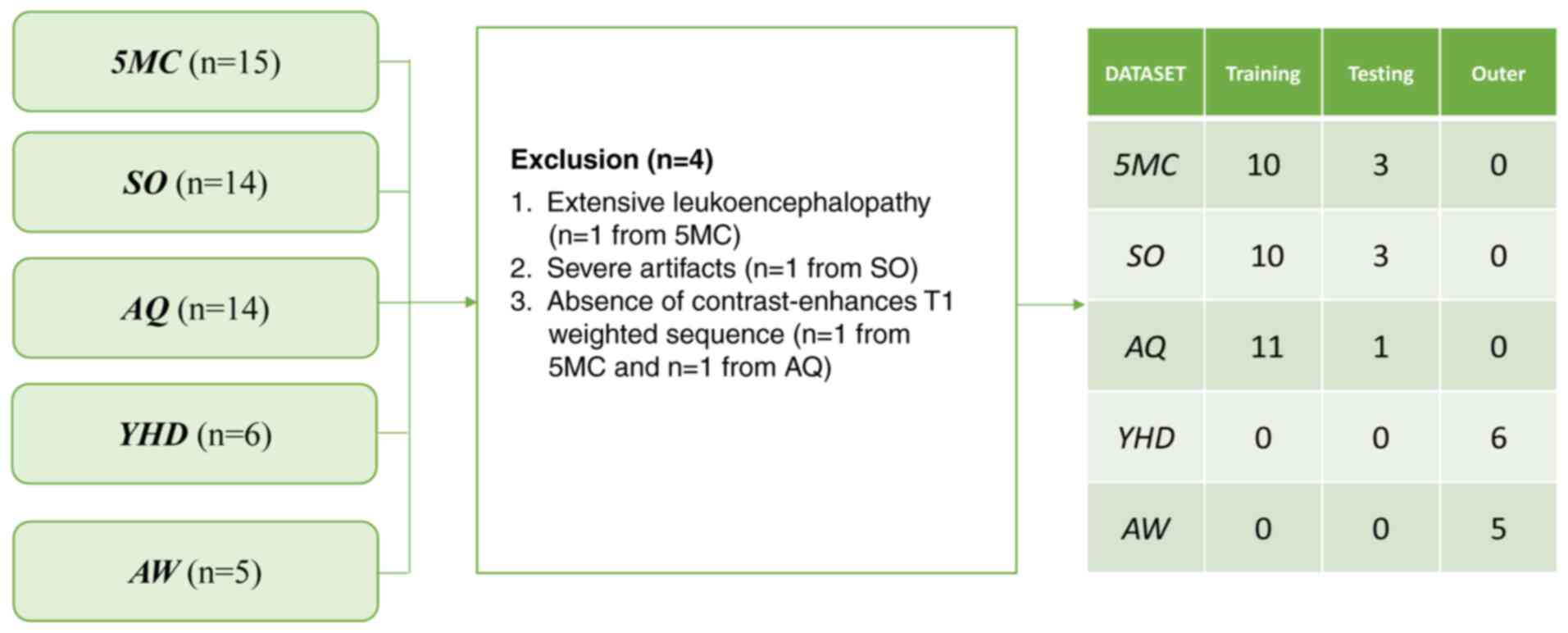
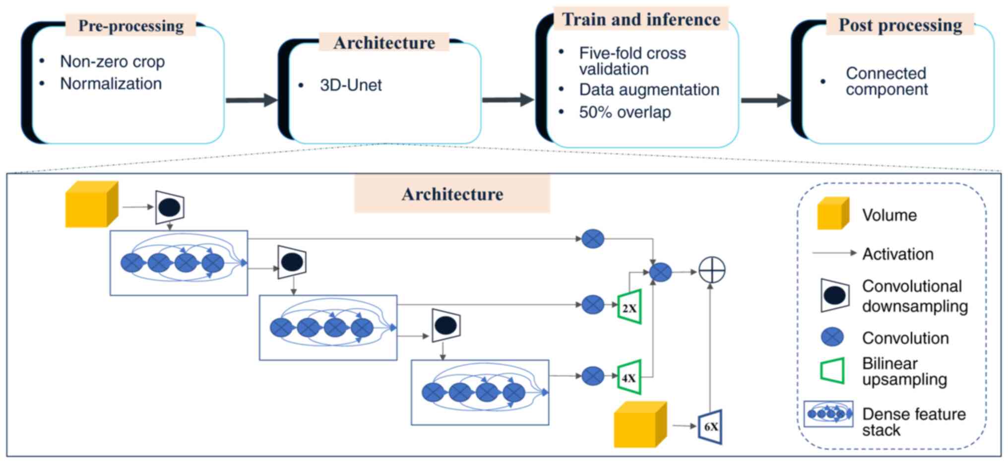
|
1
|
Villano JL, Shaikh H, Dolecek TA and
McCarthy BJ: Age, gender, and racial differences in incidence and
survival in primary CNS lymphoma. Br J Cancer. 105:1414–1418. 2011.
View Article : Google Scholar : PubMed/NCBI
|
|
2
|
Chukwueke U, Grommes C and Nayak L:
Primary central nervous system lymphomas. Hematol Oncol Clin North
Am. 36:147–159. 2022. View Article : Google Scholar : PubMed/NCBI
|
|
3
|
Morales-Martinez A, Nichelli L,
Hernandez-Verdin I, Houillier C, Alentorn A and Hoang-Xuan K:
Prognostic factors in primary central nervous system lymphoma. Curr
Opin Oncol. 34:676–684. 2022. View Article : Google Scholar : PubMed/NCBI
|
|
4
|
Schaff LR and Grommes C: Primary central
nervous system lymphoma. Blood. 140:971–979. 2022. View Article : Google Scholar : PubMed/NCBI
|
|
5
|
Lukas RV, Stupp R, Gondi V and Raizer JJ:
Primary central nervous system Lymphoma-PART 1: Epidemiology,
diagnosis, staging, and prognosis. Oncology (Williston Park).
32:17–22. 2018.PubMed/NCBI
|
|
6
|
Sangeetha SKB, Muthukumaran V, Deeba K,
Rajadurai H, Maheshwari V and Dalu GT: Multiconvolutional transfer
learning for 3D brain tumor magnetic resonance images. Comput
Intell Neurosci. 2022:87224762022. View Article : Google Scholar : PubMed/NCBI
|
|
7
|
Sadad T, Rehman A, Munir A, Saba T, Tariq
U, Ayesha N and Abbasi R: Brain tumor detection and
multi-classification using advanced deep learning techniques.
Microsc Res Tech. 84:1296–1308. 2021. View Article : Google Scholar : PubMed/NCBI
|
|
8
|
Abd-Ellah MK, Awad AI, Khalaf AAM and
Hamed HFA: A review on brain tumor diagnosis from MRI images:
Practical implications, key achievements, and lessons learned. Magn
Reson Imaging. 61:300–318. 2019. View Article : Google Scholar : PubMed/NCBI
|
|
9
|
Lu G, Zhang Y, Wang W, Miao L and Mou W:
Machine learning and deep learning CT-based models for predicting
the primary central nervous system lymphoma and glioma types: A
multicenter retrospective study. Front Neurol. 13:9052272022.
View Article : Google Scholar : PubMed/NCBI
|
|
10
|
Xia W, Hu B, Li H, Shi W, Tang Y, Yu Y,
Geng C, Wu Q, Yang L, Yu Z, et al: Deep learning for automatic
differential diagnosis of primary central nervous system lymphoma
and glioblastoma: Multi-parametric magnetic resonance imaging based
convolutional neural network model. J Magn Reson Imaging.
54:880–887. 2021. View Article : Google Scholar : PubMed/NCBI
|
|
11
|
Pennig L, Hoyer UCI, Goertz L, Shahzad R,
Persigehl T, Thiele F, Perkuhn M, Ruge MI, Kabbasch C, Borggrefe J,
et al: Primary central nervous system lymphoma: Clinical evaluation
of automated segmentation on multiparametric MRI using deep
learning. J Magn Reson Imaging. 53:259–268. 2021. View Article : Google Scholar : PubMed/NCBI
|
|
12
|
Ramadan S, Radice T, Ismail A, Fiori S and
Tarella C: Advances in therapeutic strategies for primary CNS
B-cell lymphomas. Expert Rev Hematol. 15:295–304. 2022. View Article : Google Scholar : PubMed/NCBI
|
|
13
|
Batchelor TT: Primary central nervous
system lymphoma. Hematology Am Soc Hematol Educ Program.
2016:379–385. 2016. View Article : Google Scholar : PubMed/NCBI
|
|
14
|
Bonm AV, Ritterbusch R, Throckmorton P and
Graber JJ: Clinical imaging for diagnostic challenges in the
management of gliomas: A review. J Neuroimaging. 30:139–145. 2020.
View Article : Google Scholar : PubMed/NCBI
|
|
15
|
Huang RY, Bi WL, Griffith B, Kaufmann TJ,
la Fougère C, Schmidt NO, Tonn JC, Vogelbaum MA, Wen PY, Aldape K,
et al: Imaging and diagnostic advances for intracranial
meningiomas. Neuro Oncol. 21:i44–i61. 2019. View Article : Google Scholar : PubMed/NCBI
|
|
16
|
Yadav AS, Kumar S, Karetla GR,
Cotrina-Aliaga JC, Arias-Gonzáles JL, Kumar V, Srivastava S, Gupta
R, Ibrahim S, Paul R, et al: A feature extraction using
probabilistic neural network and BTFSC-Net model with deep learning
for brain tumor classification. J Imaging. 9:102022. View Article : Google Scholar : PubMed/NCBI
|
|
17
|
ZainEldin H, Gamel SA, El-Kenawy EM,
Alharbi AH, Khafaga DS, Ibrahim A and Talaat FM: Brain tumor
detection and classification using deep learning and Sine-cosine
fitness grey wolf optimization. Bioengineering (Basel). 10:182022.
View Article : Google Scholar : PubMed/NCBI
|
|
18
|
Taher F, Shoaib MR, Emara HM, Abdelwahab
KM, Abd El-Samie FE and Haweel MT: Efficient framework for brain
tumor detection using different deep learning techniques. Front
Public Health. 10:9596672022. View Article : Google Scholar : PubMed/NCBI
|
|
19
|
Hwang K, Park J, Kwon YJ, Cho SJ, Choi BS,
Kim J, Kim E, Jang J, Ahn KS, Kim S and Kim CY: Fully automated
segmentation models of supratentorial meningiomas assisted by
inclusion of normal brain images. J Imaging. 8:3272022. View Article : Google Scholar : PubMed/NCBI
|
|
20
|
Dang K, Vo T, Ngo L and Ha H: A deep
learning framework integrating MRI image preprocessing methods for
brain tumor segmentation and classification. IBRO Neurosci Rep.
13:523–532. 2022. View Article : Google Scholar : PubMed/NCBI
|
|
21
|
Lin YY, Guo WY, Lu CF, Peng SJ, Wu YT and
Lee CC: Application of artificial intelligence to stereotactic
radiosurgery for intracranial lesions: Detection, segmentation, and
outcome prediction. J Neurooncol. 161:441–450. 2023. View Article : Google Scholar : PubMed/NCBI
|
|
22
|
Laukamp KR, Pennig L, Thiele F, Reimer R,
Görtz L, Shakirin G, Zopfs D, Timmer M, Perkuhn M and Borggrefe J:
Automated meningioma segmentation in multiparametric MRI:
comparable effectiveness of a deep learning model and manual
segmentation. Clin Neuroradiol. 31:357–366. 2021. View Article : Google Scholar : PubMed/NCBI
|
|
23
|
Chang K, Beers AL, Bai HX, Brown JM, Ly
KI, Li X, Senders JT, Kavouridis VK, Boaro A, Su C, et al:
Automatic assessment of glioma burden: A deep learning algorithm
for fully automated volumetric and bidimensional measurement. Neuro
Oncol. 21:1412–1422. 2019. View Article : Google Scholar : PubMed/NCBI
|
|
24
|
Peng J, Luo H, Zhao G, Lin C, Yi X and
Chen S: A Review of medical image segmentation algorithms based on
deep learning. Computer Engineering Appl. 57:44–57. 2021.(In
Chinese).
|
|
25
|
Latif G: DeepTumor: Framework for brain MR
image classification, segmentation and tumor detection. Diagnostics
(Basel). 12:28882022. View Article : Google Scholar : PubMed/NCBI
|
|
26
|
Kickingereder P, Isensee F, Tursunova I,
Petersen J, Neuberger U, Bonekamp D, Brugnara G, Schell M, Kessler
T, Foltyn M, et al: Automated quantitative tumour response
assessment of MRI in neuro-oncology with artificial neural
networks: A multicentre, retrospective study. Lancet Oncol.
20:728–740. 2019. View Article : Google Scholar : PubMed/NCBI
|
|
27
|
Isensee F, Jaeger PF, Kohl SAA, Petersen J
and Maier-Hein KH: nnU-Net: A self-configuring method for deep
learning-based biomedical image segmentation. Nat Methods.
18:203–211. 2021. View Article : Google Scholar : PubMed/NCBI
|
|
28
|
Pemberton HG, Wu J, Kommers I, Müller DMJ,
Hu Y, Goodkin O, Vos SB, Bisdas S, Robe PA, Ardon H, et al:
Multi-class glioma segmentation on real-world data with missing MRI
sequences: Comparison of three deep learning algorithms. Sci Rep.
13:189112023. View Article : Google Scholar : PubMed/NCBI
|
|
29
|
Ganesan P, Feng R, Deb B, Tjong FVY,
Rogers AJ, Ruipérez-Campillo S, Somani S, Clopton P, Baykaner T,
Rodrigo M, et al: Novel domain Knowledge-encoding algorithm enables
Label-efficient deep learning for cardiac CT segmentation to guide
atrial fibrillation treatment in a pilot dataset. Diagnostics
(Basel). 14:15382024. View Article : Google Scholar : PubMed/NCBI
|
|
30
|
Fazekas F, Chawluk JB, Alavi A, Hurtig HI
and Zimmerman RA: MR signal abnormalities at 1.5 T in Alzheimer's
dementia and normal aging. AJR Am J Roentgenol. 149:351–356. 1987.
View Article : Google Scholar : PubMed/NCBI
|
|
31
|
Isensee F, Petersen J, Klein A, Zimmerer
D, Jaeger PF, Kohl S, Wasserthal J, Koehler G, Norajitra T, Wirkert
S and Maier-Hein KH: nnU-Net: Self-adapting framework for
U-Net-based medical image segmentation. ArXiv. abs/1809.10486.
2018.
|
|
32
|
Perkuhn M, Stavrinou P, Thiele F, Shakirin
G, Mohan M, Garmpis D, Kabbasch C and Borggrefe J: Clinical
evaluation of a multiparametric deep learning model for
glioblastoma segmentation using heterogeneous magnetic resonance
imaging data from clinical routine. Invest Radiol. 53:647–654.
2018. View Article : Google Scholar : PubMed/NCBI
|
|
33
|
Menze BH, Jakab A, Bauer S,
Kalpathy-Cramer J, Farahani K, Kirby J, Burren Y, Porz N, Slotboom
J, Wiest R, et al: The multimodal brain tumor image segmentation
benchmark (BRATS). IEEE Trans Med Imaging. 34:1993–2024. 2015.
View Article : Google Scholar : PubMed/NCBI
|
|
34
|
Bousabarah K, Letzen B, Tefera J, Savic L,
Schobert I, Schlachter T, Staib LH, Kocher M, Chapiro J and Lin M:
Automated detection and delineation of hepatocellular carcinoma on
multiphasic contrast-enhanced MRI using deep learning. Abdom Radiol
(NY). 46:216–225. 2021. View Article : Google Scholar : PubMed/NCBI
|
|
35
|
Naser PV, Maurer MC, Fischer M,
Karimian-Jazi K, Ben-Salah C, Bajwa AA, Jakobs M, Jungk C, Jesser
J, Bendszus M, et al: Deep learning aided preoperative diagnosis of
primary central nervous system lymphoma. iScience. 27:1090232024.
View Article : Google Scholar : PubMed/NCBI
|
|
36
|
Cassinelli Petersen GI, Shatalov J, Verma
T, Brim WR, Subramanian H, Brackett A, Bahar RC, Merkaj S, Zeevi T,
Staib LH, et al: Machine learning in differentiating gliomas from
primary CNS lymphomas: A systematic review, reporting quality, and
risk of bias assessment. AJNR Am J Neuroradiol. 43:526–533. 2022.
View Article : Google Scholar : PubMed/NCBI
|
|
37
|
Ferreri AJM, Calimeri T, Cwynarski K,
Dietrich J, Grommes C, Hoang-Xuan K, Hu LS, Illerhaus G, Nayak L,
Ponzoni M and Batchelor TT: Primary central nervous system
lymphoma. Nat Rev Dis Primers. 9:292023. View Article : Google Scholar : PubMed/NCBI
|
|
38
|
Gill CM, Loewenstern J, Rutland JW, Arib
H, Pain M, Umphlett M, Kinoshita Y, McBride RB, Bederson J, Donovan
M, et al: Peritumoral edema correlates with mutational burden in
meningiomas. Neuroradiology. 63:73–80. 2021. View Article : Google Scholar : PubMed/NCBI
|
|
39
|
Reszec J, Hermanowicz A, Rutkowski R,
Turek G, Mariak Z and Chyczewski L: Expression of MMP-9 and VEGF in
meningiomas and their correlation with peritumoral brain edema.
Biomed Res Int. 2015:6468532015. View Article : Google Scholar : PubMed/NCBI
|