Spandidos Publications Logo
  • About
    • About Spandidos
    • Aims and Scopes
    • Abstracting and Indexing
    • Editorial Policies
    • Reprints and Permissions
    • Job Opportunities
    • Terms and Conditions
    • Contact
  • Journals
    • All Journals
    • Oncology Letters
      • Oncology Letters
      • Information for Authors
      • Editorial Policies
      • Editorial Board
      • Aims and Scope
      • Abstracting and Indexing
      • Bibliographic Information
      • Archive
    • International Journal of Oncology
      • International Journal of Oncology
      • Information for Authors
      • Editorial Policies
      • Editorial Board
      • Aims and Scope
      • Abstracting and Indexing
      • Bibliographic Information
      • Archive
    • Molecular and Clinical Oncology
      • Molecular and Clinical Oncology
      • Information for Authors
      • Editorial Policies
      • Editorial Board
      • Aims and Scope
      • Abstracting and Indexing
      • Bibliographic Information
      • Archive
    • Experimental and Therapeutic Medicine
      • Experimental and Therapeutic Medicine
      • Information for Authors
      • Editorial Policies
      • Editorial Board
      • Aims and Scope
      • Abstracting and Indexing
      • Bibliographic Information
      • Archive
    • International Journal of Molecular Medicine
      • International Journal of Molecular Medicine
      • Information for Authors
      • Editorial Policies
      • Editorial Board
      • Aims and Scope
      • Abstracting and Indexing
      • Bibliographic Information
      • Archive
    • Biomedical Reports
      • Biomedical Reports
      • Information for Authors
      • Editorial Policies
      • Editorial Board
      • Aims and Scope
      • Abstracting and Indexing
      • Bibliographic Information
      • Archive
    • Oncology Reports
      • Oncology Reports
      • Information for Authors
      • Editorial Policies
      • Editorial Board
      • Aims and Scope
      • Abstracting and Indexing
      • Bibliographic Information
      • Archive
    • Molecular Medicine Reports
      • Molecular Medicine Reports
      • Information for Authors
      • Editorial Policies
      • Editorial Board
      • Aims and Scope
      • Abstracting and Indexing
      • Bibliographic Information
      • Archive
    • World Academy of Sciences Journal
      • World Academy of Sciences Journal
      • Information for Authors
      • Editorial Policies
      • Editorial Board
      • Aims and Scope
      • Abstracting and Indexing
      • Bibliographic Information
      • Archive
    • International Journal of Functional Nutrition
      • International Journal of Functional Nutrition
      • Information for Authors
      • Editorial Policies
      • Editorial Board
      • Aims and Scope
      • Abstracting and Indexing
      • Bibliographic Information
      • Archive
    • International Journal of Epigenetics
      • International Journal of Epigenetics
      • Information for Authors
      • Editorial Policies
      • Editorial Board
      • Aims and Scope
      • Abstracting and Indexing
      • Bibliographic Information
      • Archive
    • Medicine International
      • Medicine International
      • Information for Authors
      • Editorial Policies
      • Editorial Board
      • Aims and Scope
      • Abstracting and Indexing
      • Bibliographic Information
      • Archive
  • Articles
  • Information
    • Information for Authors
    • Information for Reviewers
    • Information for Librarians
    • Information for Advertisers
    • Conferences
  • Language Editing
Spandidos Publications Logo
  • About
    • About Spandidos
    • Aims and Scopes
    • Abstracting and Indexing
    • Editorial Policies
    • Reprints and Permissions
    • Job Opportunities
    • Terms and Conditions
    • Contact
  • Journals
    • All Journals
    • Biomedical Reports
      • Information for Authors
      • Editorial Policies
      • Editorial Board
      • Aims and Scope
      • Abstracting and Indexing
      • Bibliographic Information
      • Archive
    • Experimental and Therapeutic Medicine
      • Information for Authors
      • Editorial Policies
      • Editorial Board
      • Aims and Scope
      • Abstracting and Indexing
      • Bibliographic Information
      • Archive
    • International Journal of Epigenetics
      • Information for Authors
      • Editorial Policies
      • Editorial Board
      • Aims and Scope
      • Abstracting and Indexing
      • Bibliographic Information
      • Archive
    • International Journal of Functional Nutrition
      • Information for Authors
      • Editorial Policies
      • Editorial Board
      • Aims and Scope
      • Abstracting and Indexing
      • Bibliographic Information
      • Archive
    • International Journal of Molecular Medicine
      • Information for Authors
      • Editorial Policies
      • Editorial Board
      • Aims and Scope
      • Abstracting and Indexing
      • Bibliographic Information
      • Archive
    • International Journal of Oncology
      • Information for Authors
      • Editorial Policies
      • Editorial Board
      • Aims and Scope
      • Abstracting and Indexing
      • Bibliographic Information
      • Archive
    • Medicine International
      • Information for Authors
      • Editorial Policies
      • Editorial Board
      • Aims and Scope
      • Abstracting and Indexing
      • Bibliographic Information
      • Archive
    • Molecular and Clinical Oncology
      • Information for Authors
      • Editorial Policies
      • Editorial Board
      • Aims and Scope
      • Abstracting and Indexing
      • Bibliographic Information
      • Archive
    • Molecular Medicine Reports
      • Information for Authors
      • Editorial Policies
      • Editorial Board
      • Aims and Scope
      • Abstracting and Indexing
      • Bibliographic Information
      • Archive
    • Oncology Letters
      • Information for Authors
      • Editorial Policies
      • Editorial Board
      • Aims and Scope
      • Abstracting and Indexing
      • Bibliographic Information
      • Archive
    • Oncology Reports
      • Information for Authors
      • Editorial Policies
      • Editorial Board
      • Aims and Scope
      • Abstracting and Indexing
      • Bibliographic Information
      • Archive
    • World Academy of Sciences Journal
      • Information for Authors
      • Editorial Policies
      • Editorial Board
      • Aims and Scope
      • Abstracting and Indexing
      • Bibliographic Information
      • Archive
  • Articles
  • Information
    • For Authors
    • For Reviewers
    • For Librarians
    • For Advertisers
    • Conferences
  • Language Editing
Login Register Submit
  • This site uses cookies
  • You can change your cookie settings at any time by following the instructions in our Cookie Policy. To find out more, you may read our Privacy Policy.

    I agree
Search articles by DOI, keyword, author or affiliation
Search
Advanced Search
presentation
Oncology Letters
Join Editorial Board Propose a Special Issue
Print ISSN: 1792-1074 Online ISSN: 1792-1082
Journal Cover
September-October 2010 Volume 1 Issue 5

Full Size Image

Sign up for eToc alerts
Recommend to Library

Journals

International Journal of Molecular Medicine

International Journal of Molecular Medicine

International Journal of Molecular Medicine is an international journal devoted to molecular mechanisms of human disease.

International Journal of Oncology

International Journal of Oncology

International Journal of Oncology is an international journal devoted to oncology research and cancer treatment.

Molecular Medicine Reports

Molecular Medicine Reports

Covers molecular medicine topics such as pharmacology, pathology, genetics, neuroscience, infectious diseases, molecular cardiology, and molecular surgery.

Oncology Reports

Oncology Reports

Oncology Reports is an international journal devoted to fundamental and applied research in Oncology.

Experimental and Therapeutic Medicine

Experimental and Therapeutic Medicine

Experimental and Therapeutic Medicine is an international journal devoted to laboratory and clinical medicine.

Oncology Letters

Oncology Letters

Oncology Letters is an international journal devoted to Experimental and Clinical Oncology.

Biomedical Reports

Biomedical Reports

Explores a wide range of biological and medical fields, including pharmacology, genetics, microbiology, neuroscience, and molecular cardiology.

Molecular and Clinical Oncology

Molecular and Clinical Oncology

International journal addressing all aspects of oncology research, from tumorigenesis and oncogenes to chemotherapy and metastasis.

World Academy of Sciences Journal

World Academy of Sciences Journal

Multidisciplinary open-access journal spanning biochemistry, genetics, neuroscience, environmental health, and synthetic biology.

International Journal of Functional Nutrition

International Journal of Functional Nutrition

Open-access journal combining biochemistry, pharmacology, immunology, and genetics to advance health through functional nutrition.

International Journal of Epigenetics

International Journal of Epigenetics

Publishes open-access research on using epigenetics to advance understanding and treatment of human disease.

Medicine International

Medicine International

An International Open Access Journal Devoted to General Medicine.

Journal Cover
September-October 2010 Volume 1 Issue 5

Full Size Image

Sign up for eToc alerts
Recommend to Library

  • Article
  • Citations
    • Cite This Article
    • Download Citation
    • Create Citation Alert
    • Remove Citation Alert
    • Cited By
  • Similar Articles
    • Related Articles (in Spandidos Publications)
    • Similar Articles (Google Scholar)
    • Similar Articles (PubMed)
  • Download PDF
  • Download XML
  • View XML
Article

Usefulness of 18F-FDG positron emission tomography/computed tomography for the diagnosis of pyothorax-associated lymphoma: A report of three cases

  • Authors:
    • Yoshiyuki Abe
    • Katsumi Tamura
    • Ikuko Sakata
    • Jiro Ishida
    • Isao Fukuba
    • Rokuro Matsuoka
    • Seiichiro Shimizu
    • Hiroshi Murakami
    • Kikuo Machida
  • View Affiliations / Copyright

    Affiliations: Tokorozawa PET Diagnostic Imaging Clinic, Tokorozawa, Saitama 359-1124, Japan. abe@toko-pet.or.jp
  • Pages: 833-836
    |
    Published online on: September 1, 2010
       https://doi.org/10.3892/ol_00000146
  • Expand metrics +
Metrics: Total Views: 0 (Spandidos Publications: | PMC Statistics: )
Metrics: Total PDF Downloads: 0 (Spandidos Publications: | PMC Statistics: )
Cited By (CrossRef): 0 citations Loading Articles...

This article is mentioned in:


Abstract

Pyothorax-associated lymphoma (PAL) is a unique and rare non-Hodgkin's lymphoma developing in the pleural cavity following a long-standing history of chronic pyothorax (CP). The development of F-18 2'-deoxy-2fluoro-D-glucose (FDG) positron emission tomography combined with computed tomography (PET/CT) has contributed to the evaluation of lymphoma staging. However, only a few studies describing FDG-PET/CT findings in PAL have been published. This study reported three cases of PAL; all 3 patients had previously undergone artificial collapse therapy for pulmonary tuberculosis. Both the first case (an 84-year-old male) and second case (an 83-year-old male) complained of abdominal pain. An ultrasound scan revealed a mass shadow in the left chest wall without abnormal findings in the abdomen, and the CT and magnetic resonance imaging scans suggested malignant lymphoma of the left chest. FDG-PET/CT imaging showed extremely intense FDG uptake only in the left pleura and chest wall. Diagnosis was CP in the two patients, showing a high maximum standardized uptake value (SUVmax: early, 14.8 and delayed, 19.4 in the first case; early, 20.8 and delayed, 27.3 in the second case, respectively). Histopathological analysis of the specimens obtained by biopsy of the PET/CT-positive pleural mass showed non-Hodgkin's, diffuse large B cell lymphoma in the two cases. The third case was a 79-year-old male with relapse after right pleuropneumonectomy for PAL (diffuse large B cell lymphoma) 4 years earlier. PET/CT showed intense FDG uptake (SUVmax: early, 19.9 and delayed, 35.7) in the right pleura and chest wall. Diagnosis was CP, suggesting the recurrence of PAL. Furthermore, abnormal intense FDG uptake was noted in the hilar, mediastinal and supraclavicular lymph nodes, as well as in the spleen. In conclusion, FDG-PET/CT imaging is useful in the evaluation of the area of invasion in PAL.

Introduction

Pyothorax-associated lymphoma (PAL) is a unique and rare disease involving malignant lymphoma. PAL appears in the pleural cavity with chronic pyothorax (CP) following artificial collapse therapy for pulmonary tuberculosis (1,2). Lymphoma comprises Hodgkin's and non-Hodgkin's types. PAL is a non-Hodgkin's lymphoma developing in the pleural cavity after a long-standing history of CP, and is strongly associated with Epstein-Barr virus (EBV) infection (3). It has been reported that approximately 90% of the cases are diffuse large B cell lymphoma and that the 5-year survival rate is approximately 20% (1). It is crucial to evaluate the area of invasion in PAL, which may determine the appropriate treatment, such as surgery, chemotherapy, irradiation and best supportive care. However, conventional imaging modalities cannot provide sufficient information for an accurate diagnosis of PAL (4).

The development of the new modality of F-18 2′-deoxy-2fluoro-D-glucose (FDG) positron emission tomography combined with computed tomography (PET/CT) has contributed to the evaluation of human cancer. Additionally, the usefulness of FDG-PET/CT for lymphoma staging is well established (5,6). However, few studies exist describing FDG-PET/CT findings in PAL (7). This study reported three patients with PAL, who had previously undergone artificial collapse therapy for pulmonary tuberculosis. The first two cases involved initial occurrences, and the last case showed recurrent disease following right pleuropneumonectomy for PAL. FDG-PET/CT imaging revealed a unique intense uptake of FDG in the area of CP and in the lymphatic and systemic metastatic lesions. Therefore, the usefulness of FDG-PET/CT imaging to evaluate the area of invasion in PAL was examined.

Patients and methods

This study was performed with informed consent of the patients and with approval of the Ethics Committee of the Tokorozawa PET Diagnostic Imaging Clinic.

Case 1

An 84-year-old male complaining of abdominal pain consulted the M Clinic in Tokyo. An ultrasound study revealed a mass shadow in the left chest wall without abnormal findings in the abdomen. CT and magnetic resonance imaging scans suggested malignant lymphoma of the chest. A physical examination showed no apparently abnormal findings or signs of systemic lymphadenopathy other than a mass in the left chest wall. Blood analysis showed an elevated serum interleukin-2 receptor (IL-2R, 1,560 Ug/ml) without other abnormalities, including tumor markers such as carcinoembryonic antigen (CEA) and CA19-9. The patient had previously undergone left artificial collapse therapy for pulmonary tuberculosis at the age of 20.

18F-FDG PET/CT scans were obtained using a Biograph Duo (Siemens CTI) at the Tokorozawa PET Diagnostic Imaging Clinic, as described in our previous study (8,9). To determine semi-quantitative FDG uptake, regions of interest (ROIs) were placed over the lesion, including the highest uptake area (circular ROI, 1 cm in diameter), and the standardized uptake value 
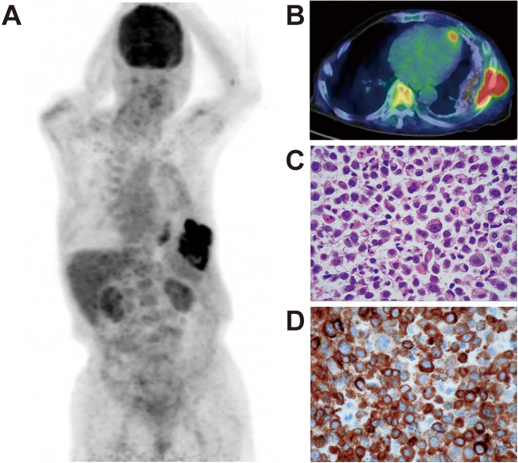
(SUV) was calculated. Early PET/CT showed intense abnormal FDG uptake only in the left pleura and chest wall, suggesting CP (SUVmax, 14.8) (Fig. 1A and B). A delayed scan showed a more intense FDG uptake (SUVmax, 19.4) in CP. No areas, other than CP, showed an abnormal uptake of FDG. The PET/CT findings strongly suggested malignancy of the chest wall without lymphatic or systemic metastasis.

Figure 1

(A and B) PET/CT of case 1 shows intense abnormal FDG uptake only in the left pleura and chest wall, showing chronic pyothorax (SUVmax, 14.8). (C) Microscopic examination shows a diffuse infiltrative growth of large atypical lymphoid cells. The atypical lymphoid cells contain irregular nuclei. (D) Immunohistochemical analysis shows neoplastic cells positive for the CD79a antigen. Magnification, ×100.

Histopathological examination was performed, and the biopsy specimens were obtained from the PET/CT-positive pleura. A microscopic examination showed a diffuse infiltrative growth of the large atypical lymphoid cells (Fig. 1C). The lymphoid cells contained irregular nuclei. Immunohistochemical analysis showed that the neoplastic cells were positive for LCA (CD45RO), CD79a (Fig. 1D), CD20 and BCL-2. The neoplastic cells were also positive for LMP-1, suggesting EBV infection. Histopathological examination confirmed the diagnosis as non-Hodgkin's, diffuse large B cell lymphoma. Due to poor general condition, the patient received best supportive care without chemotherapy or surgery despite the localized lesion of PAL.

Case 2

An 83-year-old male complaining of abdominal pain presented at the Tokorozawa Clinic in Saitama. An ultrasound study revealed a mass shadow in the left chest wall without abnormal findings in the abdomen. A physical examination showed no apparently abnormal findings or signs of systemic lymphadenopathy other than the mass of the left chest wall. Blood analysis showed elevation of the serum IL-2R (634 Ug/ml) and neuron-specific enolase (58 ng/ml). The patient had previously undergone left artificial collapse therapy for pulmonary tuberculosis at the age of 24.

18F-FDG PET/CT scans were obtained in our 
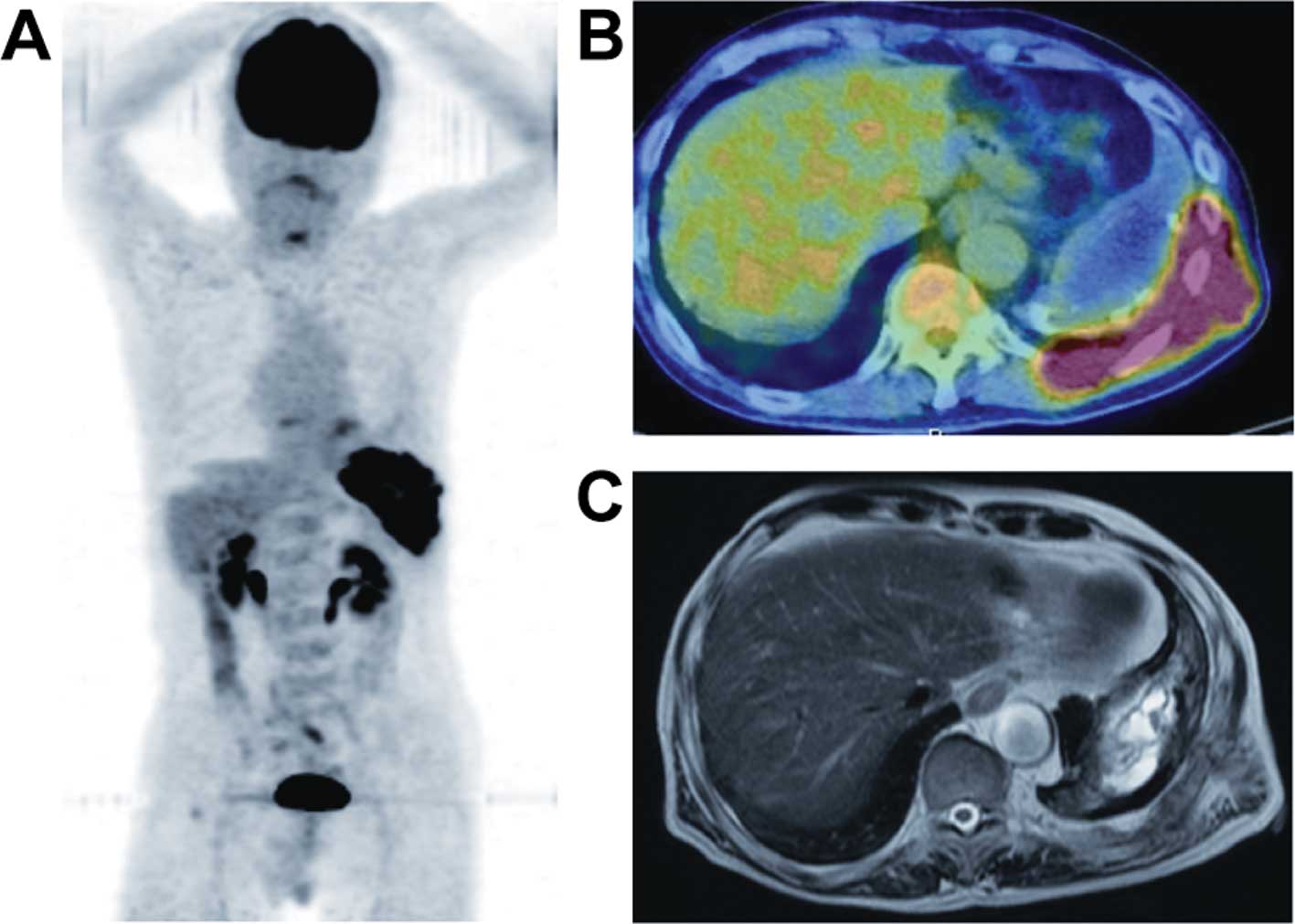
PET clinic. Early PET/CT showed intense abnormal FDG uptake only in the left pleura and chest wall, suggesting CP (SUVmax, 20.8) (Fig. 2A and B). A delayed scan showed a more intense FDG uptake (SUVmax, 27.3) in the CP. No areas, other than CP, revealed an abnormal FDG uptake. The PET/CT findings strongly suggested malignancy of the chest wall without lymphatic or systemic metastasis. An MRI scan showed high signal-intensity on T2-weighted images in the left chest, suggesting CP, as well as moderate signal-intensity around the CP, which was consistent with malignant lymphoma (Fig. 2C).

Figure 2

(A and B) PET/CT of case 2 shows intense abnormal FDG uptake in the left pleura and chest wall of chronic pyothorax (CD) (SUVmax, 20.8). (C) MRI shows high signal-intensity on T2-weighted images in the left chest, suggesting CP. A moderate signal-intensity on T2-weighted images around the CP is shown, which is consistent with malignant lymphoma.

Histopathological examination was performed on biopsy specimens obtained from the PET/CT-positive pleura. This examination confirmed the diagnosis as non-Hodgkin's, diffuse large B cell lymphoma. The patient received chemotherapy (R-CHOP: rituximab, cyclophosphamide, doxorubicin, vincristine and prednisone) despite the localized lesion of PAL, following the patient's request for non-surgical treatment.

Case 3

A 79-year-old male complaining of chest pain presented at the Kyosai Tachikawa Hospital in Tokyo. He had previously undergone right artificial collapse therapy for pulmonary tuberculosis at the age of 24. Fifty years later, he was diagnosed with PAL, diffuse large B cell lymphoma and was treated with right pleuropneumonectomy. At the present examination, a blood analysis showed elevation of the serum IL-2 receptor (2,360 U/ml), 
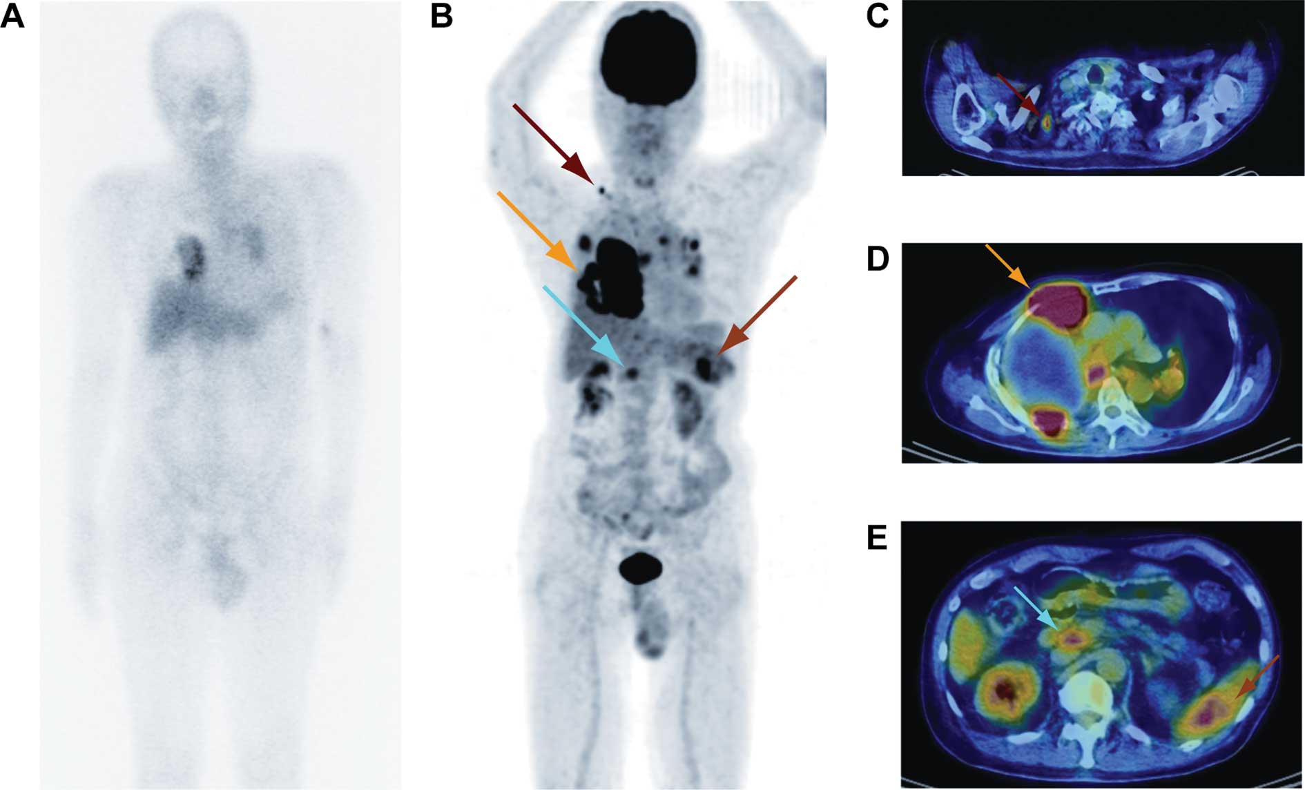
suggesting a relapse of PAL. Gallium-67 (67Ga) scintigraphy showed a faint level of abnormal uptake in the chest (Fig. 3A). Histopathological examination of specimens obtained from the pleural mass showed diffuse large B cell lymphoma, and the patient was diagnosed as having a recurrence of PAL.

Figure 3

(A) Gallium-67 (67Ga) scintigraphy of case 3 shows a faint level of abnormal uptake in the chest. (B-E) PET/CT shows intense abnormal FDG uptake in the right pleura and chest wall demonstrating chronic pyothorax (yellow arrow: SUVmax, 19.9). Abnormal intense FDG uptake is also noted in the hilar, mediastinal and supraclavicular (red arrow) and abdominal lymph nodes (blue arrow), as well as in the spleen (orange arrow).

Early 18F-FDG PET/CT showed intense abnormal FDG uptake in the right pleura and chest wall, suggesting CP (SUVmax, 19.9) (Fig. 3B-E). Abnormal intense FDG uptake was also noted in the hilar, mediastinal, supraclavicular and abdominal lymph nodes, as well as in the spleen. A delayed scan showed a more intense FDG uptake (SUVmax, 35.7) in the CP. The patient received chemotherapy (R-CHOP) numerous times, but finally refused to receive further chemotherapy.

Discussion

PAL is a non-Hodgkin's lymphoma that develops in the pleural cavity following a long-standing history of CP. It is known that approximately 90% of PAL cases involve diffuse large B cell lymphoma, and that the 5-year survival rate is approximately 20% (1). Standard treatment for PAL has yet to be established, although the prognosis is poor. Certain studies have described PAL cases in which surgery was performed (10,11). It is essential that the area of invasion in PAL be evaluated, since it may determine the appropriate choice of therapy, including surgery, chemotherapy, irradiation and best supportive care. However, conventional imaging modalities cannot provide sufficient information for the accurate diagnosis of PAL (4).

The development of FDG-PET/CT contributes to the evaluation of human cancer staging. Moreover, the usefulness of PET/CT is well established for lymphoma staging (5,6). However, only one study on PET findings in PAL has been published, by Asakura et al (7). These authors reported a PAL case in which the FDG-PET finding determined the area of PAL invasion and provided useful information for the planning of radiotherapy. We presented three cases of PAL, two of which showed initial occurrence and one recurrent case. The PET/CT scan showed intense FDG uptake only in the chest wall in the two cases showing initial occurrence of PAL. In these cases, the area of invasion determined by the PET/CT scan contributed to decisions regarding PAL treatment.

In the recurrent case of PAL, the PET/CT scan showed intense FDG uptake, not only in the chest wall, but also in the lymphatic and systemic metastatic lesions. On the other hand, 67Ga scintigraphy showed a faint level of abnormal uptake only in the chest. Certain studies have shown the usefulness of 67Ga scintigraphy in the diagnosis and assessment of the effects of treatment (4,12). However, our case showed that FDG-PET/CT imaging is a more reliable and sensitive method for staging and monitoring therapy as opposed to 67Ga scintigraphy in cases of PAL, which is in agreement with Kostakoglu et al (13). Zinzani et al performed an extensive analysis of the reliability of PET after induction treatment in patients with Hodgkin's disease and aggressive non-Hodgkin's lymphoma. Findings of these authors showed that there were no false-negative results among 75 PET scans performed in that study (14).

PAL is a non-Hodgkin's lymphoma that develops in the pleural cavity following a long-standing history of CP, and is strongly associated with EBV infection. In our first case, histopathological analysis showed that the neoplastic cells were positive for LMP-1, suggesting an association with EBV infection. Takakuwa et al reported that the downregulation of EBV nuclear antigen-2 expression may be a selection pressure for the progression of PAL (3).

In conclusion, we report three cases of PAL that showed a high FDG uptake in the lesions of PAL on the FDG-PET/CT scan. FDG-PET/CT imaging is therefore useful in the evaluation of the area of invasion, facilitation of treatment planning, as well as the assessment of treatment response in PAL.

Acknowledgements

We thank Mr. Kenji Kawai for the technical assistance.

References

1 

Nakatsuka S, Yao M, Hoshida Y, Yamamoto S, Iuchi K and Aozasa K: Pyothorax-associated lymphoma: a review of 106 cases. J Clin Oncol. 20:4255–4260. 2002. View Article : Google Scholar : PubMed/NCBI

2 

Aozasa K: Pyothorax-associated lymphoma. J Clin Exp Hematop. 46:5–10. 2006. View Article : Google Scholar

3 

Takakuwa T, Ham MF, Luo WJ, Nakatsuka S, Daibata M and Aozasa K: Loss of expression of Epstein-Barr virus nuclear antigen-2 correlates with a poor prognosis in cases of pyothorax-associated lymphoma. Int J Cancer. 118:2782–2789. 2006. View Article : Google Scholar : PubMed/NCBI

4 

Ueda T, Andreas C, Itami J, Miyakawa K, Fujimoto H, Ito H and Roos JE: Pyothorax-associated lymphoma: imaging findings. AJR Am J Roentgenol. 194:76–84. 2010. View Article : Google Scholar : PubMed/NCBI

5 

Weiler-Sagie M, Bushelev O, Epelbaum R, Dann EJ, Haim N, Avivi I, Ben-Barak A, Ben-Arie Y, Bar-Shalom R and Israel O: (18)F-FDG avidity in lymphoma readdressed: a study of 766 patients. J Nucl Med. 51:25–30. 2010. View Article : Google Scholar : PubMed/NCBI

6 

Cronin CG, Swords R, Truong MT, Viswanathan C, Rohren E, Giles FJ, O'Dwyer M and Bruzzi JF: Clinical utility of PET/CT in lymphoma. AJR Am J Roentgenol. 194:W91–W103. 2010. View Article : Google Scholar : PubMed/NCBI

7 

Asakura H, Togami T, Mitani M, Takashima H, Yokoe K, Yamamoto Y, Nishiyama Y, Monden T, Toyama Y and Ohkawa M: Usefulness of FDG-PET imaging for the radiotherapy treatment planning of pyothorax-associated lymphoma. Ann Nucl Med. 19:725–728. 2005. View Article : Google Scholar : PubMed/NCBI

8 

Ueda S, Tsuda H, Asakawa H, Shigekawa T, Fukatsu K, Kondo N, Yamamoto M, Hama Y, Tamura K, Ishida J, Abe Y and Mochizuki H: Clinicopathological and prognostic relevance of uptake level using 18F-fluorodeoxyglucose positron emission tomography computed tomography fusion imaging (18F-FDG PET/CT) in primary breast cancer. Jpn J Clin Oncol. 38:250–258. 2008.PubMed/NCBI

9 

Abe Y, Tamura K, Sakata I, Ishida J, Mukai M, Ohtaki M, Nakamura M and Machida K: Unique intense uptake demonstrated by 18F-FDG positron emission tomography/computed tomography (PET/CT) in primary pancreatic lymphoma: A case report. Oncol Lett. 4:605–607. 2010.PubMed/NCBI

10 

Fujimoto M, Haga H, Okamoto M, Obara E, Ishihara M, Mizuta N, Nishimura K and Manabe T: EBV-associated diffuse large B-cell lymphoma arising in the chest wall with surgical mesh implant. Pathol Int. 58:668–671. 2008. View Article : Google Scholar : PubMed/NCBI

11 

Santini M, Fiorello A, Vicidomini G, Busiello L and Baldi A: A surgical case of pyothorax-associated lymphoma of T-cell origin arising from the chest wall in chronic empyema. Ann Thorac Surg. 88:642–645. 2009. View Article : Google Scholar : PubMed/NCBI

12 

Aruga T, Itami J, Aruga M, Nakajima K, Shibata K, Nojo T, Yasuda S, Uno T, Hara R, Isobe K, Machida N and Ito H: Treatment for pyothorax-associated lymphoma. Radiother Oncol. 56:59–63. 2000. View Article : Google Scholar : PubMed/NCBI

13 

Kostakoglu L and Goldsmith SJ: Fluorine-18 fluorodeoxyglucose positron emission tomography in the staging and follow-up of lymphoma: is it time to shift gears? Eur J Nucl Med. 27:1564–1578. 2000. View Article : Google Scholar : PubMed/NCBI

14 

Zinzani PL, Fanti S, Battista G, Tani M, Castellucci P, Stefoni V, Alinari L, Farsad M, Musuraca G, Gabriele A, Marchi E, Nanni C, Canini R, Monetti N and Baccarani M: Predictive role of positron emission tomography (PET) in the outcome of lymphoma patients. Br J Cancer. 91:850–854. 2004.PubMed/NCBI

Related Articles

  • Abstract
  • View
  • Download
  • Twitter
Copy and paste a formatted citation
Spandidos Publications style
Abe Y, Tamura K, Sakata I, Ishida J, Fukuba I, Matsuoka R, Shimizu S, Murakami H and Machida K: Usefulness of 18F-FDG positron emission tomography/computed tomography for the diagnosis of pyothorax-associated lymphoma: A report of three cases . Oncol Lett 1: 833-836, 2010.
APA
Abe, Y., Tamura, K., Sakata, I., Ishida, J., Fukuba, I., Matsuoka, R. ... Machida, K. (2010). Usefulness of 18F-FDG positron emission tomography/computed tomography for the diagnosis of pyothorax-associated lymphoma: A report of three cases . Oncology Letters, 1, 833-836. https://doi.org/10.3892/ol_00000146
MLA
Abe, Y., Tamura, K., Sakata, I., Ishida, J., Fukuba, I., Matsuoka, R., Shimizu, S., Murakami, H., Machida, K."Usefulness of 18F-FDG positron emission tomography/computed tomography for the diagnosis of pyothorax-associated lymphoma: A report of three cases ". Oncology Letters 1.5 (2010): 833-836.
Chicago
Abe, Y., Tamura, K., Sakata, I., Ishida, J., Fukuba, I., Matsuoka, R., Shimizu, S., Murakami, H., Machida, K."Usefulness of 18F-FDG positron emission tomography/computed tomography for the diagnosis of pyothorax-associated lymphoma: A report of three cases ". Oncology Letters 1, no. 5 (2010): 833-836. https://doi.org/10.3892/ol_00000146
Copy and paste a formatted citation
x
Spandidos Publications style
Abe Y, Tamura K, Sakata I, Ishida J, Fukuba I, Matsuoka R, Shimizu S, Murakami H and Machida K: Usefulness of 18F-FDG positron emission tomography/computed tomography for the diagnosis of pyothorax-associated lymphoma: A report of three cases . Oncol Lett 1: 833-836, 2010.
APA
Abe, Y., Tamura, K., Sakata, I., Ishida, J., Fukuba, I., Matsuoka, R. ... Machida, K. (2010). Usefulness of 18F-FDG positron emission tomography/computed tomography for the diagnosis of pyothorax-associated lymphoma: A report of three cases . Oncology Letters, 1, 833-836. https://doi.org/10.3892/ol_00000146
MLA
Abe, Y., Tamura, K., Sakata, I., Ishida, J., Fukuba, I., Matsuoka, R., Shimizu, S., Murakami, H., Machida, K."Usefulness of 18F-FDG positron emission tomography/computed tomography for the diagnosis of pyothorax-associated lymphoma: A report of three cases ". Oncology Letters 1.5 (2010): 833-836.
Chicago
Abe, Y., Tamura, K., Sakata, I., Ishida, J., Fukuba, I., Matsuoka, R., Shimizu, S., Murakami, H., Machida, K."Usefulness of 18F-FDG positron emission tomography/computed tomography for the diagnosis of pyothorax-associated lymphoma: A report of three cases ". Oncology Letters 1, no. 5 (2010): 833-836. https://doi.org/10.3892/ol_00000146
Follow us
  • Twitter
  • LinkedIn
  • Facebook
About
  • Spandidos Publications
  • Careers
  • Cookie Policy
  • Privacy Policy
How can we help?
  • Help
  • Live Chat
  • Contact
  • Email to our Support Team