Introduction
Lung cancer is the most common cause of
cancer-related deaths in men and women, and is responsible for
approximately 1.3 million deaths annually. The treatments currently
available for this disease are the same for all patients. However,
some patients may respond more sensitively than others to similar
treatments, due to differences in their health status,
complications or smoking status. All of these make it difficult for
doctors to choose suitable strategies for each patient and even
harder to predict the treatment efficacy. Therefore, new biological
markers for lung cancer prediction and prognosis are urgently
needed.
Generally, lung cancer can be divided into two
subtypes, including non-small cell lung cancer (NSCLC) and small
cell lung cancer (SCLC). About 80% of the lung cancers are of the
NSCLC type. NSCLC can be further divided into adenocarcinoma,
squamous cell carcinoma, and large cell tumors. SCLC comprises only
about 19–20% of all lung cancer cases, while carcinoid tumors
account for the rest (1). Extensive
studies have led to identification of a number of DNA and protein
biomarkers related to lung cancers. Biomarkers relative to NSCLC
prediction and prognosis have been reported, such as the epidermal
growth factor receptor (EGFR)-related biomarkers (EGFR, Ki-67, pAKT
and p27) (2–9). EGFR mediates tumor cell growth,
proliferation, angiogenesis, invasion, and metastasis (2). Ki-67 expression is reported to be
tightly related to poor prognosis of NSCLC (5,6). Akt
is active in most NSCLC cells (3)
and high levels of phosphorylated Akt is often correlated with lung
cancers (4). p27 is a protein
related to cell cycle regulation, which is also found to be related
to NSCLC (7–9). All of these biomarkers described above
need to be detected by immunohistochemistry (IHC) or immunoblotting
(IB), which make the examination process time-consuming and harder
to be quantified. Therefore, biomarkers easy to be clinically
measured for NSCLC are urgently needed.
The trefoil factor (TFF) family is composed of three
thermostable, and protease-resistant proteins, named TFF1, TFF2 and
TFF3. Although mainly expressed in the epithelial cells that line
mucous membranes, TFFs are secreted proteins present in serum,
which make them easy to be detected by ELISA. TFF1 and TFF2 contain
single trefoil domains, whereas TFF2 consists of two such domains
(10,11). Although TFFs have been involved in
the protection of the gastrointestinal tract against mucosal damage
(11), their oncogenic potential
has been extensively reported, including their roles in cell
proliferation (12–15), apoptosis (12–14,16,17),
migration and invasion (14,16,18,19)
and angiogenesis (20,21). TFF proteins levels have been found
to be related to the development of breast cancer (22–33),
gastric cancer (21,22,34–38),
colon cancer (39,40), and prostate cancer (41–43).
It has also been reported that TFF proteins are related to lung
cancers (23,44–48).
Two early reports described that TFF1 levels in serum are increased
in patients with lung cancer (49)
and positive expression of TFF1 indicates worse prognosis of lung
cancer (50). Recently, TFF mRNA
and protein expression and the possibility of TFFs to serve as
potential biomarkers of cholangiocarcinoma has been investigated
(51). However, the roles of TFF1,
TFF2 and TFF3 are still unclear in the prediction and prognosis of
lung cancer.
We recently reported that sorcin, a
gemcitabine-resistance-related protein, could be a novel candidate
biomarker for predicting the response of NSCLC patients to
gemcitabine treatment (52). In
this study, we investigated the protein and mRNA levels of TFF1,
TFF2 and TFF3 in tissues of lung cancer patients and healthy
individuals, and lung cancer cell lines and normal cell lines. We
also determined the levels of secreted TFFs in the serum from lung
cancer patients as well as healthy individuals. It was found that
among the three TFF proteins, the mRNA and protein levels of TFF3
in both cultured cell lines and tissues from patients have the best
correlation with the development and prognosis of lung cancers.
ELISA and IB results indicated that the levels of TFF3 in the serum
are closely related to their mRNA and protein expression levels in
tissues. These results suggest that TFF3 levels in the serum may
serve as a promising, easily detected candidate biomarker of lung
cancer.
Materials and methods
Patients
One hundred and thirty lung cancer patients,
including 58 squamous cell lung carcinoma cases, 43 adenocarcinoma
cases, and 29 SCLC cases, were enrolled in this study prior to the
treatments including surgery, chemotherapy and radiotherapy. Sixty
healthy individuals were used as healthy controls. Information of
patients and the healthy individuals, such as ages, gender and
histological types, were obtained from medical records in the
hospital and provided in Table I.
The gender ratio (female vs. male), mean age, and age ranges
between the cancer group and the healthy group were similar,
without significant differences. The experimental protocols were
approved by the Ethics Committee of the Shandong University,
China.
 | Table IInformation of lung cancer patients
and healthy individuals. |
Table I
Information of lung cancer patients
and healthy individuals.
| n | Age (years) | Mean age
(years) |
|---|
| Non-small cell lung
carcinoma |
| Squamous cell lung
carcinoma | 58 | 24–55 | 36.2 |
|
Adenocarcinoma | 43 | 24–54 | 38.1 |
| Small cell lung
carcinoma | 29 | 23–56 | 37.4 |
| Healthy
individuals | 60 | 28–59 | 37.5 |
| Total | 190 | 23–59 | 37.3 |
Cell lines
The normal human bronchial epithelium cell line
(NuLi-1), the SCLC cell line (MS-1), the adenocarcinoma cell line
(A549), and the squamous cell carcinoma cell line (LK-2) were
provided by the Shanghai Cell Biology Institute (China). These lung
cancer cell lines were maintained in RPMI-1640 medium
(Sigma-Aldrich Co., Ltd., Irvine, CA) supplemented with 10% fetal
bovine serum (FBS), 1% L-glutamine, and 1%
penicillin/streptomycin.
Enzyme-linked immunosorbent assay
(ELISA)
Serum TFF1, TFF2 and TFF3 levels were measured by
ELISA. Antisera were prepared from rabbits immunized with human
TFFs. It was confirmed by using western blot analysis that each TFF
antibody reacted specifically and showed no cross-reactivity from
the other TFFs. Standard human TFF was used as a positive control.
PBS was used as a negative control. Blood (1 ml) was collected from
each of the 60 healthy individuals and 130 lung cancer patients,
followed by centrifugation for serum separation. Purified
polyclonal antibodies against TFF1, TFF2, or TFF3 were coated to
96-well microtiter plates, and the plates were blocked with 0.1%
bovine serum albumin in phosphate-buffered saline. Then the
blocking solution was removed, and 100 μl of assay buffer (1 M
NaCl, 0.1% bovine serum albumin, PBS) was added to each well. Fifty
microliters of the samples, PBS, or standard human TFFs was added
to the wells. After incubation overnight at room temperature, the
plates were washed, and diluted biotin-labeled anti-TFF polyclonal
antibodies were added to each appropriate well. After incubation
for 2 h, the plate was washed, and diluted horseradish
peroxidase-conjugated streptavidin (Vector Laboratories,
Burlingame, CA) was added to each well. After incubation for 2 h at
room temperature, the plates were washed, and TMB solution (Scytek
Laboratories, Inc., West Logan, UT) was added. After incubation for
10 min at room temperature, stop solution was added. The absorbance
at 450 nm was measured. Concentrations of human TFFs in the samples
were calculated from the standard curves of recombinant human
TFFs.
Immunoblot assays
Total protein samples were harvested from 190
individuals or 4 cell lines, separated on 10% SDS-PAGE gels, and
then subjected to immunoblot analyses. The primary antibodies
against TFF1, TFF2, TFF3, and actin were purchased from Santa Cruz
Biotechnology, CA, USA (anti-TFF1, cat# sc-22501, 1:200; anti-TFF2,
cat# sc-23558, 1:200; anti-TFF3, cat# sc-81467, 1:200; anti-actin,
cat# sc-130301, 1:10,000). Secondary antibodies used in this study
were donkey anti-goat IgG-HRP (cat# sc-2020, 1:5,000, Santa Cruz
Biotechnology) and goat anti-mouse IgG-HRP (Cat# sc-2005, 1:10,000,
Santa Cruz Biotechnology). Bound antibodies were detected using the
ECL system (Pierce Biotechnology). The immunoblot experiments using
the 4 cell lines were repeated at least 3 times. The mean
normalized optical density (OD) of TFF protein bands relative to
the OD of actin band from the same individual were calculated.
Quantitative reverse transcription-PCR
(RT-PCR)
Quantitative RT-PCR analysis of TFF1, TFF2 and TFF3
mRNA levels in tissues or cell lines were performed. Briefly, total
RNAs were harvested from 190 individuals or 4 cell lines using the
RNeasy kit (Qiagen) according to the manufacturer’s instructions.
The RT-PCR experiments using 4 cell lines were repeated at least 3
times.
RNA (1 μl) was reverse-transcribed into cDNA using
random primers in a Reverse Transcription II system (Promega)
according to the manufacturer’s instructions. Expression of TFF1,
TFF2 and TFF3 mRNAs was quantified by quantitative PCR using an ABI
Prism Sequence Detection System (Applied Biosystems). Primers were
given in Table II. An assay
reagent containing premixed primers and a VIC-labeled probe
(Applied Biosystems; cat. no. 4310884E) was used to quantify the
expression of endogenous GAPDH mRNA. Template-negative and
RT-negative conditions were used as controls. Amplification of TFF
cDNAs and the endogenous GAPDH cDNA were monitored by changes in
FAM and VIC fluorescence intensities, respectively, with the ABI
7900 software. The corresponding amplification plots were used to
determine the threshold cycle value, defined as the number of PCR
cycles taken for fluorescent intensity to reach a fixed threshold
for each signal. The relative amounts of TFF1, TFF2, TFF3
transcript were normalized to the amount of GAPDH mRNA in the same
sample. The levels (mean value) of TFF transcripts in lung cancer
patients and in all healthy individuals and cells were calculated.
The level of TFF transcripts in healthy individuals and cells was
assigned a value of 100.
| Table IIPrimers used in this study. |
Table II
Primers used in this study.
| Primers | Sequences | Targets |
|---|
| TFF1_F |
CCCGTGAAAGACAGAATT | TFF1 |
| TFF1_R |
GATCCCTGCAGAAGTGTCT | |
| TFF2_F |
CTCCTGGCAGCGCTCCTCGTC | TFF2 |
| TFF2_R |
GATGCCCGGGTAGCCACAGTTTCT | |
| TFF3_F |
AACCGGGGCTGCTGCTTTG | TFF3 |
| TFF3_R |
GAGGTGCCTCAGAAGGTGC | |
Statistical analyses
The experimental data, including the absorbance
values at 450 nm of antibodies in serums, levels of mRNA
transcripts, and OD of protein band in the immunoblots, are given
as mean ± standard error (SEM). Statistical software (SPSS10.0) was
used for independent sample t-tests, followed by one-way analysis
of variance. P<0.05 indicated a significant difference.
Results
Levels of TFF3 protein in the serum of
lung cancer patients are higher than in the serum of healthy
individuals
TFFs are secreted proteins present in serum, which
make them easy to be detected by the ELISA method. Therefore, ELISA
was performed to determine levels of secreted TFF proteins in the
serum of 130 lung cancer patients, prior to treatment including
surgery, chemotherapy, and radiotherapy, and in 60 healthy
individuals. The absorbance at 450 nm of the negative control (PBS)
was very low and were used as background readings, which were set
as ‘0’. As shown in Table III,
the levels (pg/ml) of secreted TFF1 proteins in the serum of lung
cancer patients (squamous cell lung carcinoma, 239.4±78.3;
adenocarcinoma, 210.3±42.2; SCLC, 222.2±95.1) are slightly higher
when compared with the levels in healthy individuals (151.8±56.3).
TFF2 levels were also slightly higher than those in healthy
individuals (squamous cell lung carcinoma, 234.2±58.9;
adenocarcinoma, 245.8±37.6; SCLC, 239.4±68.5; healthy individuals,
131.7±44.1). However, the levels of secreted TFF3 proteins in the
serum of lung cancer patients (squamous cell lung carcinoma,
592.2±110.2; adenocarcinoma, 665.8±118.6; SCLC, 983.4±229.5) are
significantly higher when compared with the levels in healthy
individuals (230.7±46.9). These results suggest that TFF3 levels in
serum of the three detected types of lung cancer patients are
significantly higher than those in healthy individuals.
| Table IIIProtein levels of TFF1, TFF2 and TFF3
in the serum of healthy individuals and lung cancer patients. |
Table III
Protein levels of TFF1, TFF2 and TFF3
in the serum of healthy individuals and lung cancer patients.
| Groups | TFF1 (pg/ml) | TFF2 (pg/ml) | TFF3 (pg/ml) |
|---|
| Healthy
individuals | 151.8±56.3 | 131.7±44.1 | 230.7±46.9 |
| Squamous cell lung
carcinoma | 239.4±78.3 | 234.2±58.9 | 592.2±110.2 |
| Adenocarcinoma | 210.3±42.2 | 245.8±37.6 | 665.8±118.6 |
| Small cell lung
carcinoma | 222.2±95.1 | 239.4±68.5 | 983.4±229.5 |
Levels of TFF3 in all three detected
types of lung cancer tissues are significantly higher than in
normal tissues from healthy individuals
To investigate if the TFF1, TFF2, TFF3 proteins have
varying expressions in several types of lung cancers and healthy
individuals, total protein samples were extracted from each of the
60 healthy individuals and 130 lung cancer patients (squamous cell
lung carcinoma cases, n=58; adenocarcinoma cases, n=43; SCLC cases,
n=29). TFF1, TFF2 and TFF3 expression levels were determined by
using western blotting, with the cellular actin protein serving as
a loading control. The mean normalized OD of TFF protein bands
relative to the OD of actin band from the same individual was
calculated and subjected to statistical analyses. Error bars show
the standard error of the mean (SEM) (P<0.05) (Fig. 1A). Representative blots from a
healthy individual and three lung cancer patients are shown in
Fig. 1B.
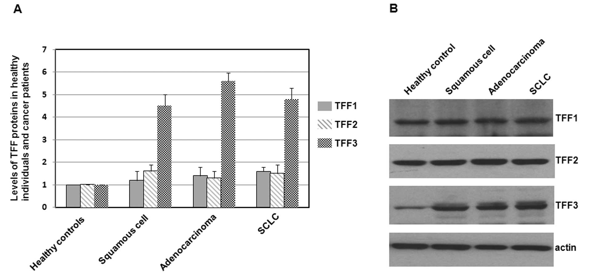 | Figure 1Immunoblots of TFF1, TFF2 and TFF3 in
healthy individuals and lung cancer patients. (A) Total proteins
were extracted from lung tissues, separated on SDS-PAGE gels, and
subjected to immunoblot analyses. The primary antibodies against
TFF1, TFF2, TFF3 and actin were purchased from Santa Cruz
Biotechnology (USA). Secondary antibodies were donkey anti-goat
IgG-HRP (cat# sc-2020, 1:5,000, Santa Cruz Biotechnology) and goat
anti-mouse IgG-HRP (cat# sc-2005, 1:10,000, Santa Cruz
Biotechnology). Bound antibodies were detected using the ECL system
(Pierce Biotechnology). The size of the TFF proteins was
approximately 7–10 kDa. Histograms show mean normalized optical
density (OD) of TFF protein bands relative to the OD of the actin
band from the same individual. Error bars show the standard error
of the mean (SEM) (P<0.05). (B) Representative blots from a
healthy individual and three lung cancer patients are shown. |
As shown in Fig. 1A,
levels of TFF1 and TFF2 in lung cancer tissues were slightly higher
or not significantly different from those in normal tissues from
the 60 healthy individuals. However, levels of TFF3 in patients of
three types of lung cancer were significantly higher than in lung
tissues of healthy individuals, suggesting a different TFF3
expression in lung cancer patients.
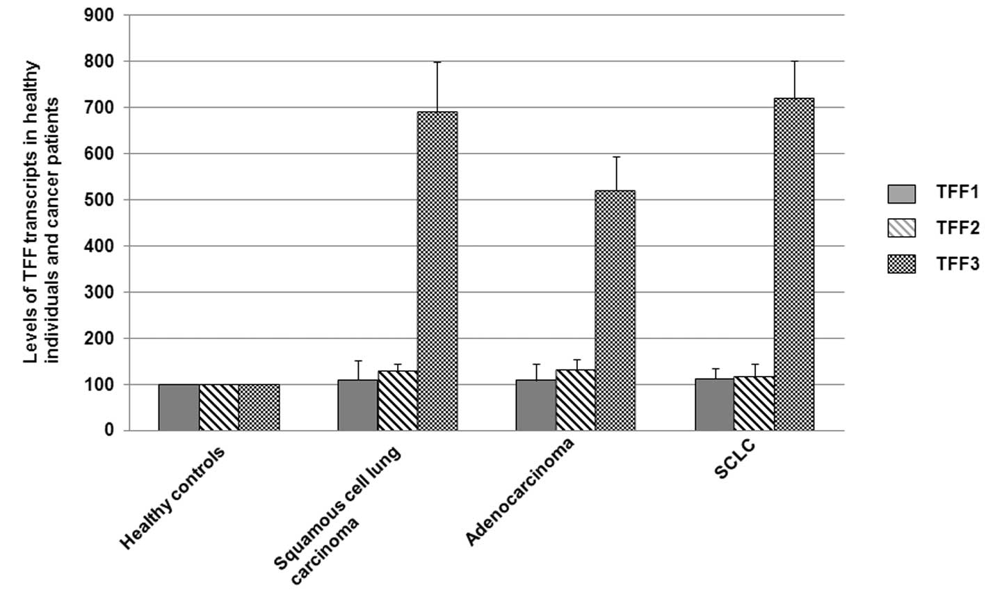
Levels of TFF3 transcripts in all three
detected types of lung cancer tissues are significantly higher than
in normal tissues from healthy individuals
High protein expression levels are often due to a
high level of gene transcription. Therefore, the mRNA transcript
levels in the lung cancer tissues (squamous cell lung carcinoma
cases, n=58; adenocarcinoma cases, n=43; SCLC cases, n=29) and
normal lung tissues from healthy individuals (n=60) were determined
by quantitative RT-PCR. The levels of TFF mRNAs (mean value) in
healthy individuals was assigned a value of 100.
As shown in Fig. 2,
levels of TFF1 and TFF2 transcripts in lung cancer tissues were
slightly higher or not significantly different from those in normal
tissues from the 60 healthy individuals. However, levels of TFF3
transcripts in patients with all three types of lung cancer were
significantly higher (P<0.05) than in lung tissues of healthy
individuals, suggesting increased TFF3 mRNA levels in lung cancer
patients in comparison to the healthy individuals.
Levels of TFF3 in lung cancer cell lines
are significantly higher than in the normal cell line
To investigate if the TFF1, TFF2 and TFF3 proteins
have varying expression in lung cancer cell lines and the normal
cell line, the total proteins were extracted and subjected to
western blot analysis, with the cellular actin protein serving as a
loading control. The mean normalized OD of TFF protein bands
relative to the OD of actin band from each cell line was all
calculated and subjected to statistical analyses. Error bars show
standard error of the mean (SEM) (P<0.05) (Fig. 3A). Representative blots from a
normal cell line and three lung cancer cell lines are shown in
Fig. 3B.
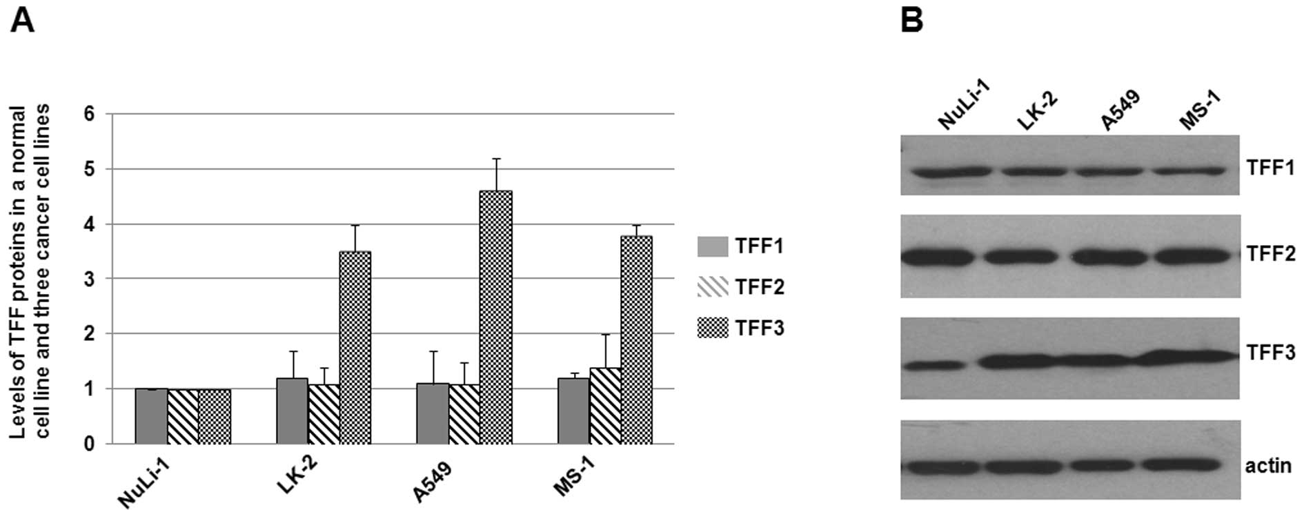 | Figure 3Immunoblots of TFF1, TFF2 and TFF3 in
a normal cell line and three lung cancer cell lines. (A) Total
proteins were harvested, separated on SDS-PAGE gels, and subjected
to immunoblot analyses. The primary antibodies against TFF1, TFF2,
TFF3 and actin were purchased from Santa Cruz Biotechnology (USA).
Secondary antibodies were donkey anti-goat IgG-HRP (cat# sc-2020,
1:5,000, Santa Cruz Biotechnology) and goat anti-mouse IgG-HRP
(cat# sc-2005, 1:10,000, Santa Cruz Biotechnology). Bound
antibodies were detected using the ECL system (Pierce
Biotechnology). The size of the TFF proteins were approximately
7–10 kDa. Experiments were repeated more than 3 times. Histograms
show mean normalized OD of TFF protein bands relative to the OD of
actin band. Error bars show standard error of the mean (SEM)
(P<0.05). (B) Representative blots were shown. Experiments were
repeated more than 3 times. |
As shown in Fig. 3A,
levels of TFF1 and TFF2 in lung cancer cell lines (LK-2, A549 and
MS-1) were slightly higher or not significantly different from
those in the normal cell line NuLi-1. However, levels of TFF3 in
the three cancer cell lines were significantly higher than in the
normal cell line NuLi-1, suggesting a different TFF3 expression in
the detected lung cancer cell lines.
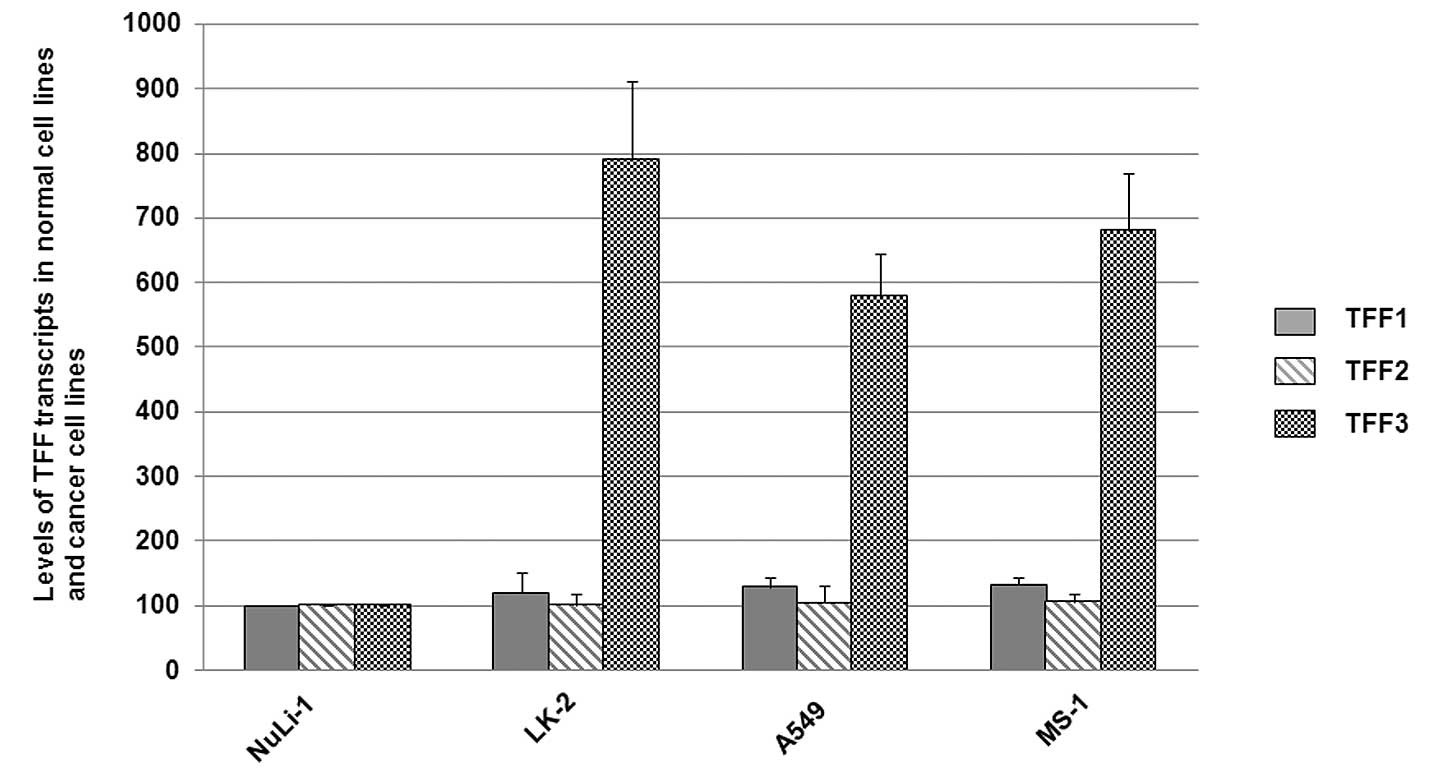
Levels of TFF3 transcripts in lung cancer
cell lines are significantly higher than in the normal cell line
NuLi-1
The mRNA transcript levels in the lung cancer cell
lines and the normal cell line were determined by quantitative
RT-PCR. The TFF transcript levels (mean value) in the normal cell
line NuLi-1 was assigned a value of 100.
As shown in Fig. 4,
levels of TFF1 and TFF2 transcripts in lung cancer cell lines
(LK-2, A549 and MS-1) were slightly higher or not significantly
different from those in the normal cell line NuLi-1. However, TFF3
transcript levels in the three cancer cell lines were significantly
higher (P<0.05) than in the normal cell line NuLi-1, suggesting
increased TFF3 mRNA levels in lung cancer cell lines in comparison
to the normal cell lines.
Discussion
Lung cancer is the most common malignancy, and the
number of cases being diagnosed is increasing. The treatments
currently available for this disease are basically the same for all
patients, including chemotherapy, radiotherapy, and surgery.
Treatment and prognosis depend on the histological type of lung
cancer and the stage of the disease. Since patients may response
quite differently to similar treatments, due to differences in
their health status, complications and smoking status, new
biological markers for lung cancer prediction and prognosis are
urgently necessary in the clinic.
In this study, we investigated the levels of TFF
proteins in the serum and lung tissues of 130 lung cancer patients,
including 58 squamous cell lung carcinoma cases, 43 adenocarcinoma
cases, and 29 SCLC cases, as well as in 60 healthy individuals. It
was found that TFF1 and TFF2 levels were similar or slightly higher
in these three subtypes of lung cancer compared to those in healthy
individuals, while TFF3 levels were significantly higher in the
detected lung cancer cases compared to healthy individuals.
Immunoblot analyses of TFF1, TFF2 and TFF3 indicated that lung
cancer tissues and lung cancer cell lines have higher expression
levels of TFF3 protein, but not of TFF1 and TFF2 proteins, compared
to tissues from healthy individuals or from normal cell line.
Quantitative RT-PCR analysis of TFF1, TFF2 and TFF3 transcripts in
tissues and cell lines indicated higher levels of TFF3, but not of
TFF1 and TFF2 transcripts in lung cancer tissues or cell lines when
compared with those in tissues of healthy individuals and normal
cells. Our results show increased TFF3 levels in the serum and lung
tissues, suggesting that TFF3 may serve as a promising, easy to
detect biomarker of lung cancer.
In our experiments, it was found that TFF1 and TFF2
levels (for the mean values of all detected patients, see Table III) in the serum of lung cancer
patients were slightly higher than in healthy individuals. However,
the levels (the mean OD values of the respective bands) of TFF1 and
TFF2 in lung cancer patient were similar to those in healthy
individuals. The inconsistency between the protein levels in serum
and in tissues might be due to pathological alterations in other
tissues of patients, which may affect the levels of secreted
proteins in the serum.
The increased levels of TFF3 in the serum of lung
cancer patients may be related to the upregulated protein
expression in lung tissues. However, the higher levels of TFF3
levels in the serum of lung cancer patients may also be attributed
to the histological changes in other tissues, especially for
patients with late stages of lung cancers, since TFF1 is mainly
expressed in the stomach and colon; TFF2 is mainly localized in the
stomach; TFF3 is principally expressed in the intestines (22,53).
We analyzed the correlation between the levels of serum TFF3 and
the stages of the investigated 130 lung cancer patients using the
statistical software. However, no significant relationship between
the cancer stages and the TFF3 levels in the serum or tissues were
found (data not shown). Therefore, TFF3 may be a promising
biomarker for lung cancer, but not appropriate for stage detection
of lung cancer patients.
It is noted that all of the proteins described in
the Introduction which are promising biomarkers of lung cancer are
detected by IHC or IB, which make the examination process
time-consuming and the quantification difficult. Therefore,
proteins easy to be measured become ideal targets to be researched
in the field of biomarkers. Recently, it has been reported that
serum levels of TFF3 are a better marker of gastric cancer than
pepsinogen (54). In the present
study, TFF3 levels in the serum of lung cancer patients differed
from those of healthy individuals, suggesting that TFF3 is a novel
biomarker easy to be measured in the clinic.
Acknowledgements
This study was supported by the Shandong Province
Science and Technology Research Fund (Grant # 2004GG2202165).
References
|
1
|
Brambilla E, Travis WD, Colby TV, Corrin B
and Shimosato Y: The new World Health Organization classification
of lung tumours. Eur Respir J. 18:1059–1068. 2001. View Article : Google Scholar : PubMed/NCBI
|
|
2
|
Mendelsohn J and Baselga J: The EGF
receptor family as targets for cancer therapy. Oncogene.
19:6550–6565. 2000. View Article : Google Scholar : PubMed/NCBI
|
|
3
|
Brognard J, Clark AS, Ni Y and Dennis PA:
Akt/protein kinase B is constitutively active in non-small cell
lung cancer cells and promotes cellular survival and resistance to
chemotherapy and radiation. Cancer Res. 61:3986–3997.
2001.PubMed/NCBI
|
|
4
|
Mukohara T, Kudoh S, Yamauchi S, et al:
Expression of epidermal growth factor receptor (EGFR) and
downstream-activated peptides in surgically excised non-small cell
lung cancer (NSCLC). Lung Cancer. 41:123–130. 2003. View Article : Google Scholar : PubMed/NCBI
|
|
5
|
D’Amico TA, Massey M, Herndon JE, Moore MB
and Harpole DH: A biologic risk model for stage I lung cancer:
immunohistochemical analysis of 408 patients with the use of ten
molecular markers. J Thorac Cardiovasc Surg. 117:736–743.
1999.PubMed/NCBI
|
|
6
|
Shiba M, Kohno H, Kakizawa K, et al: Ki-67
immunostaining and other prognostic factors including tobacco
smoking in patients with resected nonsmall cell lung carcinoma.
Cancer. 89:1457–1465. 2000. View Article : Google Scholar : PubMed/NCBI
|
|
7
|
Lloyd RV, Erickson LA, Jin L, Kulig E,
Qian X, Cheville JC and Scheithauer BW: p27kip1: a multifunctional
cyclin-dependent kinase inhibitor with prognostic significance in
human cancers. Am J Pathol. 154:313–323. 1999. View Article : Google Scholar : PubMed/NCBI
|
|
8
|
Hayashi H, Ogawa N, Ishiwa N, Yazawa T,
Inayama Y, Ito T and Kitamura H: High cyclin E and low p27/Kip1
expressions are potentially poor prognostic factors in lung
adenocarcinoma patients. Lung Cancer. 34:59–65. 2001. View Article : Google Scholar : PubMed/NCBI
|
|
9
|
Hommura F, Dosaka-Akita H, Mishina T, et
al: Prognostic significance of p27KIP1 protein and ki-67 growth
fraction in non-small cell lung cancers. Clin Cancer Res.
6:4073–4081. 2000.PubMed/NCBI
|
|
10
|
Thim L and May FE: Structure of mammalian
trefoil factors and functional insights. Cell Mol Life Sci.
62:2956–2973. 2005.PubMed/NCBI
|
|
11
|
Taupin D and Podolsky DK: Trefoil factors:
initiators of mucosal healing. Nat Rev Mol Cell Biol. 4:721–732.
2003. View
Article : Google Scholar : PubMed/NCBI
|
|
12
|
Bossenmeyer-Pourié C, Kannan R, Ribieras
S, et al: The trefoil factor 1 participates in gastrointestinal
cell differentiation by delaying G1-S phase transition and reducing
apoptosis. J Cell Biol. 157:761–770. 2002.PubMed/NCBI
|
|
13
|
Chan MW, Chan VY, Leung WK, Chan KK, To
KF, Sung JJ and Chan FK: Anti-sense trefoil factor family-3
(intestinal trefoil factor) inhibits cell growth and induces
chemosensitivity to adriamycin in human gastric cancer cells. Life
Sci. 76:2581–2592. 2005. View Article : Google Scholar : PubMed/NCBI
|
|
14
|
Yio X, Diamond M, Zhang JY, Weinstein H,
Wang LH, Werther L and Itzkowitz S: Trefoil factor family-1
mutations enhance gastric cancer cell invasion through distinct
signaling pathways. Gastroenterology. 130:1696–1706. 2006.
View Article : Google Scholar : PubMed/NCBI
|
|
15
|
Hoosein NM, Thim L, Jørgensen KH and
Brattain MG: Growth stimulatory effect of pancreatic spasmolytic
polypeptide on cultured colon and breast tumor cells. FEBS Lett.
247:303–306. 1989. View Article : Google Scholar : PubMed/NCBI
|
|
16
|
Emami S, Rodrigues S, Rodrigue CM, et al:
Trefoil factor family (TFF) peptides and cancer progression.
Peptides. 25:885–898. 2004. View Article : Google Scholar : PubMed/NCBI
|
|
17
|
Yio X, Zhang JY, Babyatsky M, et al:
Trefoil factor family-3 is associated with aggressive behavior of
colon cancer cells. Clin Exp Metastasis. 22:157–165. 2005.
View Article : Google Scholar : PubMed/NCBI
|
|
18
|
Chan VY, Chan MW, Leung WK, Leung PS, Sung
JJ and Chan FK: Intestinal trefoil factor promotes invasion in
non-tumorigenic Rat-2 fibroblast cell. Regul Pept. 127:87–94. 2005.
View Article : Google Scholar : PubMed/NCBI
|
|
19
|
Graness A, Chwieralski CE, Reinhold D,
Thim L and Hoffmann W: Protein kinase C and ERK activation are
required for TFF-peptide-stimulated bronchial epithelial cell
migration and tumor necrosis factor-alpha-induced interleukin-6
(IL-6) and IL-8 secretion. J Biol Chem. 277:18440–18446. 2002.
View Article : Google Scholar
|
|
20
|
Rodrigues S, Van Aken E, Van Bocxlaer S,
et al: Trefoil peptides as proangiogenic factors in vivo and in
vitro: implication of cyclooxygenase-2 and EGF receptor signaling.
FASEB J. 17:7–16. 2003. View Article : Google Scholar : PubMed/NCBI
|
|
21
|
Dhar DK, Wang TC, Tabara H, et al:
Expression of trefoil factor family members correlates with patient
prognosis and neoangiogenesis. Clin Cancer Res. 11:6472–6478. 2005.
View Article : Google Scholar : PubMed/NCBI
|
|
22
|
Regalo G, Wright NA and Machado JC:
Trefoil factors: from ulceration to neoplasia. Cell Mol Life Sci.
62:2910–2915. 2005. View Article : Google Scholar : PubMed/NCBI
|
|
23
|
May FE and Westley BR: Expression of human
intestinal trefoil factor in malignant cells and its regulation by
oestrogen in breast cancer cells. J Pathol. 182:404–413. 1997.
View Article : Google Scholar : PubMed/NCBI
|
|
24
|
Henry JA, Nicholson S, Hennessy C, Lennard
TW, May FE and Westley BR: Expression of the oestrogen regulated
pNR-2 mRNA in human breast cancer: relation to oestrogen receptor
mRNA levels and response to tamoxifen therapy. Br J Cancer.
61:32–38. 1990. View Article : Google Scholar : PubMed/NCBI
|
|
25
|
May FE and Westley BR: Trefoil proteins:
their role in normal and malignant cells. J Pathol. 183:4–7. 1997.
View Article : Google Scholar : PubMed/NCBI
|
|
26
|
West M, Blanchette C, Dressman H, et al:
Predicting the clinical status of human breast cancer by using gene
expression profiles. Proc Natl Acad Sci USA. 98:11462–11467. 2001.
View Article : Google Scholar : PubMed/NCBI
|
|
27
|
Speiser P, Stolzlechner J, Haider K, et
al: pS2 protein status fails to be an independent prognostic factor
in an average breast cancer population. Anticancer Res.
14:2125–2130. 1994.
|
|
28
|
Gillesby BE and Zacharewski TR: pS2 (TFF1)
levels in human breast cancer tumor samples: correlation with
clinical and histological prognostic markers. Breast Cancer Res
Treat. 56:253–265. 1999. View Article : Google Scholar : PubMed/NCBI
|
|
29
|
Tozlu S, Girault I, Vacher S, et al:
Identification of novel genes that co-cluster with estrogen
receptor alpha in breast tumor biopsy specimens, using a
large-scale real-time reverse transcription-PCR approach. Endocr
Relat Cancer. 13:1109–1120. 2006. View Article : Google Scholar
|
|
30
|
Doane AS, Danso M, Lal P, Donaton M, Zhang
L, Hudis C and Gerald WL: An estrogen receptor-negative breast
cancer subset characterized by a hormonally regulated
transcriptional program and response to androgen. Oncogene.
25:3994–4008. 2006. View Article : Google Scholar
|
|
31
|
Smid M, Wang Y, Klijn JG, et al: Genes
associated with breast cancer metastatic to bone. J Clin Oncol.
24:2261–2267. 2006. View Article : Google Scholar : PubMed/NCBI
|
|
32
|
Bosma AJ, Weigelt B, Lambrechts AC, et al:
Detection of circulating breast tumor cells by differential
expression of marker genes. Clin Cancer Res. 8:1871–1877.
2002.PubMed/NCBI
|
|
33
|
Weigelt B, Bosma AJ, Hart AA, Rodenhuis S
and van ‘t Veer LJ: Marker genes for circulating tumour cells
predict survival in metastasized breast cancer patients. Br J
Cancer. 88:1091–1094. 2003. View Article : Google Scholar : PubMed/NCBI
|
|
34
|
Ren JL, Luo JY, Lu YP, Wang L and Shi HX:
Molecular forms of trefoil factor 1 in normal gastric mucosa and
its expression in normal and abnormal gastric tissues. World J
Gastroenterol. 12:7361–7364. 2006.PubMed/NCBI
|
|
35
|
Milne AN, Carvalho R, Morsink FM, Musler
AR, de Leng WW, Ristimäki A and Offerhaus GJ: Early-onset gastric
cancers have a different molecular expression profile than
conventional gastric cancers. Mod Pathol. 19:564–572. 2006.
View Article : Google Scholar
|
|
36
|
Sonoda H, Yamamoto K, Kushima R, Yamamoto
H, Naitoh H, Okabe H and Tani T: Detection of lymph node
micrometastasis in pN0 early gastric cancer: Efficacy of duplex
RT-PCR with MUC2 and TFF1 in mucosal cancer. Oncol Rep. 16:411–416.
2006.PubMed/NCBI
|
|
37
|
Suárez C, Vizoso F, Rodríguez JC, et al:
Prognostic significance of cytosolic pS2 protein content in gastric
cancer. Int J Biol Markers. 16:37–44. 2001.PubMed/NCBI
|
|
38
|
Mori K, Aoyagi K, Ueda T, et al: Highly
specific marker genes for detecting minimal gastric cancer cells in
cytology negative peritoneal washings. Biochem Biophys Res Commun.
313:931–937. 2004. View Article : Google Scholar : PubMed/NCBI
|
|
39
|
Tuna B, Sökmen S, Sarioğlu S, Füzün M,
Küpelioğlu A and Ellidokuz H: PS2 and HSP70 expression in rectal
adenocarcinomas: an immunohistochemical investigation of 45 cases.
Appl Immunohistochem Mol Morphol. 14:31–36. 2006. View Article : Google Scholar : PubMed/NCBI
|
|
40
|
Taupin D, Ooi K, Yeomans N and Giraud A:
Conserved expression of intestinal trefoil factor in the human
colonic adenoma-carcinoma sequence. Lab Invest. 75:25–32.
1996.PubMed/NCBI
|
|
41
|
Welsh JB, Sapinoso LM, Su AI, et al:
Analysis of gene expression identifies candidate markers and
pharmacological targets in prostate cancer. Cancer Res.
61:5974–5978. 2001.PubMed/NCBI
|
|
42
|
Luo J, Duggan DJ, Chen Y, et al: Human
prostate cancer and benign prostatic hyperplasia: molecular
dissection by gene expression profiling. Cancer Res. 61:4683–4688.
2001.PubMed/NCBI
|
|
43
|
Vestergaard EM, Borre M, Poulsen SS, Nexø
E and Tørring N: Plasma levels of trefoil factors are increased in
patients with advanced prostate cancer. Clin Cancer Res.
12:807–812. 2006. View Article : Google Scholar : PubMed/NCBI
|
|
44
|
Buache E, Etique N, Alpy F, et al:
Deficiency in trefoil factor 1 (TFF1) increases tumorigenicity of
human breast cancer cells and mammary tumor development in
TFF1-knockout mice. Oncogene. 30:3261–3273. 2011. View Article : Google Scholar : PubMed/NCBI
|
|
45
|
Shen J, Liu J, Xie Y, Diwan BA and Waalkes
MP: Fetal onset of aberrant gene expression relevant to pulmonary
carcinogenesis in lung adenocarcinoma development induced by in
utero arsenic exposure. Toxicol Sci. 95:313–320. 2007. View Article : Google Scholar : PubMed/NCBI
|
|
46
|
Mathelin C, Tomasetto C and Rio MC:
Trefoil factor 1 (pS2/TFF1), a peptide with numerous functions.
Bull Cancer. 92:773–781. 2005.PubMed/NCBI
|
|
47
|
Liu S, Stromberg A, Tai HH and Moscow JA:
Thiamine transporter gene expression and exogenous thiamine
modulate the expression of genes involved in drug and prostaglandin
metabolism in breast cancer cells. Mol Cancer Res. 2:477–487.
2004.PubMed/NCBI
|
|
48
|
dos Santos Silva E, Ulrich M, Döring G,
Botzenhart K and Gött P: Trefoil factor family domain peptides in
the human respiratory tract. J Pathol. 190:133–142. 2000.PubMed/NCBI
|
|
49
|
Higashiyama M, Doi O, Kodama K, Yokuchi H,
Inaji H and Tateishi R: Estimation of serum level of pS2 protein in
patients with lung adenocarcinoma. Anticancer Res. 16:2351–2355.
1996.PubMed/NCBI
|
|
50
|
Higashiyama M, Doi O, Kodama K, Yokouchi
H, Inaji H, Nakamori S and Tateishi R: Prognostic significance of
pS2 protein expression in pulmonary adenocarcinoma. Eur J Cancer.
30:792–797. 1994. View Article : Google Scholar : PubMed/NCBI
|
|
51
|
Kosriwong K, Menheniott TR, Giraud AS,
Jearanaikoon P, Sripa B and Limpaiboon T: Trefoil factors: tumor
progression markers and mitogens via EGFR/MAPK activation in
cholangiocarcinoma. World J Gastroenterol. 17:1631–1641. 2011.
View Article : Google Scholar : PubMed/NCBI
|
|
52
|
Qu Y, Yang Y, Liu B and Xiao W:
Comparative proteomic profiling identified sorcin being associated
with gemcitabine resistance in non-small cell lung cancer. Med
Oncol. 27:1303–1308. 2010. View Article : Google Scholar : PubMed/NCBI
|
|
53
|
Madsen J, Nielsen O, Tornøe I, Thim L and
Holmskov U: Tissue localization of human trefoil factor 1, 2 and 3.
J Histochem Cytochem. 55:505–513. 2007. View Article : Google Scholar
|
|
54
|
Aikou S, Ohmoto Y, Gunji T, et al: Tests
for serum levels of trefoil factor family proteins can improve
gastric cancer screening. Gastroenterology. 141:837–845. 2011.
View Article : Google Scholar : PubMed/NCBI
|