Introduction
Lung cancer is a significant health problem not only
in China, but worldwide. According to the information presented in
the online database GLOBOCAN 2008 (http://globocan.iarc.fr/factsheet.asp) of the
International Agency for Research on Cancer, lung cancer was the
most common and most aggressive cancer worldwide in 2008,
accounting for 16.5% of the total cancer cases and 22.5% of the
total cancer-related deaths in 182 countries. In China, the
incidence and mortality rates of lung cancer also rank first among
all types of cancer, which were predicted to be 33.5/100,000 and
28.7/100,000, respectively, in 2008 based on the data from 36
cancer registries and the Third National Death Survey
(2004–2005)(1).
Approximately 85% of lung cancer cases are non-small
cell lung cancer (NSCLC), which includes adenocarcinoma (AD),
squamous cell carcinoma (SCC), large cell carcinoma and
bronchioloalveolar carcinoma (2).
Despite extensive research and clinical efforts, prognosis of NSCLC
remains poor. Nevertheless, biomarkers have been recognized to play
a critical role in early diagnosis, therapy guidance and prognosis
monitoring of NSCLC (3). Over the
last two decades, several biomarkers have been identified for
NSCLC, but none have been routinely used in clinical settings
(4). It is, therefore, urgent to
explore more reliable and feasible novel biomarkers for lung
cancer.
Adenylate cyclase-associated protein 1 (CAP1) is an
actin monomer-binding protein coded by the CAP1 gene (5), which was originally cloned from
budding yeast and is located in the downstream of the ras gene
(6). Human homology of CAP1 was
identified in the early 1990s (7).
Both mammal and yeast CAPs interact with actin (8) and play a role in actin turnover
(9). Given the critical role for
actin filament reorganization in cell migration and the regulatory
role for CAP1 in actin filament reorganization (10,11),
it is logical to hypothesize that CAP1 may be associated with tumor
metastasis. Currently, however, studies on the correlation between
CAP1 expression and tumor metastasis are scarce (12). The objective of the present study
was to evaluate the potential value of CAP1 in early diagnosis and
prognostic prediction of progressive lung cancer.
Materials and methods
Ethical considerations
The study was reviewed and approved by the
Institutional Ethics Committee of Tongji University School of
Medicine in Shanghai and was conducted in compliance with the
Helsinki Declaration. Written informed consent was obtained from
all subjects.
Subjects and biopsy specimen
collection
Twenty-four lung cancer patients and 6 control
subjects with non-neoplastic lung condition(s) who underwent
surgical lung resection of neoplastic and non-neoplastic lesions,
respectively, at the Shanghai 10th People’s Hospital, China,
between 2007 and 2009 were recruited. Cancer types and stages were
determined based on results from laboratory tests, X-rays, CT
scans, bone scans and MRI scans. Biopsy specimens were collected at
surgery, snap-frozen in liquid nitrogen and stored at −80°C until
analyses.
Tissue bank specimen acquisition
Tissue blocks of 82 formalin-fixed and
paraffin-embedded lung cancer specimens were acquired from the
Tissue Bank in the Pathology Department of Tongji University School
of Medicine. The documented general demographic data and cancer
stages assessed based on the tumor-node-metastasis (TNM)
classification system were obtained.
Lung cancer cell line
For in vitro analyses, a low invasive lung
cancer cell line 95-C and an invasive lung cancer cell line 95-D
were purchased from the American Type Culture Collection (ATCC,
Manassas, VA, USA). For maintenance, cells were cultured in
RPMI-1640 supplemented with 10% fetal bovine serum (FBS) at 37°C in
a humidified atmosphere of 5% CO2 and 95% air.
Real-time PCR
Total RNA was isolated from neoplastic and
non-neoplastic lung biopsy specimens using an RNA extraction kit
from Isogen (Nippon Gene Co., Ltd., Toyama, Japan). RNA samples
were treated with DNase I (Promega Corp., Madison, WI, USA) to
remove genomic DNA. First-strand cDNAs were synthesized using a
commercial First-Strand cDNA Synthesis kit as instructed by the
manufacturer. PCR amplifications of the test gene CAP1 and the
reference gene glyceraldehyde-3-phosphate dehydrogenase (GAPDH)
were performed using, respectively, a pair of primers (forward,
5′-ACT CGC TGC TTG CTG GTC-3′ and reverse: 5′-ATG GGT GCC AAC AAA
TCG-3′) designed based on the human CAP1 mRNA sequence (GenBank
accession number BT007152) and a pair of primers (forward, 5′-GAA
GGT GAA GGT CGG AGTC-3′ and reverse, 5′-CCC GAA TCA CAT TCT CCA AGA
A-3) designed based on the human GAPDH cDNA sequence (GenBank
access number X01677). All reactions were carried out with the
SYBR-Green PCR Core Reagents kit (Perkin-Elmer Applied Biosystems,
Foster City, CA, USA). The PCR amplification parameters were: 50°C
for 2 min (one cycle), 95°C for 10 min (one cycle), 95°C for 15 sec
and 60°C for 1 min (40 cycles). The emission intensity of the
SYBR-Green fluorescence was measured in a real-time fashion with
the ABI Prism 7700 Sequence Detector from Perkin-Elmer Applied
Biosystems. Relative quantitation of CAP1 mRNA abundance was
performed using the DataAssist software (Life Technologies, Grand
Island, NY, USA).
Western blot analysis
Whole cell and tissues lysates were prepared from
cultured 95-C and 95-D cells and biopsy lung specimens,
respectively. Following total protein quantitation, the lysates
were loaded onto 10% polyacrylamide-SDS gels, separated by
electrophoresis and blotted onto N-C membrane blots using a
semi-dry transfer system. Blots were incubated with a mouse
anti-human CAP1 antibody and a mouse anti-human actin antibody
(Sigma, St. Louis, MO, USA) at 4°C overnight. After washing, blots
were incubated with horseradish peroxidase-conjugated secondary
antibodies at room temperature for 45 min. The immunoreactive
signals for both CAP1 and actin were visualized using the ECL
system from GE Healthcare UK Ltd. (Buckinghamshire, UK) and
subjected to densitometric analyses with the ImageJ software
(National Institutes of Health, Bethesda, MD, USA). Relative levels
of CAP1 (i.e., after adjustment against actin) were determined
based on the densitometric data.
Immunohistochemical and
immunocytochemical staining
Lung cancer specimen blocks obtained from the tissue
bank were sectioned at 5 μM and mounted onto glass slides. Low and
high invasive lung cancer 95-C and 95-D cells were grown on culture
slides (BD Biosciences, San Diego, CA, USA) and fixed in 3.7%
formaldehyde in PBS. Both tissue sections and cultured cells were
incubated with a primary CAP1 antibody at 4°C overnight. After
rinsing three times in Tris-buffered saline (0.01 M Tris-HCl pH
7.5, 150 mM NaCl), tissue sections and cultured cells were
incubated with FITC-labeled secondary antibody (Invitrogen,
Carlsbad, CA, USA) at room temperature for 30 min. After washing,
the slides were mounted with a DAPI-containing mounting solution to
identify cell nuclei. Fluorescent signals were visualized under an
Axiovert 200 microscope and images were captured using an AxioCam
CCD camera and the AxioVision software (Leica Microsystems,
Wetzlar, Germany). The human peritumoral vascular endothelial cells
were used as a positive control.
Staining results were assessed by two pathologists,
blind to the specimen identity, independently. The immunoreactive
intensity in the test tissue specimens and cultured tumor (95-C and
95-D) cells was graded by the difference against the intensity in
endothelial cells in the positive control group: 0, negative; 1
(weak), weaker than epithelial cells; 2 (moderate), the same as
epithelial cells; 3 (strong), stronger than epithelial cells. A
staining score of 2 or 3 was considered CAP1-positive.
RNA interference
Invasive 95-D cells were seeded onto collagen
I-coated 6-well plates at 5×10⁴ cells/well. Following
overnight culture, cells were transfected with CAP1 specific siRNA
(siCAP1) or non-silencing control siRNA in serum-free RPMI-1640
using Oligofectamine (Invitrogen). The target sequence was
AAACCGAGTCCTCAAAGAGTA. Both specific and control siRNA molecules
were synthesized by GenePharma Co. (Shanghai, China). Four hours
after transfection, an equal volume of medium supplemented with 20%
fetal bovine serum (FBS) was added. Transfectants were cultured
until further analysis.
Migration assay
Invasive 95-D cells transfected with siCAP or
control siRNA were seeded onto the upper chambers of 6-well
transwell plates (BD Biosciences, San Jose, CA, USA) in triplicates
in RPMI-1640 supplemented with 0.5% FBS. Culture medium containing
10% FBS was added to the bottom chamber. After a 24-h culture,
cells on the upper surface of the chamber membrane were removed and
those on the lower surface of the membrane were stained by
hematoxylin and eosin (H&E). The number of migrated cells was
counted under a light microscope (×400).
Statistical analysis
Categorical data expressed as percentage were
analyzed by the χ2 test. Correlations between CAP1
expression and various clinicopathological variables was analyzed
by multivariate logistic regression analysis. The statistical
software SPSS version 17.0 (SPSS Inc., Chicago, IL, USA) was used.
Differences were considered significant when P<0.05.
Results
Demographic characteristics
In the present study, 24 lung cancer patients and 6
non-neoplastic control subjects were recruited and 82 tissue bank
specimens of lung cancer were included. Of the 24 lung cancer
patients, 4 had advanced (metastasis), 10 were of the SCC and 14 of
the AD type. The base-line demographic data on the 82 tissue bank
cases are presented in Table I.
 | Table IGeneral demographics and CAP1
immunoreactive signals in the 82 patients whose lung tumor tissue
specimens were banked in the institutional tissue bank. |
Table I
General demographics and CAP1
immunoreactive signals in the 82 patients whose lung tumor tissue
specimens were banked in the institutional tissue bank.
| Variables | Total N (%) | CAP1-negative | CAP1-positive | P-value |
|---|
| Age (years) | | | | 0.204 |
| ≤65 | 43 (52.4) | 15 | 28 | |
| >65 | 39 (47.6) | 19 | 20 | |
| Gender | | | | 0.701 |
| Female | 27 (32.9) | 12 | 15 | |
| Male | 55 (67.1) | 22 | 33 | |
| Histologic type | | | | 0.526 |
| AD | 40 (48.8) | 18 | 22 | |
| SCC | 31 (37.8) | 16 (non-AD) | 26 (non-AD) | |
| Others | 11 (13.4) | | | |
| Tumor size (T)
(cm) | | | | 0.231 |
| ≤3 | 37 (45.1) | 18 | 19 | |
| >3 | 45 (54.9) | 16 | 29 | |
| Node metastasis
(N) | | | | 0.01 |
| 0–1 | 42 (51.2) | 22 | 20 | |
| 2–3 | 40 (48.8) | 12 | 28 | |
| Distant metastasis
(M) | | | | 0.126 |
| 0 | 63 (76.8) | 29 | 34 | |
| 1 | 9 (23.2) | 5 | 14 | |
| TNM | | | | 0.04 |
| I | | 17 | 8 | |
| II–IV | | 17 | 40 | |
CAP1 mRNA abundance
To assess the association of CAP1 expression with
lung cancer type and stage, we first performed real-time PCR to
analyze relative CAP1 mRNA abundance in non-neoplastic and lung
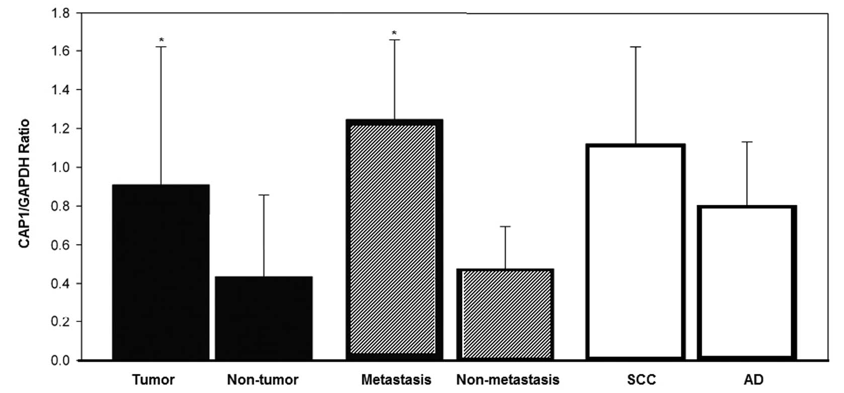
cancer biopsy specimens. The results are shown in Fig. 1. The relative CAP1 mRNA abundance,
expressed as the CAP1/GAPDH ratio, was significantly higher in
neoplastic tissues than in control specimens (P=0.028), and in
metastatic than in non-metastatic lung cancer specimens (P=0.016).
When a comparison was made between histological types, SCC
specimens had slightly higher CAP1 mRNA abundance than AD
specimens, but the difference failed to reach a statistically
significant level (P=0.227).
CAP1 protein levels
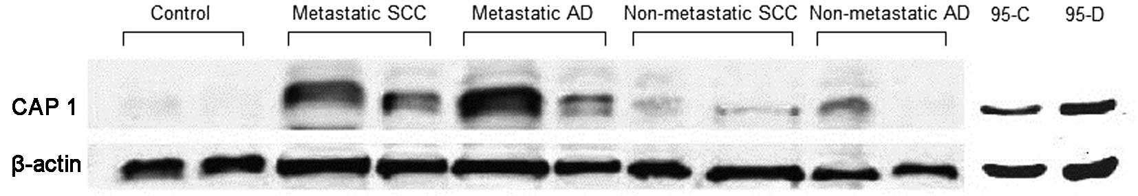
In addition, mRNA abundance, CAP1 protein levels in
biopsy specimens and cultured cells were determined by western blot
analysis. Shown in Fig. 2 is an
image of a representative blot. When expressed as a densitometric
ratio of CAP1 band to β-actin band, the relative CAP1 protein level
was significantly elevated in lung cancer patients (0.7527±0.2767)
as compared to non-neoplastic control subjects (0.3476±0.1713,
P=0.002); it was also significantly elevated in metastatic lung
tumors (0.8941±0.1442) as compared to non-metastatic lung tumors
(0.4701±0.2647, P=0.002); it was slightly higher in SCC than in AD
specimens (0.7440±0.2911 vs. 0.7601±0.2757, P=0.891), but the
difference was not significant (P>0.05). In cultured cell lines,
the CAP1 protein level was significantly higher in invasive 95-D
cells than that in non-invasive 95-C cells (P<0.05).
Correlation between immunoreactive CAP1
signals and cancer metastasis
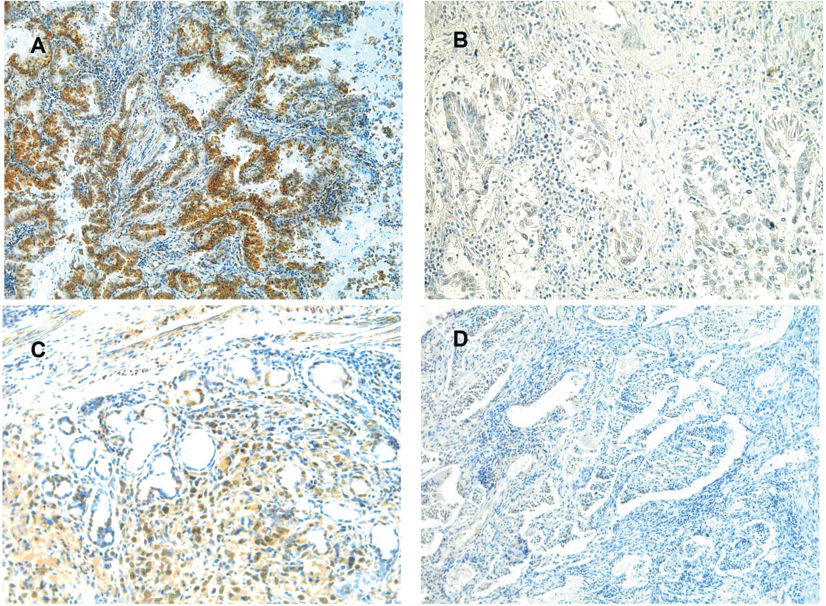
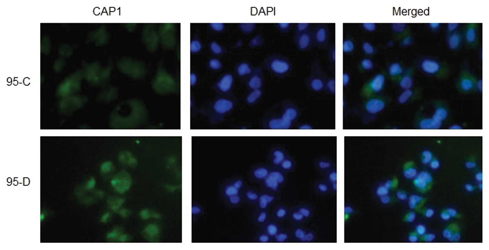
Immunoreactive CAP1 was detected in the cytoplasm of
cancer cells in all 82 tissue bank samples (Fig. 3) and cultured 95-C and 95-D cells
and also in the cytoplasm of stromal cells (Fig. 4). Using the signal in the positive
control endothelial cells as a reference, the 82 bank samples were
classified into a CAP1-negative group, where the tumor staining
score was 0 or 1, and a CAP1-positive group, where the tumor
staining score was 2 or 3. As shown in Table I, a CAP1-positive status was
significantly associated with lymph node metastasis (P=0.04) and
TNM stage (P=0.01) as demonstrated by the χ2 test.
To assess the association of CAP1 expression
simultaneously with various demographic variables and tumor
properties in lung cancer cases, multivariate logistic regression
analysis was performed. These results indicated that the expression
level of CAP1 was associated with TNM stage of lung cancer
(P=0.01), particularly the lymph node invasion (P=0.04) (Table I).
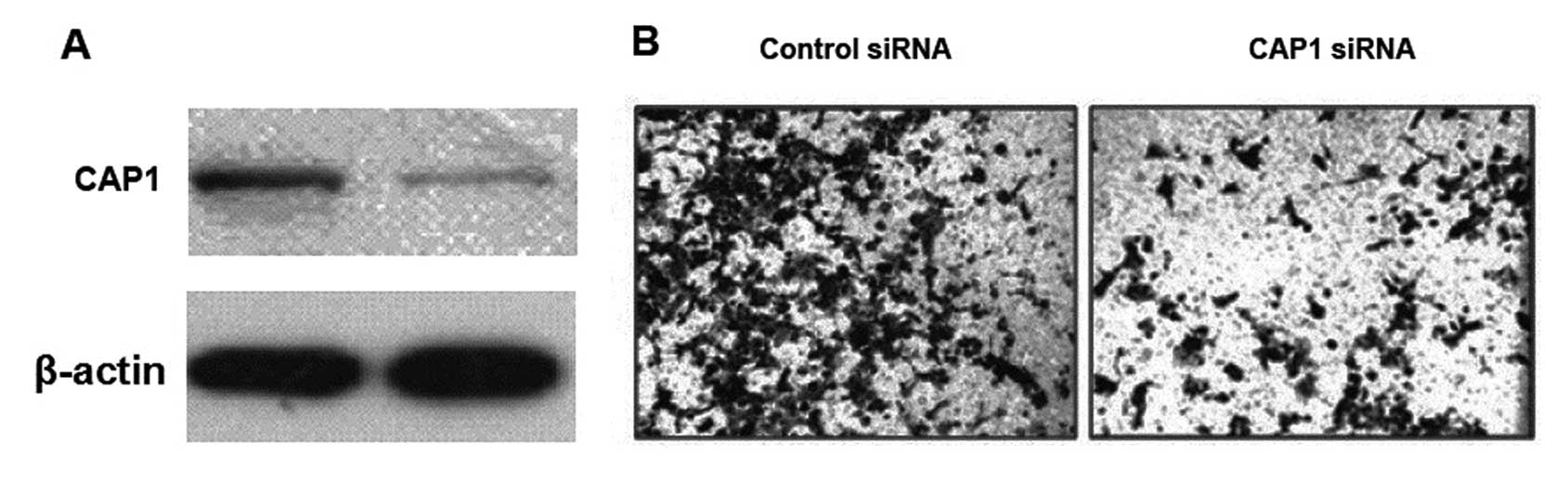
Reduced migration of invasive lung cancer
cells following CAP1 knockdown
To validate the positive correlation between CAP1
and metastasis of lung tumors, we attempted to silence the CAP1
gene in invasive lung cancer 95-D cells with a small interfering
RNA (siRNA) molecule specific for human CAP1 and to determine the
migration capacity of the CAP1-siRNA carrying cells in
vitro. As shown in Fig. 5, the
CAP1 gene was adequately silenced by sequence-specific CAP1-siRNA
and correspondingly the number of cells that migrated across the
transwell membrane was significantly reduced in the CAP1
gene-silenced 95-D cells.
Discussion
As a first step toward evaluating the association of
CAP1 with histological type and clinical stage of lung cancer, we
first assessed the CAP1 gene expression at the mRNA level in 24
lung cancer patients and 6 non-cancer control subjects by real-time
PCR. The results clearly demonstrated that the CAP1 gene
transcription was significantly elevated in lung cancer patients
and the elevation was more pronounced in metastasis (Fig. 1).
Although it is generally assumed that mRNA abundance
is predictive of the corresponding protein level, this assumption
may not be valid for all human genes. In a study aimed at assessing
the abundance of 165 protein spots representing 98 individual genes
using 76 lung adenocarcinomas and 9 non-neoplastic lung tissue
specimens, marked variation in the mRNA/protein correlation
coefficient was found among proteins and between stage I and stage
III lung adenocarcinomas (13). To
further analyze whether the observed elevation in CAP1 mRNA
abundance was correlated with increased CAP1 protein synthesis, we
next performed western blot analysis on the CAP1 protein content in
biopsy specimens and cultured lung cancer cells. In accordance with
the pattern of changes in CAP1 mRNA abundance in various specimens,
the CAP1 protein content was significantly higher in cancer than in
non-cancer subjects and higher in metastatic than in non-metastatic
cancer patients with no difference between SCC and AD (Fig. 2). Since the sample size used in the
present study was small and there are no reports in the literature
on the CAP1 mRNA/protein correlation coefficient, more thorough
studies with larger sample sizes are warranted to validate the
concurrent elevation in CAP1 mRNA and protein levels in lung
cancer.
To further evaluate the correlation between CAP1
expression and histological type and clinical stage of lung cancer,
we performed immunostaining on 82 tissue bank specimens and
cultured 95-C and 95-D cells. Despite the lack of reports on
intracellular localization of CAP1 protein in lung cancer cells,
non-nuclear localization of CAP1 has been observed at the site of
rapid actin turnover in non-muscle cells (9). In the present study, stronger
immunoreactive CAP1 signals were detected in the perinuclear
cytoplasm of lung cancer cells in the biopsy specimens (Fig. 3) and cultured lung cancer cells
(Fig. 4). This intracellular
localization suggests that CAP1 may be associated with
lamellipodium formation and cell migration. Following
classification of the 82 tissue bank specimens as CAP1-negative and
CAP1-positive, two groups based on their immunoreactive signal
densities relative to the density in endothelial cells in the
positive control group and a subsequent multivariate regression
analysis, the CAP1 immunoreactive signal was positively correlated
with metastasis, particularly metastasis to lymph nodes (Table I). Given that lymph node metastasis
of lung cancer is associated with poor clinical outcomes (14,15),
this finding suggests that immunostaining of the CAP1 protein in
lung cancer tissue may provide additional information predictive of
patient prognosis.
siRNA-based RNA interference (RNAi) is a
post-transcriptional process triggered by the introduction of
double-stranded RNA (dsRNA) which leads to gene silencing in a
sequence-specific manner (16). As
one of the most notable discoveries of the past decade in
functional genomics, RNAi has become an important tool for
analyzing gene functions in eukaryotes. Using this advanced
technology, we successfully silenced the CPA1 gene in invasive lung
cancer 95-D cells (Fig. 4), which
resulted in a significant reduction in the capacity of 95-D cells
to migrate (Fig. 5). This
observation further supports a functional role for CAP1 in the
process of lung cancer cell metastasis.
At present, the molecular mechanisms underlying the
effect of CAP1 on lung cancer cell migration and metastasis are
largely unknown. Cofilin is a family of actin-binding proteins
which disassembles actin filaments. Cofilin activity is present in
the malignant, invasive cancer cells (17) and may function as a factor to
regulate cancer cell migration/invasion phenotypes. We previously
demonstrated that CAP1 is a downstream target of cofilin (18), suggesting that the CAP1
overexpression-associated lung cancer cell migration and metastasis
may involve a cofilin-mediated signaling pathway. Nevertheless,
this requires further evaluation.
Acknowledgements
The present study was supported by a research grant
from the Shanghai Science Committee (09ZR1424500).
References
|
1
|
Chang S, Dai M, Ren JS, Chen YH and Guo
LW: Estimates and prediction on incidence, mortality and prevalence
of lung cancer in China in 2008. Zhonghua Liu Xing Bing Xue Za Zhi.
33:391–394. 2012.(In Chinese).
|
|
2
|
Ramalingam SS, Owonikoko TK and Khuri FR:
Lung cancer: new biological insights and recent therapeutic
advances. CA Cancer J Clin. 61:91–112. 2011. View Article : Google Scholar : PubMed/NCBI
|
|
3
|
Cho JY and Sung HJ: Proteomic approaches
in lung cancer biomarker development. Expert Rev Proteomics.
6:27–42. 2009. View Article : Google Scholar : PubMed/NCBI
|
|
4
|
Valle RP, Chavany C, Zhukov TA and
Jendoubi M: New approaches for biomarker discovery in lung cancer.
Expert Rev Mol Diagn. 3:55–67. 2003. View Article : Google Scholar : PubMed/NCBI
|
|
5
|
Fedor-Chaiken M, Deschenes RJ and Broach
JR: SRV2, a gene required for RAS activation of adenylate cyclase
in yeast. Cell. 61:329–340. 1990. View Article : Google Scholar : PubMed/NCBI
|
|
6
|
Field J, Vojtek A, Ballester R, et al:
Cloning and characterization of CAP, the S.
cerevisiae gene encoding the 70 kd adenylyl cyclase-associated
protein. Cell. 61:319–327. 1990.
|
|
7
|
Matviw H, Yu G and Young D: Identification
of a human cDNA encoding a protein that is structurally and
functionally related to the yeast adenylyl cyclase-associated CAP
proteins. Mol Cell Biol. 12:5033–5040. 1992.PubMed/NCBI
|
|
8
|
Freeman NL, Chen Z, Horenstein J, Weber A
and Field J: An actin monomer binding activity localizes to the
carboxyl-terminal half of the Saccharomyces cerevisiae
cyclase-associated protein. J Biol Chem. 270:5680–5685. 1995.
View Article : Google Scholar : PubMed/NCBI
|
|
9
|
Moriyama K and Yahara I: Human CAP1 is a
key factor in the recycling of cofilin and actin for rapid actin
turnover. J Cell Sci. 115:1591–1601. 2002.PubMed/NCBI
|
|
10
|
Hubberstey AV and Mottillo EP:
Cyclase-associated proteins: CAPacity for linking signal
transduction and actin polymerization. FASEB J. 16:487–499. 2002.
View Article : Google Scholar : PubMed/NCBI
|
|
11
|
Loisel TP, Boujemaa R, Pantaloni D and
Carlier MF: Reconstitution of actin-based motility of
Listeria and Shigella using pure proteins. Nature.
401:613–616. 1999. View
Article : Google Scholar : PubMed/NCBI
|
|
12
|
Yamazaki K, Takamura M, Masugi Y, et al:
Adenylate cyclase-associated protein 1 overexpressed in pancreatic
cancers is involved in cancer cell motility. Lab Invest.
89:425–432. 2009. View Article : Google Scholar : PubMed/NCBI
|
|
13
|
Chen G, Gharib TG, Huang CC, et al:
Discordant protein and mRNA expression in lung adenocarcinomas. Mol
Cell Proteomics. 1:304–313. 2002. View Article : Google Scholar : PubMed/NCBI
|
|
14
|
Renyi-Vamos F, Tovari J, Fillinger J, et
al: Lymphangiogenesis correlates with lymph node metastasis,
prognosis, and angiogenic phenotype in human non-small cell lung
cancer. Clin Cancer Res. 11:7344–7353. 2005. View Article : Google Scholar : PubMed/NCBI
|
|
15
|
Sun JG, Wang Y, Chen ZT, et al: Detection
of lymphangiogenesis in non-small cell lung cancer and its
prognostic value. J Exp Clin Cancer Res. 28:212009. View Article : Google Scholar : PubMed/NCBI
|
|
16
|
Macrae IJ, Zhou K, Li F, et al: Structural
basis for double-stranded RNA processing by Dicer. Science.
311:195–198. 2006. View Article : Google Scholar : PubMed/NCBI
|
|
17
|
Van Troys M, Huyck L, Leyman S, Dhaese S,
Vandekerkhove J and Ampe C: Ins and outs of ADF/cofilin activity
and regulation. Eur J Cell Biol. 87:649–667. 2008.PubMed/NCBI
|
|
18
|
Wang W, Eddy R and Condeelis J: The
cofilin pathway in breast cancer invasion and metastasis. Nat Rev
Cancer. 7:429–440. 2007. View
Article : Google Scholar : PubMed/NCBI
|