Introduction
In many developed countries, cancer is one of the
most common causes of death. The incidence of colorectal cancer
(CRC) has recently increased in Japan, in concert with changing
lifestyles (1). The major cause of
death from cancer is distant metastases. Identification of genes
responsible for development and progression of CRC and
understanding their clinical significance are critical for the
establishment of adequate treatments for this disease (2,3).
Inhibin β A (INHBA) is a member of the
transforming growth factor β (TGF-β) superfamily (4). INHBA forms a disulfide-linked
homodimer known as activin A, which was originally described in
1978 for its role in the hypothalamic-pituitary-gonadal axis
(5,6). It is able to strongly induce embryonic
stem cell differentiation (7). Its
expression is increased in carcinoma tissues, as established by
studies of activin A levels in esophageal (8), pancreatic (9), prostate (10), and ovarian (11,12)
cancers, and patients with endometrial and cervical carcinomas have
high serum levels of activin A (13).
The aim of this study was to analyze the correlation
between INHBA expression in CRC tissues obtained from
patients and clinicopathological factors. In addition, we performed
an in vitro study in which gene knockdown techniques and the
introduction of INHBA were used to investigate the relevance
of INHBA expression and its relationship with
clinicopathological characteristics.
Materials and methods
Cell culture
Human CRC cell lines (CaR1, CCK81 and DLD-1) were
obtained and cultured in minimum essential medium (MEM; Invitrogen
Life Technologies, Carlsbad, CA, USA) containing 10% fetal bovine
serum (FBS; Gibco-BRL, Carlsbad, CA, USA) and antibiotics at 37°C
in a humidified atmosphere containing 5% CO2. Caco-2
cells were cultured in MEM containing 20% FBS. For the small
interference RNA (siRNA) knockdown experiment, RNA duplexes that
targeted human INHBA (5′ end) were synthesized
(Hs_INHBA_4HPsiRNA; Qiagen, Valencia, CA, USA). AllStars Neg siRNA
was used as a negative control (sense sequence,
UUCUCCGAACGUGUCACGU; Qiagen). CRC cell lines were transfected with
15 μmol/l siRNA using HiPerFect transfection reagent (Qiagen). The
growth rate of the cell culture was measured by counting cells
using a CellTac kit (Nihon Koden, Tokyo, Japan). Triple
transfection was performed using all the siRNA duplexes together.
Plasmids containing human INHBA NM_002192 (OriGene Inc.,
Rockville, MA, USA) were transfected into CCK81, DLD-1 and Caco-2
cells using Lipofectamine™ 2000 (Invitrogen Life Technologies). An
empty vector was used as a mock control. Values are presented as
means ± standard deviation (SD) of 3 independent experiments.
Clinical tissue samples
From 1992 to 2002, 126 patients (75 men and 51
women) diagnosed with CRC underwent surgical resection at the
Medical Institute of Bioregulation at Kyushu University. Primary
CRC specimens and their adjacent normal colorectal mucosa were
obtained from patients after obtaining their informed consent and
in accordance with the institutional guidelines. Each patient was
definitively diagnosed as having CRC on the basis of
clinicopathological findings. The resected surgical specimens were
equally divided into two halves; one half was frozen in liquid
nitrogen and preserved at −80°C for RNA study, and the other half
was fixed in formalin, processed through graded ethanol, and
embedded in paraffin. The formalin-fixed sections were stained with
hematoxylin and eosin and elastic van Gieson, and the degree of
histological differentiation, lymphatic invasion, and venous
invasion was microscopically examined. None of the patients
received chemotherapy or radiotherapy before surgery.
Clinicopathological factors were assessed according to the
tumor-node-metastasis (TNM) classification criteria as defined by
the International Union Against Cancer (14,15).
The patients were followed up with blood examination, including for
levels of tumor markers such as serum carcinoembryonic antigen and
cancer antigen, and underwent imaging investigations such as
abdominal ultrasonography and/or computer tomography as well as
chest radiography every 3–6 months.
RNA study
Total RNA was extracted from the frozen tissues, and
reverse transcription was performed (16,17).
Two human INHBA oligonucleotide primers used for PCR were
designed as 238-bp INHBA fragments [5′-CCTCGGAGATCATCACG
TTT-3′ (forward) and 5′-CCCTTTAAGCCCACTTCCTC-3′ (reverse)]. The
forward primer was located in exon 1, and the reverse primer was
located in exon 2. As an internal control, a PCR assay was
performed using primers specific to glyceraldehyde-3-phosphate
dehydrogenase (GAPDH). These GAPDH primers,
5′-TTGGTATCGTGGAAGGACTCA-3′ (forward) and
5′-TGTCATCATATTGGCAGGTT-3′ (reverse), produced a 270-bp amplicon.
Real-time PCR monitoring was performed using the LightCycler system
(Roche Diagnostics, Tokyo, Japan) for complementary DNA (cDNA)
amplification of INHBA and GAPDH. The amplification
protocol consisted of 40 cycles of denaturation at 95°C for 10 sec,
annealing at 60°C for 10 sec, and elongation at 72°C for 10 sec.
The PCR products were then subjected to a temperature gradient from
55°C to 95°C at 0.1°C sec−1 with continuous fluorescence
monitoring to produce product melting curves. The mRNA expression
ratio of tumor to normal tissues was calculated and normalized
against GAPDH mRNA expression.
Statistical analysis
For continuous variables used in an in vitro
analysis, data are expressed as means ± SD and were analyzed using
the Wilcoxon rank test. The relationship between mRNA expression
and clinicopathological factors was analyzed using the Chi-square
and Student's t-tests. Kaplan-Meier survival curves were plotted
and compared using a generalized log-rank test. Univariate and
multivariate analyses for the identification of factors prognostic
for overall survival were performed using the Cox proportional
hazard regression model. All tests were analyzed using JMP software
(SAS Institute Inc., Cary, NC, USA). P-values of <0.05 were
considered statistically significant.
Results
INHBA mRNA expression in CRC clinical
tissue specimens and clinicopathological characteristics
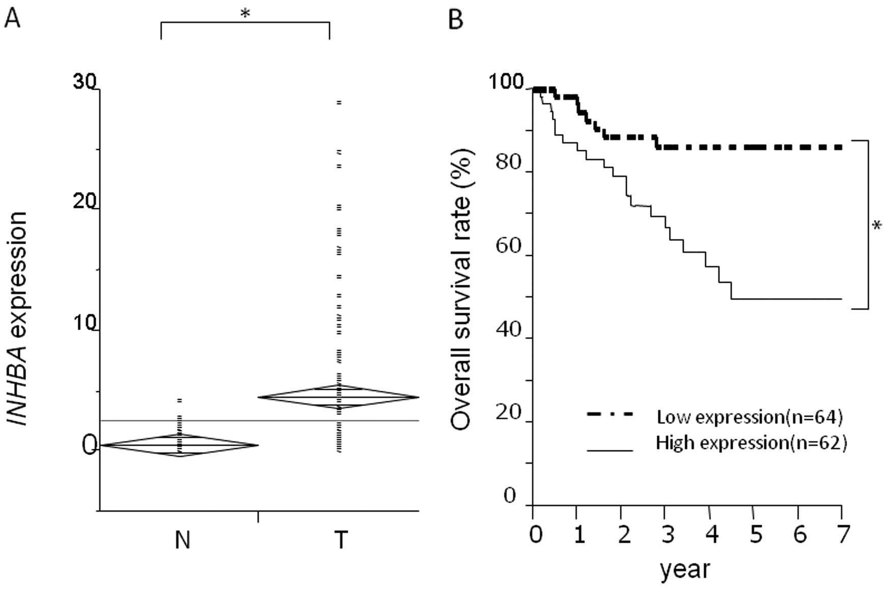
We performed quantitative real-time RT-PCR analysis
using primary CRC and adjacent noncancerous CRC tissues. RT-PCR of
126 paired clinical samples showed that 116 (92.1%) of these
samples exhibited higher INHBA mRNA levels in tumor tissues
than in paired normal tissues (Fig.
1A). INHBA expression was calculated as
INHBA/GAPDH expression. For the evaluation of
clinicopathological factors, the tissue samples were divided into 2
groups according to INHBA expression. Patients with tumors
that had a more than median INHBA expression were assigned
to the high expression group (n=63); the others were assigned to
the low expression group (n=63). Clinicopathological factors
related to the INHBA expression status of the 126 patients
are summarized in Table I. The data
indicated that INHBA expression was correlated with the tumor stage
(P<0.0001), lymph node metastasis (P<0.0001), lymphatic
invasion (P=0.0013), venous invasion (P<0.0001) and liver
metastasis (P=0.0024). Other factors were not significantly
correlated with INHBA expression.
 | Table IClinicopathological factors and
INHBA mRNA expression in 126 CRC patients. |
Table I
Clinicopathological factors and
INHBA mRNA expression in 126 CRC patients.
| INHBA/GAPDH
expression | |
|---|
|
| |
|---|
| Factors | High n=63, n (%) | Low n=63, n (%) | P-value |
|---|
| Age (years) | | | |
| ≤68 | 31 (49.2) | 30 (47.6) | 0.87 |
| >68 | 32 (50.8) | 33 (52.6) | |
| Gender | | | |
| Male | 41 (65.1) | 35 (55.6) | 0.26 |
| Female | 22 (34.9) | 28 (44.4) | |
| Histological
grade | | | |
| Well, mod | 58 (92.1) | 61 (96.8) | 0.22 |
| Others | 5 (7.9) | 2 (3.2) | |
| Tumor stage | | | |
| T0–T2 | 6 (9.5) | 29 (46.0) | <0.0001a |
| T3–T4 | 57 (90.5) | 34 (54.0) | |
| Lymph node
metastasis | | | |
| Absent | 21 (33.3) | 47 (74.6) | <0.0001a |
| Present | 42 (66.7) | 16 (25.4) | |
| Lymphatic
invasion | | | |
| Absent | 37 (58.7) | 44 (69.8) | 0.0013a |
| Present | 26 (41.3) | 19 (30.2) | |
| Venous
invasion | | | |
| Absent | 43 (68.3) | 60 (95.2) | <0.0001a |
| Present | 20 (31.7) | 3 (4.8) | |
| Liver
metastasis | | | |
| Absent | 49 (77.8) | 60 (95.2) | 0.0024a |
| Present | 14 (22.2) | 3 (4.8) | |
| Peritoneal
dissemination | | | |
| Absent | 59 (93.7) | 62 (98.4) | 0.15 |
| Present | 4 (6.3) | 1 (1.6) | |
Relationship between INHBA expression and
prognosis
The data showed that the overall survival rate was
significantly lower in the high expression group than in the low
expression group (P=0.0016) (Fig.
1B). The median follow-up period was 3.33±2.67 years. Table II shows the results of the
univariate and multivariate analyses of factors related to overall
survival. Univariate analysis showed that the following factors
were significantly related to overall survival: histological grade
(P=0.0139), tumor stage (P=0.0006), lymph node metastasis
(P<0.0001), lymphatic invasion (P<0.0001), venous invasion
(P=0.0011), liver metastasis (P<0.0001) and INHBA mRNA
expression (P=0.0007). Multivariate analysis indicated that
lymphatic invasion and liver metastasis were independent predictors
of overall survival. INHBA mRNA high expression was not an
independent predictor.
| Table IIUnivariate and multivariate analysis
of the clinicopathological factors affecting survival rate. |
Table II
Univariate and multivariate analysis
of the clinicopathological factors affecting survival rate.
| | Univariate
analysis | Multivariate
analysis |
|---|
| |
|
|
|---|
| Factors | No. of
patients | 5-year survival
rate (%) | P-value | Relative risk (95%
CI) | P-value |
|---|
| Age (years) |
| ≤68 | 60 | 76.5 | 0.180 | | |
| >68 | 66 | 61.8 | | | |
| Gender |
| Male | 75 | 67.4 | 0.389 | | |
| Female | 51 | 73.1 | | | |
| Histological
grade |
| Well, mod | 119 | 71.8 | 0.0139a | 1.75
(0.81–3.25) | 0.139 |
| Others | 7 | 33.3 | | | |
| Tumor stage |
| T0–T2 | 36 | 93.1 | 0.0006b | 1.26
(0.60–3.34) | 0.566 |
| T3–T4 | 90 | 58.9 | | | |
| Lymph node
metastasis |
| Absent | 69 | 85.9 | <0.0001b | 1.20
(0.75–2.03) | 0.461 |
| Present | 57 | 50.0 | | | |
| Lymphatic
invasion |
| Absent | 71 | 84.1 | <0.0001b | 2.23
(1.40–3.73) | 0.0006b |
| Present | 55 | 51.8 | | | |
| Venous
invasion |
| Absent | 103 | 77.5 | 0.0011b | 1.41
(0.91–2.11) | 0.112 |
| Present | 23 | 36.2 | | | |
| Liver
metastasis |
| Absent | 17 | 79.5 | <0.0001b | 2.56
(1.67–3.97) | 0.0000b |
| Present | 109 | 20.3 | | | |
| INHBA
expression |
| High | 63 | 49.9 | 0.0007b | 1.16
(0.73–1.92) | 0.546 |
| Low | 63 | 86.5 | | | |
Relationship between INHBA expression and
lymph node metastasis
Table III shows
the univariate and multivariate analyses of factors affecting lymph
node metastasis. Univariate analysis showed that the following
factors were significantly related to lymph node metastasis:
histological grade (P=0.0341), tumor stage (P=0.0028), lymphatic
invasion (P=0.0215) and INHBA mRNA high expression
(P=0.0074). Multivariate analysis indicated that inclusion in the
INHBA mRNA high expression group [relative risk (RR), 3.95;
95% confidence interval (CI), 1.71–9.35; P=0.0014] was an
independent predictor of lymph node metastasis, as was lymphatic
invasion (RR, 3.25; 95% CI, 1.39–7.72; P=0.0067).
| Table IIIResults of the univariate and
multivariate analysis of clinicopathological factors affecting
lymph node metastasis. |
Table III
Results of the univariate and
multivariate analysis of clinicopathological factors affecting
lymph node metastasis.
| Univariate
analysis | Multivariate
analysis |
|---|
|
|
|
|---|
| Factors | RR | 95% CI | P-value | RR | 95% CI | P-value |
|---|
| Age (years)
(≤68/>68) | 0.593 | 0.63–3.54 | 0.324 | - | - | - |
| Gender
(Male/female) | 0.571 | 0.172–1.65 | 0.322 | - | - | - |
| Histological grade
(Well, mod/others) | 5.62 | 1.02–28.2 | 0.0341a | 4.91 | 0.73–98.0 | 0.16 |
| Depth
(T0–T2/T3–T4) | 5.53 | 1.89–18.5 | 0.0028b | 2.25 | 0.93–5.50 | 0.072 |
| Lymphatic invasion
(Absent/present) | 3.68 | 1.27–12.3 | 0.0215a | 3.25 | 1.39–7.72 | 0.0067b |
| Venous invasion
(Absent/present) | 2.95 | 0.914–8.91 | 0.0585 | - | - | - |
| INHBA mRNA
expression (Low/high) | 5.93 | 1.81–26.8 | 0.0074b | 3.95 | 1.71–9.35 | 0.0014b |
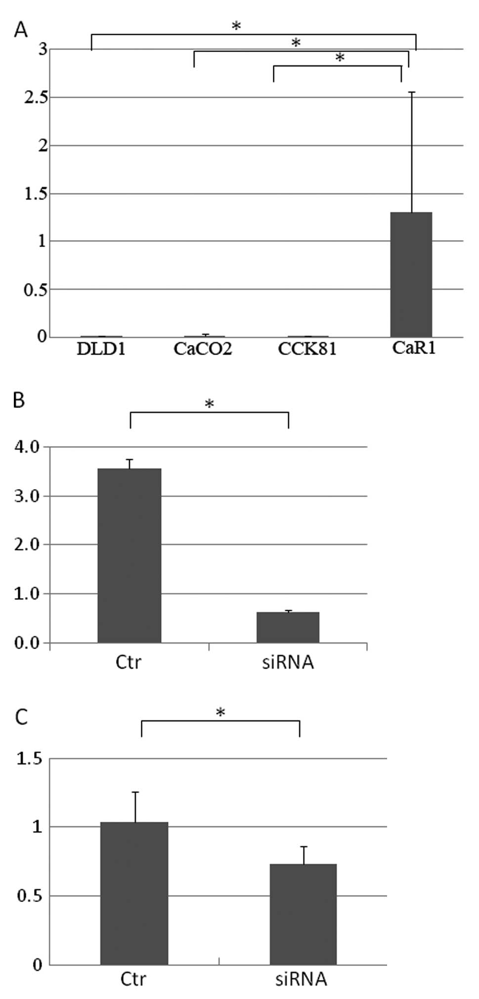
In vitro assessment of knockdown and
transfection of INHBA
The INHBA expression study indicated that
CaR1 cells had higher levels of expression than other CRC cell
lines such as DLD1, Caco-2 and CCK81 (Fig. 2A). We performed a knockdown
experiment of INHBA expression using the CaR1 cell line. After 48 h
of siRNA transfection, quantitative real-time RT-PCR was used to
confirm the reduction in INHBA expression due to siRNA
treatment (Fig. 2B). A
proliferation assay indicated that the knockdown resulted in a
reduction in the number of CaR1 cells at 72 h (P<0.05; Fig. 2C). We induced INHBA
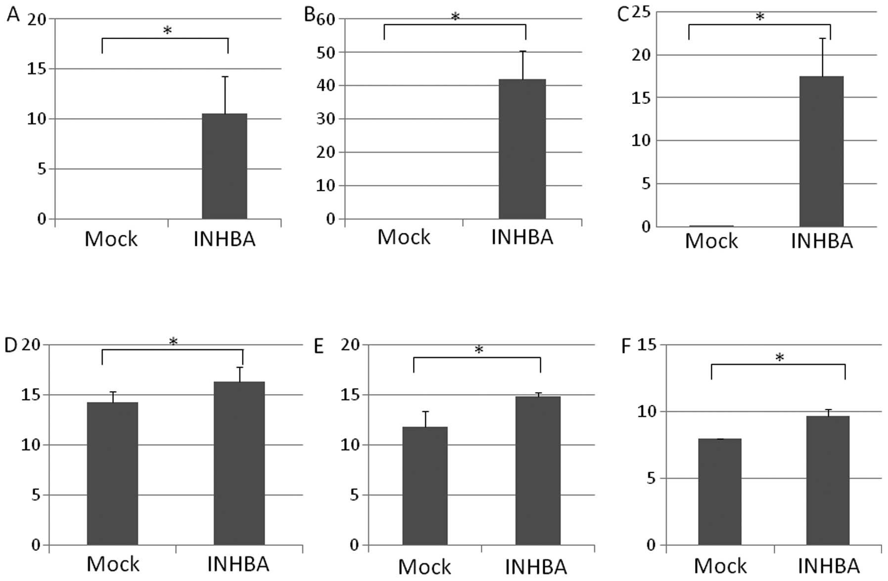
expression in the cell lines (DLD1, Caco-2 and CCK81) using a
plasmid technique, and quantitative real-time RT-PCR confirmed
successful induction in them (Fig.
3A–C). Proliferation assays indicated that high INHBA
expression increased the cell numbers of CRC cells, which have low
INHBA expression by default (P<0.05; Fig. 3D–F) at 48 h.
Discussion
INHBA is a subunit of both activin and inhibin, two
closely related glycoproteins with opposing biological effects,
which belong to the TGF-β superfamily (18–20).
The TGF-β superfamily comprises a structurally similar, although
functionally diverse group of proteins that play important roles in
embryonic development as well as in the functions of terminally
differentiated tissues. Activins play fundamental roles in cell
differentiation and development and are known to induce cellular
responses via activin receptors and the SMAD2/3 pathway, while
inhibits function to antagonize activins through either competition
of receptor binding or β-glycan.
In the present study, we determined that INHBA is
highly expressed in CRC tissues when compared with that in the
corresponding normal tissues. In addition, high INHBA expression in
CRC tissues was a predictor of poor prognosis when compared with
low INHBA expression. Clinicopathological factors related to the
INHBA expression status indicated that the high expression
group displayed a worse histological grade, higher tumor stage,
more lymph node metastasis, poorer lymphatic invasion, greater
vascular invasion and more extensive liver metastasis. Therefore,
INHBA expression was not an independent prognostic factor but may
be strongly related to one of the other prognostic factors of CRC.
Thus, this study of the association of high INHBA expression with
other prognostic factors indicated that high INHBA expression is an
independent prognostic factor for lymph node metastasis. Inclusion
in the INHBA mRNA high expression group was an independent
predictor of lymph node metastasis, as was lymphatic invasion. The
overexpression and knockdown experiments were performed in
vitro. These experiments showed that high INHBA
expression induced cell growth, whereas low INHBA expression
induced an opposite effect.
It is useful to determine the necessity for
intensive follow-up and adjuvant therapy for CRC by predicting
recurrence and metastases after curative surgical resection
(21–23). In the present study,
clinicopathological analysis revealed that patients who had CRCs
with high INHBA expression had a poor prognosis for overall
survival than those with low expression. The data indicated that
INHBA expression is presumably a novel predictor of CRC
prognosis. Several adjuvant chemotherapies are helpful at certain
disease stages, particularly in CRC (23–27).
For such cases, an informative prognostic marker, which is
independent of the traditional TNM classification, is extremely
important and can contribute to diagnosis and treatment. Adjuvant
chemotherapy for CRC is necessary for highly suspicious recurrent
cases. In such cases, analysis of INHBA expression may be
useful for predicting CRC prognosis, and INHBA is also
proposed to be a therapeutic target in treatment for patients with
poor prognosis.
References
|
1
|
Kohno SI, Luo C, Nawa A, et al: Oncolytic
virotherapy with an HSV amplicon vector expressing
granulocyte-macrophage colony-stimulating factor using the
replication-competent HSV type 1 mutant HF10 as a helper virus.
Cancer Gene Ther. 14:918–926. 2007. View Article : Google Scholar
|
|
2
|
Hermsen M, Postma C, Baak J, et al:
Colorectal adenoma to carcinoma progression follows multiple
pathways of chromosomal instability. Gastroenterology.
123:1109–1119. 2002. View Article : Google Scholar : PubMed/NCBI
|
|
3
|
Leslie A, Pratt NR, Gillespie K, et al:
Mutations of APC, K-ras, and p53 are associated with specific
chromosomal aberrations in colorectal adenocarcinomas. Cancer Res.
63:4656–4661. 2003.PubMed/NCBI
|
|
4
|
Gaddy-Kurten D, Tsuchida K and Vale W:
Activins and the receptor serine kinase superfamily. Recent Prog
Horm Res. 50:109–129. 1995.
|
|
5
|
Vale W, Rivier C, Hsueh A, et al: Chemical
and biological characterization of the inhibin family of protein
hormones. Recent Prog Horm Res. 44:1–34. 1988.PubMed/NCBI
|
|
6
|
Lorenzen JR, Channing CP and Schwartz NB:
Partial characterization of FSH suppressing activity
(folliculostatin) in porcine follicular fluid using the metestrous
rat as an in vivo bioassay model. Biol Reprod. 19:635–640. 1978.
View Article : Google Scholar : PubMed/NCBI
|
|
7
|
Asashima M, Ariizumi T and Malacinski GM:
In vitro control of organogenesis and body patterning by activin
during early amphibian development. Comp Biochem Physiol B Biochem
Mol Biol. 126:169–178. 2000. View Article : Google Scholar : PubMed/NCBI
|
|
8
|
Yoshinaga K, Mimori K, Yamashita K,
Utsunomiya T, Inoue H and Mori M: Clinical significance of the
expression of activin A in esophageal carcinoma. Int J Oncol.
22:75–80. 2003.PubMed/NCBI
|
|
9
|
Kleeff J, Ishiwata T, Friess H, Büchler MW
and Korc M: Concomitant over-expression of activin/inhibin β
subunits and their receptors in human pancreatic cancer. Int J
Cancer. 77:860–868. 1998.PubMed/NCBI
|
|
10
|
Thomas TZ, Wang H, Niclasen P, et al:
Expression and localization of activin subunits and follistatins in
tissues from men with high grade prostate cancer. J Clin Endocrinol
Metab. 82:3851–3858. 1997. View Article : Google Scholar : PubMed/NCBI
|
|
11
|
Woodruff TK: Role of inhibins and activins
in ovarian cancer. Cancer Treat Res. 107:293–302. 2002.PubMed/NCBI
|
|
12
|
Zheng W, Luo MP, Welt C, et al: Imbalanced
expression of inhibin and activin subunits in primary epithelial
ovarian cancer. Gynecol Oncol. 69:23–31. 1998. View Article : Google Scholar : PubMed/NCBI
|
|
13
|
Petraglia F, Florio P, Luisi S, et al:
Expression and secretion of inhibin and activin in normal and
neoplastic uterine tissues. High levels of serum activin A in women
with endometrial and cervical carcinoma. J Clin Endocrinol Metab.
83:1194–1200. 1998.PubMed/NCBI
|
|
14
|
Miyoshi N, Ishii H, Sekimoto M, Doki Y and
Mori M: RGS16 is a marker for prognosis in colorectal
cancer. Ann Surg Oncol. 16:3507–3514. 2009. View Article : Google Scholar
|
|
15
|
Sobin LH and Fleming ID: TNM
Classification of Malignant Tumors. 5th edition (1997). Union
Internationale Contre le Cancer and the American Joint Committee on
Cancer. Cancer. 80:1803–1804. 1997. View Article : Google Scholar : PubMed/NCBI
|
|
16
|
Mimori K, Mori M, Shiraishi T, et al:
Clinical significance of tissue inhibitor of metalloproteinase
expression in gastric carcinoma. Br J Cancer. 76:531–536. 1997.
View Article : Google Scholar : PubMed/NCBI
|
|
17
|
Mori M, Staniunas RJ, Barnard GF, et al:
The significance of carbonic anhydrase expression in human
colorectal cancer. Gastroenterology. 105:820–826. 1993.PubMed/NCBI
|
|
18
|
Burger HG and Igarashi M: Inhibin:
definition and nomenclature, including related substances.
Endocrinology. 122:1701–1702. 1988. View Article : Google Scholar : PubMed/NCBI
|
|
19
|
Murata M, Eto Y, Shibai H, Sakai M and
Muramatsu M: Erythroid differentiation factor is encoded by the
same mRNA as that of the inhibin βA chain. Proc
Natl Acad Sci USA. 85:2434–2438. 1988. View Article : Google Scholar : PubMed/NCBI
|
|
20
|
Brown CW, Houston-Hawkins DE, Woodruff TK
and Matzuk MM: Insertion of Inhbb into the Inhba locus rescues the
Inhba-null phenotype and reveals new activin functions. Nat Genet.
25:453–457. 2000. View
Article : Google Scholar : PubMed/NCBI
|
|
21
|
Wolpin BM and Mayer RJ: Systemic treatment
of colorectal cancer. Gastroenterology. 134:1296–1310. 2008.
View Article : Google Scholar : PubMed/NCBI
|
|
22
|
Kornmann M, Formentini A, Ette C, et al:
Prognostic factors influencing the survival of patients with colon
cancer receiving adjuvant 5-FU treatment. Eur J Surg Oncol.
34:1316–1321. 2008. View Article : Google Scholar : PubMed/NCBI
|
|
23
|
Bathe OF, Dowden S, Sutherland F, et al:
Phase II study of neoadjuvant 5-FU + leucovorin + CPT-11 in
patients with resectable liver metastases from colorectal
adenocarcinoma. BMC Cancer. 4:322004.
|
|
24
|
Lacy AM, Garcia-Valdecasas JC, Delgado S,
Castells A, Taurá P, Piqué JM and Visa J: Laparoscopy-assisted
colectomy versus open colectomy for treatment of non-metastatic
colon cancer: a randomised trial. Lancet. 359:2224–2229. 2002.
View Article : Google Scholar : PubMed/NCBI
|
|
25
|
Weeks JC, Nelson H, Gelber S, et al:
Short-term quality-of-life outcomes following laparoscopic-assisted
colectomy vs open colectomy for colon cancer: a randomized trial.
JAMA. 287:321–328. 2002. View Article : Google Scholar : PubMed/NCBI
|
|
26
|
Clinical Outcomes of Surgical Therapy
Study Group. A comparison of laparoscopically assisted and open
colectomy for colon cancer. N Engl J Med. 350:2050–2059. 2004.
View Article : Google Scholar
|
|
27
|
Jayne DG, Guillou PJ, Thorpe H, et al:
Randomized trial of laparoscopic-assisted resection of colorectal
carcinoma: 3-year results of the UK MRC CLASICC Trial Group. J Clin
Oncol. 25:3061–3068. 2007.PubMed/NCBI
|