Introduction
Hepatocellular carcinoma (HCC) is a major health
problem; it is the fifth most common type of cancer worldwide and
the third most common cause of cancer-related mortality (1–3). The
prognosis for untreated HCC is generally poor and the curative
treatments for this disease consist of surgical resection,
radiofrequency ablation and liver transplantation (1–3).
Non-curative therapies for HCC include transcatheter arterial
chemoembolization (TACE), transcatheter arterial chemotherapy
infusion (TACI), continuous arterial chemoinfusion therapy,
radioembolization, molecular targeting therapies such as sorafenib
and radiation therapy (1–12).
TACE is a procedure whereby an embolic agent is
injected into the tumor feeding artery to deprive it of its major
nutrient source by means of embolization; this results in ischemic
necrosis of the targeted tumor (11,12).
The survival benefit of TACE for unresectable HCC was established
in two randomized controlled trials (RCTs) and in one meta-analysis
(13–15). Thus, TACE plays an important role in
treating unresectable HCC. It is clearly defined as a first-line
therapy with an improved 2-year survival rate as compared with
conservative therapy (16).
The Barcelona Clinic Liver Cancer (BCLC)
classification is regarded as one of the most reliable staging and
treatment strategy staging systems for HCC as it considers liver
function, tumor status and performance status (PS) (16,17).
The BCLC intermediate stage (BCLC-B) includes Child-Pugh A and B
patients with multifocal HCC, defined as >3 tumors of any size
or 2–3 tumors with a maximal diameter >3 cm and a single HCC
(>5 cm) (24,36). To be categorized as
intermediate-stage HCC, patients should be asymptomatic and have
extrahepatic spread or no vascular invasion. The BCLC
classification indicates that these patients are optimal candidates
for TACE (16,17).
In general, chemotherapeutic agents such as
doxorubicin, epirubicin, cisplatin, mitomycin, 5-fluorouracil,
zinostatin stimalamer and miriplatin are combined in TACE for the
treatment of HCC; however, their treatment efficacy remains unclear
(18). In cases where TACE was
technically impossible due to anatomical reasons, a poor liver
functional reserve or cessation of blood flow in the tumor feeding
arteries recognized using lipiodol (iodine addition products of the
ethyl esters of fatty acids obtained from poppy seed oil)
chemolization alone, TACI was often performed in Japan. Indeed,
Takayasu et al(11) reported
in their large prospective Japanese nationwide study that out of
11,030 unresectable HCC patients who underwent transcatheter
arterial therapies as an initial treatment, 2,523 patients (22.9%)
were treated with TACI.
In our department, we have routinely performed TACE
or TACI using an epirubicin-mitomycin-lipiodol (EML) emulsion for
HCC when carrying out angiography (19,20).
Epirubicin alone or in combination with other chemotherapeutic
agents such as mitomycin has often been used in transcatheter
arterial chemotherapy for HCC in Asian countries including Japan
(18,21). However, to the best of our
knowledge, whether TACE using an EML emulsion could benefit
survival compared with TACI using an EML emulsion remains elusive.
The aim of the present study was, therefore, to compare clinical
outcomes between TACE and TACI, both using an EML emulsion, in
patients with intermediate-stage HCC.
Materials and methods
Patients
We performed TACE therapy as an initial treatment in
148 treatment-naive patients diagnosed with intermediate-stage HCC
in the Department of Gastroenterology and Hepatology, Osaka Red
Cross Hospital, Japan, between January 2004 and December 2012. Of
these patients, 145 were treated with TACE using an EML emulsion
and 3 were treated with TACE using a miriplatin-lipiodol emulsion.
During the same period, we performed TACI therapy as an initial
treatment in 82 treatment-naive patients diagnosed with
intermediate-stage HCC in our department. Of these patients, 81
were treated with TACI using an EML emulsion and 1 patient was
treated with TACI using a miriplatin-lipiodol emulsion. Thus, a
total of 226 patients with intermediate-stage HCC [the TACE group
(n=145) and the TACI group (n=81)] were analyzed in the present
study. Patients diagnosed with HCC rupture at initial therapy were
not included in the present study since they were treated with
transcatheter arterial embolization without chemolization alone. We
compared the overall survival (OS) and the treatment efficacy in
the two groups.
Written informed consent was obtained from all
patients prior to each therapy, and the study protocol complied
with all the provisions of the Declaration of Helsinki. The present
study was approved by the Ethics Committee of Osaka Red Cross
Hospital, Japan, and the need for written informed consent in the
present study was waived since the data were analyzed
retrospectively and anonymously. The present study comprised a
retrospective analysis of patient records registered in our
database and all treatments were conducted in an open-label
manner.
HCC diagnosis
HCC was diagnosed using abdominal ultrasound and
dynamic computed tomography (CT) scans (hyperattenuation during the
arterial phase in all or some part of the tumor and hypoattenuation
in the portal-venous phase) and/or magnetic resonance imaging
(MRI), based mainly on the recommendations of the American
Association for the Study of Liver Diseases (16). Arterial- and portal-phase dynamic CT
images were obtained at ~30 and 120 sec, respectively, after the
injection of the contrast material. When carrying out angiography,
we also confirmed intermediate-stage HCC using CT during hepatic
arteriography (CTHA) and arterial-portography (CTAP) (22,23).
TACE and TACI procedures
In our angiography room, a catheter was advanced to
the superior mesenteric artery and CTAP was performed to
investigate the site and the size of the HCCs. Furthermore, we
confirmed the patency of the portal vein at post-mesenteric
portography. Then, a catheter was advanced to the celiac artery and
a micocatheter was advanced to the common hepatic artery or proper
hepatic artery through a catheter. This approach was used to
perform CTHA and digital subtraction angiography with the purpose
of investigating tumor vascularity and identifying the feeding
vessels. After the completion of these procedures, a microcatheter
was advanced as close as possible to the feeding vessels of
targeted tumors. This was followed by an intra-arterial infusion
via the feeding arteries according to tumor size and liver function
of an emulsion containing epirubicin (Farmorubicin; Pfizer) at a
mean dose of 39.7±10.4 mg, mitomycin (Mitomycin C; Kyowa Hakko
Kirin Company, Ltd., Tokyo, Japan) at a mean dose of 9.1±3.2 mg and
lipiodol at a mean dose of 5.7±2.8 ml in the TACE group; in the
TACI group, the emulsion contained epirubicin at a mean dose of
37.2±9.9 mg, mitomycin at a mean dose of 9.0±2.7 mg and lipiodol at
a mean dose of 4.8±1.9 ml (19,20,24).
For patients treated with TACE after the infusion of an EML
emulsion, gelatin sponge particles were injected slowly into the
feeding arteries to prevent reflux into untreated segments. The
sites of injection of the embolizing agents were segmental or
subsegmental in all patients treated with TACE. When patients had
poor liver function, the dosages of the anticancer agents and
lipiodol were reduced. The decision as to whether TACE or TACI was
performed was mainly based on the recommendations of the attending
physicians, who considered tumor-related factors, vascular anatomy
and liver function. When selective catheterization of the tumor
feeding arteries was technically impossible, TACI was performed.
Additional embolization using gelatin sponge particles was not
performed in patients when cessation of the blood flow in tumor
feeding arteries was recognized using an infusion of an EML
emulsion alone.
Assessment of treatment efficacy
Treatment efficacy was evaluated using CT findings
within 2 months after the initial treatment. We regarded lipiodol
accumulation in targeted tumors seen on CT scans as indicating
necrosis. This was due to the fact that it had been previously
reported in several studies that the lipiodol retention areas
observed on CT corresponded to necrotic areas (25–27).
Complete response (CR) was defined as the disappearance of all
targeted tumors or 100% tumor necrosis, partial response (PR) was
defined as a ≥50% reduction in tumor size and/or necrosis, and
progressive disease (PD) was defined as >25% tumor enlargement
and/or the appearance of any new HCC tumors. Stable disease (SD)
was defined as disease that did not qualify for classification as
CR, PR or PD.
Follow-up
Follow-up after each therapy consisted of periodic
blood tests and monitoring of tumor markers, including
α-fetoprotein and des-γ-carboxy prothrombin (DCP). Dynamic CT scans
and/or MRI were obtained every 2–4 months after each therapy. Chest
CT, whole abdominal CT, brain MRI and bone scintigraphy were
performed when extrahepatic HCC recurrence was suspected. When
disease progression of the treated HCC lesions was observed after
the initial therapy and/or new hepatic lesions were observed, the
most appropriate therapies were performed if the liver functional
reserve was adequate and if patients did not refuse such therapies.
They included transcatheter arterial therapies in most cases.
However, when the treated lesion was well controlled after the
initial therapy and the new lesion appeared in the liver,
percutaneous ablative therapies were also considered. In cases that
were refractory to transcatheter arterial therapies or those
involving extrahepatic metastases, molecular targeting therapy such
as sorafenib was also considered.
Statistical analysis
Data were analyzed using univariate and multivariate
analyses. Continuous variables were compared using the unpaired
t-test and categorical variables were compared using Fisher’s exact
test. For analysis of OS, follow-up ended at the time of mortality
from any cause, and the remaining patients were censored at the
last follow-up visit. The cumulative OS rates were calculated using
the Kaplan-Meier method, and tested using the log-rank test.
Factors with a P-value <0.05 in univariate analysis were
subjected to multivariate analysis using the Cox proportional
hazards model. These statistical methods were used to estimate the
interval from initial treatment. Data were analyzed using SPSS
software (SPSS Inc., Chicago, IL, USA) for Microsoft Windows. Data
are expressed as the mean ± standard deviation. Values of P<0.05
were considered to indicate statistically significant
differences.
Results
Baseline characteristics
The baseline characteristics of the patients in the
two groups are shown in Table I.
The median observation period was 1.8 years (range, 0.2–9.0 years)
in the TACE group and 2.0 years (range, 0.2–8.7 years) in the TACI
group. The mean age in the TACE group (72.5±9.1 years) tended to be
higher (P=0.083) than that in the TACI group (70.3±9.3 years).
Maximum tumor diameter was significantly larger (P<0.001) in the
TACE group (5.4±3.0 cm) than in the TACI group (3.5±2.0 cm). The
proportion of patients with bilobar disease was significantly lower
(P=0.002) in the TACE group than in the TACI group. The prothrombin
time (PT) and platelet count were significantly higher in the TACE
group than in the TACI group. The proportion of patients with
Child-Pugh class A disease was significantly higher in the TACE
group than in the TACI group. These findings indicated that
patients in the TACE group had a superior liver functional reserve
to those in the TACI group.
 | Table IBaseline characteristics between the
TACE group and the TACI group. |
Table I
Baseline characteristics between the
TACE group and the TACI group.
| Variables | TACE group
(n=145) | TACI group
(n=81) | P-value |
|---|
| Age (years) | 72.5±9.1 | 70.3±9.3 | 0.083a |
| Gender,
male/female | 94/51 | 57/24 | 0.462b |
| Maximum tumor size
(cm) | 5.4±3.0 | 3.5±2.0 | <0.001a |
| Tumor number,
>5/≤5 | 31/114 | 23/58 | 0.257b |
| Tumor distribution,
bilobar/unilobar | 56/89 | 49/32 | 0.002b |
| Child-Pugh
classification, A/B | 100/45 | 46/35 | 0.082b |
| Causes of liver
disease |
| B/C/non B non C/B
and C | 12/93/39/1 | 7/56/18/0 | 0.843b |
| Efficacy of initial
treatment |
| CR/PR/SD/PD | 30/86/28/1 | 6/48/27/0 | 0.009b |
| AST (IU/l) | 61.8±32.5 | 67.9±48.0 | 0.260a |
| ALT (IU/l) | 48.9±34.3 | 54.7±48.5 | 0.294a |
| ALP (IU/l) | 417.3±221.0 | 430.6±183.0 | 0.646a |
| GGT (IU/l) | 140.1±202.1 | 120.8±138.4 | 0.446a |
| Serum albumin
(g/dl) | 3.63±0.52 | 3.61±0.56 | 0.793a |
| Total bilirubin
(mg/dl) | 1.07±0.85 | 1.19±0.73 | 0.273a |
| Prothrombin time
(%) | 84.2±18.2 | 78.2±16.5 | 0.015a |
| Platelets
(x104/mm3) | 13.8±7.2 | 11.3±5.4 | 0.006a |
| AFP (ng/ml) | 1310.0±3693.9 | 3692.5±16082.5 | 0.192a |
| DCP (mAU/ml) | 9071.8±34906.2 | 5009.4±27603.3 | 0.339a |
Median survival time and cumulative OS
rates
The median survival time (MST) and the 1-, 2-, 3-
and 5-year cumulative OS rates were 2.68 years and 81.5, 63.4, 43.9
and 32.7%, respectively in the TACE group, and 2.64 years and 85.0,
60.0, 43.2 and 26.0%, respectively in the TACI group; there was no
significant difference (P=0.691) in these parameters between the
two groups (Fig. 1).
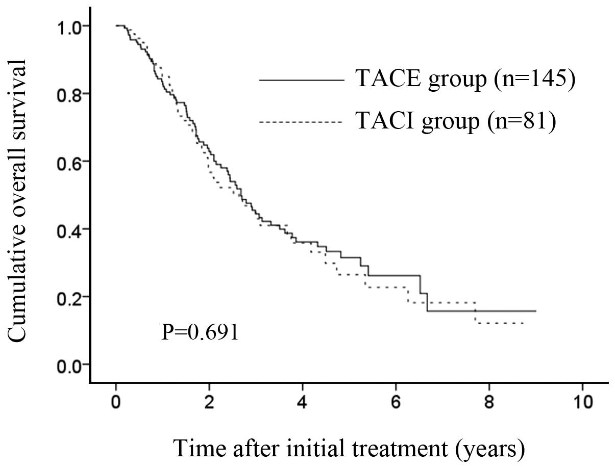 | Figure 1Median survival time (MST) and
cumulative overall survival (OS) for patients in the TACE and TACI
groups. The MST was 2.68 years in the TACE group and 2.64 years in
the TACI group. The 1-, 2-, 3- and 5-year cumulative OS rates were
81.5, 63.4, 43.9 and 32.7%, respectively, in the TACE group, and
85.0, 60.0, 43.2 and 26.0%, respectively, in the TACI group
(P=0.691). |
Treatment efficacy at initial treatment
in the two groups
In the TACE group, a CR was achieved in 30 patients,
a PR in 86 patients, SD in 28 patients and PD in one patient. Thus,
the objective response rate (ORR) in the TACE group was 80.0%
(116/145 patients). In the TACI group, a CR was achieved in 6
patients, a PR in 48 patients, SD in 27 patients; no patient had
PD. Thus, the ORR in the TACI group was 66.7% (54/81 patients). In
terms of treatment efficacy, the TACE group achieved significantly
improved treatment efficacy relative to the TACI group
(P=0.009).
Univariate and multivariate analyses of
factors contributing to OS
Univariate analysis identified the following factors
as being significantly associated with OS for all cases (n=226):
the Child-Pugh classification (P<0.001); tumor number ≤5
(P=0.011); tumor distribution (P=0.003); a maximum tumor size ≤4 cm
(P=0.005); a serum albumin level ≥3.7 g/dl (P=0.001); a total
bilirubin level ≥1.0 mg/dl, a PT >80% (P=0.044) and a DCP
>100 mAU/ml (P<0.001) (Table
II). The hazard ratios and 95% confidence intervals calculated
using multivariate analysis for the eight factors with P-values of
<0.05 in the univariate analysis are detailed in Table II. The Child-Pugh classification
(P=0.017); tumor number ≤5 (P=0.045) and DCP >100 mAU/ml
(P=0.002) were found to be significant predictors linked to OS in
multivariate analysis.
| Table IIUnivariate and multivariate analysis
contributing to overall survival. |
Table II
Univariate and multivariate analysis
contributing to overall survival.
| | | Multivariate
analysis |
|---|
| | |
|
|---|
| Variables | n | Univariate
analysis | Hazard ratio (95%
CI) | P-valuea |
|---|
| Gender, male vs.
female | 151/75 | 0.415 | | |
| Age (years), >70
vs. ≤70 | 123/103 | 0.113 | | |
| TACE vs. TACI | 145/81 | 0.691 | | |
| Child-Pugh, A vs.
B | 146/80 | <0.001 | 0.526
(0.310–0.892) | 0.017 |
| Tumor number, >5
vs. ≤5 | 54/172 | 0.011 | 0.631
(0.403–0.989) | 0.045 |
| Tumor distribution,
bilobar vs. unilobar | 105/121 | 0.003 | 1.125
(0.736–1.721) | 0.586 |
| Maximum tumor size,
>4 cm vs. ≤4 cm | 106/120 | 0.005 | 0.864
(0.583–1.279) | 0.465 |
| Objective response
at initial therapy, yes/no | 170/56 | 0.165 | | |
| AST (IU/l), >50
vs. ≤50 | 122/104 | 0.648 | | |
| ALT (IU/l), >40
vs. ≤40 | 116/110 | 0.158 | | |
| ALP (IU/l), >380
vs. ≤380 | 106/120 | 0.077 | | |
| GGT (IU/l), >80
vs. ≤80 | 101/125 | 0.381 | | |
| Serum albumin level
(g/dl), ≥3.7 vs. <3.7 | 113/113 | 0.001 | 1.260
(0.795–1.996) | 0.325 |
| Total bilirubin
(mg/dl), ≥1.0 vs. <1.0 | 101/125 | 0.023 | 0.888
(0.603–1.307) | 0.546 |
| Platelet count
(x104/mm3), >12 vs. ≤12 | 110/116 | 0.651 | | |
| Prothrombin time
(%), >80 vs. ≤80 | 125/101 | 0.044 | 0.935
(0.593–1.475) | 0.772 |
| Serum AFP (ng/ml),
>100 vs. ≤100 | 80/146 | 0.053 | | |
| DCP (mAU/ml),
>100 vs. ≤100 | 156/70 | <0.001 | 0.483
(0.304–0.765) | 0.002 |
Causes of mortality
Seventy-nine patients in the TACE group (54.5%) died
during the follow-up period. The causes of mortality were HCC
progression in 48 patients, liver failure in 23 patients and
miscellaneous causes in 8 patients. Fifty patients in the TACI
group (61.7%) died during the follow-up period and the causes of
mortality were HCC progression in 28 patients, liver failure in 15
patients and miscellaneous causes in 7 patients.
Adverse events in the two groups
In both groups, symptoms associated with
postembolization syndrome such as fever, appetite loss, abdominal
pain and nausea were transient and were mostly resolved within 2
weeks after initial treatment (28). In the TACE group, serious adverse
events (SAEs) were observed in 8 patients (5.5%). These 8 patients
had one of the following SAEs: acute respiratory distress syndrome
(ARDS), hepatic encephalopathy, cholangitis, hyponatremia,
hyperbilirubinemia, aspiration pneumonia, liver abscess and
refractory ascites. All SAEs were managed successfully, although in
1 patient who developed ARDS, management in the intensive care unit
was required. Thus, TACE-related mortality was 0%. In the TACI
group, SAEs were observed in 2 patients (2.5%) and included
gastrointestinal bleeding and liver abscess in 1 patient each.
These SAEs were successfully managed and TACI-related mortality was
also 0%.
Subgroup analysis according to the
Child-Pugh classification
Marginal significance was observed between the two
groups in terms of the Child-Pugh classification (P=0.082), and we,
therefore, performed subgroup analyses according to this
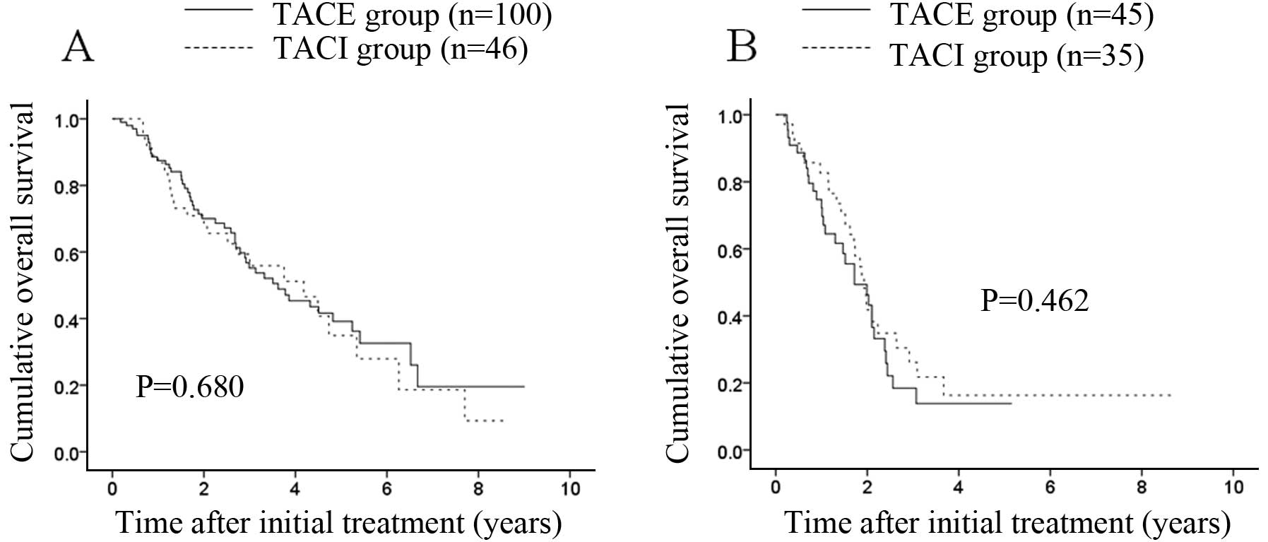
classification. No significant difference (P=0.680) was observed
between the two groups in terms of OS in patients with Child-Pugh
class A disease [100 patients (69.0%) in the TACE group and 45
patients (55.6%) in the TACI group]; the MST was 3.62 years in the
TACE group and 4.18 years in the TACI group (Fig. 2A). Similarly, no significant
difference (P=0.462) was found between the two groups in terms of
OS in patients with Child-Pugh class B disease [45 patients (31.0%)
in the TACE group and 36 (44.4%) in the TACI group]; the MST was
1.72 years in the TACE group and 1.93 years in the TACI group
(Fig. 2B).
Subgroup analysis according to maximum
tumor size
The maximum tumor size was significantly larger in
the TACE group than in the TACI group (P<0.001). Consequently,
we performed subgroup analyses according to maximum tumor size. No
significant difference (P=0.801) was observed between the two
groups in terms of OS in patients with a maximum tumor size >4
cm [82 patients (56.6%) in the TACE group and 24 (29.6%) in the
TACI group]; the MST was 2.15 years in the TACE group and 1.93
years in the TACI group (Fig. 3A).
Similarly, no significant difference was found between the two
groups in terms of OS (P=0.269) in patients with a maximum tumor
size ≤4 cm [63 patients (43.4%) in the TACE group and 57 (70.4%) in
the TACI group]; the MST was 3.51 years in the TACE group and 2.92
years in the TACI group (Fig.
2B).
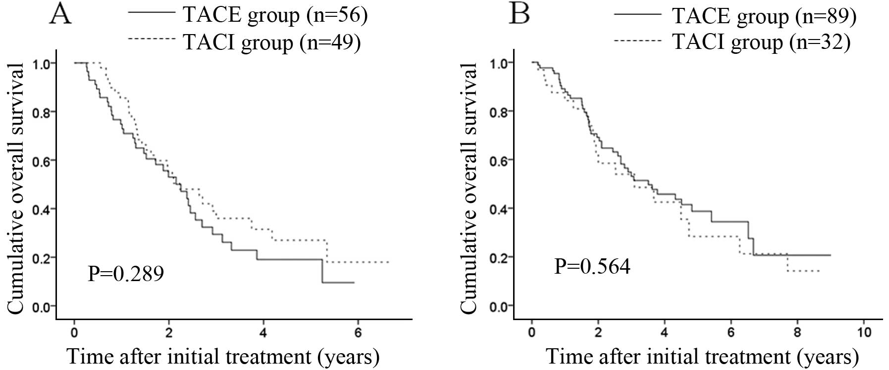
Subgroup analysis according to tumor
distribution
The proportion of patients with bilobar disease was
significantly lower in the TACE group than in the TACI group
(P=0.002). Hence, we performed subgroup analysis in terms of tumor
distribution. There were 56 (38.6%) patients with bilobar disease
in the TACE group and 49 (60.5%) in the TACI group. In terms of OS,
there was no significant difference (P=0.289) between the two
groups; the MST was 2.25 years in the TACE group and 2.23 years in
the TACI group (Fig. 4A). There
were 89 (61.4%) patients with unilobar disease in the TACE group
and 32 (39.5%) in the TACI group. In terms of OS, there was no
significant difference (P=0.564) between the two groups; the MST
was 3.51 years in the TACE group and 3.09 years in the TACI group
(Fig. 4B).
Discussion
Okusaka et al(29) conducted an RCT that compared
clinical outcomes between unresectable HCC patients treated with
TACE using zinostatin stimalamer (n=79) and those treated with TACI
using zinostatin stimalamer (n=82). They concluded that treatment
intensification involving the addition of embolization did not
improve survival over TACI with zinostatin stimalamer. Similarly,
Ikeda et al(30) carried out
a retrospective comparative study regarding clinical outcomes
between HCC patients treated with TACE using cisplatin suspended in
lipiodol (n=74) and patients treated with TACI using cisplatin
suspended in lipiodol (n=94). They reported that TACE using
cisplatin suspended in lipiodol had a higher treatment efficacy
than TACI using cisplatin suspended in lipiodol, but that it did
not significantly improve the survival of patients with HCC.
However, it remains unclear as to whether or not TACE using an EML
emulsion can deliver a survival benefit over TACI using an EML
emulsion; hence the reason for the present comparative study. To
the best of our knowledge, this is the first study to compare
clinical outcomes in intermediate-stage HCC patients using TACE and
TACI, both with an EML emulsion.
In the present study, the MST was 2.68 years for
patients in the TACE group and 2.64 years for patients in the TACI
group. Takayasu et al(12)
reported that the MST was 2.83 years in 8510 HCC patients who
underwent TACE. Our findings were similar to theirs, although their
study population analysis included patients with early-,
intermediate- and advanced-stage HCC (12). Here, in all cases and in all
subgroup analyses, the difference between the TACE and the TACI
group did not reach statistical significance in terms of OS. Our
results suggest that TACI using an EML emulsion could achieve a
comparable survival benefit to TACE using an EML emulsion, although
the treatment efficacy after initial therapy in the TACE group was
significantly higher than that in the TACI group.
Intermediate-stage HCC includes heterogeneous patient populations
with varying tumor size, tumor distribution, tumor number and liver
function (31). Thus, all patients
with intermediate-stage HCC might not derive a similar survival
benefit from TACE. In fact, in patients with Child-Pugh class B
disease, the MST in the TACI group was longer than that in the TACE
group in the present study. In patients where TACE was technically
impossible for anatomical reasons, or in patients with poor liver
function whose liver function was expected to deteriorate if TACE
was performed, TACI using an EML emulsion was an acceptable
alternative.
Epirubicin and doxorubicin are anthracycline-based
anticancer drugs (18). Both drugs
have been conventionally used in TACE or TACI for the treatment of
patients with HCC (18). Since
epirubicin can easily undergo glucuronidation, it is less toxic
than doxorubicin (18).
Furthermore, TACE or TACI using cisplatin may cause renal
dysfunction or a hypersensitivity reaction, and TACE or TACI using
zinostatin stimalamer causes severe vascular endothelial damage and
loss of the hepatic artery for infusion (18,32).
This is the reason why we have routinely used an epirubicin
containing regimen in transcatheter arterial chemotherapy for
HCC.
In our multivariate analysis, Child-Pugh
classification, tumor number and DCP value were revealed as being
significant predictors linked to OS. As mentioned earlier, Takayasu
et al(12) conducted a large
nationwide prospective cohort study in 8,610 HCC patients treated
with TACE. Their multivariate analysis revealed that the degree of
liver damage, tumor marker, maximum tumor size, tumor number and
portal vein invasion were significantly associated with OS
(12); our results were consistent
with those reported in that study. Preserving liver function in HCC
patients treated with transcatheter arterial therapies may be one
of the key factors for optimizing clinical outcome (33). However, it is of interest that
objective tumor response after initial therapy was not a
significant predictor linked to OS in the present study. Even if
treatment response after initial therapy was poor, performance of
the most appropriate therapy at disease progression may be
associated with favorable clinical outcomes.
In the present study, molecular targeted therapies
were performed (sorafenib was approved for its use in 2009 in
Japan) in 14 patients (9.7%) in the TACE group and in 3 patients
(3.7%) in the TACI group. In practice, the point at which
transcatheter arterial therapies should be replaced by targeted
molecular therapy in patients with HCC refractory to transcatheter
arterial therapies is a critical issue (34). We did not examine this issue in the
present study; future studies will therefore be required.
In this study, TACE or TACI-related mortality was 0%
in both groups. TACE-related mortality has been reported to range
from 0.5 to 7% (12,35,36).
Relative to these other studies, our transcatheter arterial therapy
procedure was safe.
The present study had several limitations. First, it
was a retrospective study. Second, the choice of TACE or TACI in
the treatment of HCC was mainly based on the decision of the
attending physicians, leading to bias. Third, patient
characteristics in the two groups were not well balanced for
analysis, also leading to bias. Future prospective studies with
well-balanced cohorts are, therefore, required to overcome these
limitations. However, our results demonstrated that patients in the
TACI group had a similar prognosis to patients in the TACE group.
In conclusion, TACI using an EML emulsion can be considered as one
of the therapeutic options for the treatment of intermediate-stage
HCC.
Acknowledgements
The authors thank Haruko Takada for the data
collection.
References
|
1
|
de Lope CR, Tremosini S, Forner A, Reig M
and Bruix J: Management of HCC. J Hepatol. 56(Suppl 1): S75–S87.
2012.
|
|
2
|
Livraghi T, Mäkisalo H and Line PD:
Treatment options in hepatocellular carcinoma today. Scand J Surg.
100:22–29. 2011.PubMed/NCBI
|
|
3
|
El-Serag HB: Epidemiology of viral
hepatitis and hepatocellular carcinoma. Gastroenterology.
142:1264–1273. 2012. View Article : Google Scholar : PubMed/NCBI
|
|
4
|
Llovet JM, Ricci S, Mazzaferro V, Hilgard
P, Gane E, Blanc JF, de Oliveira AC, Santoro A, Raoul JL, Forner A,
Schwartz M, Porta C, Zeuzem S, Bolondi L, Greten TF, Galle PR,
Seitz JF, Borbath I, Häussinger D, Giannaris T, Shan M, Moscovici
M, Voliotis D and Bruix J; SHARP Investigators Study Group.
Sorafenib in advanced hepatocellular carcinoma. N Engl J Med.
359:378–390. 2008. View Article : Google Scholar : PubMed/NCBI
|
|
5
|
Abou-Alfa GK, Schwartz L, Ricci S, Amadori
D, Santoro A, Figer A, De Greve J, Douillard JY, Lathia C, Schwartz
B, Taylor I, Moscovici M and Saltz LB: Phase II study of sorafenib
in patients with advanced hepatocellular carcinoma. J Clin Oncol.
24:4293–4300. 2006. View Article : Google Scholar : PubMed/NCBI
|
|
6
|
Cheng AL, Kang YK, Chen Z, Tsao CJ, Qin S,
Kim JS, Luo R, Feng J, Ye S, Yang TS, Xu J, Sun Y, Liang H, Liu J,
Wang J, Tak WY, Pan H, Burock K, Zou J, Voliotis D and Guan Z:
Efficacy and safety of sorafenib in patients in the Asia-Pacific
region with advanced hepatocellular carcinoma: a phase III
randomised, double-blind, placebo-controlled trial. Lancet Oncol.
10:25–34. 2009. View Article : Google Scholar : PubMed/NCBI
|
|
7
|
Wigg AJ, Palumbo K and Wigg DR:
Radiotherapy for hepatocellular carcinoma: systematic review of
radiobiology and modeling projections indicate reconsideration of
its use. J Gastroenterol Hepatol. 25:664–671. 2010. View Article : Google Scholar
|
|
8
|
Mazzaferro V, Sposito C, Bhoori S, Romito
R, Chiesa C, Morosi C, Maccauro M, Marchianò A, Bongini M, Lanocita
R, Civelli E, Bombardieri E, Camerini T and Spreafico C: Yttrium-90
radioembolization for intermediate-advanced hepatocellular
carcinoma: a phase 2 study. Hepatology. 57:1826–1837. 2013.
View Article : Google Scholar : PubMed/NCBI
|
|
9
|
Vogl TJ, Lammer J, Lencioni R, Malagari K,
Watkinson A, Pilleul F, Denys A and Lee C: Liver, gastrointestinal,
and cardiac toxicity in intermediate hepatocellular carcinoma
treated with PRECISION TACE with drug-eluting beads: results from
the PRECISION V randomized trial. AJR Am J Roentgenol.
197:W562–W570. 2011. View Article : Google Scholar
|
|
10
|
Ueshima K, Kudo M, Takita M, Nagai T,
Tatsumi C, Ueda T, Kitai S, Ishikawa E, Yada N, Inoue T, Hagiwara
S, Minami Y and Chung H: Hepatic arterial infusion chemotherapy
using low-dose 5-fluorouracil and cisplatin for advanced
hepatocellular carcinoma. Oncology. 78(Suppl 1): 148–153. 2010.
View Article : Google Scholar : PubMed/NCBI
|
|
11
|
Takayasu K, Arii S, Ikai I, Kudo M,
Matsuyama Y, Kojiro M and Makuuchi M; Liver Cancer Study Group of
Japan. Overall survival after transarterial lipiodol infusion
chemotherapy with or without embolization for unresectable
hepatocellular carcinoma: propensity score analysis. AJR Am J
Roentgenol. 194:830–837. 2010. View Article : Google Scholar
|
|
12
|
Takayasu K, Arii S, Ikai I, Omata M, Okita
K, Ichida T, Matsuyama Y, Nakanuma Y, Kojiro M and Makuuchi M:
Prospective cohort study of transarterial chemoembolization for
unresectable hepatocellular carcinoma in 8510 patients.
Gastroenterology. 131:461–469. 2006. View Article : Google Scholar : PubMed/NCBI
|
|
13
|
Llovet JM, Real MI, Montaña X, Planas R,
Coll S, Aponte J, Ayuso C, Sala M, Muchart J, Solà R, Rodés J and
Bruix J; Barcelona Liver Cancer Group. Arterial embolisation or
chemoembolisation versus symptomatic treatment in patients with
unresectable hepatocellular carcinoma: a randomised controlled
trial. Lancet. 359:1734–1739. 2002. View Article : Google Scholar
|
|
14
|
Lo CM, Ngan H, Tso WK, Liu CL, Lam CM,
Poon RT, Fan ST and Wong J: Randomized controlled trial of
transarterial lipiodol chemoembolization for unresectable
hepatocellular carcinoma. Hepatology. 35:1164–1171. 2002.
View Article : Google Scholar : PubMed/NCBI
|
|
15
|
Cammà C, Schepis F, Orlando A, Albanese M,
Shahied L, Trevisani F, Andreone P, Craxì A and Cottone M:
Transarterial chemoembolization for unresectable hepatocellular
carcinoma: meta-analysis of randomized controlled trials.
Radiology. 224:47–54. 2002.PubMed/NCBI
|
|
16
|
Bruix J and Sherman M; Practice Guidelines
Committee, American Association for the Study of Liver Diseases.
Management of hepatocellular carcinoma. Hepatology. 42:1208–1236.
2005. View Article : Google Scholar
|
|
17
|
Bruix J and Sherman M; American
Association for the Study of Liver Diseases. Management of
hepatocellular carcinoma: an update. Hepatology. 53:1020–1022.
2011. View Article : Google Scholar : PubMed/NCBI
|
|
18
|
Nishikawa H, Osaki Y, Kita R and Kimura T:
Hepatic arterial infusion chemotherapy for advanced hepatocellular
carcinoma in Japan. Cancers. 4:165–183. 2012. View Article : Google Scholar : PubMed/NCBI
|
|
19
|
Nishikawa H, Osaki Y, Kita R, Kimura T,
Inuzuka T, Takeda H, Nakajima J, Matsuda F, Sakamoto A, Henmi S,
Hatamaru K, Saito S and Nasu A: Transcatheter arterial infusion
chemotherapy prior to radiofrequency thermal ablation for single
hepatocellular carcinoma reduces the risk of intrahepatic distant
recurrence. Int J Oncol. 41:903–909. 2012.
|
|
20
|
Nishikawa H, Arimoto A, Wakasa T, Kita R,
Kimura T and Osaki Y: Effect of transcatheter arterial
chemoembolization prior to surgical resection for hepatocellular
carcinoma. Int J Oncol. 42:151–160. 2013.PubMed/NCBI
|
|
21
|
Intra-arterial administration of
epirubicin in the treatment of nonresectable hepatocellular
carcinoma. Epirubicin Study Group for Hepatocellular Carcinoma.
Cancer Chemother Pharmacol. 19:183–189. 1987.PubMed/NCBI
|
|
22
|
Nishikawa H, Inuzuka T, Takeda H, Nakajima
J, Sakamoto A, Henmi S, Matsuda F, Eso Y, Ishikawa T, Saito S, Kita
R, Kimura T and Osaki Y: Percutaneous radiofrequency ablation
therapy for hepatocellular carcinoma: a proposed new grading system
for the ablative margin and prediction of local tumor progression
and its validation. J Gastroenterol. 46:1418–1426. 2011. View Article : Google Scholar
|
|
23
|
Hayashi M, Matsui O, Ueda K, Kawamori Y,
Gabata T and Kadoya M: Progression to hypervascular hepatocellular
carcinoma: correlation with intranodular blood supply evaluated
with CT during intraarterial injection of contrast material.
Radiology. 225:143–149. 2002. View Article : Google Scholar
|
|
24
|
Satake M, Uchida H, Arai Y, Anai H,
Sakaguchi H, Nagata T, Yamane T, Kichikawa K, Osaki Y, Okazaki M,
Higashihara H, Nakamura H, Osuga K, Nakao N and Hirota S:
Transcatheter arterial chemoembolization (TACE) with lipiodol to
treat hepatocellular carcinoma: survey results from the TACE study
group of Japan. Cardiovasc Intervent Radiol. 31:756–761. 2008.
View Article : Google Scholar
|
|
25
|
Minami Y and Kudo M: Therapeutic response
assessment of transcatheter arterial chemoembolization for
hepatocellular carcinoma: ultrasonography, CT and MR imaging.
Oncology. 84(Suppl 1): 58–63. 2013. View Article : Google Scholar
|
|
26
|
Kudo M, Kubo S, Takayasu K, Sakamoto M,
Tanaka M, Ikai I, Furuse J, Nakamura K and Makuuchi M; Liver Cancer
Study Group of Japan (Committee for Response Evaluation Criteria in
Cancer of the Liver, Liver Cancer Study Group of Japan). Hepatol
Res. 40:686–692. 2010. View Article : Google Scholar
|
|
27
|
Imaeda T, Yamawaki Y, Seki M, Goto H,
Iinuma G, Kanematsu M, Mochizuki R, Doi H, Saji S and Shimokawa K:
Lipiodol retention and massive necrosis after
lipiodol-chemoembolization of hepatocellular carcinoma: correlation
between computed tomography and histopathology. Cardiovasc
Intervent Radiol. 16:209–213. 1993. View Article : Google Scholar
|
|
28
|
Jun CH, Ki HS, Lee HK, Park KJ, Park SY,
Cho SB, Park CH, Joo YE, Kim HS, Choi SK and Rew JS: Clinical
significance and risk factors of postembolization fever in patients
with hepatocellular carcinoma. World J Gastroenterol. 19:284–289.
2013. View Article : Google Scholar : PubMed/NCBI
|
|
29
|
Okusaka T, Kasugai H, Shioyama Y, Tanaka
K, Kudo M, Saisho H, Osaki Y, Sata M, Fujiyama S, Kumada T, Sato K,
Yamamoto S, Hinotsu S and Sato T: Transarterial chemotherapy alone
versus transarterial chemoembolization for hepatocellular
carcinoma: a randomized phase III trial. J Hepatol. 51:1030–1036.
2009. View Article : Google Scholar
|
|
30
|
Ikeda M, Maeda S, Shibata J, Muta R,
Ashihara H, Tanaka M, Fujiyama S and Tomita K: Transcatheter
arterial chemotherapy with and without embolization in patients
with hepatocellular carcinoma. Oncology. 66:24–31. 2004. View Article : Google Scholar : PubMed/NCBI
|
|
31
|
Raoul JL, Sangro B, Forner A, Mazzaferro
V, Piscaglia F, Bolondi L and Lencioni R: Evolving strategies for
the management of intermediate-stage hepatocellular carcinoma:
available evidence and expert opinion on the use of transarterial
chemoembolization. Cancer Treat Rev. 37:212–220. 2011. View Article : Google Scholar
|
|
32
|
Ishikawa T: Future perspectives on the
treatment of hepatocellular carcinoma with cisplatin. World J
Hepatol. 1:8–16. 2009. View Article : Google Scholar : PubMed/NCBI
|
|
33
|
Nishikawa H and Osaki Y: Clinical
significance of therapy using branched-chain amino acid granules in
patients with liver cirrhosis and hepatocellular carcinoma. Hepatol
Res. Jul 2–2013. View Article : Google Scholar : (Epub ahead of
print).
|
|
34
|
Ikeda M, Mitsunaga S, Shimizu S, Ohno I,
Takahashi H, Okuyama H, Kuwahara A, Kondo S, Morizane C, Ueno H,
Satake M, Arai Y and Okusaka T: Efficacy of sorafenib in patients
with hepatocellular carcinoma refractory to transcatheter arterial
chemoembolization. J Gastroenterol. Jun 23–2013.(Epub ahead of
print).
|
|
35
|
Poon RT, Fan ST, Lo CM, Liu CL, Ngan H, Ng
IO and Wong J: Hepatocellular carcinoma in the elderly: results of
surgical and nonsurgical management. Am J Gastroenterol.
94:2460–2466. 1999. View Article : Google Scholar : PubMed/NCBI
|
|
36
|
Yau T, Yao TJ, Chan P, Epstein RJ, Ng KK,
Chok SH, Cheung TT, Fan ST and Poon RT: The outcomes of elderly
patients with hepatocellular carcinoma treated with transarterial
chemoembolization. Cancer. 115:5507–5515. 2009. View Article : Google Scholar : PubMed/NCBI
|