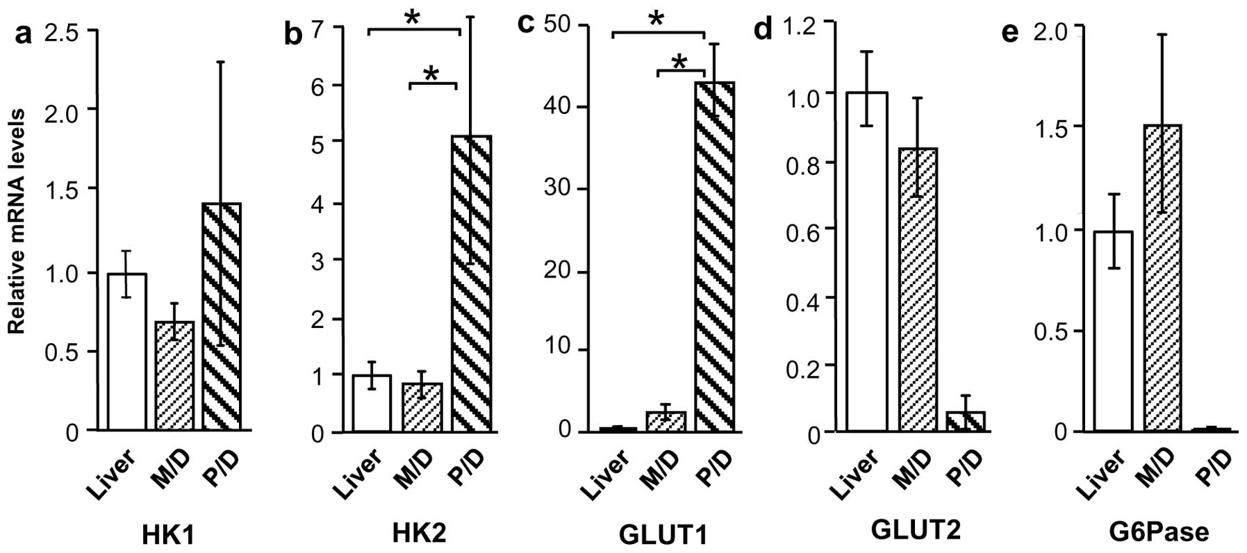
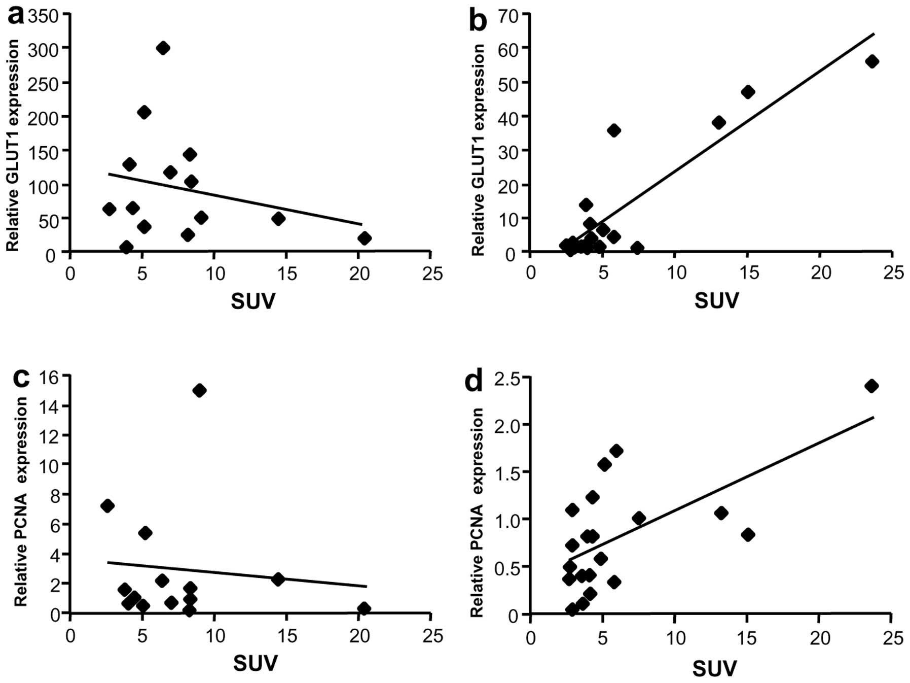
|
1
|
Fass L: Imaging and cancer. Mol Oncol.
2:115–152. 2008. View Article : Google Scholar
|
|
2
|
Mawlawi O and Townsend DW: Multimodality
imaging: an update on PET/CT technology. Eur J Nucl Med Mol
Imaging. 36(Suppl 1): 15–29. 2009. View Article : Google Scholar : PubMed/NCBI
|
|
3
|
Schaefer O and Langer M: Detection of
recurrent rectal cancer with CT, MRI and PET/CT. Eur Radiol.
17:2044–2054. 2007. View Article : Google Scholar : PubMed/NCBI
|
|
4
|
Sugiyama M, Sakahara H, Torizuka T, Kanno
T, Nakamura F, Futatsubashi M and Nakamura S: 18F-FDG PET in the
detection of extrahepatic metastases from hepatocellular carcinoma.
J Gastroenterol. 39:961–968. 2004. View Article : Google Scholar : PubMed/NCBI
|
|
5
|
Iwata Y, Shiomi S, Sasaki N, et al:
Clinical usefulness of positron emission tomography with
fluorine-18-fluorodeoxyglucose in the diagnosis of liver tumors.
Ann Nucl Med. 14:121–126. 2000. View Article : Google Scholar : PubMed/NCBI
|
|
6
|
Khan MA, Combs CS, Brunt EM, et al:
Positron emission tomography scanning in the evaluation of
hepatocellular carcinoma. J Hepatol. 32:792–797. 2000. View Article : Google Scholar : PubMed/NCBI
|
|
7
|
Trojan J, Schroeder O, Raedle J, et al:
Fluorine-18 FDG positron emission tomography for imaging of
hepatocellular carcinoma. Am J Gastroenterol. 94:3314–3319. 1999.
View Article : Google Scholar : PubMed/NCBI
|
|
8
|
Nagaoka S, Itano S, Ishibashi M, et al:
Value of fusing PET plus CT images in hepatocellular carcinoma and
combined hepatocellular and cholangiocarcinoma patients with
extrahepatic metastases: preliminary findings. Liver Int.
26:781–788. 2006. View Article : Google Scholar
|
|
9
|
Akiyoshi T, Oya M, Fujimoto Y, et al:
Comparison of preoperative whole-body positron emission tomography
with MDCT in patients with primary colorectal cancer. Colorectal
Dis. 11:464–469. 2009. View Article : Google Scholar : PubMed/NCBI
|
|
10
|
Seo S, Hatano E, Higashi T, et al:
Fluorine-18 fluorodeoxyglucose positron emission tomography
predicts tumor differentiation, P-glycoprotein expression, and
outcome after resection in hepatocellular carcinoma. Clin Cancer
Res. 13:427–433. 2007. View Article : Google Scholar
|
|
11
|
Lee JD, Yun M, Lee JM, et al: Analysis of
gene expression profiles of hepatocellular carcinomas with regard
to 18F-fluorodeoxyglucose uptake pattern on positron emission
tomography. Eur J Nucl Med Mol Imaging. 31:1621–1630. 2004.
View Article : Google Scholar : PubMed/NCBI
|
|
12
|
Hatano E, Ikai I, Higashi T, et al:
Preoperative positron emission tomography with
fluorine-18-fluorodeoxyglucose is predictive of prognosis in
patients with hepatocellular carcinoma after resection. World J
Surg. 30:1736–1741. 2006. View Article : Google Scholar : PubMed/NCBI
|
|
13
|
Byström P, Berglund A, Garske U, et al:
Early prediction of response to first-line chemotherapy by
sequential [18F]-2-fluoro-2-deoxy-D-glucose positron emission
tomography in patients with advanced colorectal cancer. Ann Oncol.
20:1057–1061. 2009.PubMed/NCBI
|
|
14
|
Chau I and Cunningham D: Treatment in
advanced colorectal cancer: what, when and how? Br J Cancer.
100:1704–1719. 2009. View Article : Google Scholar : PubMed/NCBI
|
|
15
|
de Langen AJ, van den Boogaart V,
Lubberink M, et al: Monitoring response to antiangiogenic therapy
in non-small cell lung cancer using imaging markers derived from
PET and dynamic contrast-enhanced MRI. J Nucl Med. 52:48–55.
2011.PubMed/NCBI
|
|
16
|
Lee JH, Park JY, Kim do Y, et al:
Prognostic value of 18F-FDG PET for hepatocellular carcinoma
patients treated with sorafenib. Liver Int. 31:1144–1149. 2011.
View Article : Google Scholar : PubMed/NCBI
|
|
17
|
Torizuka T, Tamaki N, Inokuma T, et al: In
vivo assessment of glucose metabolism in hepatocellular carcinoma
with FDG-PET. J Nucl Med. 36:1811–1817. 1995.PubMed/NCBI
|
|
18
|
Caracó C, Aloj L, Chen LY, Chou JY and
Eckelman WC: Cellular release of [18F]2-fluoro-2-deoxyglucose as a
function of the glucose-6-phosphatase enzyme system. J Biol Chem.
275:18489–18494. 2000.
|
|
19
|
Kameyama R, Yamamoto Y, Izuishi K, Sano T
and Nishiyama Y: Correlation of 18F-FLT uptake with equilibrative
nucleoside transporter-1 and thymidine kinase-1 expressions in
gastrointestinal cancer. Nucl Med Commun. 32:460–465. 2011.
View Article : Google Scholar : PubMed/NCBI
|
|
20
|
Izuishi K, Yamamoto Y, Sano T, et al:
Molecular mechanism underlying the detection of colorectal cancer
by 18F-2-fluoro-2-deoxy-d-glucose-positron emission tomography. J
Gastrointest Surg. 16:394–400. 2012. View Article : Google Scholar : PubMed/NCBI
|
|
21
|
Murakami K: FDG-PET for hepatobiliary and
pancreatic cancer: Advances and current limitations. World J Clin
Oncol. 2:229–236. 2011.PubMed/NCBI
|
|
22
|
Lin WY, Tsai SC and Hung GU: Value of
delayed 18F-FDG-PET imaging in the detection of hepatocellular
carcinoma. Nucl Med Commun. 26:315–321. 2005. View Article : Google Scholar : PubMed/NCBI
|
|
23
|
Zhao FQ and Keating AF: Functional
properties and genomics of glucose transporters. Curr Genomics.
8:113–128. 2007. View Article : Google Scholar : PubMed/NCBI
|
|
24
|
Paudyal B, Paudyal P, Oriuchi N, Tsushima
Y, Nakajima T and Endo K: Clinical implication of glucose transport
and metabolism evaluated by 18F-FDG PET in hepatocellular
carcinoma. Int J Oncol. 33:1047–1054. 2008.PubMed/NCBI
|
|
25
|
Roh MS, Jeong JS, Kim YH, Kim MC and Hong
SH: Diagnostic utility of GLUT1 in the differential diagnosis of
liver carcinomas. Hepatogastroenterology. 51:1315–1318.
2004.PubMed/NCBI
|
|
26
|
Wood IS and Trayhurn P: Glucose
transporters (GLUT and SGLT): expanded families of sugar transport
proteins. Br J Nutr. 89:3–9. 2003. View Article : Google Scholar : PubMed/NCBI
|
|
27
|
Brown GK: Glucose transporters: structure,
function and consequences of deficiency. J Inherit Metab Dis.
23:237–246. 2000. View Article : Google Scholar : PubMed/NCBI
|
|
28
|
Leturque A, Brot-Laroche E, Le Gall M,
Stolarczyk E and Tobin V: The role of GLUT2 in dietary sugar
handling. J Physiol Biochem. 61:529–537. 2005. View Article : Google Scholar : PubMed/NCBI
|
|
29
|
Ong LC, Jin Y, Song IC, Yu S, Zhang K and
Chow PK: 2-[18F]-2-deoxy-D-glucose (FDG) uptake in human tumor
cells is related to the expression of GLUT-1 and hexokinase II.
Acta Radiol. 49:1145–1153. 2008.
|
|
30
|
Paudyal B, Oriuchi N, Paudyal P, et al:
Clinicopathological presentation of varying 18F-FDG uptake and
expression of glucose transporter 1 and hexokinase II in cases of
hepatocellular carcinoma and cholangiocellular carcinoma. Ann Nucl
Med. 22:83–86. 2008. View Article : Google Scholar
|