Introduction
Hepatocellular carcinoma (HCC) is the sixth most
common solid tumor in humans worldwide and the third most common
cause of cancer-related death (1).
Chemotherapy is unsafe for many HCC patients, since most HCCs
develop on the basis of cirrhosis, and chronically damaged or
cirrhotic underlying livers poorly tolerate conventional
chemotherapy. There is thus the need for non-toxic, novel therapies
for HCC patients. Sorafenib is a multi-kinase inhibitor with high
efficacy against a variety of cancers as confirmed in preclinical
models (2). It suppresses cell
proliferation and induces apoptosis in HCC cell lines (3,4) and
has become the first approved drug for advanced HCC by the positive
results of clinical trials (5,6).
Sorafenib has been reported to inhibit experimental HCC cell growth
and angiogenesis by inhibiting Raf kinase as well as receptor
tyrosine kinases, such as VEGF and PDGF receptors (3,7).
Unlike traditional systemic chemotherapy, sorafenib has shown
survival benefits but only minimal tumor shrinkage (8). Moreover, substantial severe, although
rare, adverse events such as cardiac ischemia, hand-foot syndrome,
neutropenia and hypertension are associated with the use of this
drug. Thus, there is increasing interest in manipulating its
actions to reduce its toxicity, as well as in examining the effects
of this drug in combination with other agents (9).
All-trans retinoic acid, an analog of vitamin
A, is currently being extensively studied for its potential as a
therapeutic and chemopreventive agent since it has been used
successfully in the treatment of acute promyelocytic leukemia (APL)
(10) and other hematologic
diseases (11,12). It induces cellular differentiation
of numerous malignant tumors and inhibits their growth (13). Notably, epidemiological evidence
indicates that low levels of serum retinol are correlated with HCC
risk (14,15), suggesting a potential role of
retinoids in the chemoprevention of this cancer. Consistent with
this view, in vitro and in vivo preclinical evidence
indicated that ATRA was able to inhibit proliferation and induce
apoptosis in human hepatoma cells (Hep3B) (16) and suppressed tumorigenicity in a
nude mouse model (17). The
antiproliferative, differentiation and/or apoptotic effects of ATRA
on rat hepatocytes (18) or
HepG2 cells (19,20),
have also been demonstrated. The precise mechanism through which
ATRA exerts its chemopreventive effect remains controversial.
Several studies have indicated that the antitumor effects of ATRA
in various types of cancers are associated with its ability to
restore gap junction function of otherwise gap junctional
communication-impaired tumor cells (21,22).
Gap junctions, composed of connexins (Cxs), connect
the cytoplasm of neighboring cells, thereby mediating the direct
exchange of cytoplasmic signaling molecules smaller than 1 kDa,
such as secondary message Ca2+, cyclic adenosine
monophosphate (cAMP) and inositol triphosphate (IP3)
between adjacent cells. This process of exchange of molecules
between neighboring cells via gap junctions is termed gap
junctional intercellular communication (GJIC). GJIC has been
implicated in many cellular functions, including cancer biology and
chemotherapy (23,24). Emerging evidence indicates a
GJIC-dependent enhancing effect on the toxicity of various
chemotherapeutic agents in tumor cells (25–27).
In this context, an intercellular diffusion of toxic/apoptotic
signals through gap junction channels is considered to be involved.
The degree of the additive bystander toxicity generally correlates
with the level of GJIC, although it is present at low levels or
even completely absent in tumor cells (28,29).
Therefore, increasing gap junction activity in tumor cells provides
a new target by which to enhance antineoplastic therapies.
In view of these observations, there has been
considerable interest in using the combination of sorafenib and
ATRA for the prevention and treatment of HCC. The aim of this study
was to investigate whether the combination of sorafenib plus ATRA
exerts more pronounced growth-inhibitory effects on human HCC cells
and to examine possible mechanisms for such an effect,
predominantly focusing on the modulation of GJIC by the combination
of these two agents.
Materials and methods
Materials
Sorafenib was obtained from the Bayer Corp. (West
Haven, CT, USA). All-trans retinoic acid (ATRA),
18-α-glycyrrhetinic acid (18-α-GA), oleamide, dimethyl sulfoxide
(DMSO), 3-(4,5-dimethylthiazol-2-yl)-2,5-diphenyltetrazolium
bromide (MTT), anti-Cx32 and anti-Cx43 mouse IgG were from Sigma
(St. Louis, MO, USA). Dulbecco’s modified Eagle’s medium (DMEM),
fetal bovine serum, TRIzol, cell labeling dyes CM-DiI and
calcein-AM (acetoxymethyl ester), anti-Cx26 mouse IgG and
fluorescein isothiocyanate (FITC)-anti-mouse IgG were from
Invitrogen (Carlsbad, CA, USA). Secondary antibodies for western
blotting were from Amersham Biosciences Corp. (Piscataway, NJ,
USA). All other reagents were from Sigma unless stated
otherwise.
Cell lines and cell culture
Human HCC cell line HepG2 was obtained
from the American Type Culture Collection (Manassas, VA, USA), and
SMMC-7721 was purchased from the cell bank of the Shanghai
Institutes for Biological Sciences. Both cell lines were grown at
37°C in a humidified atmosphere containing 5% (v/v) CO2
in DMEM supplemented with 10% fetal bovine serum, and 100 U/ml
streptomycin and 100 mg/ml penicillin.
Drug treatment
Sorafenib and ATRA were dissolved in DMSO to a stock
concentration of 10 and 50 mM, respectively. A stock concentration
of 18-α-GA (10 mM) or oleamide (25 mM) was also diluted in DMSO and
stored at −20°C. Just before each experiment, aliquots were thawed
and diluted to the desirable concentration with DMEM. The final
concentration of DMSO as solvent was always <0.1%.
MTT assay
HepG2 (2×10⁴ cells/well) and
SMMC-7721 (8×10³ cells/well) cells were seeded into
96-well plates for 1 day, and then exposed to sorafenib and ATRA
either alone or in combination at the indicated concentrations for
48 h. Cells incubated with DMSO at the same concentration (always
less than 0.1% v/v) were used as a control. MTT (5 mg/ml in PBS)
was then added to each well, and the dishes were incubated at 37°C
for 4 h, and the medium containing MTT was then removed. The
formazan crystals in the viable cells were solubilized with 100 μl
DMSO, and the absorbance at 490 nm of each well was read using a
microplate enzyme-linked immunosorbent assay (ELISA) reader (MRX
II; Dynex Technologies, Chantilly, VA, USA). All experiments were
performed at least three times, with five wells for each
concentration of the tested compounds (n=5 per experiment). The
cell viability was calculated as follows: (OD of experimental group
- OD of blank group) / (OD of control group - OD of blank
group).
Hoechst 33258 staining
The staining method was carried out according to the
method recommended by the manufacturer. In brief, after treatment
for 48 h, HepG2 and SMMC-7721 cells cultured in 6-well
plates were washed with PBS and fixed with 4% paraformaldehyde for
30 min at room temperature. Fixed cells were washed with PBS,
stained with Hoechst 33258 (Sigma, 5 μg/ml) for 30 min at room
temperature in the dark, and the apoptotic cells were identified by
condensation and fragmentation of nuclei as examined by a
fluorescence microscope (Olympus, Tokyo, Japan). The apoptotic rate
of the cell population was calculated as the ratio of apoptotic
cells to total cells counted ×100. A minimum of 500 cells were
counted for each treatment.
Early stage of cell apoptosis by flow
cytometric analysis
Early stage of cell apoptosis was measured by
Annexin V-FITC and propidium iodide (PI) (BD Biosciences Clontech,
USA) labeling technique and flow cytometric analyses. Cells were
plated onto 12-well plates and grown to confluence. They were then
treated in the absence (vehicle control) or presence of sorafenib
(10 μM), ATRA (10 μM for HepG2 cells and 0.1 μM for SMMC-7721
cells), or sorafenib + ATRA for 8 h. Cells were harvested, washed
with cold PBS and then resuspended in 100 μl binding buffer
containing 5 μl Annexin V-FITC and 10 μl of PI. After an incubation
time of 10 min at room temperature in the dark, stained cells were
analyzed by flow cytometry. Unstained and single stained controls
were included in each experiment. This assay was carried out in
triplicate.
Determination of GJIC: ‘parachute’
dye-coupling assay
GJIC was determined by a ‘parachute’ technique as we
previously described (27,30). Briefly, cells were cultured in
12-well plates to 80–85% confluence. Donor cells from one well were
incubated with a freshly made solution of 5 μM calcein-AM and 2.5
μM CM-DiI in growth medium for 30 min at 37°C. CM-DiI is a
nontransferable membrane dye that does not spread to coupled cells,
while calcein-AM is converted intracellularly into the gap
junction-permeable dye calcein. Unincorporated dye was removed by
three consecutive washes with culture medium. The donor cells were
then trypsinized and seeded onto the receiver cells at a 1:150
donor/receiver ratio. The donor cells were allowed to attach to the
monolayer of receiver cells and form gap junctions for 4 h at 37°C,
and then examined with a fluorescence microscope (Olympus). The
average number of receiver cells containing calcein per donor cell
was considered as a measure of the degree of GJIC.
Western blotting
Cells were washed three times with cold PBS and
lysed in lysis buffer (Tris-HCl pH 7.4 20 mM, NaCl 150 mM, EDTA 1
mM, EGTA 1 mM, Triton 1%, sodium pyrophosphate 2.5 mM,
Na3VO4 1 mM, β-glycerophosphate 1 mM,
protease inhibitors 1:1,000), followed by a brief sonication. The
suspension was then centrifuged at 12,000 rpm for 30 min at 4°C,
and proteins from the supernatant were extracted. Protein
determination was performed using a DC protein assay kit (Bio-Rad
Chemical Co.). Samples (25 μg) from cells were applied to
SDS-polyacrylamide gels of 10% (w/v) acrylamide, followed by
electrophoresis and blotting. Membranes were blocked in 5% nonfat
milk for 30 min at room temperature and probed with appropriate
antibodies at the dilution recommended by the suppliers. The
immunoreactive bands were visualized using an enhanced
chemiluminescence detection kit (Amersham, Aylesbury, UK).
Analysis of immunofluorescence
Cells were seeded onto sterile slides with
coverslips in 24-well plates and the indicated treatments were
carried out. The cells were then briefly washed three times with
PBS, and fixed with 0.1% Triton X-100–4% paraformaldehyde for 30
min. Coverslips were blocked with 2% bovine serum albumin (BSA) in
PBS and probed with anti-Cx32 (1:100) or anti-Cx43 (1:200), the
primary antibodies diluted in 2% BSA in PBS overnight at 4°C. Cells
were washed, followed by the addition of FITC anti-mouse IgG at a
1:200 dilution in 2% BSA in PBS for 2 h in the dark at room
temperature. Nuclear staining was performed with DAPI at 37°C for 5
min. After rinsing, the coverslips were mounted on slides, and the
cells were examined under a fluorescence microscope (Olympus). Six
to eight randomly selected fields were examined in each of three
separate experiments.
RNA isolation and reverse
transcriptase-polymerase chain reaction (RT-PCR)
Total RNA was extracted using Trizol reagent
according to the manufacturer’s instructions. Complementary DNA
(cDNA) was synthesized from 1 μg RNA using the standard procedure
with avian myeloblastosis virus reverse transcriptase (Promega) to
generate 20 μl of cDNA at 42°C for 60 min. For polymerase chain
reaction (PCR) quantification, 2 μl of cDNA reaction was amplified
in a 20 μl standard PCR reaction. PCR was initiated at 94°C for 3
min followed by 30 cycles consisting of 45 sec at 94°C, 45 sec at
58°C, and 45 sec at 72°C, with the final cycle extended to 10 min
at 72°C, followed by termination at 4°C. The following primers were
used: for human Cx32, forward primer 5′-TCC CTGCAGCTCATCCTAGT-3′
and reverse primer 5′-CCC TGAGATGTGGACCTTGT-3′, product size 156
bp; for human Cx43, forward primer 5′-AGGAGTTCAATCACTTGGCG-3′ and
reverse primer 5′-GCAGGATTCGGAAAATGAAA-3′, product size 168 bp; for
human β-actin, forward primer 5′-TCCTCCTGAGCGCAAGTACTC-3′ and
reverse primer 5′-GCATTTGCGGTGGACGAT-3′, product size 130 bp. The
detection of β-actin transcripts provided an internal control in
PCR, standardizing the quantity of input cDNA. PCR products were
analyzed on an ethidium bromide-stained 1.5% agarose gel.
Statistical analysis
The data are represented as means ± SEM for the
number of individual experiment specified in each figure legend.
All statistical analyses used SigmaPlot 10.0 software (Jandel
Scientific, San Rafael, CA, USA). Comparison of numerical data was
achieved with the unpaired Student’s t-test; differences with
p<0.05 were considered significant.
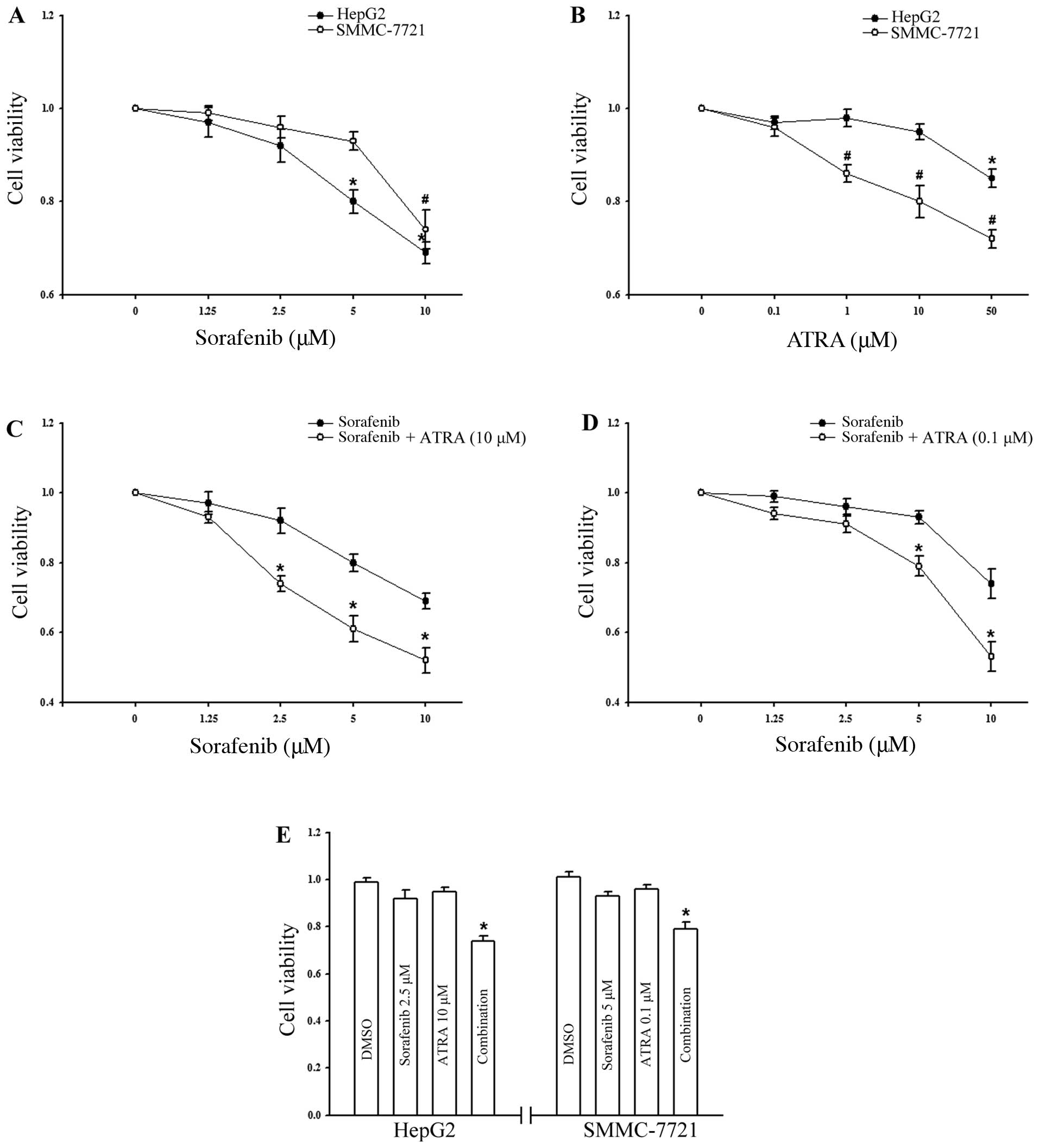
Results
Inhibition of HCC cell growth by
sorafenib plus ATRA
To assess the growth-inhibitory effects of combining
sorafenib with ATRA, HepG2 human HCC cells, which are
frequently used in evaluations of the effect of sorafenib, were
treated with the tested agents, either individually or in
combination, and were then examined by MTT assay. The results shown
in Fig. 1A and B demonstrated that
neither sorafenib nor ATRA alone had a significant effect on
HepG2 cell viability at the tested concentrations
(sorafenib up to 2.5 μM; ATRA up to 10 μM). We then focused on the
effect of ATRA at a non-toxic concentration on sorafenib-induced
cell inhibition. Fig. 1C shows the
data for the combination of varying doses of sorafenib and a fixed
dose of ATRA, demonstrating that the growth inhibitory activity of
each treatment was enhanced by the addition of ATRA (p<0.05).
Similar results were also observed for the human HCC cell line
SMMC-7721 of Asian origin (Fig.
1D); the response to sorafenib was less sensitive while the
response to ATRA was markedly sensitive (Fig. 1A and B). It is noteworthy that in
both cell lines, the combination of these two agents both at
non-toxic concentrations significantly inhibited cell growth when
compared to the effect of each single agent treatment (p<0.05,
Fig. 1E).
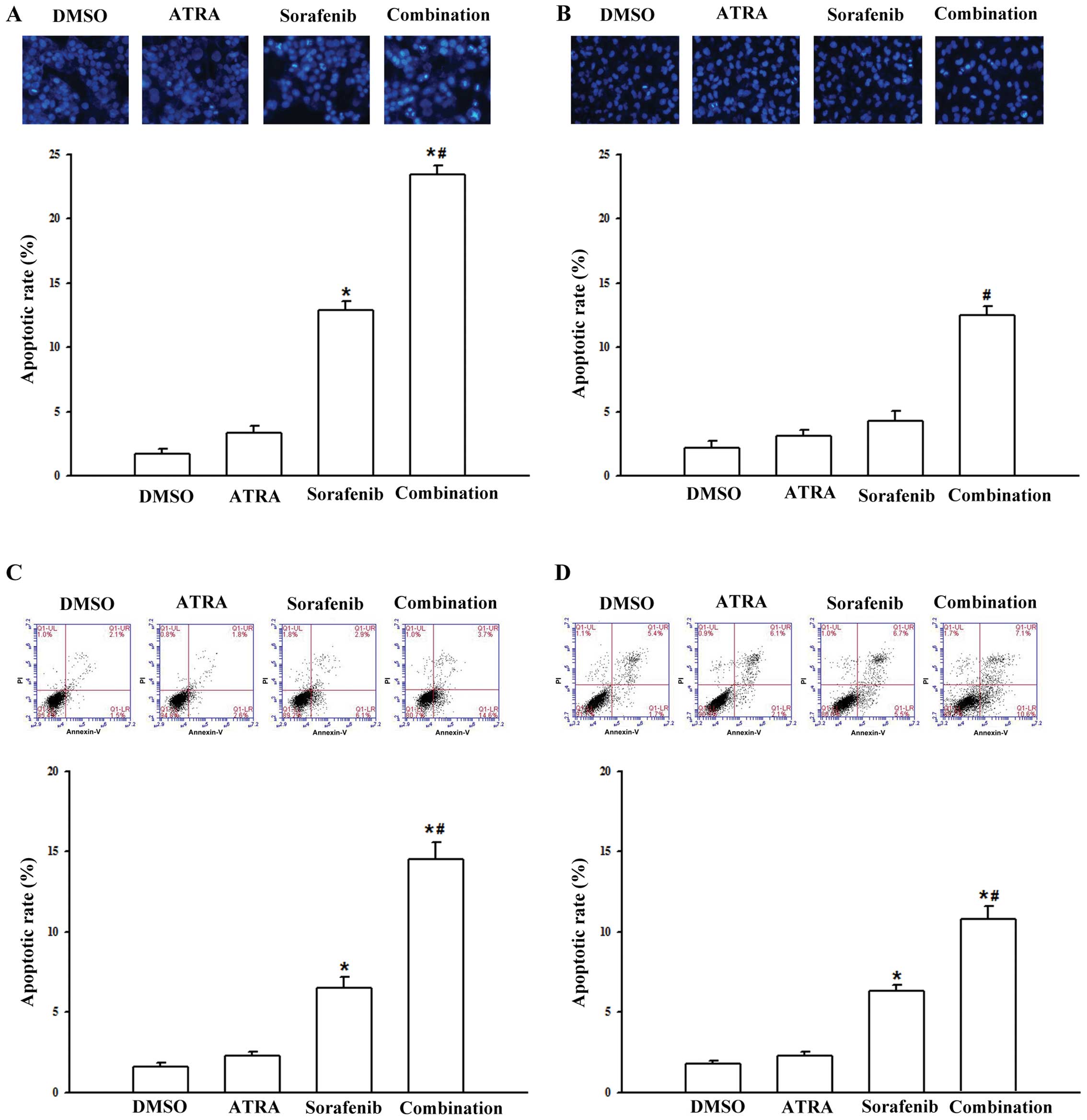
Induction of apoptosis by sorafenib plus
ATRA
We next investigated the effect of the combination
treatment of sorafenib plus ATRA on cell apoptosis. Morphological
change showing the presence of late apoptosis was assessed by
Hoechest 33258 staining. Data showed that treatment with sorafenib
alone, at a low concentration (5 μM) for 48 h was sufficient to
trigger slight apoptosis in the HepG2 cell line
(12.9±0.6%), but led to no significant effect on SMMC-7721 cells.
However, simultaneous exposure to sorafenib and ATRA, at its
maximum non-toxic concentrations, respectively (10 μM for HepG2
cells and 0.1 μM for SMMC-7721 cells), resulted in a pronounced
increase in apoptosis (Fig. 2A and
B). Annexin V/PI double staining followed by flow cytometric
analysis, which provides more sensitivity and precision, was
conducted to detect the early stage of cell apoptosis. As shown in
Fig. 2C and D, to yield a modest
toxic effect, sorafenib, at a high concentration of 10 μM, was
used. Compared with either agent alone, the apoptotic rate was
substantially greater following the treatment of the cells with
sorafenib and ATRA concomitantly for a short treatment time (8 h).
These highly concordant results indicated that ATRA synergistically
potentiated sorafenib to induce apoptosis in both cell lines.
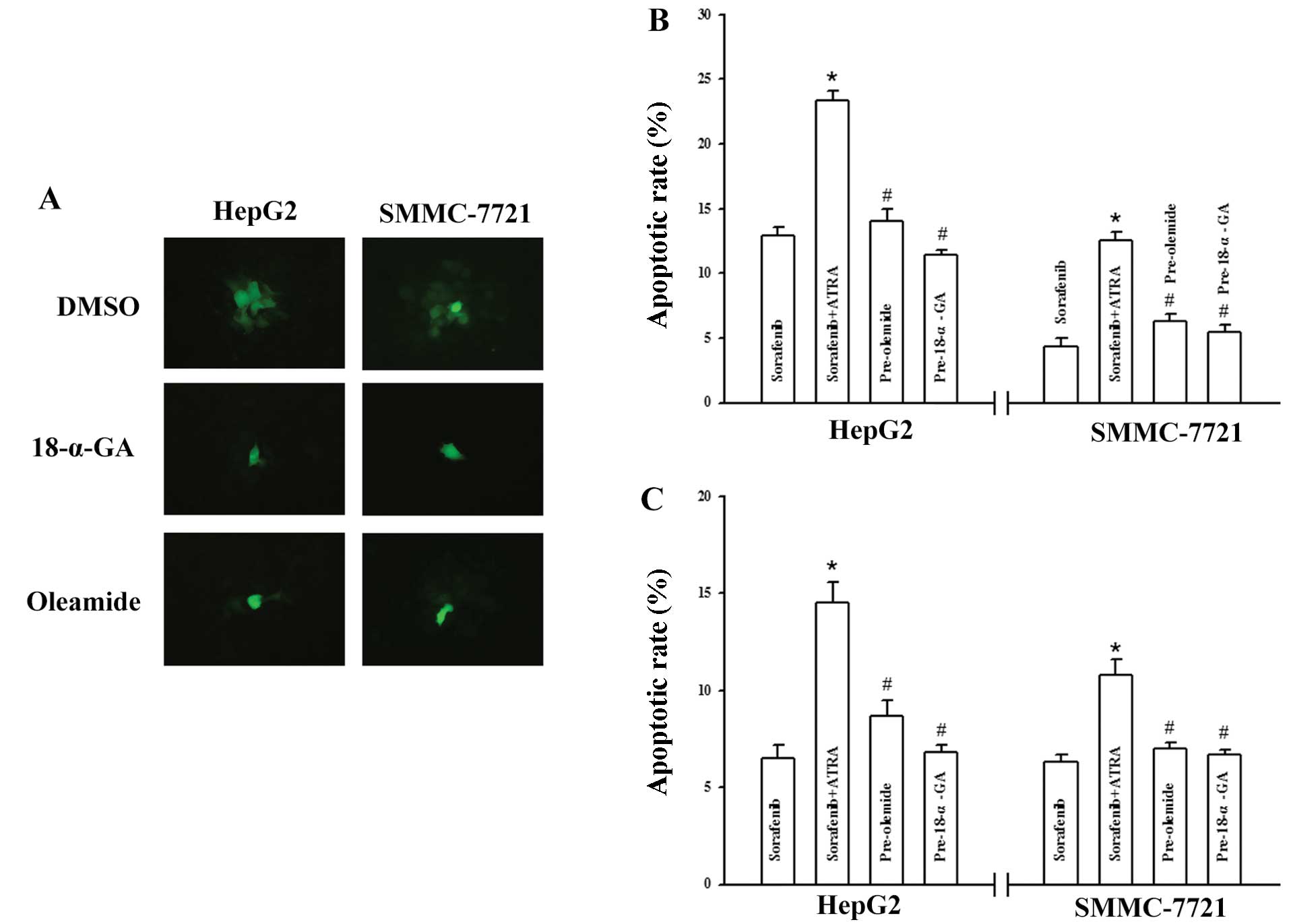
Involvement of GJIC in sorafenib plus
ATRA-mediated apoptosis
GJIC plays a critical role in cancer therapy and
previous findings suggest that ATRA augments gap junction function
in a variety of normal and malignant cell types (21,22,31);
therefore, we aimed to ascertain whether the mechanism involved in
sorafenib plus ATRA-mediated apoptosis is related to alteration of
GJIC. Although few drugs have been identified as highly potent and
selective gap junction channel-blocking molecules (32), two extensively used drugs as GJIC
blockers, 18-α-GA (33,34) and oleamide (35,36),
serve as a powerful tool with which to investigate the role of GJIC
in the process. As shown in Fig.
3A, HepG2 and SMMC-7721 cells were
communication-competent and transferred calcein to numerous cells
distant to the ‘donor cell’, and incubation with 18-α-GA (10 μM) or
oleamide (25 μM) for 1 h, prevented GJIC in both cell types as
assessed by the dye transfer assay. Moreover, pretreatment of the
two HCC cell lines with either inhibitor completely reversed the
induction of apoptosis caused by the combination of the agents as
measured by Hoechst 33258 staining and flow cytometric analysis
(Fig. 3B and C).
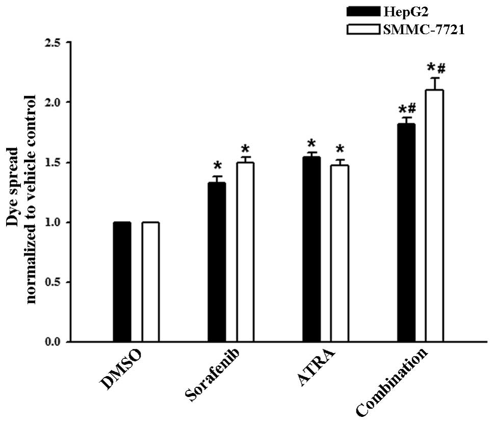
Enhancement of GJIC by sorafenib plus
ATRA
The blockade of the sorafenib plus ATRA-induced
apoptosis in HCC cells by GJIC inhibitors indicated that the
enhanced growth inhibitory effect was mediated by GJIC. To test
this hypothesis, the effects on GJIC by sorafenib plus ATRA or
either agent alone were investigated. Results of the dye transfer
assay are summarized in Fig. 4.
Treatement with sorafenib (5 μM) as well as ATRA (10 μM)
substantially resulted in up to a 33 and 54% increase in the dye
spread between the HepG2 cells respectively, and the
most pronounced effects were expectedly achieved by the combination
treatment. A similar, yet more marked effect was observed when
SMMC-7721 cells were treated with sorafenib (5 μM), ATRA (0.1 μM),
or in combination for 48 h, with the rate of improvement being
50±4.1, 47±5.3 and 110±10.2%, respectively (Fig. 4).
Effect on Cx expression by sorafenib plus
ATRA
Since the dye transfer assay measures the
permeability of gap junction, the enhancement in GJIC activity
could be a consequence of either gating state or the number of Cx
channels on the cell membrane (28). We thus investigated the modulation
of Cx expression in the two HCC cell lines. As shown in Fig. 5A, both cell types had no endogenous
Cx26 expression virtually, whereas a moderate level of Cx32 or Cx43
protein was detected. The results were consistent with prior
studies (31,37). We then focused on the expression
modulation of these two structural proteins. In the
HepG2 cell line, no Cx32 expression change was detected
following each treatment, whereas treatment with sorafenib or ATRA
alone elicited a slight stimulation of Cx43 expression. A
significant increase in the Cx43 amount was achieved following
treatment with the combination of sorafenib plus ATRA (Fig. 5B and C). By contrast, the
enhancement of GJIC activity in SMMC-7721 cells was associated with
an increase in Cx32 expression, but not Cx43 expression, and this
effect was obvious following the combination treatment (Fig. 5D and E).
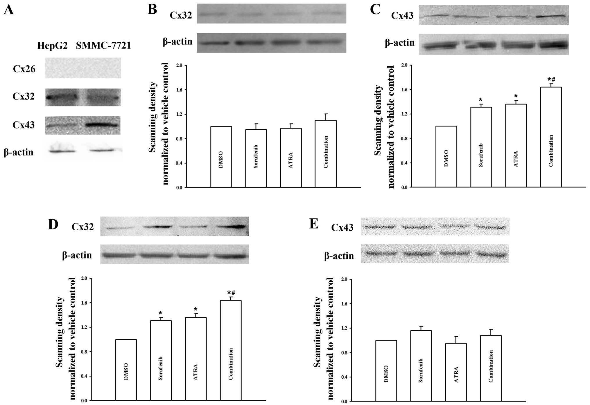 | Figure 5Effects of sorafenib and ATRA on the
protein expression of Cxs. (A) Expression of different Cx isoforms
in HepG2 and SMMC-7721 cells, as shown by western
blotting for Cx26, Cx32 and Cx43 proteins, respectively. (B and C)
Sorafenib (5 μM), ATRA (10 μM), or in combination increased the
expression of Cx43, but not Cx32, after treatment for 48 h in
HepG2 cells. (D and E) Sorafenib (5 μM), ATRA (0.1 μM),
or in combination increased the expression of Cx32, but not Cx43,
after treatment for 48 h in SMMC-7721 cells. Bar graphs were
plotted according to the densitometry of the Cx/actin band
densities, normalized to vehicle controls treated with DMSO. Data
represent the mean ± SEM of three independent experiments.
*p<0.05 vs. vehicle control; #p<0.05
vs. single-agent treatment. |
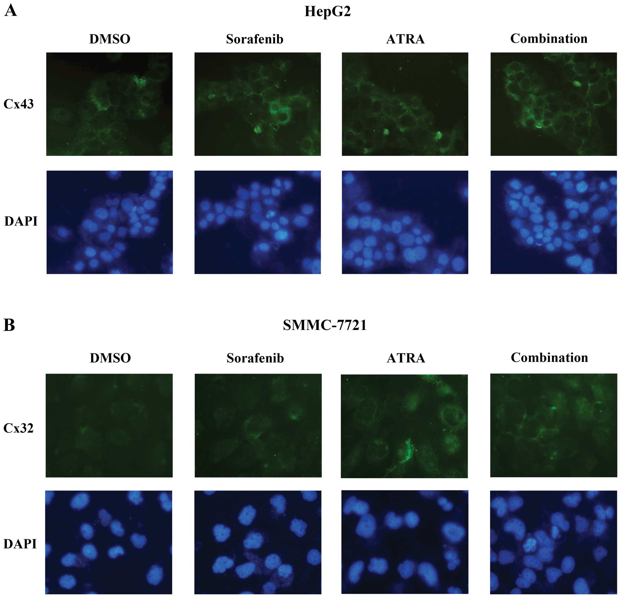
Effect on Cx distribution by sorafenib
plus ATRA
Since increased intracellular levels of Cxs do not
necessarily imply that these proteins are also properly assembled
to form functional gap junction channels on the plasma membrane, we
therefore evaluated Cx relocalization and positioning in both HCC
cell lines. As shown in Fig. 6A,
HepG2 cells displayed a small number of Cx43-specific
positive spots, predominantly along the plasma membrane at
cell-cell contacts, confirming the presence of functional GJIC in
intact cells (Fig. 3A). Following
the treatment of sorafenib or ATRA, Cx43 staining increased and was
located throughout the cell membrane and cytoplasm. The most marked
increment of Cx43 in both compartments was observed following the
combination treatment for 48 h (Fig.
6A), consistent with the immunoblot data (Fig. 5C). In the SMMC-7721 cells, the
treatment produced similar findings demonstrating an increase in
the membrane-associated levels of Cx32 staining (Fig. 6B). These results suggest that the
number of gap junctions composed of its structural Cx was
paralleled by an increase in the amount of protein.
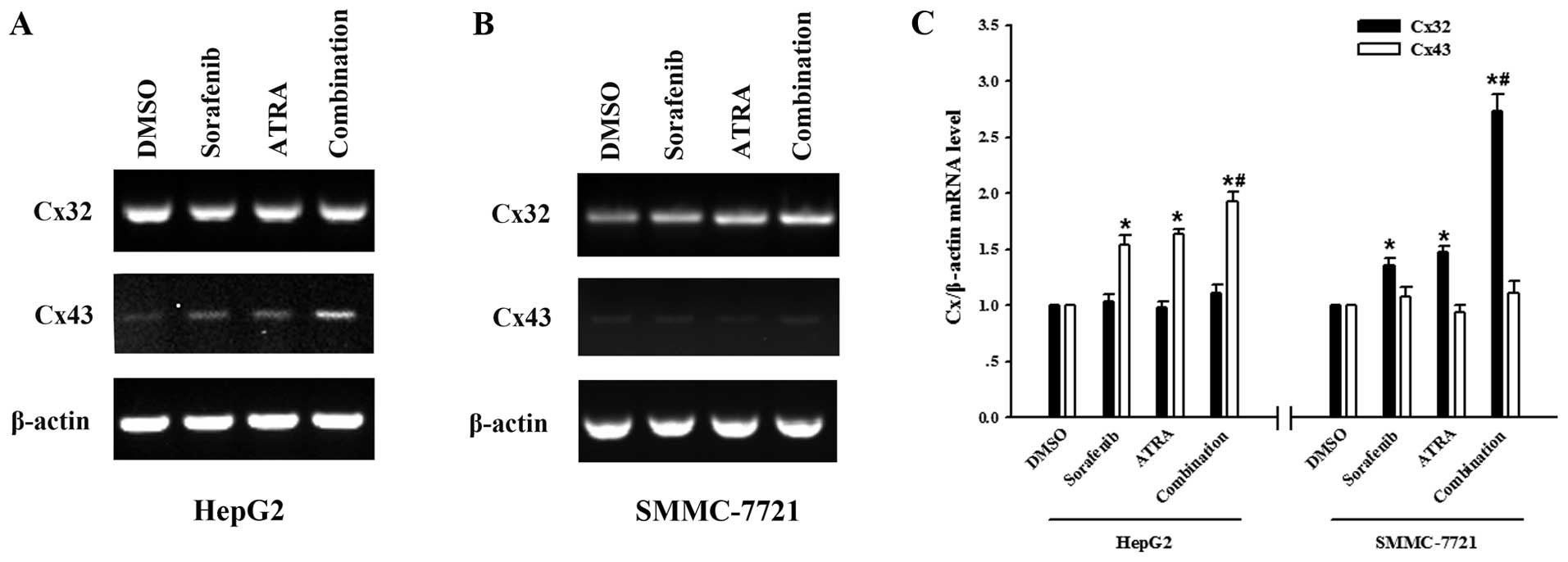
Modulation of Cx32 and Cx43 mRNA
expression
To further test whether the modulation of Cx
expression occurred at the transcriptional level, we measured the
expression of Cx32 and Cx43 mRNA in cells treated with sorafenib
and ATRA, either alone or in combination. As shown in Fig. 7A and C, HepG2 cells
displayed no appreciable change in the amplified PCR product for
the Cx32 transcript in either sample, whereas enhancement of Cx43
mRNA expression to different degrees was detected. In regards to
the SMMC-7721 cells, densitometric analysis demonstrated stable
mRNA expression of Cx43 following each treatment, whereas the
combination of sorafenib and ATRA caused a most significant
augmentation in the Cx32 mRNA level, when compared with either
agent alone (Fig. 7B and C). This
trend was highly consistent with the effect as demonstrated for the
drug-induced increases in Cx protein level (Fig. 5).
Discussion
The present study investigated the effects of
sorafenib in combination with ATRA on the inhibition of growth and
induction of apoptosis in human HCC cells, and the involvement of
gap junctional communication in facilitating this effect. The
results showed that an upregulation of GJIC between treated cells
was associated with the observed pronounced growth-inhibitory
effects by ATRA in combination with low and clinically relevant
concentrations of sorafenib. Thus, the combination treatment may be
a candidate for clinical application, either to permit the use of
lower and less toxic doses of sorafenib, or to supplement standard
sorafenib doses in order to enhance clinical responses, that have,
to date, been poor.
Although clinical trials (5,6) have
established sorafenib as the first standard systemic therapy for
advanced and unresectable HCC, the survival benefit of sorafenib is
still unsatisfactory, and patients with cirrhotic livers exhibit
poor tolerance to this drug. Consequently, the development of novel
approaches that exhibit a non-toxic effect or have low toxicity
when using sorafenib for the treatment of HCC, particularly the
establishment of effective combination therapies, is required. In
this regard, ATRA is of particular interest due to its strong
antitumor activity and convenience for use. The use of ATRA plus
chemotherapeutic agents for the treatment of solid tumors has been
previously investigated (38);
however, ATRA was shown to be extremely toxic hampering its use in
clinical practice (39). This issue
prompted us to focus on the activity of ATRA at a non-toxic
concentration in our study.
At the low concentrations tested in this study, ATRA
showed no significant cytotoxic effect on the HepG2 cell
line, but effectively inhibited SMMC-7721 cell growth in a
dose-dependent manner (Fig. 1B).
These results are in agreement with prior data showing that
HepG2 cells were more resistant to ATRA treatment, and
were only susceptible to an extremely high concentration of ATRA
(20). Data from the MTT assay
showed that sorafenib alone had a significant effect on cell
viability only at relatively high concentrations in both HCC cell
lines (Fig. 1A). Of note, when
combined with ATRA at a non-toxic concentration, there was
substantial enhancement of the effect of sorafenib in inducing both
cell growth inhibition and apoptosis (Figs. 1 and 2). The concentration of sorafenib used in
the combination study with ATRA was approximately the dose that is
used in most in vitro studies (2.5–10 μM) (3,40,41),
and within peak plasma levels of sorafenib (5–10 μg/ml) that can be
achieved by oral administration (8,42). The
use of these doses led to a moderate inhibition of cell growth and
induction of apoptosis by sorafenib, and allowed us to reveal
clinically relevant modulation of the effects of sorafenib in
combination with ATRA.
We further demonstrated that the increased apoptosis
induced by sorafenib plus ATRA was GJIC-dependent, since
pre-treatment with GJIC inhibitors dramatically blocked the
apoptosis induced by sorafenib plus ATRA (Fig. 3). Subsequent experiments revealed
that treatment with sorafenib and ATRA alone led to a moderately
positive effect on gap junction function, while combined treatment
resulted in a strong synergistic enhancement in GJIC activity
(Fig. 4), further strengthening
this conclusion. More importantly, the dose at which sorafenib or
ATRA positively modulated GJIC, was minimally toxic for both cell
lines. Low toxicity but effective concentrations of either drug in
clinical practice are needed, and the effect of cell toxicity on
GJIC must be excluded.
The drug-induced gap junction stimulation in tumor
cells is important in cancer therapy, since aberrant GJIC is widely
regarded to correlate with tumorigenic phenotypes (28,29).
However, changes in the status of gap junction function with
tumorigenesis are complex and heterogeneous. In some cases, such as
ovarian adenocarcinomas and cervical cancer, GJIC is dramatically
reduced or essentially absent (43,44).
In other cases, GJIC is maintained or only modestly reduced. For
example, in the liver, GJIC involves at least three different Cxs,
Cx32, Cx26 and Cx43, depending on the cell type or cell position in
the lobule (45). In hepatoma cell
lines, Cx26 is normally not detected, while Cx43 is upregulated and
Cx32 is upregulated or downregulated depending on the type of tumor
(31,37,46).
Consistent with these previous reports, only Cx32 and Cx43 proteins
were detectable in both HCC cell lines (Fig. 5A). HepG2 and SMMC-7721
cells are gap junctional communication-competent cells, as
evidenced by the dye transfer assay to verify the presence of
functional GJIC (Fig. 3A). Thus, in
this situation, one would expect GJIC-mediated cell growth control
to play a role.
GJIC-mediated cell sensitivity to anticancer therapy
has been demonstrated in many systems (25–27).
The mechanism involves the transport of anticancer agents or their
active metabolites to adjacent cells through gap junctions, thereby
targeting a greater proportion of the cell population. Most
relevant to our study, upregulation of GJIC by cyclic-AMP and ATRA
has been shown to act synergistically with a variety of
chemotherapeutic agents to cause cell death (47). Some toxic metabolites of prodrugs
(e.g., ganciclovir triphosphate and 5-fluorouracil) can pass
between cells through gap junctions (48,49).
Since the bystander toxic effect depends on the gap junction level,
and the functionality of this pathway in human cancers probably
provides an important determinant of the clinical response to
anticancer agents, prevention of GJIC downregulation and
restoration of GJIC in tumor cells could be a rational
chemopreventive approach. In our case, although GJIC was moderately
enhanced by either sorafenib or ATRA alone, the role of GJIC in
cell growth control was not triggered and the growth-inhibitory
effect by either agent was slight. However, when the two agents
were concurrently administered, there was substantial enhancement
of the toxic effect by the two agents at which condition the GJIC
was markedly enhanced. We hypothesized that the toxic signals
generated in one cell can enter another via GJIC and thus enhance
the likelihood of cell death in a cell that might not otherwise be
affected by either agent alone, and in turn, increased drug
sensitivity may be generated in a positive feedback mechanism.
In addition to GJIC-mediated toxic effects, gap
junctions also play an important role in the control of cell growth
and differentiation. Evidence in support of this indicates that the
disruption of GJIC and abnormal expression of Cxs have been found
in transformed and cancer cell lines (28,29),
while GJIC recovery in these cells results in growth normalization
and suppression of tumorigenicity (28,50).
Moreover, studies have shown that the antitumor effects of various
antineoplastic agents, including ATRA, are associated with
restoration of GJIC function and Cx expression in a number of solid
tumors (21,22,37).
These results suggest that Cxs may be defined as tumor suppressors
and that restoration of GJIC may be a unique antitumor therapeutic
strategy. Herein, the ability of sorafenib at a low concentration
to enhance GJIC is important, since this study is the first to
explore the upregulation of gap junction function in HCC cell lines
by sorafenib, which will add a new component to its mechanistic
frame.
In exploring the mechanisms by which GJIC was
enhanced, we found a slight increase in expression of Cx43, but not
Cx32, by a transcriptional mechanism following sorafenib or ATRA
treatment alone in HepG2 cells. SMMC-7721 is a poor
differentiated primary human HCC cell line derived from an elderly
Chinese patient. The cells possess high capacity for tumorigenicity
and low capacity for metastasis (51), for which the GJIC characteristic has
not been entirely ruled out. In contrast to the HepG2
cell line, a significant increase in both protein and mRNA levels
of Cx32, but not Cx43, was observed after 48 h of each drug
treatment in SMMC-7721 cells. Hence, the increase in Cx43
expression in HepG2 cells, and Cx32 expression in
SMMC-7721 cells, could be interpreted as one main cell event
responsible for gap junction upregulation of the targeted cells.
The reasons responsible for these cell-type discrepancies are
unknown, although they may be relative to cell type-specific
transcriptional regulators. Since differential expression of Cxs in
a variety of tissues is generally believed to reflect cell-specific
regulation of junctional coupling and functional demands for GJIC
in different cell types, it is possible that a particular Cx may
function as a tumor modulating protein in one or several specific
cell types but not in others. Although the patterns of action were
not entirely identical, a common feature of the cell response to
treatment was that sorafenib and ATRA acted synergistically, under
each condition, to induce a more significant increase in both
protein and mRNA levels of its structural Cx. The upregulation of
Cx expression is also related to the increase in the number of Cx
channels on the cell membrane as evidenced by the
immunofluorescence assay (Fig. 6).
These data strongly suggest that enhanced GJIC was correlated with
upregulation of Cx relocalization and protein amount by a
transcriptional mechanism. In future studies, the observed
sorafenib effects should be confirmed in other cancer cell lines
with different Cx expression profiles and GJIC levels.
Taken together, the data, presented here for the
first time, suggest that sorafenib and ATRA act synergistically to
enhance inhibition of cell growth and induction of apoptosis via
the upregulation of Cx expression and relocalization resulting in
the enhancement of GJIC in HCC cell lines. Thus, the combination
treatment represents a future therapeutic option for the treatment
of HCC.
Acknowledgements
This study was supported by the Natural Science
Foundation of Anhui, China (no. 1208085MH170).
References
|
1
|
Verslype C, Rosmorduc O and Rougier P:
Hepatocellular carcinoma: ESMO-ESDO Clinical Practice Guidelines
for diagnosis, treatment and follow-up. Ann Oncol. 23(Suppl 7):
vii41–vii48. 2012. View Article : Google Scholar : PubMed/NCBI
|
|
2
|
Wilhelm SM, Carter C, Tang L, et al: BAY
43-9006 exhibits broad spectrum oral antitumor activity and targets
the RAF/MEK/ERK pathway and receptor tyrosine kinases involved in
tumor progression and angiogenesis. Cancer Res. 64:7099–7109. 2004.
View Article : Google Scholar : PubMed/NCBI
|
|
3
|
Liu L, Cao Y, Chen C, et al: Sorafenib
blocks the RAF/MEK/ERK pathway, inhibits tumor angiogenesis, and
induces tumor cell apoptosis in hepatocellular carcinoma model
PLC/PRF/5. Cancer Res. 66:11851–11858. 2006. View Article : Google Scholar : PubMed/NCBI
|
|
4
|
Zhao X, Tian C, Puszyk WM, et al: OPA1
downregulation is involved in sorafenib-induced apoptosis in
hepatocellular carcinoma. Lab Invest. 93:8–19. 2013. View Article : Google Scholar : PubMed/NCBI
|
|
5
|
Llovet JM, Ricci S, Mazzaferro V, et al:
Sorafenib in advanced hepatocellular carcinoma. N Engl J Med.
359:378–390. 2008. View Article : Google Scholar : PubMed/NCBI
|
|
6
|
Cheng AL, Kang YK, Chen Z, et al: Efficacy
and safety of sorafenib in patients in the Asia-Pacific region with
advanced hepatocellular carcinoma: a phase III randomised,
double-blind, placebo-controlled trial. Lancet Oncol. 10:25–34.
2009. View Article : Google Scholar : PubMed/NCBI
|
|
7
|
Wilhelm SM, Adnane L, Newell P, Villanueva
A, Llovet JM and Lynch M: Preclinical overview of sorafenib, a
multikinase inhibitor that targets both Raf and VEGF and PDGF
receptor tyrosine kinase signaling. Mol Cancer Ther. 7:3129–3140.
2008. View Article : Google Scholar : PubMed/NCBI
|
|
8
|
Abou-Alfa GK, Schwartz L, Ricci S, et al:
Phase II study of sorafenib in patients with advanced
hepatocellular carcinoma. J Clin Oncol. 24:4293–4300. 2006.
View Article : Google Scholar : PubMed/NCBI
|
|
9
|
Nojiri K, Sugimoto K, Shiraki K, et al:
Sorafenib and TRAIL have synergistic effect on hepatocellular
carcinoma. Int J Oncol. 42:101–108. 2013.PubMed/NCBI
|
|
10
|
Shen ZX, Shi ZZ, Fang J, et al:
All-trans retinoic
acid/As2O3combination yields a high quality
remission and survival in newly diagnosed acute promyelocytic
leukemia. Proc Natl Acad Sci USA. 101:5328–5335. 2004.PubMed/NCBI
|
|
11
|
Ferrara FF, Fazi F, Bianchini A, et al:
Histone deacetylase-targeted treatment restores retinoic acid
signaling and differentiation in acute myeloid leukemia. Cancer
Res. 61:2–7. 2001.PubMed/NCBI
|
|
12
|
Maeda Y, Yamaguchi T, Hijikata Y, et al:
Clinical efficacy of all-trans retinoic acid for treating
adult T cell leukemia. J Cancer Res Clin Oncol. 134:673–677.
2008.
|
|
13
|
Sun SY, Wan H, Yue P, Hong WK and Lotan R:
Evidence that retinoic acid receptor beta induction by retinoids is
important for tumor cell growth inhibition. J Biol Chem.
275:17149–17153. 2000. View Article : Google Scholar : PubMed/NCBI
|
|
14
|
Newsome PN, Beldon I, Moussa Y, et al: Low
serum retinol levels are associated with hepatocellular carcinoma
in patients with chronic liver disease. Aliment Pharmacol Ther.
14:1295–1301. 2000. View Article : Google Scholar : PubMed/NCBI
|
|
15
|
Yuan JM, Gao YT, Ong CN, Ross RK and Yu
MC: Prediagnostic level of serum retinol in relation to reduced
risk of hepatocellular carcinoma. J Natl Cancer Inst. 98:482–490.
2006. View Article : Google Scholar : PubMed/NCBI
|
|
16
|
Kim DG, Jo BH, You KR and Ahn DS:
Apoptosis induced by retinoic acid in Hep 3B cells in vitro. Cancer
Lett. 107:149–159. 1996. View Article : Google Scholar : PubMed/NCBI
|
|
17
|
Hsu SL, Lin HM and Chou CK: Suppression of
the tumorigenicity of human hepatoma hep3B cells by long-term
retinoic acid treatment. Cancer Lett. 99:79–85. 1996. View Article : Google Scholar : PubMed/NCBI
|
|
18
|
Falasca L, Favale A, Gualandi G, Maietta G
and Conti Devirgiliis L: Retinoic acid treatment induces apoptosis
or expression of a more differentiated phenotype on different
fractions of cultured fetal rat hepatocytes. Hepatology.
28:727–737. 1998. View Article : Google Scholar : PubMed/NCBI
|
|
19
|
Falasca L, Marcellini P, Ara C, Rufo A and
Devirgiliis LC: Growth inhibition and induction of specific hepatic
phenotype expression by retinoic acid in HEPG2 cells. Anticancer
Res. 19:3283–3292. 1999.PubMed/NCBI
|
|
20
|
Arce F, Gatjens-Boniche O, Vargas E,
Valverde B and Diaz C: Apoptotic events induced by naturally
occurring retinoids ATRA and 13-cis retinoic acid on human
hepatoma cell lines Hep3B and HepG2. Cancer Lett. 229:271–281.
2005. View Article : Google Scholar : PubMed/NCBI
|
|
21
|
Watanabe J, Nomata K, Noguchi M, et al:
All-trans retinoic acid enhances gap junctional
intercellular communication among renal epithelial cells in vitro
treated with renal carcinogens. Eur J Cancer. 35:1003–1008.
1999.
|
|
22
|
Wang J, Dai Y, Huang Y, et al:
All-trans retinoic acid restores gap junctional
intercellular communication between oral cancer cells with
upregulation of Cx32 and Cx43 expressions in vitro. Med Oral Patol
Oral Cir Bucal. 18:e569–e577. 2013.
|
|
23
|
Harris AL: Connexin channel permeability
to cytoplasmic molecules. Prog Biophys Mol Biol. 94:120–143. 2007.
View Article : Google Scholar : PubMed/NCBI
|
|
24
|
Herve JC and Derangeon M:
Gap-junction-mediated cell-to-cell communication. Cell Tissue Res.
352:21–31. 2013. View Article : Google Scholar : PubMed/NCBI
|
|
25
|
Krutovskikh VA, Piccoli C and Yamasaki H:
Gap junction intercellular communication propagates cell death in
cancerous cells. Oncogene. 21:1989–1999. 2002. View Article : Google Scholar : PubMed/NCBI
|
|
26
|
Jensen R and Glazer PM:
Cell-interdependent cisplatin killing by Ku/DNA-dependent protein
kinase signaling transduced through gap junctions. Proc Natl Acad
Sci USA. 101:6134–6139. 2004. View Article : Google Scholar : PubMed/NCBI
|
|
27
|
Hong X, Wang Q, Yang Y, et al: Gap
junctions propagate opposite effects in normal and tumor testicular
cells in response to cisplatin. Cancer Lett. 317:165–171. 2012.
View Article : Google Scholar : PubMed/NCBI
|
|
28
|
Mesnil M: Connexins and cancer. Biol Cell.
94:493–500. 2002. View Article : Google Scholar
|
|
29
|
Leithe E, Sirnes S, Omori Y and Rivedal E:
Downregulation of gap junctions in cancer cells. Crit Rev Oncog.
12:225–256. 2006. View Article : Google Scholar : PubMed/NCBI
|
|
30
|
Yang Y, Cao MH, Wang Q, Yuan DD, Li L and
Tao L: The effects of 2-aminoethoxydiphenyl borate and
diphenylboronic anhydride on gap junctions composed of Connexin43
in TM(4) sertoli cells. Biol Pharm Bull. 34:1390–1397. 2011.
View Article : Google Scholar : PubMed/NCBI
|
|
31
|
Ara C, Massimi M and Devirgiliis Conti L:
Retinoic acid modulates gap junctional intercellular communication
in hepatocytes and hepatoma cells. Cell Mol Life Sci. 59:1758–1765.
2002. View Article : Google Scholar : PubMed/NCBI
|
|
32
|
Bai D, del Corsso C, Srinivas M and Spray
DC: Block of specific gap junction channel subtypes by
2-aminoethoxydiphenyl borate (2-APB). J Pharmacol Exp Ther.
319:1452–1458. 2006. View Article : Google Scholar : PubMed/NCBI
|
|
33
|
Guo Y, Martinez-Williams C, Gilbert KA and
Rannels DE: Inhibition of gap junction communication in alveolar
epithelial cells by 18alpha-glycyrrhetinic acid. Am J Physiol.
276:L1018–L1026. 1999.PubMed/NCBI
|
|
34
|
Bolognesi M, Zampieri F, Di Pascoli M, et
al: Increased myoendothelial gap junctions mediate the enhanced
response to epoxyeicosatrienoic acid and acetylcholine in
mesenteric arterial vessels of cirrhotic rats. Liver Int.
31:881–890. 2011. View Article : Google Scholar : PubMed/NCBI
|
|
35
|
Guan X, Cravatt BF, Ehring GR, et al: The
sleep-inducing lipid oleamide deconvolutes gap junction
communication and calcium wave transmission in glial cells. J Cell
Biol. 139:1785–1792. 1997. View Article : Google Scholar : PubMed/NCBI
|
|
36
|
Boger DL, Patterson JE, Guan X, Cravatt
BF, Lerner RA and Gilula NB: Chemical requirements for inhibition
of gap junction communication by the biologically active lipid
oleamide. Proc Natl Acad Sci USA. 95:4810–4815. 1998. View Article : Google Scholar : PubMed/NCBI
|
|
37
|
Liu CL, Huang YS, Hosokawa M, Miyashita K
and Hu ML: Inhibition of proliferation of a hepatoma cell line by
fucoxanthin in relation to cell cycle arrest and enha-nced gap
junctional intercellular communication. Chem Biol Interact.
182:165–172. 2009. View Article : Google Scholar : PubMed/NCBI
|
|
38
|
Kalemkerian GP, Jiroutek M, Ettinger DS,
Dorighi JA, Johnson DH and Mabry M: A phase II study of
all-trans-retinoic acid plus cisplatin and etoposide in
patients with extensive stage small cell lung carcinoma: an Eastern
Cooperative Oncology Group Study. Cancer. 83:1102–1108.
1998.PubMed/NCBI
|
|
39
|
Lee JS, Newman RA, Lippman SM, et al:
Phase I evaluation of all-trans-retinoic acid in adults with solid
tumors. J Clin Oncol. 11:959–966. 1993.PubMed/NCBI
|
|
40
|
Dasmahapatra G, Yerram N, Dai Y, Dent P
and Grant S: Synergistic interactions between vorinostat and
sorafenib in chronic myelogenous leukemia cells involve Mcl-1 and
p21CIP1down-regulation. Clin Cancer Res. 13:4280–4290.
2007. View Article : Google Scholar
|
|
41
|
Eicher C, Dewerth A, Kirchner B, Warmann
SW, Fuchs J and Armeanu-Ebinger S: Treatment effects of the
multikinase inhibitor sorafenib on hepatoblastoma cell lines and
xenografts in NMRI-Foxn1 nu mice. Liver Int. 32:574–581. 2012.
View Article : Google Scholar : PubMed/NCBI
|
|
42
|
Strumberg D, Richly H, Hilger RA, et al:
Phase I clinical and pharmacokinetic study of the Novel Raf kinase
and vascular endothelial growth factor receptor inhibitor BAY
43-9006 in patients with advanced refractory solid tumors. J Clin
Oncol. 23:965–972. 2005. View Article : Google Scholar
|
|
43
|
Umhauer S, Ruch RJ and Fanning J: Gap
junctional intercellular communication and connexin 43 expression
in ovarian carcinoma. Am J Obstet Gynecol. 182:999–1000. 2000.
View Article : Google Scholar : PubMed/NCBI
|
|
44
|
Gershon E, Plaks V and Dekel N: Gap
junctions in the ovary: expression, localization and function. Mol
Cell Endocrinol. 282:18–25. 2008. View Article : Google Scholar : PubMed/NCBI
|
|
45
|
Zhang JT and Nicholson BJ: The topological
structure of connexin 26 and its distribution compared to connexin
32 in hepatic gap junctions. J Membr Biol. 139:15–29. 1994.
View Article : Google Scholar : PubMed/NCBI
|
|
46
|
Kawasaki Y, Omori Y, Li Q, et al:
Cytoplasmic accumulation of connexin32 expands cancer stem cell
population in human HuH7 hepatoma cells by enhancing its
self-renewal. Int J Cancer. 128:51–62. 2011. View Article : Google Scholar : PubMed/NCBI
|
|
47
|
Carystinos GD, Alaoui-Jamali MA, Phipps J,
Yen L and Batist G: Upregulation of gap junctional intercellular
communication and connexin 43 expression by cyclic-AMP and
all-trans-retinoic acid is associated with glutathione
depletion and chemosensitivity in neuroblastoma cells. Cancer
Chemother Pharmacol. 47:126–132. 2001. View Article : Google Scholar : PubMed/NCBI
|
|
48
|
Matono S, Tanaka T, Sueyoshi S, Yamana H,
Fujita H and Shirouzu K: Bystander effect in suicide gene therapy
is directly proportional to the degree of gap junctional
intercellular communication in esophageal cancer. Int J Oncol.
23:1309–1315. 2003.PubMed/NCBI
|
|
49
|
Lawrence TS, Rehemtulla A, Ng EY, Wilson
M, Trosko JE and Stetson PL: Preferential cytotoxicity of cells
transduced with cytosine deaminase compared to bystander cells
after treatment with 5-flucytosine. Cancer Res. 58:2588–2593.
1998.PubMed/NCBI
|
|
50
|
Edwards GO, Jondhale S, Chen T and Chipman
JK: A quantitative inverse relationship between connexin32
expression and cell proliferation in a rat hepatoma cell line.
Toxicology. 253:46–52. 2008. View Article : Google Scholar : PubMed/NCBI
|
|
51
|
Wang ZJ, Song L, Guo LC, Yin M and Sun YN:
Induction of differentiation by panaxydol in human hepatocarcinoma
SMMC-7721 cells via cAMP and MAP kinase dependent mechanism.
Yakugaku Zasshi. 131:993–1000. 2011. View Article : Google Scholar : PubMed/NCBI
|