Introduction
Cholangiocarcinoma (CCA) is a rare but highly
aggressive cancer, accounting for ~3% of all gastrointestinal
malignancies (1). CCA is
characterized by poor responsiveness to chemotherapy and
radiotherapy in the majority of cases (2,3).
Surgical resection is the only potentially curative option.
Therefore, novel biomarkers are urgently needed for CCA management
and treatment.
The human epidermal growth factor receptor (EGFR)
family consists of four members: HER1 (EGFR), HER2 (c-erbB-2), HER3
(c-erbB-3) and HER4 (c-erbB-4) (4).
These receptors activate multiple downstream pathways in response
to extracellular ligands, regulating diverse processes that include
differentiation, migration, proliferation and survival (5,6).
Aberrations in EGFR family members play a role in the development
and progression of many human cancers (7–9).
Extensive studies have reported either the overexpression or
amplification of EGFR and HER2 in different malignancies (10,11).
Accordingly, current evidence suggests that HER3 expression is
associated with increased cellular proliferation whereas HER4
activation seems to mainly mediate antiproliferative effects
(12–14).
In CCA, overexpression of EGFR and HER2 are thought
to be prognostic factors and targets of novel biologic agents
(15). Of note, a series of studies
have revealed overexpression of EGFR and HER2, amplification and
mutations of these genes (15–17).
Although the clinical significance of such overexpression is not
fully clear, some case reports and phase II trials have reported
promising results targeting EGFR in CCA (18). By contrast, the data of HER3 and
HER4 is very limited. Most recently, Lee et al (19) reported that HER3 is overexpressed in
a subset of extrahepatic cholangiocarcinoma (EHCC) patients and
HER3 overexpression is correlated with decreased patient survival.
The biological function and prognostic role of HER4 has thus far
not been investigated in CCA.
According to the 7th edition of the Union for
International Cancer Control-American Joint Committee on Cancer
(UICC/AJCC), CCAs are classified as intrahepatic
cholangiocarcinomas (IHCCs) and EHCCs, the latter being further
divided into perihilar CCAs and distal CCAs. Although linked
anatomically and histopathologically, it is unclear whether CCA
patients from different sites share common pathogenetic features.
It has been reported that CCA differentially expresses cell
cycle-regulatory proteins based on tumor location and morphology
(20).
In the present study, we comprehensively
characterized and compared differential gene expression of all EGFR
family members across the full spectrum of CCAs. We aimed to define
differential expression of EGFR family members based on anatomic
site of origin to establish new potential prognostic factors in
CCA.
Patients and methods
Patients and tissue microarray (TMA)
construction
The present study consisted of 175 CCAs (male, 106;
female, 69) who underwent surgical resections between 2004 to 2011
at the Qilu Hospital of Shandong University (Jinan, China).
Follow-up time ranges from 3 to 98 months (mean, 26 months). A
total of two TMAs were constructed. Two cores (1.0 mm in diameter)
were taken from each representative tumor focus and the morphology
was evaluated by two pathologists (B.H. and X.Y.). Detailed
clinical and pathological profiles were obtained from medical
records and maintained in a secure relational database with TMA
data. Tumor staging and histological classification were assessed
according to AJCC 7th Edition of TNM Staging (21). Patient demographics are shown in
Table I. Informed written consent
was obtained from the CCA patients. The study and the consent
procedures were approved by the Institutional Review Board at the
School of Medicine of Shandong University. All cases were
anatomically classified into two groups: IHCC and EHCC. Hilar and
distal CCA were classified as EHCC. The numbers of IHCC and EHCC
cases were 65 and 110, respectively.
 | Table ISummary of CCA patient
demographics. |
Table I
Summary of CCA patient
demographics.
| Parameters | IHCC (%) | EHCC (%) |
|---|
| Age (years) | | |
| <60 | 35 (53.8) | 73 (66.4) |
| ≥60 | 30 (46.2) | 37 (33.6) |
| Gender | | |
| Male | 29 (44.6) | 77 (70.0) |
| Female | 36 (55.4) | 33 (30.0) |
| Tumor size
(cm)a | | |
| <5 | 22 (33.8) | 68 (61.8) |
| ≥5 | 43 (66.2) | 42 (38.2) |
| Histological
differentiation | | |
| Well | 17 (26.2) | 56 (50.9) |
| Moderate | 31 (47.7) | 37 (33.6) |
| Poor | 17 (26.2) | 17 (15.5) |
| Perineural
invasion | | |
| Negative | 57 (87.7) | 80 (72.7) |
| Positive | 8 (12.3) | 30 (27.3) |
| Microvascular
invasion | | |
| Negative | 51 (78.5) | 97 (88.2) |
| Positive | 14 (21.5) | 13 (11.8) |
| Venous
invasion | | |
| Negative | 62 (95.4) | 103 (93.6) |
| Positive | 3 (4.6) | 7 (6.4) |
| T stage | | |
| I + II | 51 (78.5) | 57 (51.8) |
| III + IV | 14 (21.5) | 53 (78.2) |
| N stage | | |
| Negative | 50 (76.9) | 78 (70.9) |
| Positive | 15 (23.1) | 32 (29.1) |
| UICC stage | | |
| I + II | 40 (61.5) | 63 (57.3) |
| III + IV | 25 (38.5) | 47 (42.7) |
Immunohistochemistry (IHC)
IHC was performed as previously described (22). Briefly, the slides were
deparaffinized by successive passages through xylene and ethanol.
Antigen retrieval was performed by microwave pretreatment in 0.01 M
citrate buffer (pH 6.0) for 15 min. The primary antibodies used
were anti-EGFR (1:500; Dako), anti-HER2 (1:500; Dako) anti-HER3
(sc-415, 1:500; Santa Cruz Biotechnology, Santa Cruz, CA, USA) and
anti-HER4 (sc-283, 1:500; Santa Cruz Biotechnology). The slides
were incubated overnight at 4°C. For visualization,
3,3-diaminobenzidine tetrahydrochloride was used as chromogen. The
slides were evaluated by two independent pathologists (B.H. and
X.Y.) who were blinded to the clinical data. For EGFR and HER2,
only the membrane immunostaining was scored following a four-step
scale (scores 0, 1+, 2+ and 3+). For HER2, we followed the
consensus panel recommendations on HER2 scoring for breast cancer
(8). Slides with a score of 2+ or
3+ were classified as positive or expressed, in contrast to slices
with a score of 0 or 1+, which were defined as negative. For HER3
and HER4, nuclear and cytoplasmic staining was evaluated using the
Rajkumar score (23), which was
built by multiplying the scores of 2 parameters, the staining
intensity (range, 0–3) and the percentage of positive cells (range,
0–4; 0, 0–10%; 1, 11–25%; 2, 26–50%; 3, 51–75%; and 4, 76–100%).
Slides with scores of ≥8 were classified as overexpression and
slides with scores <8 as non-overexpression.
Fluorescence in situ hybridization
(FISH)
FISH analysis for EGFR and HER2 gene
aberrations was performed as previously described (10). Briefly, the GLP EGFR/CSP 7
probe and GLP HER2/CSP17 (Beijing GP Medical Technologies,
Beijing, China) were utilized and slides were examined using an
ImagingZ1 microscope (Carl Zeiss, Oberkochen, Germany). FISH
signals were scored manually (100× oil immersion) in
morphologically intact and non-overlapping nuclei by a pathologist
(B.H.) and a minimum of 50 cancer cells from each site were
recorded. Cancer sites with very weak or no signals were recorded
as insufficiently hybridized. A previously documented method was
utilized to validate genetic aberrations of EGFR and HER2 (22). A case was scored as amplification
when >10% of tumor cells displayed either definite cluster of
locus probe signals or EGFR (HER2):CEP 17 ratio >2.
Cell culture and reagents
The CCA cell lines RBE, HuCCT-1, QBC939 and one
breast cancer cell line MCF-7 were obtained from the Cell Bank of
the Chinese Academy of Sciences (Shanghai, China) and cultured
following the manufacturer’s instructions.
Western blot analysis
Western blot analysis was performed as previously
described (22). Briefly, the
membrane was incubated overnight at 4°C with primary antibody for
anti-HER4 (sc-283, 1:1,000; Santa Cruz Biotechnology). The
secondary antibody was a goat anti-rabbit antibody at a dilution of
1:5,000 and the signals were detected with RapidStep™ ECL reagent
(Millipore Corp., Billerica, MA, USA). Three independent
experiments were performed. For analysis of the western blot
images, the ImageJ software (1.37v; Wayne Rasband, NIH, Bethesda,
MD, USA) was used.
In vitro overexpression of HER4
Human HER4 cDNA was subcloned into the pcDNA3.1
eukaryotic expression vectors. HER4 and empty control plasmids were
independently transfected into HuCCT-1 cells using Lipofectamine
(Invitrogen, Carlsbad, CA, USA) according to the manufacturer’s
protocol.
siRNA knockdown
siRNA transfection on CCA cell line RBE was carried
out using Lipofectamine 2000 (Invitrogen) according to the
manufacturer’s protocol. Three specific siRNAs for each gene were
designed and synthesized by GenePharma (Shanghai, China),
respectively. The most effective single siRNA-HER4 (sense strand,
5′-GCGCAGGAAA CAUCUAUAUTT-3′ and antisense strand, 5′-AUAUAGAUG
GUUUCCUGCGCTT-3′) was used for further experiments. Non-specific
negative control siRNAs were also used (sense strand,
5′-UUCUCCCAACGUGUCACG-3′ and antisense strand,
5′-ACGUGACACGUUCGGAGAATT-3′). The mock group was defined as the
ones supplemented with the transfection reagent only.
Cell proliferation, migration and
invasion assays
Cell proliferation was measured by MTT assays as
previously described (22). For
each assay, 20 μl MTT (methyl thiazolyl tetrazolium) was added to
each well and incubated for 4 h. For cell migration assay, a wound
was created by a p200 pipette tip on cells grown to confluence
using 6-well plates; the cell-free space was measured on images
captured at both 0 and 48 h. The invasion assays were performed as
previously described (22). All
experiments were performed in triplicates.
Statistical analysis
The SPSS statistical software package, standard
version 17.0 (SPSS, Chicago, IL, USA) was used for statistical
analysis. The association between expression of EGFR family members
and the clinicopathological variables was analyzed by using
Chi-square test. Cumulative overall survival rates were calculated
by Kaplan-Meier method and statistical significance for survival
curves comparison was analyzed by log-rank test. Univariate and
multivariate survival analyses were performed using the Cox
multiple hazards model to estimate hazard ratio (HR) and 95%
confidence interval (CI) of each outcome. Differences for all the
tests were regarded as statistically significant when the P-value
from a two-tailed test was P<0.05.
Results
Expression and genetic aberrations of
EGFR family members in CCAs
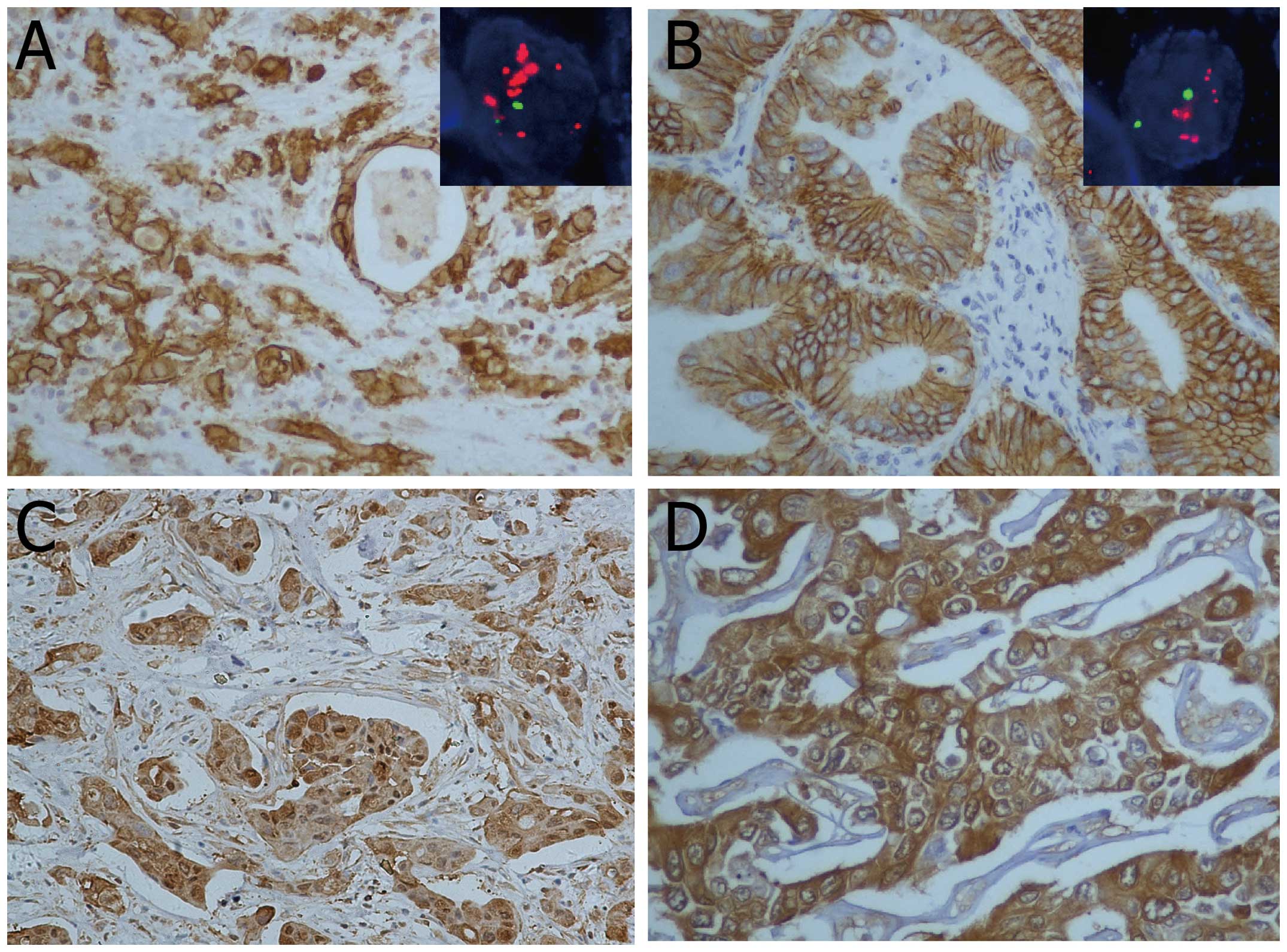
Representative cases of positive staining by IHC for
each member are shown in Fig. 1.
EGFR, HER3 and HER4 were overexpressed in 20 (30.8%), 8 (12.3%) and
41 (63.1%) of the 65 IHCCs, and in 23 (20.9%), 13 (11.8%) and 62
(56.4%) of the 110 EHCCs, respectively. Overexpression of HER2 was
exclusively identified in EHCCs, among which the rate was 4.5%
(5/110). FISH analysis for EGFR and HER2 was available for 169 and
171 cases, respectively. In all, amplification of EGFR was
identified in 1 (1.6%) of the 63 IHCC cases, comparable with that
of 2.8% (3/106) in patients with EHCC. By contrast, HER2
amplification was observed in 8 of the 108 (7.4%) EHCC, but was
absent in IHCC cases. Of note, a significant association was
identified between EGFR amplification and EGFR overexpression
(P=0.002). Similarly, HER2 amplification was strongly associated
with HER2 overexpresssion (P<0.001).
Overall, overexpression of any EGFR family member
was found in 47 (72.3%) of 65 patients in IHCC, and 76 (69.1%) of
110 cases in EHCC, respectively. The combination of EGFR and HER4
was the most common, found in 23.1% of IHCC tumors (n=15) and 12.7%
of EHCC tumors (n=14). In contrast, none of the CCA tumors
co-expressed EGFR and HER2.
Associations between EGFR family members
with clinicopathological variables
In IHCC, EGFR overexpression was significantly
associated with poor histological differentiation (P=0.033), but
not with age (P=0.507), tumor size (P=0.485), lymph node metastasis
(P=1.000), vascular invasion (P=0.900), perineural invasion
(P=0.432) or UICC stage (P=0.702) (Table II). In all, 9 EGFR-positive cases
were observed in 17 (52.9%) poor differentiation samples, whereas
only 11 out of 48 (22.9%) cases with well and/or moderate
differentiation demonstrated EGFR positivity. Poorly differentiated
tumors were significantly associated with HER3 overexpression
(P=0.047). By contrast, HER4 expression was associated with UICC
I/II stage (P=0.046), but not with well differentiation (P=0.746).
No significant association of EGFR amplification was identified
with tumor size, histological type, clinical stage, presence of
vascular or perineural invasion or lymph node metastases.
| Table IIAssociation of expression of EGFR
family members with clinicopathological parameters in IHCC. |
Table II
Association of expression of EGFR
family members with clinicopathological parameters in IHCC.
| EGFR | HER3 | HER4 |
|---|
|
|
|
|
|---|
| Parameters | Not overexpressed
(%) | Overexpressed
(%) | P-value | Not overexpressed
(%) | Overexpressed
(%) | P-value | Not overexpressed
(%) | Overexpressed
(%) | P-value |
|---|
| Age (years) | | | | | | | | | |
| <60 | 23 (65.7) | 12 (34.3) | | 31 (88.6) | 4 (11.4) | | 14 (40.0) | 21 (60.0) | |
| ≥60 | 22 (73.3) | 8 (26.7) | 0.507 | 26 (86.7) | 4 (13.3) | 1.000 | 10 (33.3) | 20 (66.7) | 0.579 |
| Gender | | | | | | | | | |
| Male | 18 (62.1) | 11 (37.9) | | 23 (79.3) | 6 (20.7) | | 11 (37.9) | 18 (62.1) | |
| Female | 27 (75.0) | 9 (25.0) | 0.262 | 34 (94.4) | 2 (5.6) | 0.143 | 13 (36.1) | 23 (63.9) | 0.880 |
| Tumor size | | | | | | | | | |
| <5 cm | 14 (63.6) | 8 (36.4) | | 19 (86.4) | 3 (13.6) | | 7 (31.8) | 15 (68.2) | |
| ≥5 cm | 31 (72.1) | 12 (27.9) | 0.485 | 38 (88.4) | 5 (11.6) | 1.000 | 17 (39.5) | 26 (60.5) | 0.542 |
| Histological
differentiation | | | | | | | | | |
| Well | 15 (88.2) | 2 (11.8) | | 17 (100) | 0 (0.0) | | 7 (41.2) | 10 (58.8) | |
| Moderate | 22 (71.0) | 9 (29.0) | | 27 (87.1) | 4 (12.9) | | 12 (38.7) | 19 (61.3) | |
| Poor | 8 (47.1) | 9 (52.9) | 0.033 | 13 (76.5) | 4 (23.5) | 0.047 | 5 (29.4) | 12 (70.6) | 0.746 |
| Perineural
invasion | | | | | | | | | |
| Negative | 38 (66.7) | 19 (33.3) | | 50 (87.7) | 7 (12.3) | | 20 (35.1) | 37 (64.9) | |
| Positive | 7 (87.5) | 1 (12.5) | 0.432 | 7 (87.5) | 1 (12.5) | 1.000 | 4 (50.0) | 4 (50.0) | 0.669 |
| Vascular
invasion | | | | | | | | | |
| Negative | 36 (70.6) | 15 (29.4) | | 44 (86.3) | 7 (13.7) | | 20 (39.2) | 31 (60.8) | |
| Positive | 9 (64.3) | 5 (35.7) | 0.900 | 13 (92.9) | 1 (7.1) | 0.838 | 4 (28.6) | 10 (71.4) | 0.465 |
| T stage | | | | | | | | | |
| I + II | 35 (68.6) | 16 (31.4) | | 44 (86.3) | 7 (13.7) | | 16 (31.4) | 35 (68.6) | |
| III + IV | 10 (71.4) | 4 (28.6) | 1.000 | 13 (92.9) | 1 (7.1) | 0.838 | 8 (57.1) | 6 (42.9) | 0.077 |
| Lymph node
metastasis | | | | | | | | | |
| Negative | 35 (70.0) | 15 (30.0) | | 43 (86.0) | 7 (14.0) | | 19 (38.0) | 31 (62.0) | |
| Positive | 10 (66.7) | 5 (33.3) | 1.000 | 14 (93.3) | 1 (6.7) | 0.756 | 5 (33.3) | 10 (66.7) | 0.743 |
| UICC stage | | | | | | | | | |
| I + II | 27 (67.5) | 13 (32.5) | | 33 (82.5) | 7 (17.5) | | 11 (27.5) | 29 (72.5) | |
| III + IV | 18 (72.0) | 7 (28.0) | 0.702 | 24 (96.0) | 1 (4.0) | 0.221 | 13 (52.0) | 12 (48.0) | 0.046 |
In EHCC, EGFR overexpression was significantly
associated with well histological differentiation (P<0.001) and
lymph node metastasis (P=0.006). Overexpression of HER2 was
significantly associated with TI/TII stage (P=0.050), but not with
histological differentiation or lymph node metastasis (Table III). No significant correlation
was identified between HER3 or HER4 overexpression with other
clinicopathological factors.
| Table IIIAssociation of expression of EGFR
family members with clinicopathological parameters in EHCC. |
Table III
Association of expression of EGFR
family members with clinicopathological parameters in EHCC.
| EGFR | HER2 | HER3 | HER4 |
|---|
|
|
|
|
|
|---|
| Parameters | Not overexpressed
(%) | Overexpressed
(%) | P-value | Not overexpressed
(%) | Overexpressed
(%) | P-value | Not overexpressed
(%) | Overexpressed
(%) | P-value | Not overexpressed
(%) | Overexpressed
(%) | P-value |
|---|
| Age (years) | | | | | | | | | | | | |
| <60 | 59 (80.8) | 14 (19.2) | | 68 (93.2) | 5 (6.8) | | 64 (87.7) | 9 (12.3) | | 29 (39.7) | 44 (60.3) | |
| ≥60 | 28 (75.7) | 9 (24.3) | 0.531 | 37 (100) | 0 (0.0) | 0.252 | 33 (89.2) | 4 (10.8) | 0.889 | 19 (51.4) | 18 (48.6) | 0.245 |
| Gender | | | | | | | | | | | | |
| Male | 63 (81.8) | 14 (18.2) | | 73 (94.8) | 4 (5.2) | | 69 (89.6) | 8 (10.4) | | 35 (45.5) | 42 (54.5) | |
| Female | 24 (72.7) | 9 (27.3) | 0.283 | 32 (97.0) | 1 (3.0) | 1.000 | 28 (84.8) | 5 (15.2) | 0.698 | 13 (39.4) | 20 (60.6) | 0.557 |
| Tumor size
(cm) | | | | | | | | | | | | |
| <3 | 52 (77.6) | 16 (22.4) | | 63 (92.6) | 5 (7.4) | | 63 (92.6) | 5 (7.4) | | 30 (44.1) | 38 (55.9) | |
| ≥3 | 35 (83.3) | 7 (16.7) | 0.390 | 42 (100) | 0 (0.0) | 0.184 | 34 (81.0) | 8 (19.0) | 0.123 | 18 (42.3) | 24 (57.1) | 0.897 |
| Histological
differentiation | | | | | | | | | | | | |
| Well | 53 (94.6) | 3 (5.4) | | 54 (96.4) | 2 (3.6) | | 51 (91.1) | 5 (8.9) | | 26 (46.4) | 30 (53.6) | |
| Moderate | 26 (70.3) | 11 (29.7) | | 34 (91.9) | 3 (8.1) | | 32 (86.5) | 5 (13.5) | | 15 (40.5) | 22 (59.5) | |
| Poor | 8 (47.1) | 9 (52.9) | <0.001 | 17 (100) | 0 (0.0) | 0.453 | 14 (82.4) | 3 (17.6) | 0.688 | 7 (41.2) | 10 (58.8) | 0.834 |
| Perineural
invasion | | | | | | | | | | | | |
| Negative | 63 (78.8) | 17 (21.3) | | 77 (96.3) | 3 (3.8) | | 70 (87.5) | 10 (12.5) | | 34 (42.5) | 46 (57.5) | |
| Positive | 24 (80.0) | 6 (20.0) | 0.886 | 28 (93.3) | 2 (6.7) | 0.889 | 27 (90.0) | 3 (10.0) | 0.889 | 14 (46.7) | 16 (53.3) | 0.695 |
| Vascular
invasion | | | | | | | | | | | | |
| Negative | 77 (79.4) | 20 (20.6) | | 92 (94.8) | 5 (5.2) | | 88 (90.7) | 9 (9.3) | | 40 (41.2) | 57 (58.8) | |
| Positive | 10 (76.9) | 3 (23.1) | 1.000 | 13 (100) | 0 (0.0) | 1.000 | 9 (69.2) | 4 (30.8) | 0.072 | 8 (61.5) | 5 (38.5) | 0.166 |
| T stage | | | | | | | | | | | | |
| I + II | 42 (80.8) | 10 (19.2) | | 47 (90.4) | 5 (9.6) | | 46
(88.5) | 6 (11.5) | | 22 (42.3) | 30 (57.7) | |
| III + IV | 45 (77.6) | 13 (22.4) | 0.682 | 58 (100) | 0 (0.0) | 0.050 | 51 (87.9) | 7 (12.1) | 0.833 | 26 (44.8) | 32 (55.2) | 0.790 |
| Lymph node
metastasis | | | | | | | | | | | | |
| Negative | 67 (85.9) | 11 (14.1) | | 73 (93.6) | 5 (6.4) | | 71 (91.0) | 7 (9.0) | | 31 (39.7) | 47 (60.3) | |
| Positive | 20 (62.5) | 12 (37.5) | 0.006 | 32 (100.0) | 0 (0.0) | 0.336 | 26 (81.3) | 6 (18.7) | 0.264 | 17 (53.1) | 15 (46.9) | 0.199 |
| UICC stage | | | | | | | | | | | | |
| I + II | 49 (77.8) | 14 (22.2) | | 58 (92.1) | 5 (7.9) | | 59 (93.6) | 4 (6.4) | | 26 (41.3) | 37 (58.7) | |
| III + IV | 38 (80.9) | 9 (19.1) | 0.695 | 47 (100) | 0 (0.0) | 0.130 | 38 (80.8) | 9 (19.2) | 0.078 | 22 (46.8) | 25 (53.2) | 0.562 |
Univariate and multivariate analyses in
CCA
In IHCC, univariate analysis revealed that EGFR
overexpression was a prognostic factor (P=0.016). Additionally,
tumor size (P=0.022) and lymph node metastasis (P<0.001) were
also significantly related to overall survival. Notably, in a
multivariate analysis, EGFR overexpression remained an independent
prognostic factor [HR (95% CI): 3.689 (1.253–10.587), P=0.018]
(Table IV).
| Table IVUnivariate and multivariate analysis
of variables associated with survival in IHCC patients. |
Table IV
Univariate and multivariate analysis
of variables associated with survival in IHCC patients.
| | | Multivariate
analysis |
|---|
| | |
|
|---|
| Variables | Coding | Univariate
analysis | HR (95% CI) | P-value |
|---|
| Tumor size | <5 vs. ≥5 | 0.012 | 3.571
(1.524–8.370) | 0.003 |
| Lymph node
metastasis | Neg vs. pos | <0.001 | 4.248
(1.761–10.249) | 0.001 |
| EGFR IHC | Neg vs. pos | 0.016 | 3.689
(1.253–10.587) | 0.018 |
In EHCC, 4 factors including EGFR overexpression
were identified as prognostic factors by univariate analysis. In
multivariate analysis, as shown in Table V, only lymph node status was an
independent prognostic factor [HR (95% CI): 2.429 (1.120–5.266),
P=0.025]. In contrast, EGFR expression lost its predictive
value.
| Table VUnivariate and multivariate analysis
of variables associated with survival in EHCC patients. |
Table V
Univariate and multivariate analysis
of variables associated with survival in EHCC patients.
| | | Multivariate
analysis |
|---|
| | |
|
|---|
| Variables | Coding | Univariate
analysis | HR (95% CI) | P-value |
|---|
| Histological
differentiation | Well/moderate vs.
poor | <0.001 | Non
significant | |
| pT stage | I + II vs. III +
IV | 0.006 | Non
significant | |
| Lymph node
metastasis | Neg vs. pos | <0.001 | 2.429
(1.120–5.266) | 0.025 |
| EGFR IHC | Neg vs. pos | 0.026 | Non
significant | |
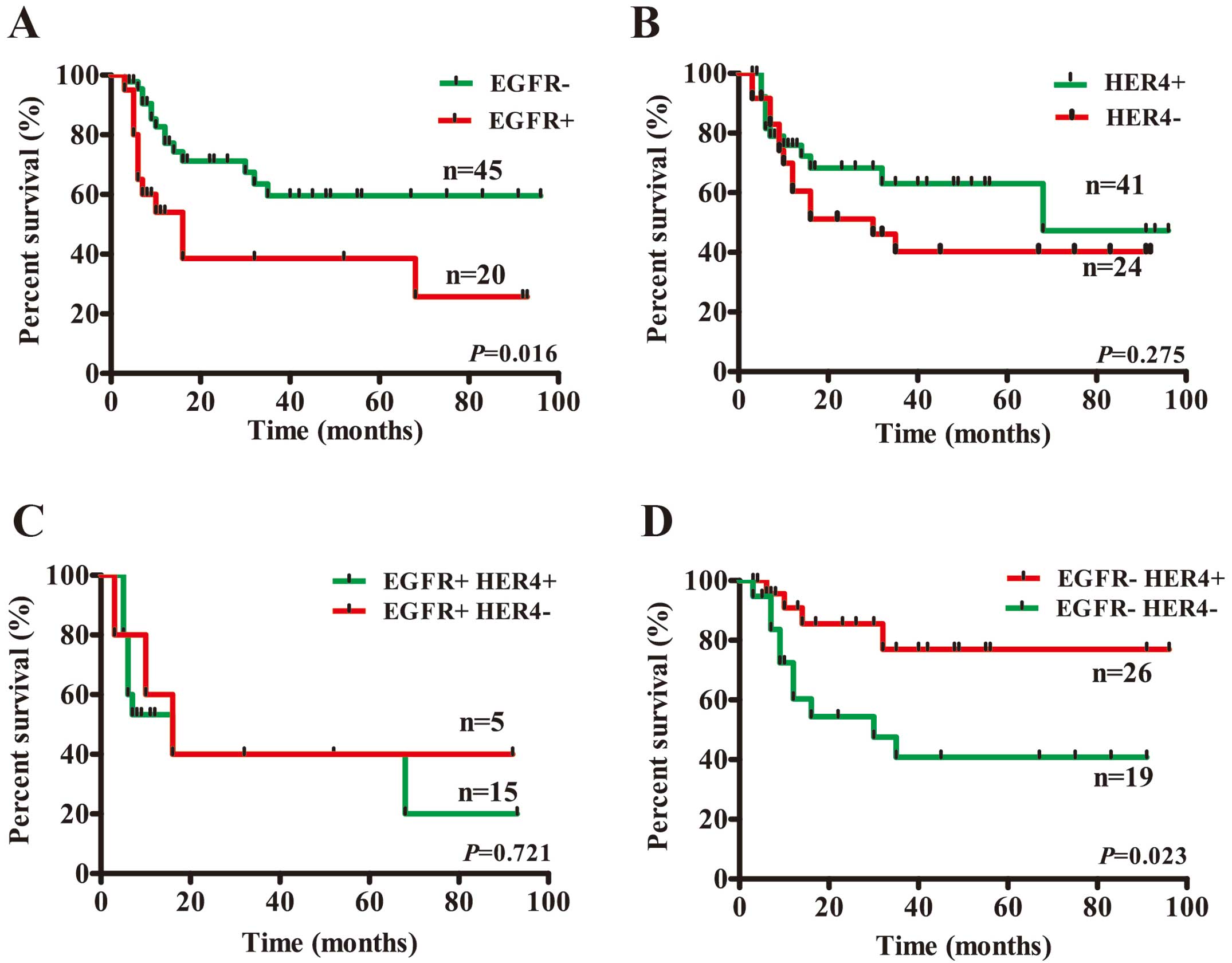
Overall, no statistical significance was identified
between HER4 expression and overall survival in CCA by univariate
analysis. However, HER4 expression was identified as a prognostic
factor (P=0.023) in EGFR-negative IHCC cases (Table VI). While in EGFR-positive cases,
HER4 expression showed no influence on survival rate (P=0.721)
(Fig. 2). In EHCC, no significant
correlation was present between HER4 expression and overall
survival both in EGFR+ and EGFR− cases (data not shown).
| Table VIUnivariate analysis of HER4
expression in patients with IHCC stratified by EGFR. |
Table VI
Univariate analysis of HER4
expression in patients with IHCC stratified by EGFR.
| IHCC | |
|---|
|
| |
|---|
| N | Survival % | P-value |
|---|
| EGFR− |
| HER4− | 19 | 47.4 | |
| HER4+ | 26 | 84.6 | 0.023 |
| EGFR+ |
| HER4− | 5 | 40.0 | |
| HER+ | 15 | 40.0 | 0.721 |
Due to a very limited number of HER2-positive cases,
survival analysis was not performed for HER2 expression either in
univariate or multivariate analysis.
HER4 inhibits cellular proliferation,
migration and invasion in CCA cell lines
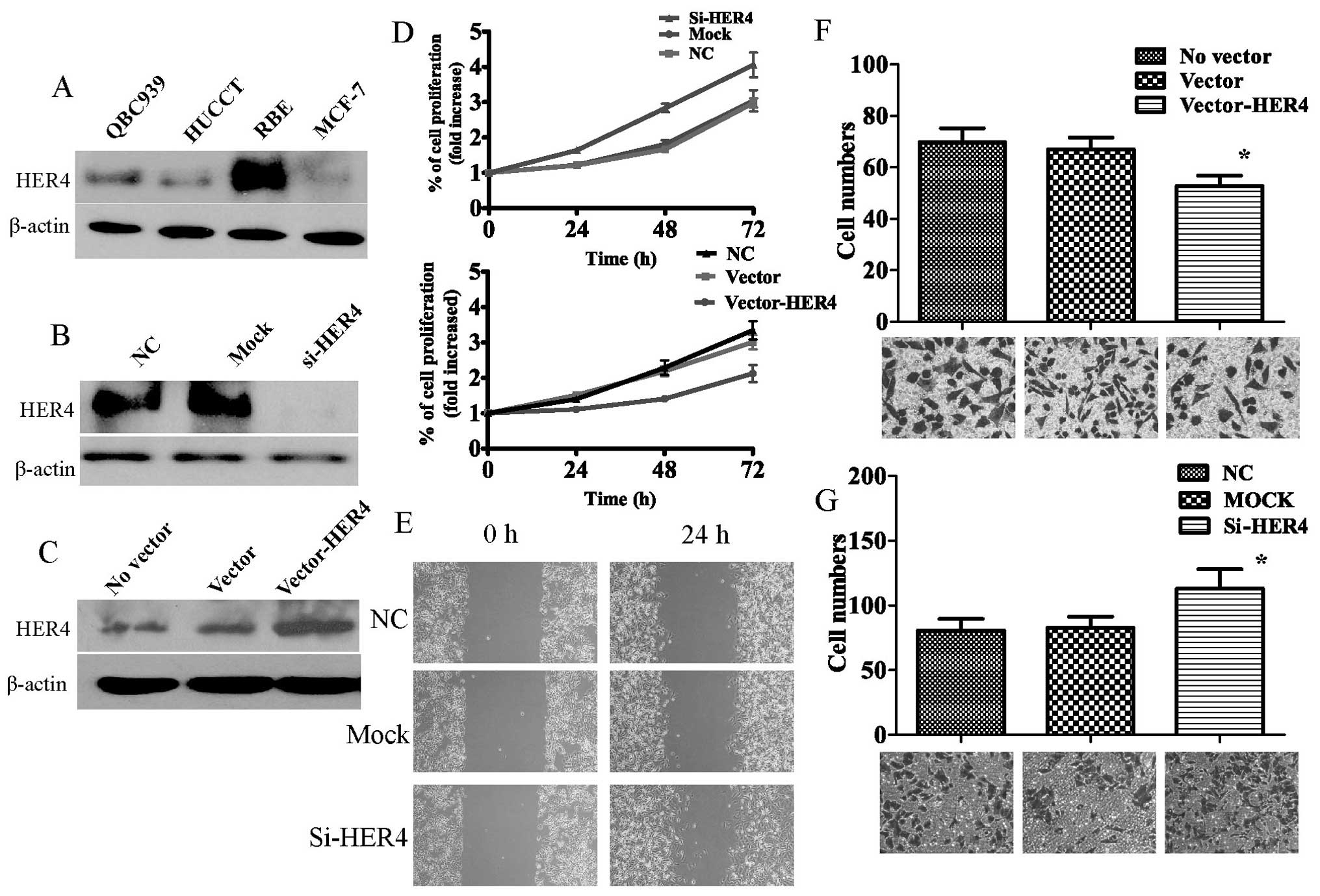
Using the MTT assay, we found that siRNA knockdown
of HER4 in RBE cells significantly increased cell proliferation at
24 and 48 h after treatment compared with negative controls (n=3,
P<0.05; Fig. 3). Wound healing
assay indicated that siRNA-HER4-transfected RBE cells displayed a
significant decrease in cell migration ability compared to control
conditions (P<0.05; Fig. 3). To
further examine the effect of HER4 on cell invasion,
siHER4-transfected RBE and PcDNA3.1-HER4-transfected HuCCT-1 cells
were cultured in a Transwell apparatus. The percentage of migrated
cells was significantly less in siHER4-treated groups when compared
to the negative control groups (for both P<0.05).
PcDNA3.1-HER4-transfected HuCCT-1 cell line showed increased
percentage of migrated cells (both P<0.05; Fig. 3).
Discussion
To the best of our knowledge, this is the largest
cohort of CCA patients reported thus far, investigating aberrations
of all four EGFR family members. Previously, only one study
reported expression and clinical significance of all EGFR family
members in a small cohort (n=56) of IHCC (24). In the present study, we firstly
confirmed a strong link of EGFR to the biological aggressiveness in
Chinese CCA patients. The prevalence of EGFR overexpression in CCA
has been presented in various reports, ranging from 8.1 to 81%
(15,17,25,26).
This broad range might be explained by the lack of a standardized
methodology, different standards of interpretation or differences
in tumor location. Similar to Yoshikawa et al (15), we detected the frequency of EGFR
aberration in our cohort was 30.8% in IHCC and 20.9% in EHCC cases.
The EGFR overexpression is linked to poor histological
differentiation and lymph node metastasis. More importantly, EGFR
expression is an independent prognostic indicator in Chinese
patients with IHCC. In a systematic review of prognostic biomarkers
in CCA, Ruys et al (27)
showed that EGFR, along with other biomarkers fascin, MUC1, MUC4
and p27 is associated with survival in patients with resected CCA.
To date, EGFR expression in CCA makes it a promising target for
EGFR-directed therapy. Preliminary studies suggest that combination
chemotherapy using anti-EGFR antibody, along with conventional
therapeutic agents, increase the efficacy of treatment (28). In a recent clinical phase II trial,
Chiorean et al (29)
suggested that anti-EGFR therapy remained an important option in
advanced biliary cancers but only with a molecular ‘targeted’
approach.
In the present study, another key finding was that
HER4 expression is a prognostic factor in EGFR-negative IHCC cases.
To date, the role of HER4 expression in cancer remains
controversial. Some studies demonstrated that HER4 acts as a tumor
suppressor in aggressive cancers such as the breast, prostate,
pancreas and larynx (8,30–33).
HER4 expression is antiproliferative and clinically associated with
increased survival, reduced recurrence, as well as antagonizing the
effect of HER2. By contrast, some reports suggested HER4
overexpression in medulloblastomas and ependymomas possesses
oncogenic activities (34,35). In the present study, we provided
several lines of evidence to show the antitumor role of HER4
expression in CCA. Firstly, HER4 expression seems to be related to
earlier clinical stage and lower tendency of lymph node metastasis,
although these did not reach significant difference. Secondly,
in vitro data demonstrated that HER4 knockdown in RBE cells
promotes cellular proliferation, motility and invasion. Thirdly, a
prognostic role of HER4 expression was identified in a subset of
IHCC cases. These findings reinforce the importance of molecular
subclassification (whether through HER4 status or other relevant
biomarkers) of EGFR-negative CCA patients in clinical trials. The
reason why HER4 expression was not a prognostic factor in EHCC
might be partially explained by variables associated with their
anatomic behavior and methods of surgery. Our data suggested HER4
might play a tumor suppressor role in a selected subset of Chinese
CCA patients.
One aim of the present study was to examine the
entire spectrum of CCA to define anatomic site-related similarities
and differences. Of note, the frequencies of overexpression of
EGFR, HER3 and HER4 in EHCC are similar to those of IHCC cases.
However, we found that both HER2 overexpression and HER2
amplification were exclusively identified in EHCC cases. Overall,
HER2 overexpression and amplification has been found in a range
between 5 and 76% in CCA (16,24,25,36).
Some authors suggested that HER2 overexpression is due to gene
deregulation rather than gene amplification as in some studies
there is no strict correlation between protein expression and gene
amplification (37). However,
consistent with Harder et al (11), our data suggested that in CCA cases
with high HER2 expression there is also a good correlation between
overexpression and amplification. Considering the lack of
correlation between HER2 overexpression with other clinical
variables, out data suggested that HER2 overexpression might play a
role in a subset of EHCC cases, but has limited significance in the
development and progression of CCA.
HER3 overexpression was present in 12.3% (8/65) of
IHCC and 11.8% (13/110) of EHCC. The prevalence in this study is
significantly lower than almost all of those in previous studies
(19,24). HER3 overexpression has been reported
to be associated with a poorer outcome in melanomas (38) and lung and gastric cancers (38–40).
In CCA, Lee et al (19)
suggested that HER3 overexpression is correlated with decreased
patient survival and represents a prognostic indicator of patients
with EHCC. Notably, they observed the co-overexpression of HER2 and
HER3 in subsets of EHCC patients. However, HER3 expression did not
show significant prognostic value in the present study. Thus, the
exact biological role of HER3 in CCA merits further
investigation.
In summary, our results revealed expression of the
EGFR family members in CCA development and progression. CCAs
differentially express HER2 protein based on tumor location. For
the first time, our study suggested that HER4 is a prognostic
factor in subset of EGFR-negative CCA patients with IHCC.
Determination of HER4 expression status allows stratification of
CCA patients into different survival categories.
Acknowledgements
The present study was supported by the National
Natural Science Foundation of China (grant nos. 81072110 and
81171951), the Independent Innovation Foundation of Shandong
University (grant no. 2010TB012), the Scientific Research
Foundation for Returned Scholars, Ministry of Education of
China.
References
|
1
|
Malhi H and Gores GJ: Cholangiocarcinoma:
modern advances in understanding a deadly old disease. J Hepatol.
45:856–867. 2006. View Article : Google Scholar : PubMed/NCBI
|
|
2
|
Khan SA, Thomas HC, Davidson BR and
Taylor-Robinson SD: Cholangiocarcinoma. Lancet. 366:1303–1314.
2005. View Article : Google Scholar : PubMed/NCBI
|
|
3
|
Anderson C and Kim R: Adjuvant therapy for
resected extrahepatic cholangiocarcinoma: a review of the
literature and future directions. Cancer Treat Rev. 35:322–327.
2009. View Article : Google Scholar : PubMed/NCBI
|
|
4
|
Zhang H, Berezov A, Wang Q, et al: ErbB
receptors: from oncogenes to targeted cancer therapies. J Clin
Invest. 117:2051–2058. 2007. View
Article : Google Scholar : PubMed/NCBI
|
|
5
|
Zaczek A, Brandt B and Bielawski KP: The
diverse signaling network of EGFR, HER2, HER3 and HER4 tyrosine
kinase receptors and the consequences for therapeutic approaches.
Histol Histopathol. 20:1005–1015. 2005.PubMed/NCBI
|
|
6
|
Verma S, Miles D, Gianni L, et al:
Trastuzumab emtansine for HER2-positive advanced breast cancer. N
Engl J Med. 367:1783–1791. 2012. View Article : Google Scholar : PubMed/NCBI
|
|
7
|
Franklin WA, Veve R, Hirsch FR, Helfrich
BA and Bunn PA Jr: Epidermal growth factor receptor family in lung
cancer and premalignancy. Semin Oncol. 29:3–14. 2002. View Article : Google Scholar : PubMed/NCBI
|
|
8
|
Suo Z, Risberg B, Kalsson MG, et al: EGFR
family expression in breast carcinomas. c-erbB-2 and c-erbB-4
receptors have different effects on survival. J Pathol. 196:17–25.
2002. View Article : Google Scholar : PubMed/NCBI
|
|
9
|
Wei Q, Shui Y, Zheng S, et al: EGFR, HER2
and HER3 expression in primary colorectal carcinomas and
corresponding metastases: Implications for targeted radionuclide
therapy. Oncol Rep. 25:3–11. 2011.PubMed/NCBI
|
|
10
|
Awaya H, Takeshima Y, Furonaka O, Kohno N
and Inai K: Gene amplification and protein expression of EGFR and
HER2 by chromogenic in situ hybridisation and immunohistochemistry
in atypical adenomatous hyperplasia and adenocarcinoma of the lung.
J Clin Pathol. 58:1076–1080. 2005. View Article : Google Scholar : PubMed/NCBI
|
|
11
|
Harder J, Waiz O, Otto F, et al: EGFR and
HER2 expression in advanced biliary tract cancer. World J
Gastroenterol. 15:4511–4517. 2009. View Article : Google Scholar : PubMed/NCBI
|
|
12
|
Jaiswal BS, Kljavin NM, Stawiski EW, et
al: Oncogenic ERBB3 mutations in human cancers. Cancer Cell.
23:603–617. 2013. View Article : Google Scholar : PubMed/NCBI
|
|
13
|
Fujiwara S, Ibusuki M, Yamamoto S,
Yamamoto Y and Iwase H: Association of ErbB1–4 expression in
invasive breast cancer with clinicopathological characteristics and
prognosis. Breast Cancer. Oct 26–2012.(Epub ahead of print).
|
|
14
|
Memon AA, Sorensen BS, Melgard P, Fokdal
L, Thykjaer T and Nexo E: Expression of HER3, HER4 and their ligand
heregulin-4 is associated with better survival in bladder cancer
patients. Br J Cancer. 91:2034–2041. 2004. View Article : Google Scholar : PubMed/NCBI
|
|
15
|
Yoshikawa D, Ojima H, Iwasaki M, et al:
Clinicopathological and prognostic significance of EGFR, VEGF, and
HER2 expression in cholangiocarcinoma. Br J Cancer. 98:418–425.
2008. View Article : Google Scholar : PubMed/NCBI
|
|
16
|
Kim HJ, Yoo TW, Park DI, et al: Gene
amplification and protein overexpression of HER-2/neu in human
extrahepatic cholangiocarcinoma as detected by chromogenic in situ
hybridization and immunohistochemistry: its prognostic implication
in node-positive patients. Ann Oncol. 18:892–897. 2007. View Article : Google Scholar
|
|
17
|
Shafizadeh N, Grenert JP, Sahai V and
Kakar S: Epidermal growth factor receptor and HER-2/neu status by
immunohistochemistry and fluorescence in situ hybridization in
adenocarcinomas of the biliary tree and gallbladder. Hum Pathol.
41:485–492. 2010. View Article : Google Scholar : PubMed/NCBI
|
|
18
|
Lubner SJ, Mahoney MR, Kolesar JL, et al:
Report of a multicenter phase II trial testing a combination of
biweekly bevacizumab and daily erlotinib in patients with
unresectable biliary cancer: a phase II Consortium study. J Clin
Oncol. 28:3491–3497. 2010. View Article : Google Scholar : PubMed/NCBI
|
|
19
|
Lee HJ, Chung JY, Hewitt SM, Yu E and Hong
SM: HER3 overexpression is a prognostic indicator of extrahepatic
cholangiocarcinoma. Virchows Arch. 461:521–530. 2012. View Article : Google Scholar : PubMed/NCBI
|
|
20
|
Jarnagin WR, Klimstra DS, Hezel M, et al:
Differential cell cycle-regulatory protein expression in biliary
tract adenocarcinoma: correlation with anatomic site, pathologic
variables, and clinical outcome. J Clin Oncol. 24:1152–1160. 2006.
View Article : Google Scholar
|
|
21
|
Edge SB and Compton CC: The American Joint
Committee on Cancer: the 7th edition of the AJCC cancer staging
manual and the future of TNM. Ann Surg Oncol. 17:1471–1474. 2010.
View Article : Google Scholar : PubMed/NCBI
|
|
22
|
Wang L, Zhang J, Yang X, et al: SOX4 is
associated with poor prognosis in prostate cancer and promotes
epithelial-mesenchymal transition in vitro. Prostate Cancer
Prostatic Dis. 16:301–307. 2013. View Article : Google Scholar : PubMed/NCBI
|
|
23
|
Rajkumar T, Stamp GW, Pandha HS, Waxman J
and Gullick WJ: Expression of the type 1 tyrosine kinase growth
factor receptors EGF receptor, c-erbB2 and c-erbB3 in bladder
cancer. J Pathol. 179:381–385. 1996. View Article : Google Scholar : PubMed/NCBI
|
|
24
|
Ito Y, Takeda T, Sasaki Y, et al:
Expression and clinical significance of the erbB family in
intrahepatic cholangiocellular carcinoma. Pathol Res Pract.
197:95–100. 2001. View Article : Google Scholar : PubMed/NCBI
|
|
25
|
Nakazawa K, Dobashi Y, Suzuki S, Fujii H,
Takeda Y and Ooi A: Amplification and overexpression of c-erbB-2,
epidermal growth factor receptor, and c-met in biliary tract
cancers. J Pathol. 206:356–365. 2005. View Article : Google Scholar : PubMed/NCBI
|
|
26
|
Endreffy E, Burg K, Gyurkovits K, Kalman
M, Laszlo A and Rasko I: Allele frequencies of cystic
fibrosis-linked markers and F508 deletion in affected Hungarian
families. Acta Paediatr Hung. 32:101–113. 1992.PubMed/NCBI
|
|
27
|
Ruys AT, Groot Koerkamp B, Wiggers JK,
Klumpen HJ, Ten Kate FJ and van Gulik TM: Prognostic biomarkers in
patients with resected cholangiocarcinoma: a systematic review and
meta-analysis. Ann Surg Oncol. 21:487–500. 2013. View Article : Google Scholar : PubMed/NCBI
|
|
28
|
Lee J, Park SH, Chang HM, et al:
Gemcitabine and oxaliplatin with or without erlotinib in advanced
biliary-tract cancer: a multicentre, open-label, randomised, phase
3 study. Lancet Oncol. 13:181–188. 2012. View Article : Google Scholar : PubMed/NCBI
|
|
29
|
Chiorean EG, Ramasubbaiah R, Yu M, et al:
Phase II trial of erlotinib and docetaxel in advanced and
refractory hepatocellular and biliary cancers: Hoosier Oncology
Group GI06–101. Oncologist. 17:132012.PubMed/NCBI
|
|
30
|
Barnes NL, Khavari S, Boland GP, Cramer A,
Knox WF and Bundred NJ: Absence of HER4 expression predicts
recurrence of ductal carcinoma in situ of the breast. Clin Cancer
Res. 11:2163–2168. 2005. View Article : Google Scholar : PubMed/NCBI
|
|
31
|
Edwards J, Traynor P, Munro AF, Pirret CF,
Dunne B and Bartlett JM: The role of HER1-HER4 and EGFRvIII in
hormone-refractory prostate cancer. Clin Cancer Res. 12:123–130.
2006. View Article : Google Scholar : PubMed/NCBI
|
|
32
|
Thybusch-Bernhardt A, Beckmann S and Juhl
H: Comparative analysis of the EGF-receptor family in pancreatic
cancer: expression of HER-4 correlates with a favourable tumor
stage. Int J Surg Investig. 2:393–400. 2001.
|
|
33
|
Bussu F, Ranelletti FO, Gessi M, et al:
Immunohistochemical expression patterns of the HER4 receptors in
normal mucosa and in laryngeal squamous cell carcinomas:
antioncogenic significance of the HER4 protein in laryngeal
squamous cell carcinoma. Laryngoscope. 122:1724–1733. 2012.
View Article : Google Scholar
|
|
34
|
Gilbertson RJ, Perry RH, Kelly PJ, Pearson
AD and Lunec J: Prognostic significance of HER2 and HER4
coexpression in childhood medulloblastoma. Cancer Res.
57:3272–3280. 1997.PubMed/NCBI
|
|
35
|
Rickert CH: Prognosis-related molecular
markers in pediatric central nervous system tumors. J Neuropathol
Exp Neurol. 63:1211–1224. 2004.PubMed/NCBI
|
|
36
|
Settakorn J, Kaewpila N, Burns GF and
Leong AS: FAT, E-cadherin, β catenin, HER 2/neu, Ki67
immuno-expression, and histological grade in intrahepatic
cholangiocarcinoma. J Clin Pathol. 58:1249–1254. 2005.
|
|
37
|
Buchler P, Reber HA, Buchler MC, et al:
Therapy for pancreatic cancer with a recombinant humanized
anti-HER2 antibody (herceptin). J Gastrointest Surg. 5:139–146.
2001. View Article : Google Scholar : PubMed/NCBI
|
|
38
|
Reschke M, Mihic-Probst D, van der Horst
EH, et al: HER3 is a determinant for poor prognosis in melanoma.
Clin Cancer Res. 14:5188–5197. 2008. View Article : Google Scholar : PubMed/NCBI
|
|
39
|
Yi ES, Harclerode D, Gondo M, et al: High
c-erbB-3 protein expression is associated with shorter survival in
advanced non-small cell lung carcinomas. Mod Pathol. 10:142–148.
1997.PubMed/NCBI
|
|
40
|
Begnami MD, Fukuda E, Fregnani JH, et al:
Prognostic implications of altered human epidermal growth factor
receptors (HERs) in gastric carcinomas: HER2 and HER3 are
predictors of poor outcome. J Clin Oncol. 29:3030–3036. 2011.
View Article : Google Scholar : PubMed/NCBI
|