Introduction
Endometrial cancer (EC) is the most common
gynecological malignancy in Poland, with nearly 5,500 new cases a
year and well-established risk factors (1,2). The
majority of cases are designated as type I estrogen-dependent
tumors according to the Bokhman’s dualistic model of endometrial
tumorigenesis (3). Another 10–20%
of uterine malignancies, designated as type II carcinomas, follow
the estrogen-unrelated pathway and arise in the background of
atrophic endometrium (4,5). Genetic instability and molecular
alterations have been systematically investigated in ECs, and it is
known that four major genetic changes are generally responsible for
endometrial carcinogenesis: silencing of the PTEN
tumor-suppressor gene, presence of microsatellite instability due
to alterations of the mismatch repair genes, mutation of the
K-ras proto-oncogene and alteration of the β-catenin
gene. Subsequent steps in studying the molecular aspects of
carcinogenesis should be carried out with the aim to provide a
better understanding of the influence of known gene mutations on
expression of other genes as well as on metabolic processes that
have not yet been investigated (6–9).
The invasive phenotype is crucial for the ability of
cancer cells to infiltrate the surrounding tissue and to
metastasize. During neoplastic transformation, cancer cells cross
the basement membrane (BM), the extracellular matrix (ECM) and
vessel walls (10,11). ECM components such as glycoproteins,
proteoglycans and other proteins responsible for cellular signaling
play a crucial role in carcinogenesis (12,13).
The most important changes during carcinogenesis are observed in
the BM. The BM separates from the stroma which leads to defects in
its continuity (14). BM is a layer
50–100 nm thick, composed of different proteins (collagen type IV,
laminin, nidogen and perlecan). Collagen type IV and laminin are
responsible for stabilization of the BM, whereas nidogen and
perlecan play a crucial role in extracellular signaling. In
addition to those four major proteins, many other proteins are
involved in forming the BM, for example: fibronectin, fibulin,
agryne, tenascins, other types of collagen and osteonectin, all of
which are responsible for ECM specificity (15–21).
Advancement in molecular genetics in the field of
microarrays and macroarrays has made the analysis of gene
interactions possible. Techniques based on the large scale
measurement of mRNA expression can be easily applied for evaluating
the complexity and influence of gene expression on cellular
metabolism and processes leading to neoplastic transformation
(22,23). Continual advancement of such
techniques, along with rigorous analysis, is the key to identifying
the most relevant genetic changes and correlating them with patient
outcome. Such research is crucial for the development of tests with
the ability to detect cancer early, as well as for the treatment of
EC (24–26).
The aim of the present study was to investigate the
expression profiles, as measured by cDNA macroarrays, of genes
coding for ECM proteins in EC.
Materials and methods
Study group
The present study was approved by the local ethics
committee, and all patients signed informed consent forms for
surgery and tissue sampling for scientific investigation.
Endometrial tissue specimens were collected from 49 patients who
underwent surgery between 2002 and 2005 at The Second Department of
Gynecology, Medical University of Lublin, Poland. Forty patients
underwent surgery due to EC (cases) and 9 patients due to uterine
leiomyomas (controls).
Macroarray analysis
RNA was isolated according to TRI
reagent® protocol (Sigma-Aldrich, USA) and then frozen
at −80°C for further preparation. The quality of RNA samples was
assessed by gel electrophoresis, and quantitative analysis was
performed using a GeneQuant spectrophotometer (Pharmacia, Sweden).
Absorption was measured at a 260-nm wavelength, and 1 OD was
equivalent to 40 μg of RNA. Reverse transcription of RNA and cDNA
labeling were performed with the BD RiboQuant™ RPA system (BD
Bioscences-Pharmingen, San Jose, CA, USA) and CDS Primer Mix (BD
Biosciences, Clontech, Palo Alto, CA, USA). PCR reaction was then
performed using the BD Atlas™ NucleoSpin® extraction
kit. In order to perform gene expression profiling, Atlas Human
Cancer 1.2 array membranes were used (cDNA Expression Array
PT3547-3E; BD Biosciences, Clontech; cat. no. 7851-1). These
macroarray membranes contain 1,176 hybridization points for various
genes, of which 31 are specific for ECM proteins. Hybridization
reactions were performed according to the kit manual, and then
membranes were transferred to the exposure cassette BioMax
TranScreen HE with Kodak BioMax MS film (Kodak, USA). Exposure
times varied and depended on the isotopic activity of the cDNA
(between 4 and 7 days). Film development was performed with Kodak
liquid developers (Kodak). Results were digitally read with
AtlasImage™ software (BD Biosciences, Clontech).
Statistical analysis
Statistical analyses, including Shapiro-Wilk,
Kolmogorov-Smirnov and Lilliefors, Student’s t-test, Chi-squared
and Mann-Whitney U tests, and Kruskal-Wallis rank test, were
performed with STATISTICA StatSoft v8.0. Numerical measures were
summarized as median values and quartiles. Statistical significance
was set at α=0.05.
Results
Clinicopathological features
The demographic patient data is presented in
Table I. We found that patients
with stage III ECs were statistically significantly younger
compared to those with stage IC and II Ecs (p<0.02 and
p<0.03, respectively). We also observed that patients with
normal endometrium had their last menstruation at a younger age
compared to those with ECs in stages IB and IC (p<0.03 and
p<0.04, respectively). A statistically significant difference
was also found when comparing the mean age of patients with normal
endometrium to that of patients with ECs. This difference was
likely due to the younger age of patients who underwent surgery for
uterine leiomyomas.
 | Table IPatient data stratified by the FIGO
staging system. |
Table I
Patient data stratified by the FIGO
staging system.
| FIGO stage | Patients, n
(%) | Age ± SD
(years) | Parity ± SD
(n) | FM ± SD | LMP ± SD |
|---|
| IB | 16 (32.6) | 61.6±10.24 | 2.1±1.09 | 13.9±1.84 | 51.2±3.36 |
| IC | 14 (28.6) | 69.1±8.99 | 2.9±2.27 | 14.8±1.75 | 51.3±3.24 |
| II | 5 (10.2) | 67.7±4.99 | 2.0±0.82 | 14.5±1.00 | 51.5±2.38 |
| III | 5 (10.2) | 52.2±3.77a | 1.2±1.26 | 14.0±0.82 | 50.7±2.22 |
| Normal
endometrium | 9 (18.4) | 50.1±7.56b | 2.0±1.12 | 14.3±1.66 | 47.6±3.03c |
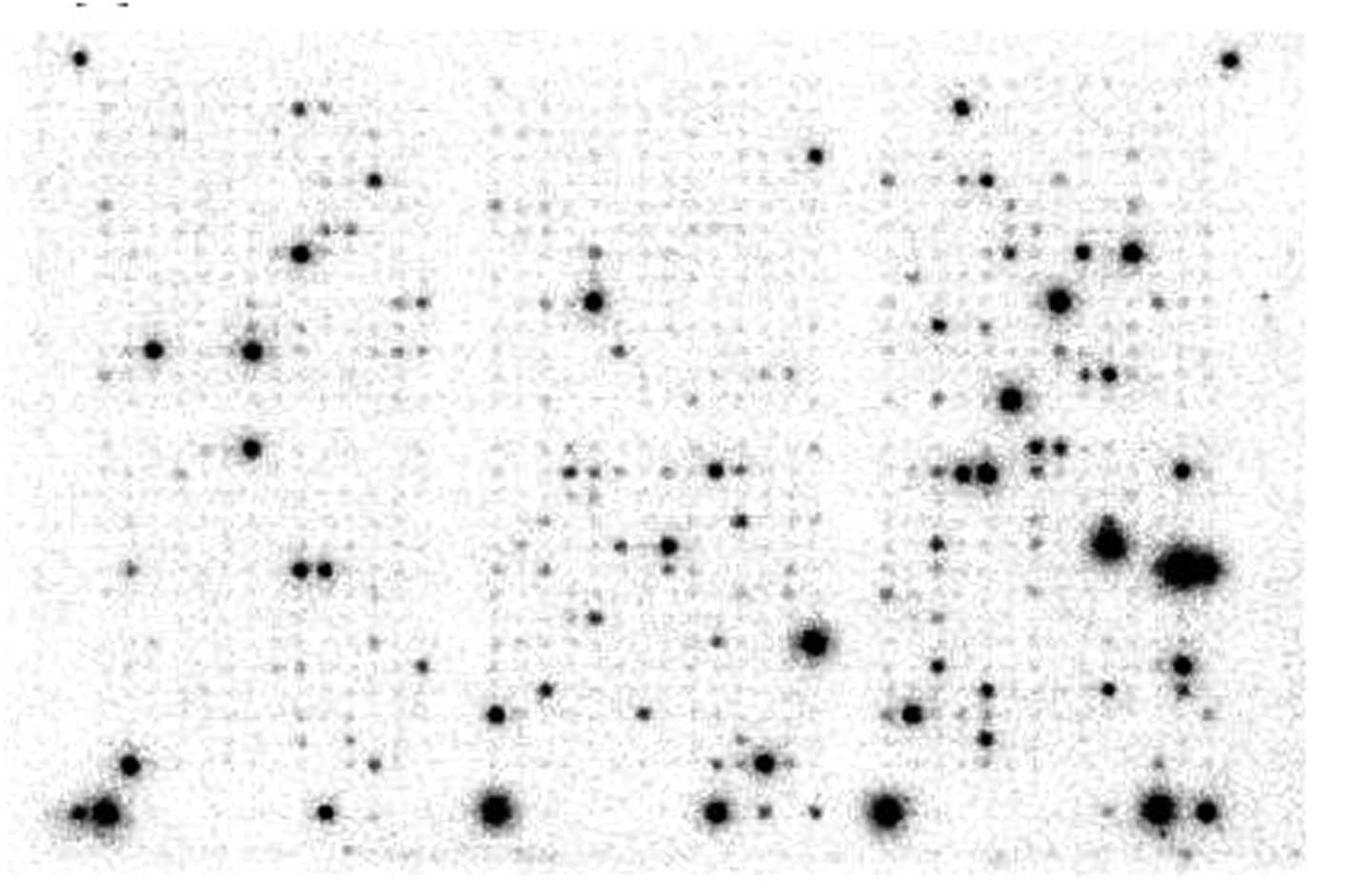
Macroarray study
The level of ECM protein gene expression in normal
endometrial tissue was compared to expression of these genes in EC
specimens. Results of the gene expression in ECs are presented in
Table II. In general,
statistically significant differences were found in regards to the
expression of aggrecan, collagen type VIII chain α1, collagen type
XI chain α2, vitronectin, nidogen and tenascin R gene expression
stratified by clinical stage according to the International
Federation of Gynecology and Obstetrics (FIGO) classification. In
all cases analyzed, gene overexpression was noted in advanced stage
ECs compared to early stage tumors. Example of a macroarray
membrane after isotopic labeling is shown in Fig. 1.
| Table IIStatistical descriptive analysis of
the investigated samples according to FIGO classification. |
Table II
Statistical descriptive analysis of
the investigated samples according to FIGO classification.
| Gene | FIGO stage | Median | Lower quartile | Upper quartile | Range | Analysis |
|---|
|
|
|---|
| Min | Max | H | P-value |
|---|
| Aggrecan | I | 0.00 | 0.00 | 0.00 | 0.00 | 0.22 | 6.60 | 0.04a |
| II | 0.00 | 0.00 | 0.00 | 0.00 | 0.18 | | |
| III | 0.07 | 0.00 | 2.24 | 0.00 | 2.89 | | |
| Collagen type VIII
α1 | I | 0.00 | 0.00 | 0.00 | 0.00 | 0.16 | 9.06 | <0.01a |
| II | 0.00 | 0.00 | 0.00 | 0.00 | 0.00 | | |
| III | 0.00 | 0.00 | 0.39 | 0.00 | 0.46 | | |
| Collagen type XI
α2 | I | 0.00 | 0.00 | 0.00 | 0.00 | 0.00 | 14.36 | <0.01a |
| II | 0.00 | 0.00 | 0.00 | 0.00 | 0.00 | | |
| III | 0.00 | 0.00 | 0.37 | 0.00 | 1.11 | | |
| Vitronectin | I | 0.00 | 0.00 | 0.00 | 0.00 | 0.00 | 14.36 | <0.01a |
| II | 0.00 | 0.00 | 0.00 | 0.00 | 0.00 | | |
| III | 0.00 | 0.00 | 0.34 | 0.00 | 0.53 | | |
| Nidogen | I | 0.00 | 0.00 | 0.00 | 0.00 | 0.00 | 7.00 | 0.03a |
| II | 0.00 | 0.00 | 0.00 | 0.00 | 0.00 | | |
| III | 0.00 | 0.00 | 0.00 | 0.00 | 1.11 | | |
| Tenascin R | I | 0.00 | 0.00 | 0.00 | 0.00 | 0.00 | 7.00 | 0.03a |
| II | 0.00 | 0.00 | 0.00 | 0.00 | 0.00 | | |
| III | 0.00 | 0.00 | 0.00 | 0.00 | 1.01 | | |
Discussion
A variety of genetic alterations, in particular
those affecting proteins bound to the cell membrane and responsible
for cell adhesion and signaling transduction, are responsible for
human carcinogenesis. More accurate tools enabling deeper insight
into the molecular mechanisms play a crucial role in predicting
cancer development and progression. It is of utmost importance to
investigate the interactions between different genes, not only
changes in expression of individual ones. The ideal tools for gene
expression profiling appear to be cDNA macroarrays or microarrays.
Along with proper statistical analysis of acquired data it will
became the ‘golden standard’.
Aggrecan
Aggrecan is a major structural component of
cartilage, and consists of a protein core of ~220 kDa, with
covalently attached glycosaminoglycan side chains, responsible for
its unique biochemical properties, integrity and functionality
(27,28). Aggrecan is a member of the
proteoglycans and influences the adhesive and mitotic activity of
cancer cells. Eshchenko et al (29) found that expression of another
proteoglycan, syndecan, was significantly higher in breast cancer
cells compared to that in normal tissues, whereas aggrecan was
expressed at the same level in both types of tissues. On the other
hand, Japanese researchers (30)
reported that mRNA alternative splicing is associated with the
malignant transformation of chondrocytes. In the present study, a
statistically significant increase in expression was found in
cancer cells in stage III according to the FIGO classification.
Notably, the expression level was not found to be altered by the
histological type of EC (p=0.43). In addition, aggrecan was
expressed only in G2 and G3 uterine carcinomas. These results
suggest that aggrecan may be an important marker of clinical cancer
progression. In order to confirm this result, investigation using
more samples should be performed, and the association between
aggrecan protein levels and patient survival should also be
evaluated.
Vitronectin
Integrins are heterodimeric glycoproteins that have
been found to undergo dynamic temporal and spatial changes in
distribution in the endometrium during the menstrual cycle in
women. They participate in a wide range of physiological processes,
including embryogenesis, wound healing, the immune response and the
behavior of malignant cells (31).
Moreover, the expression of vitronectin
(αvβ3) in melanoma cells has been associated
with increased invasiveness (32).
Vitronectin is one of the integrins strictly linked to
cycle-dependent changes of endometrial cells. Highest expression
was found in the luteal phase whether it was not found after
menopause or in cancer tissues (33,34).
Vitronectin expression was also discovered in patients suffering
from endometriosis and infertility. Lessey et al (35) investigated 241 endometrium specimens
of patients affected by endometriosis between day 19 and 21 of the
menstrual cycle and found that lack of β3 expression, a
subunit of vitronectin, was responsible for the progression of
endometriosis (p=0.02). Our results of integrin
αvβ3 gene expression, which was observed only
in stage III EC cells, suggest that vitronectin plays a role in
myoinvasion and increases the ability of cancer cells to
metastasize.
Tenascin
Tenascins are a family of large multimeric ECM
proteins present in the connective tissue of vertebrates. To date,
four tenascins termed tenascin C, R, X and W have been identified
in humans. Contrary to many other ECM proteins, tenascins promote
only weak cell adhesion and do not activate cell spreading. They
have been classified as anti-adhesive, adhesion-modulating, or even
repellent ECM proteins (36). These
proteins are also used in bioengineering due to their ability to
influence the cell shape (20).
Tenascin C (myotendinous antigen, cytotactin) is
synthesized in the central nervous system as well as in peripheral
nerves. The most significant expression has been observed during
embryogenesis, organo-genesis and during regeneration of mature
tissues. Tenascin C is mostly responsible for blocking the adhesion
of various cells to fibronectin fibers, which makes cell migration
possible in embryogenesis or partially injured nerve regeneration
and axon development (36–38).
Tenascin R (restrictin, janusin) is
only synthesized in the central nervous system (39). The biological functions of this
protein include the development of new nerve connections and
neurite migration in ECM (40,41).
We found that the tenascin R gene was expressed only in stage III
human uterine carcinomas, which may be an important sign for
acquiring an invasive cancer phenotype.
Nidogen
Nidogen, a glycoprotein, is a binding molecule that
links together BM components and is responsible for establishing
proper connections within this subtile structure. Nidogen is a
single polypeptide chain, folded into two N-terminal globular
domains, a C-terminal globular domain and connecting rod-like
segments and makes up 3% of extracellular masses (42). There are two separate nidogen
particles, encoded by two different genes [nidogen 1 (NID1) and
nidogen 2 (NID2)] with molecules of a different size (18,43).
Nidogen 2 is more common and it is found mostly in vessels. Both
types are responsible for the proper functioning of lungs and blood
vessels (44). Additional functions
assigned to nidogen include increased cell adhesion ability,
neutrophil chemotaxis regulation and necrosis; it also influences
neoangiogenesis and trophoblastic development (18,45).
To date, altered expression of nidogen has only been observed in
the kidney and pancreatic cancer cells (46). According to the functions of this
protein and our data, it may be concluded that due to the finding
of expression of this gene only in stage III EC cells it may be
responsible for increased neoangiogenesis; in addition, it may play
a role in defending against infiltration of cancer cells into the
BM. Probably, those two processes are synergistic and consist of
simultaneous neoangiogenesis in cancer tissues and the necrosis of
invaded healthy ECM cells.
Collagens
The collagen family of structural proteins make up
25% of connective tissue components. There are 25 different types
of collagens in the human body responsible for stabilizing the BM
and ECM. They play a pivotal role in proper functioning and spatial
organization of organs, tissues and blood vessels (15).
Type VIII collagen is produced by intraepithelial
cells, keratinocytes and fatty cells. Increased concentrations of
this collagen were found in migrating smooth muscle cells and
intraepithelial vessel cells. It is hypothesized that it increases
the production of metalloproteinases (MMPs), which increase cell
migration ability (47,48). Collagen type VIII mRNA
overexpression was observed in the region surrounding new blood
vessel formation (49).
Type XI collagen is a fibrillar protein forming the
scaffold for skin, bones, tendons and ligaments (15). In 2008, Halsted et al
(50) published the results of an
immunohistochemical study of 72 breast cancer samples and healthy
tissues from the same patients. They found decreased expression of
α chain collagen XI in cancer tissues when comparing to the
expression in healthy tissues (p<0.01) and in metastasis
compared to primary tumors (p=0.01). Based on these observations,
the authors concluded that decreased expression of collagen may be
helpful in identifying patients presenting with metastases.
Presently, we found increased expression of α types
VIII and XI collagens in EC tissues compared to levels in normal
tissues, yet statistical significance was found only between normal
and stage III ECs.
Finally, the results of our macroarray study of ECM
gene expression profiles may shed some light on the investigational
methods for cancer prognosis. Gene expression levels of the
above-mentioned proteins are likely to be important markers of
clinical cancer progression, metastasis formation and
neoangiogenesis. These functions are responsible for cancer
progression and, if investigated further, may be additional
diagnostic and prognostic tools for women suspected of or suffering
from ECs.
Acknowledgements
This study was supported by a grant from the Lublin
Medical University of Lublin, Lublin, Poland (grant no. 326/14 to
A.S.).
References
|
1
|
Didkowska J, Wojciechowska U and Zatoński
W: Cancer in Poland in 2011. National M. Sklodowska-Curie Cancer
Center; Warsaw: pp. 13–21. 2012
|
|
2
|
Ferlay J, Steliarova-Foucher E,
Lortet-Tieulent J, et al: Cancer incidence and mortality patterns
in Europe: estimates for 40 countries in 2012. Eur J Cancer.
49:1374–1403. 2013. View Article : Google Scholar : PubMed/NCBI
|
|
3
|
Bokhman JV: Two pathogenetic types of
endometrial carcinoma. Gynecol Oncol. 15:10–17. 1983. View Article : Google Scholar : PubMed/NCBI
|
|
4
|
Potischman N, Hoover RN, Brinton LA, et
al: Case-control study of endogenous steroid hormones and
endometrial cancer. J Natl Cancer Inst. 88:1127–1135. 1996.
View Article : Google Scholar : PubMed/NCBI
|
|
5
|
Lax SF, Pizer ES, Ronnett BM and Kurman
RJ: Comparison of estrogen and progesterone receptor, Ki-67, and
p53 immunoreactivity in uterine endometrioid carcinoma and
endometrioid carcinoma with squamous, mucinous, secretory, and
ciliated cell differentiation. Hum Pathol. 29:924–931. 1998.
View Article : Google Scholar : PubMed/NCBI
|
|
6
|
Lax SF: Molecular genetic pathways in
various types of endometrial carcinoma: from a phenotypical to a
molecular-based classification. Virchows Arch. 444:213–223. 2004.
View Article : Google Scholar : PubMed/NCBI
|
|
7
|
Liu FS: Molecular carcinogenesis of
endometrial cancer. Taiwan J Obstet Gynecol. 46:26–32. 2007.
View Article : Google Scholar
|
|
8
|
Wu H, Goel V and Haluska FG: PTEN
signaling pathways in melanoma. Oncogene. 22:3113–3122. 2003.
View Article : Google Scholar : PubMed/NCBI
|
|
9
|
Semczuk A, Berbeć H, Kostuch M, Cybulski
M, Wojcierowski J and Baranowski W: K-ras gene point
mutations in human endometrial carcinoma: correlation with
clinicopathological features and patients’ outcome. J Cancer Res
Clin Oncol. 124:695–700. 1998.
|
|
10
|
Egeblad M and Werb Z: New functions for
the matrix metalloproteinases in cancer progression. Nat Rev
Cancer. 2:163–174. 2002. View
Article : Google Scholar
|
|
11
|
Duffy MJ, Maguire TM, Hill A, McDermott E
and O’Higgins N: Metalloproteinases: role in breast carcinogenesis,
invasion and metastasis. Breast Cancer Res. 2:252–257. 2000.
View Article : Google Scholar : PubMed/NCBI
|
|
12
|
McCawley LJ and Matrisian LM: Matrix
metalloproteinases: multifunctional contributors to tumor
progression. Mol Med Today. 6:149–156. 2000. View Article : Google Scholar : PubMed/NCBI
|
|
13
|
Nabeshima K, Inoue T, Shimao Y and
Sameshima T: Matrix metalloproteinases in tumor invasion: role for
cell migration. Pathol Int. 52:255–264. 2002. View Article : Google Scholar : PubMed/NCBI
|
|
14
|
Gimona M: The microfilament system in the
formation of invasive adhesions. Semin Cancer Biol. 18:23–34. 2008.
View Article : Google Scholar
|
|
15
|
Rhodes JM and Simons M: The extracellular
matrix and blood vessel formation: not just a scaffold. J Cell Mol
Med. 11:176–205. 2007. View Article : Google Scholar : PubMed/NCBI
|
|
16
|
Vanacore RM, Shanmugasundararaj S,
Friedman DB, Bondar O, Hudson BG and Sundaramoorthy M: The α1.α2
network of collagen IV. Reinforced stabilization of the
noncollagenous domain-1 by noncovalent forces and the absence of
Met-Lys cross-links. J Biol Chem. 279:44723–44730. 2004.
|
|
17
|
Aumailley M, Nurcombe V, Edgar D, Paulsson
M and Timpl R: The cellular interactions of laminin fragments. Cell
adhesion correlates with two fragment-specific high affinity
binding sites. J Biol Chem. 262:11532–11538. 1987.PubMed/NCBI
|
|
18
|
Erickson AC and Couchman JR: Still more
complexity in mammalian basement membranes. J Histochem Cytochem.
48:1291–1306. 2000. View Article : Google Scholar : PubMed/NCBI
|
|
19
|
Girós A, Morante J, Gil-Sanz C, Fairén A
and Costell M: Perlecan controls neurogenesis in the developing
telencephalon. BMC Dev Biol. 5:292007.PubMed/NCBI
|
|
20
|
Pankov R and Yamada KM: Fibronectin at a
glance. J Cell Sci. 115:3861–3863. 2002. View Article : Google Scholar : PubMed/NCBI
|
|
21
|
Shin H, Jo S and Mikos AG: Biomimetic
materials for tissue engineering. Biomaterials. 24:4353–4364. 2003.
View Article : Google Scholar : PubMed/NCBI
|
|
22
|
Ohtsuka S, Iwase K, Kato M, et al: An mRNA
amplification procedure with directional cDNA cloning and
strand-specific cRNA synthesis for comprehensive gene expression
analysis. Genomics. 84:715–729. 2004. View Article : Google Scholar : PubMed/NCBI
|
|
23
|
Gamberoni G, Storari S and Volinia S:
Finding biological process modifications in cancer tissues by
mining gene expression correlations. BMC Bioinformatics. 9:62006.
View Article : Google Scholar : PubMed/NCBI
|
|
24
|
Ewis AA, Zhelev Z, Bakalova R, et al: A
history of microarrays in biomedicine. Expert Rev Mol Diagn.
5:315–328. 2005. View Article : Google Scholar : PubMed/NCBI
|
|
25
|
Braunschweig T, Chung JY and Hewitt SM:
Tissue microarrays: bridging the gap between research and the
clinic. Expert Rev Proteomics. 2:325–336. 2005. View Article : Google Scholar : PubMed/NCBI
|
|
26
|
Smid-Koopman E, Blok LJ, Helmerhorst TJ,
et al: Gene expression profiling in human endometrial cancer tissue
samples: utility and diagnostic value. Gynecol Oncol. 93:292–300.
2004. View Article : Google Scholar : PubMed/NCBI
|
|
27
|
Skandalis SS, Theocharis AD, Vynios DH, et
al: Cartilage aggrecan undergoes significant compositional and
structural alterations during laryngeal cancer. Biochim Biophys
Acta. 1760:1046–1053. 2006. View Article : Google Scholar
|
|
28
|
East CJ, Stanton H, Golub SB, Rogerson FM
and Fosang AJ: ADAMTS-5 deficiency does not block aggrecanolysis at
preferred cleavage sites in the chondroitin sulfate-rich region of
aggrecan. J Biol Chem. 282:8632–8640. 2007. View Article : Google Scholar : PubMed/NCBI
|
|
29
|
Eshchenko TY, Rykova VI, Chernakov AE,
Sidorov SV and Grigorieva EV: Expression of different proteoglycans
in human breast tumors. Biochemistry. 72:1016–1020. 2007.PubMed/NCBI
|
|
30
|
Matsui Y, Araki N, Tsuboi H, et al:
Differential expression of aggrecan mRNA isoforms by chondrosarcoma
cells. Anticancer Res. 22:4169–4172. 2002.PubMed/NCBI
|
|
31
|
Albelda SM: Role of integrins and other
cell adhesion molecules in tumor progression and metastasis. Lab
Invest. 68:4–17. 1993.PubMed/NCBI
|
|
32
|
Albelda SM, Mette SA, Elder DE, et al:
Integrin distribution in malignant melanoma: association of the
β3 subunit with tumor progression. Cancer Res.
50:6757–6764. 1990.
|
|
33
|
Lessey BA, Albelda S, Buck CA, et al:
Distribution of integrin cell adhesion molecules in endometrial
cancer. Am J Pathol. 146:717–726. 1995.PubMed/NCBI
|
|
34
|
Castelbaum AJ, Ying L, Somkuti SG, Sun J,
Ilesanmi AO and Lessey BA: Characterization of integrin expression
in a well differentiated endometrial adenocarcinoma cell line
(Ishikawa). J Clin Endocrinol Metab. 82:136–142. 1997.PubMed/NCBI
|
|
35
|
Lessey BA, Castelbaum AJ, Sawin SW, et al:
Aberrant integrin expression in the endometrium of women with
endometriosis. J Clin Endocrinol Metab. 79:643–649. 1994.PubMed/NCBI
|
|
36
|
Chiquet-Ehrismann R: Tenascins. Int J
Biochem Cell Biol. 36:986–990. 2004. View Article : Google Scholar
|
|
37
|
Wehrle B and Chiquet M: Tenascin is
accumulated along developing peripheral nerves and allows neurite
outgrowth in vitro. Development. 110:401–415. 1990.PubMed/NCBI
|
|
38
|
Tucker RP: Abnormal neural crest cell
migration after the in vivo knockdown of tenascin-C expression with
morpholino antisense oligonucleotides. Dev Dyn. 222:115–119. 2001.
View Article : Google Scholar : PubMed/NCBI
|
|
39
|
Joester A and Faissner A: The structure
and function of tenascins in the nervous system. Matrix Biol.
20:13–22. 2001. View Article : Google Scholar : PubMed/NCBI
|
|
40
|
Zacharias U, Leuschner R, Nörenberg U and
Rathjen FG: Tenascin-R induces actin-rich microprocesses and
branches along neurite shafts. Mol Cell Neurosci. 21:626–633. 2002.
View Article : Google Scholar : PubMed/NCBI
|
|
41
|
Becker CG, Schweitzer J, Feldner J, Becker
T and Schachner M: Tenascin-R as a repellent guidance molecule for
developing optic axons in zebrafish. J Neurosci. 23:6232–6237.
2003.PubMed/NCBI
|
|
42
|
Fox JW, Mayer U, Nischt R, et al:
Recombinant nidogen consists of three globular domains and mediates
binding of laminin to collagen type IV. EMBO J. 10:3137–3146.
1991.PubMed/NCBI
|
|
43
|
Chung AE, Dong LJ, Wu C and Durkin ME:
Biological functions of entactin. Kidney Int. 43:13–19. 1993.
View Article : Google Scholar : PubMed/NCBI
|
|
44
|
Kimura N, Toyoshima T, Kojima T and
Shimane M: Entactin-2: a new member of basement membrane protein
with high homology to entactin/nidogen. Exp Cell Res. 241:36–45.
1998. View Article : Google Scholar : PubMed/NCBI
|
|
45
|
Yelian FD, Edgeworth NA, Dong LJ, Chung AE
and Armant DR: Recombinant entactin promotes mouse primary
trophoblast cell adhesion and migration through the Arg-Gly-Asp
(RGD) recognition sequence. J Cell Biol. 121:923–929.
1993.PubMed/NCBI
|
|
46
|
Oivula J, Lohi J, Tani T, et al: Renal
cell carcinomas and pancreatic adenocarcinomas produce nidogen in
vitro and in vivo. J Pathol. 187:455–461. 1999. View Article : Google Scholar : PubMed/NCBI
|
|
47
|
Shuttleworth CA: Type VIII collagen. Int J
Biochem Cell Biol. 29:1145–1148. 1997. View Article : Google Scholar
|
|
48
|
Hou G, Mulholland D, Gronska MA and
Bendeck MP: Type VIII collagen stimulates smooth muscle cell
migration and matrix metalloproteinase synthesis after arterial
injury. Am J Pathol. 156:467–476. 2000. View Article : Google Scholar : PubMed/NCBI
|
|
49
|
Rüger B, Dunbar PR, Hasan Q, et al: Human
mast cells produce type VIII collagen in vivo. Int J Exp Pathol.
75:397–404. 1994.
|
|
50
|
Halsted KC, Bowen KB, Bond L, et al:
Collagen α1(XI) in normal and malignant breast tissue. Mod Pathol.
21:1246–1254. 2008.
|