Introduction
Mammary neoplasms are the most common tumors
observed in female dogs and are the main cause of mortality and
morbidity (1). Mammary tumors in
female dogs provide a suitable model for the study of cancer
biology (2–4) and also for therapeutic agents, since
these animals have tumors with epidemiological and clinical
characteristics, as well as biological behavior similar to mammary
carcinomas in women (5–7).
Many studies have attempted to individualize the
behavior of breast carcinomas by tumor-specific characteristics,
trying to predict whether or not a tumor will respond to proposed
treatment (8,9). Estrogen receptor (ER) and progesterone
receptor (PR) are both predictive and prognostic factors used in
the clinic to customize the management of each breast cancer case
(10). ER-positive and PR-positive
mammary tumors have a better prognosis and capacity to respond to
therapy (11).
In this sense, the identification of therapeutic
agents that can interact with specific molecular markers and
inhibit tumor progression are potentially useful. Exogenous
administration of melatonin appears to play an important role in
tumor growth inhibition (12,13).
The oncostatic properties of melatonin can be exerted through
interference at different levels of the signaling pathways of
estrogen (14). Melatonin may
inhibit estradiol hormone precursors produced in the ovary, thereby
reducing the mitogenic response of estrogen-dependent breast tumor
cells (15). Particularly in breast
cancer, estrogen and melatonin are co-regulators of cell
proliferation. Estrogen promotes proliferation and invasiveness,
whereas melatonin suppresses these effects by decreasing the
mitogenic response in estrogen-dependent tumor cells (16). Thus, the antiestrogenic properties
of melatonin are the basis of its oncostatic action in ER-positive
breast cancer tumors (17,18) and can be an excellent adjunct to
drugs commonly used for the prevention of breast cancer (19).
In addition, melatonin acts by inducing apoptosis.
The induction of apoptosis by melatonin has been described in the
last few years in particular cancer types involving different
action mechanisms (20,21). Melatonin exerts significant
inhibition of apoptotic processes in normal tissues or
non-neoplastic diseases and there is evidence that melatonin may
promote apoptosis in several cancer cell lines (22). Eck et al (23) found that sequential treatment with
melatonin and all-trans-retinoic acid inhibits the
proliferation and induces apoptosis of MCF-7 cells by decreasing
the protein levels of the death suppressor, Bcl-2, and increasing,
although with different time courses, the levels of death
promoters, Bax and Bak (23).
Furthermore, melatonin may exert its physiologic
effects through membrane receptors. This hormone binds to and
activates MT1 and MT2 G-protein-coupled receptors (24). The activated MT1 and MT2 receptors
can reduce proliferation of tumor cells (25). Much of the inhibitory effect of
melatonin on breast cancer cell proliferation is related with its
binding to receptors MT1 and MT2 (26,27).
Thus, melatonin and its receptors may provide a promising avenue
for establishing new therapeutic approaches in human cancer
(12). Furthermore, there are no
studies in the literature, which relate canine mammary tumors with
melatonin receptors. Therefore, we explored the potential
therapeutic value of melatonin in canine estrogen-positive and
estrogen-negative mammary tumors, and related its action to MT1 and
MT2 expression.
Materials and methods
Sample characterization
Tumor samples were collected from 24 female dogs
with mammary neoplasia during the years of 2011 and 2012. After
tumor excision, the animals were followed up from 1 to 18 months,
with a median of 540 days. During the follow-up time, the
veterinarians evaluated tumor metastasis and recurrence, as well as
the cause of death of the animals.
For histopathologic diagnostics, the tumor biopsies
collected were classified according to Misdorp et al
(28) by the Armed Forces Institute
of Pathology (AFIP). The parameters employed for the classification
of clinical tumor staging were in accordance with the TNM system
(size, lymph node involvement, metastasis) established by WHO for
canine mammary gland tumors [modified (29)]: tumor mass size (T): T1, <3 cm;
T2, between 3 and 5 cm; T3, >5 cm; lymph node involvement (N):
N0, no apparent involvement; N1, unilateral involvement; N2,
bilateral involvement; and distant metastasis (M): M0, no evident
metastasis; M1, distant metastasis including non-regional lymph
nodes. Clinical staging was assigned as I, II, III or IV according
to the tumor extension and prognostic establishment.
The presence of local tumor recurrence, metastasis
and death were described and the overall survival was determined
from the date of diagnosis until the date of last follow-up or
death. The cause of death was evaluated by the attending
veterinarian and only female dogs that died of the illness were
included in the group for the study. The dogs that had died of
respiratory failure were diagnosed with lung metastasis as shown by
X-ray. This study was approved by the Ethics Committee of the
Faculdade de Medicina de São José do Rio Preto (protocol no.
001-005222/2010).
Immunohistochemistry technique
Tumor samples were embedded into paraffin blocks and
cut to provide 3-μm sections. The samples were prepared on
silanized glass slides before the paraffin was removed. The
sections were rehydrated in an ascending range of alcohol
concentrations and incubated with 3% hydrogen peroxide for 30 min.
Antigenic recovery was made in a recipient at 95°C in buffer for 35
min for each specific antibody, and then the slides were covered
with bovine serum albumin (BSA) and incubated with the primary
antibody (Table I). After cooling,
the slides were covered with BSA for 30 min and incubated at 4°C
overnight with the antibodies. After being washed with
phosphate-buffered saline (PBS) for 15 min, incubation was carried
out with Starr Trek Universal HRP Detection kit (Medical Biocare,
Concord, CA, USA), consisting of the secondary antibody
‘anti-mouse, rabbit and goat immunoglobulin with biotin’ for 1 h
and ‘streptoavidin complex with peroxidase’ for 30 min, followed by
washes with PBS for 15 min and 0.5% of 3,3′-diaminobenzidine
tetrahydrochloride (DAB; Signet® Laboratories, Dedham,
MA, USA) was applied to the slides for 2–5 min at 20–22°C. The
slides were counterstained with Harris’ hematoxylin for 40 min.
Negative controls were obtained by omitting the primary antibody,
and human kidney or breast cancer tissues served as the internal
positive control in every assay.
 | Table IAntibodies and dilutions. |
Table I
Antibodies and dilutions.
| Antibody | Specificity | Clone | Dilution | Company |
|---|
| ER | Monoclonal
(mouse) | 1D5 | 1:150 | Santa Cruz |
| PR | Monoclonal
(rabbit) | SP42 | 1:400 | Abcam |
| Her2/neu | Polyclonal
(rabbit) | C-18 | 1:800 | Santa Cruz |
| Ki-67 | Monoclonal
(rabbit) | SP6 | 1:200 | Biocare
Medical |
| P53 | Monoclonal
(mouse) | FPS392 | 1:100 | Santa Cruz |
| Caspase-3 | Polyclonal
(rabbit) | N/A | 1:1,000 | Biocare
Medical |
Immunohistochemistry quantification
The slides were photographed and the proteins were
quantified using ImageJ software (NIH, Bethesda, MD, USA) at ×40
magnification under a Nikon Eclipse E200 microscope. For each
sample, three regions of the tumor tissue were selected and 20
points in the tumor cells were marked in each region. In this way
60 different points were analyzed in each sample to obtain an
average relative intensity of immunoreactivity. The values were
expressed in arbitrary units (a.u.) and the mean optical density
(MOD) showing the specific immunostaining intensity at
immunoreactive areas. Cases were considered positive for estrogen
receptors when >10% tumor cells had cytoplasmic staining
(30).
Cell culture
In vitro study
The cell culture was performed with tumor biopsies
of 10 dogs out of the 24 with mammary tumors from the
immunohistochemistry and PCR study. The tumor biopsies were sliced
into microfragments and incubated at 37°C in 5% CO2 in
Dulbecco’s modified Eagle’s medium (DMEM; Cultilab, São Paulo,
Brazil) supplemented with 20% fetal bovine serum (Cultilab), 1%
streptomycin/penicillin/fungizone (Sigma-Aldrich, St. Louis, MO,
USA), 200 μl hydrocortisone (Sigma-Aldrich) and 10 μl epidermal
growth factor (EGF; Sigma-Aldrich). The cells were submitted to
immunocytochemistry for confirmation of the epithelium origin, with
the anti-cytokeratin antibody, resulting in a positive protein
staining.
Cell viability by the
3-(4.5-dimethylthiazol-2-yl)-2.5-diphe-nyltetrazolium bromide (MTT)
assay
The cells from each tumor biopsy were plated into
96-well plates containing 3×10³ cells/well and divided
into 2 groups: control (untreated) and treatment with different
concentrations of melatonin (Sigma-Aldrich) (0.5, 1, 2, 5 and 10
mM) for 24 h. Melatonin was diluted in 0.05% ethanol. In the
control cells, equivalent amounts of ethanol were added as vehicle.
Thereafter, 10 μl of MTT solution (Invitrogen Life Technologies,
Carlsbad, CA, USA) was added to each well and the plates were
incubated at 37°C for 4 h. To solubilize the MTT formazan crystals,
the cells were incubated with dimethylsulfoxide (DMSO;
Sigma-Aldrich) for 10 min at 37°C. Absorbance was measured at 540
nm by an ELISA reader (Thermo Plate, Waltham, MA, USA). Medium was
used as background and subtracted from the samples. Cell viability
(%) was calculated for all groups compared to the control sample.
All experimental samples were in triplicate.
Immunocytochemistry
To confirm the efficacy of the treatment with
melatonin, immunocytochemistry was performed with the
anti-caspase-3 and anti-Ki-67 antibodies. For the procedure, the
canine mammary tumor cell line CMT-U229 was treated with 1 mM of
melatonin. Two treatment groups were established: Group I (control)
containing only cells in culture medium and group II treated also
with 1 mM of melatonin. The cells were thus incubated for 24 h.
Initially, the whole content was taken from the bottles and the
cells were washed with PBS. They were then incubated with
paraformaldehyde 4% fixative and rinsed with PBS. Next, they were
incubated with 3% hydrogen peroxide for 30 min. Antigenic recovery
was carried out in a recipient at 95°C in buffer for 35 min for
each specific antibody, and then the slides were covered with BSA
and incubated with the anti-caspase-3 and anti-Ki-67 primary
antibodies (Table I). After
cooling, the slides were covered with BSA for 30 min and incubated
at 4°C overnight with the antibodies. They were then washed with
PBS for 15 min and incubated with the EasyPath kit (Biocare
Medical, Concord, CA, USA) composed of the secondary antibody. In
the next stage, they were stabilized at room temperature, washed
with PBS buffer solution, and incubated with the EasyPath kit
(Erviegas, São Paulo, SP, Brazil) containing the secondary antibody
(biotinylated anti-mouse, rabbit, goat immunoglobulins). They were
once again rinsed with PBS and incubated with the tertiary antigen
(peroxidase-streptavidin conjugates) and then rinsed one last time
with PBS. The result was revealed using chromogenic substrate (DAB;
Signet Laboratories), 1 drop/ml and hematoxylin for
counter-staining. Finally, the apoptosis and cell proliferation
indices were analyzed.
Apoptosis and cell proliferation
indices
Tumors were categorized in relation to apoptosis and
cellular proliferation according to the staining of caspase-3 and
Ki-67, respectively. For analysis of immunocytochemistry slides,
five areas were photographed at ×40 magnification (center, bottom,
top, left and right regions). Caspase-3- and Ki-67-positive cells
were counted using Image J. At least 75–100 neoplastic cells were
counted. The cut-off of positivity was set as 5% of positive
neoplastic cells.
Gene expression of MT1 and MT2
Quantitative RT-PCR tests were performed in
triplicate using a System Step One Plus (Applied Biosystems, Foster
City, CA, USA). The PCR reactions contained 100 ng cDNA, 10 μl
TaqMan Universal Master Mix, 8 μl of DEPC solution, 1 μl TaqMan
Gene Expression for MT1, MT2 and RPL8 (Applied
Biosystems); and 100 ng cDNA, 10 μl TaqMan Universal Master Mix
(Applied Biosystems), 8 μl of DEPC solution (Applied Biosystems),
100 nmol of each primer, 250 nmol of the probe for RPS19 were
subjected to the following amplification scheme: 50°C for 2 min,
95°C for 10 min, followed by 40 cycles of 95°C for 15 sec and 60°C
for 1 min.
Endogenous control RPS19 (Applied Biosystems)
and RPL8 were used for normalization. The relative
expression values of the genes of interest were determined with
Data Assist v3.0, by the quantification method related to the
average of the normalizing genes used as endogenous control (ΔΔCt)
(31). All samples were tested in
triplicate and the negative control was included in each
reaction.
The gene RPS19 was researched, selected in
PUBMED (http://www.ncbi.nlm.nih.gov/entrez), and synthesized
from canine messenger RNA already sequenced and confirmed. The
design used the program Primer Express. Primers used for
amplification were: RPS19 (endogenous control) sense
(5′-GCCTTCCTCAAAAAGTCTGGG-3′), antisense (5′-GCT
TGCTCCCTACGATGAGAAC-3′) and probe (5′-CCCTGAA TGGGTGGAC-3′)
(GenBank: NG_006583.3). For analyses of the expression of other
genes, TaqMan assays (Applied Biosystems) were used: MT1
(Cf02705306_g1), MT2 (Cf02730504_g1) and RPL8 (endogenous control)
(Cf02663820_m1).
For analyses of MT1 and MT2
expression, the relative quantitation (RQ) value for the control
group (used as reference) was established as unity and are
expressed in log10 scale as zero.
Statistical analysis
The results were submitted for descriptive analysis
to determine normality. For samples with normal distribution, the
Student’s t-test or ANOVA was used, followed by the Bonferroni
test. The Kruskal-Wallis test was used for samples with non-normal
distributions. For all tests, p<0.05 was considered significant.
GraphPad Prism4 (GraphPad Software, La Jolla, CA) and DataAssist
3.0 (Applied Biosystems) were used for the analyses.
Results
Clinical data
The age of the animals varied from 7 to 14 years
(mean, 10 years). There was a prevalence of malignant tumors, which
were represented by 9 (37.5%) tubulopapillary carcinomas, 5 (20.8%)
carcinomas of mixed type, 1 (4.2%) carcinosarcoma, 1 (4.2%) in
situ carcinoma, 1 (4.2%) sarcoma, 1 (4.2%) adenocarcinoma and 1
(4.2%) solid carcinoma with tubular areas. Benign changes were
represented by 2 (8.3%) benign mixed tumors, 1 (4.2%) high-grade
dysplasia, 1 (4.2%) papilloma and 1 (4.2%) adenoma. Among the
clinicopathological characteristics, there was a prevalence of
tumors with clinical stage III and IV (46%); 21 (88%) tumors had
the absence of ulceration, 13 (54%) tumors had time course (range
among the observation of the anomaly by the owner/injury diagnosis
by the veterinarian and removal by surgery) greater than 6 months,
17 (71%) tumors showed moderate vascularization, 19 (79%) had the
absence of tumor recurrence and 22 (92%) did not show metastasis.
All data are documented in Table
II.
| Table IICorrelation between the five antibody
stains with the clinical and pathological factors of the female
dogs with mammary tumors. |
Table II
Correlation between the five antibody
stains with the clinical and pathological factors of the female
dogs with mammary tumors.
| Clinicopathological
factors | No. of female dogs
(%) | ER | PR | Ki-67 | P53 | Her2/neu |
|---|
| Age (years) |
| ≥10 | 14 (58) | 165.7±4.145 | 153.2±4.040 | 162.6±6.074 | 153.9±2.695 | 166.8±5.046 |
| <10 | 10 (42) | 148.0±6.011 | 152.5±5.278 | 160.5±6.425 | 160.0±5.363 | 158.3±8.227 |
| P-value | |
0.0193a | 0.9131 | 0.8215 | 0.2817 | 0.3564 |
| Time course |
| ≥6 months | 11 (46) | 156.2±5.486 | 150.4±4.560 | 166.7±5.039 | 154.9±4.030 | 160.3±7.864 |
| >6 months | 13 (54) | 158.1±5.275 | 155.1±4.750 | 157.4±7.237 | 157.1±3.977 | 165.0±5.516 |
| P-value | | 0.7987 | 0.4908 | 0.3181 | 0.7052 | 0.6165 |
| Clinical stage |
| I and II | 11 (54) | 158.9±4.677 | 154.7±5.635 | 167.7±5.688 | 157.0±3.944 | 171.5±5.437 |
| III and IV | 13 (46) | 155.9±5.782 | 151.5±3.909 | 154.5±6.628 | 155.3±4.045 | 157.1±6.353 |
| P-value | | 0.6978 | 0.6379 | 0.1561 | 0.7621 | 0.1087 |
| Number of
nodules |
| Multiple | 12 (50) | 153.1±5.477 | 152.3±5.165 | 158.1±8.094 | 157.0±4.437 | 161.3±5.520 |
| Single | 12 (50) | 161.4±5.003 | 153.6±4.268 | 165.3±4.370 | 155.2±3.550 | 164.3±7.592 |
| P-value | | 0.2769 | 0.8378 | 0.4401 | 0.7556 | 0.7528 |
| Tumor
vascularization |
| Abundant | 7 (29) | 163.8±6.918 | 150.0±5.949 | 164.0±7.370 | 159.4±3.613 | 163.6±6.683 |
| Moderate | 17 (71) | 154.5±4.386 | 152.8±3.993 | 160.7±5.799 | 154.7±3.651 | 162.5±6.003 |
| P-value | | 0.2645 | 0.7022 | 0.7530 | 0.4579 | 0.9175 |
| Tumor
ulceration |
| Yes | 3 (12) | 164.7±11.04 | 161.2±4.100 | 172.7±6.015 | 161.7±2.159 | 167.5±4.310 |
| No | 21 (88) | 156.2±4.009 | 151.8±3.652 | 160.1±5.082 | 155.3±3.141 | 162.2±5.232 |
| P-value | | 0.4598 | 0.3504 | 0.3721 | 0.4548 | 0.7105 |
| Metastasis |
| Yes | 2 (8) | 145.8±31.97 | 130.1±2.308 | 173.9±8.266 | 144.2±12.27 | 171.5±13.85 |
| No | 22 (92) | 158.3±3.401 | 155.0±3.221 | 160.6±4.883 | 157.1±2.821 | 162.0±4.910 |
| P-value | | 0.3663 | 0.0327 | 0.4298 | 0.2070 | 0.5816 |
| Censure |
| Living | 21 (87) | 154.6±3.904 | 152.6±3.502 | 159.8±5.055 | 155.4±3.137 | 163.5±5.200 |
| Death | 3 (13) | 175.5±4.685 | 155.1±11.33 | 174.6±5.368 | 160.8±3.491 | 158.5±6.580 |
| P-value | | 0.0628 | 0.8144 | 0.2930 | 0.5328 | 0.7282 |
| Tumor
recurrence |
| Yes | 5 (21) | 152.5±10.82 | 153.5±5.798 | 162.0±11.70 | 156.7±6.109 | 146.4±9.696 |
| No | 19 (79) | 158.5±3.895 | 152.8±3.923 | 161.6±5.057 | 155.9±3.214 | 167.2±4.863 |
| P-value | | 0.5238 | 0.9391 | 0.9690 | 0.9096 | 0.0645 |
Immunohistochemistry study
Correlation between the expression of
prognostic markers and clinical and pathological characteristics of
the female dogs
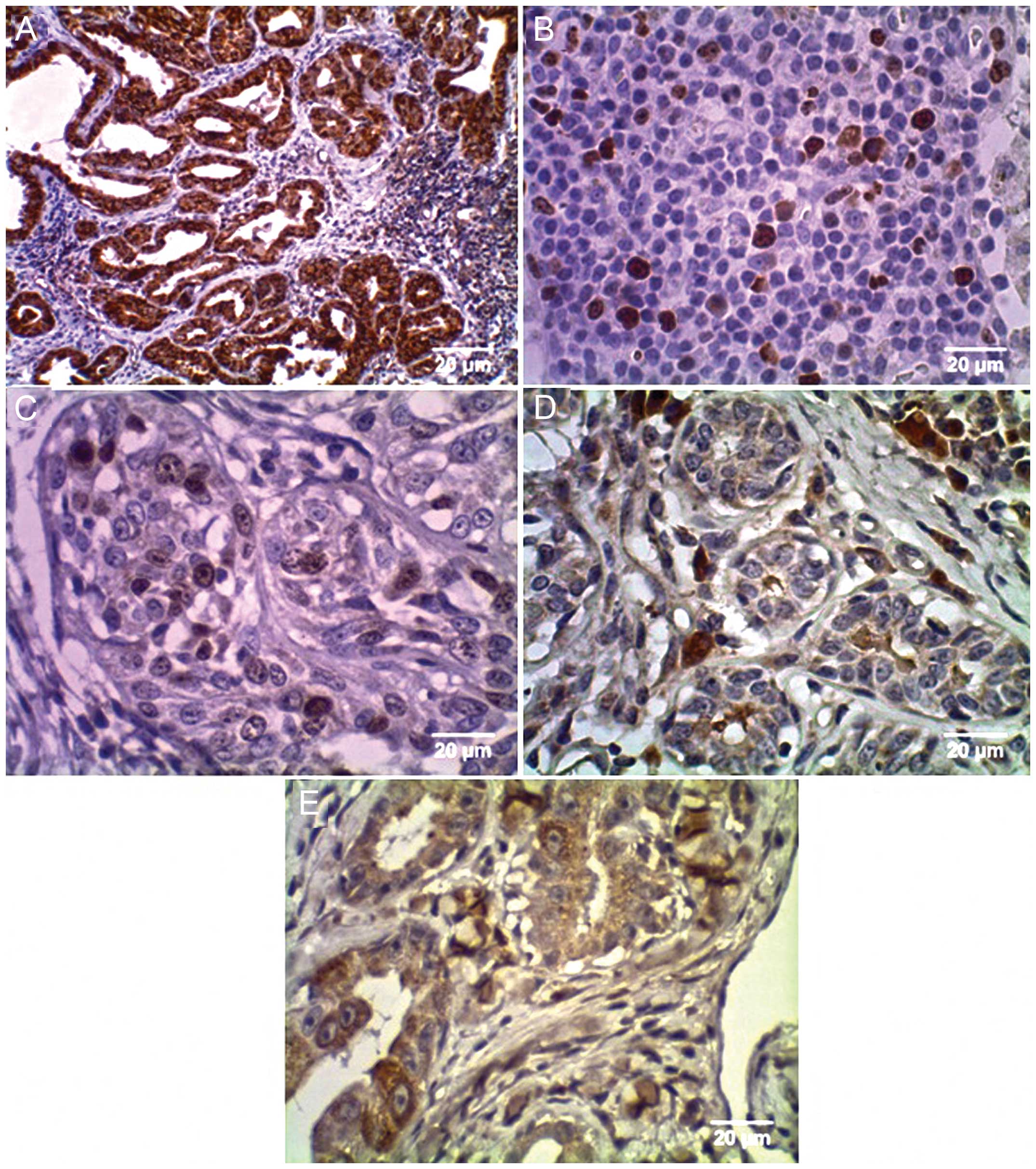
The panel of classic prognostic markers, analyzed by
the immunohistochemical expression of ER, PR, Her2/neu, p53 and
cell proliferation marker, Ki-67, were determined for all animal
casex (Fig. 1).
Thirteen of 24 (54%) tumors were positive for ER,
with immunostaining in the nucleus and cytoplasm (Fig. 1). PR immunostaining was cytoplasmic;
Ki-67 and p53 were evident in the nucleus of the neoplastic cells
while Her2/neu presented staining in the membrane of tumor cells
(Fig. 1).
There was no correlation between ER expression and
clinical characteristics and pathology including time course,
clinical stage, number of nodules, tumor vascularization,
ulceration and metastasis (p>0.05; Table II). However, the high expression of
ER had a statistically significant correlation with age (p=0.02;
Table II). The expression of ER,
PR, HER2/neu, p53 and Ki-67 by immunohistochemistry was compared
with the clinical development of the female dogs, including
metastasis and death. PR expression was increased in the female
dogs that did not present with metastasis (p=0.03; Table II). There was no significant
statistical relationship between the analyzed variables and the
expression of the enzymes HER2/neu, p53 and Ki-67 (p>0.05;
Table II).
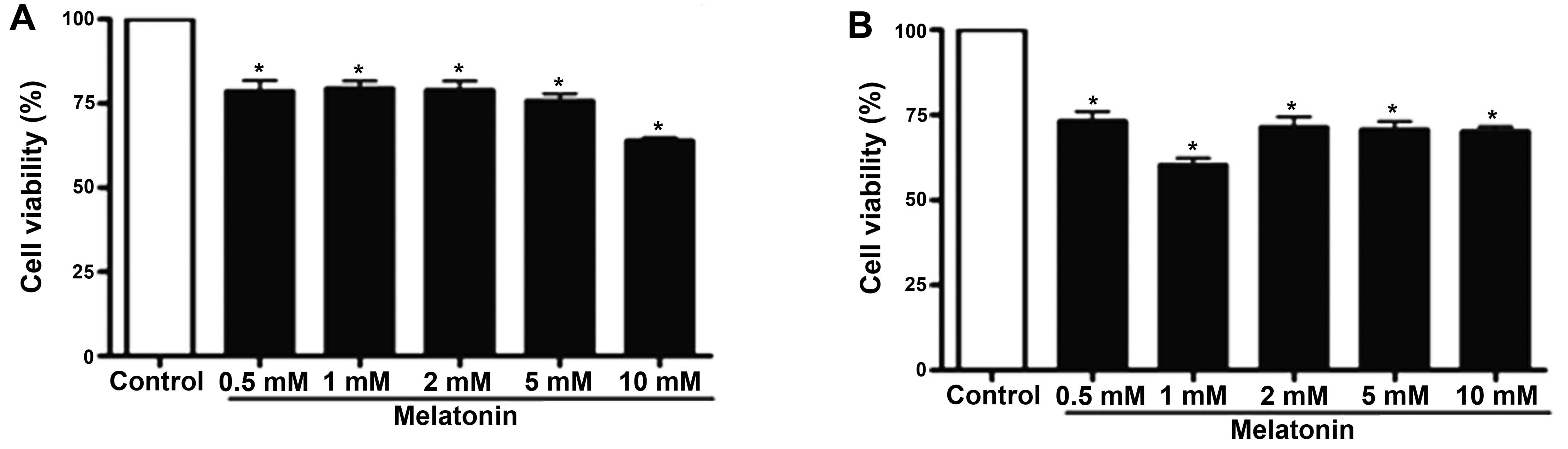
MTT assay
In vitro study
Ten tumor specimens were analyzed from the 24
samples used for immunohistochemistry and PCR. The canine mammary
tumors were divided into ER-positive and ER-negative tumors. To
target the antitumor activity by melatonin, we evaluated the cell
viability in primary culture of canine mammary neoplasms by MTT
assay following melatonin treatment. As shown in Fig. 2, cell viability was decreased in
both the ER-positive and ER-negative tumors at all melatonin
concentrations tested when compared to the control (p<0.05)
after 24 h. However, in the ER-positive tumors, the pharmacological
concentration of 1 mM of melatonin significantly decreased the cell
viability and the same occurred with the highest concentration of
melatonin (10 mM) in ER-negative tumors (p<0.05; Fig. 2).
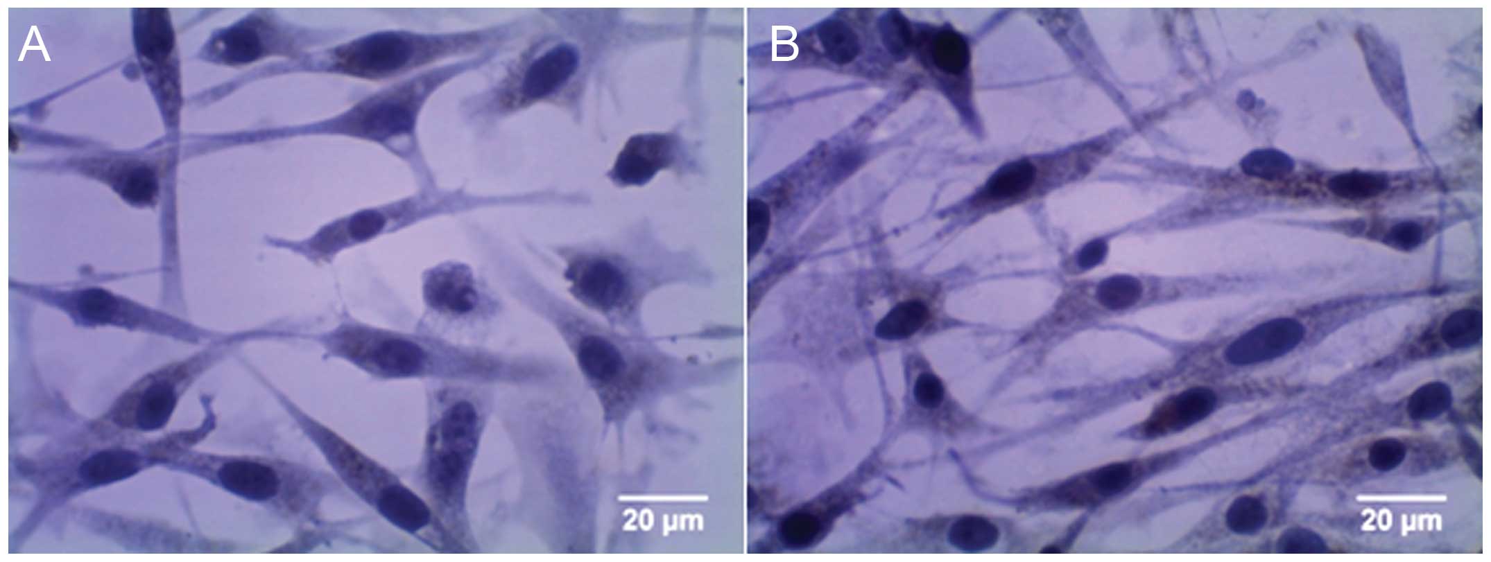
Apoptosis and cell proliferation
The level of caspase-3 immunostaining and the rate
of apoptosis in the group treated with 1 mM of melatonin were
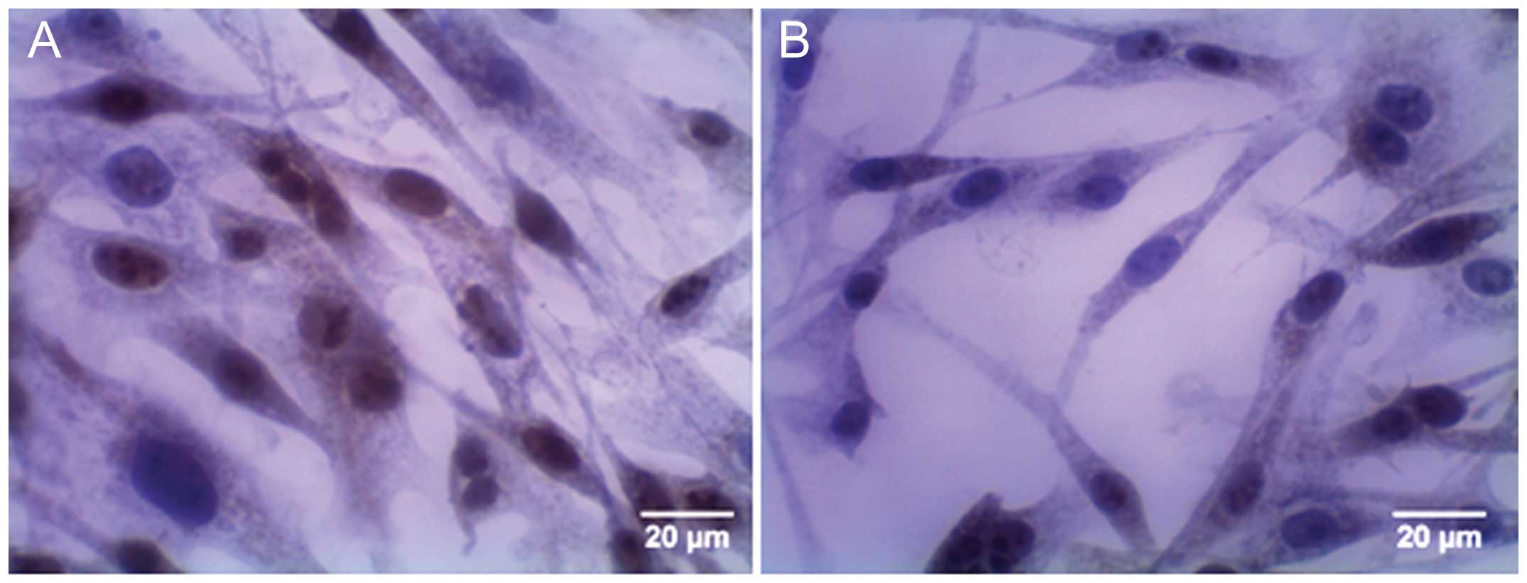
higher than these values in the control cell group (Fig. 3). Regarding Ki-67, numerous positive
cells were observed in the control group, whereas few positive
cells were observed in the group treated with 1 mM of melatonin
(Fig 4).
MT1 and MT2 gene expression analysis
In order to compare the expression of the receptors
MT1 and MT2 with ER expression and with the cellular response to
melatonin, analysis of the relative expression of the MT1
and MT2 genes by quantitative PCR was carried out and the
data were compared with the expression of ER in 24 canine mammary
tumors and the cellular response to melatonin in the 10
samples.
The RQ value for the control group (used as a
calibrator) was established as 1 and is shown on a log10 scale as
zero.
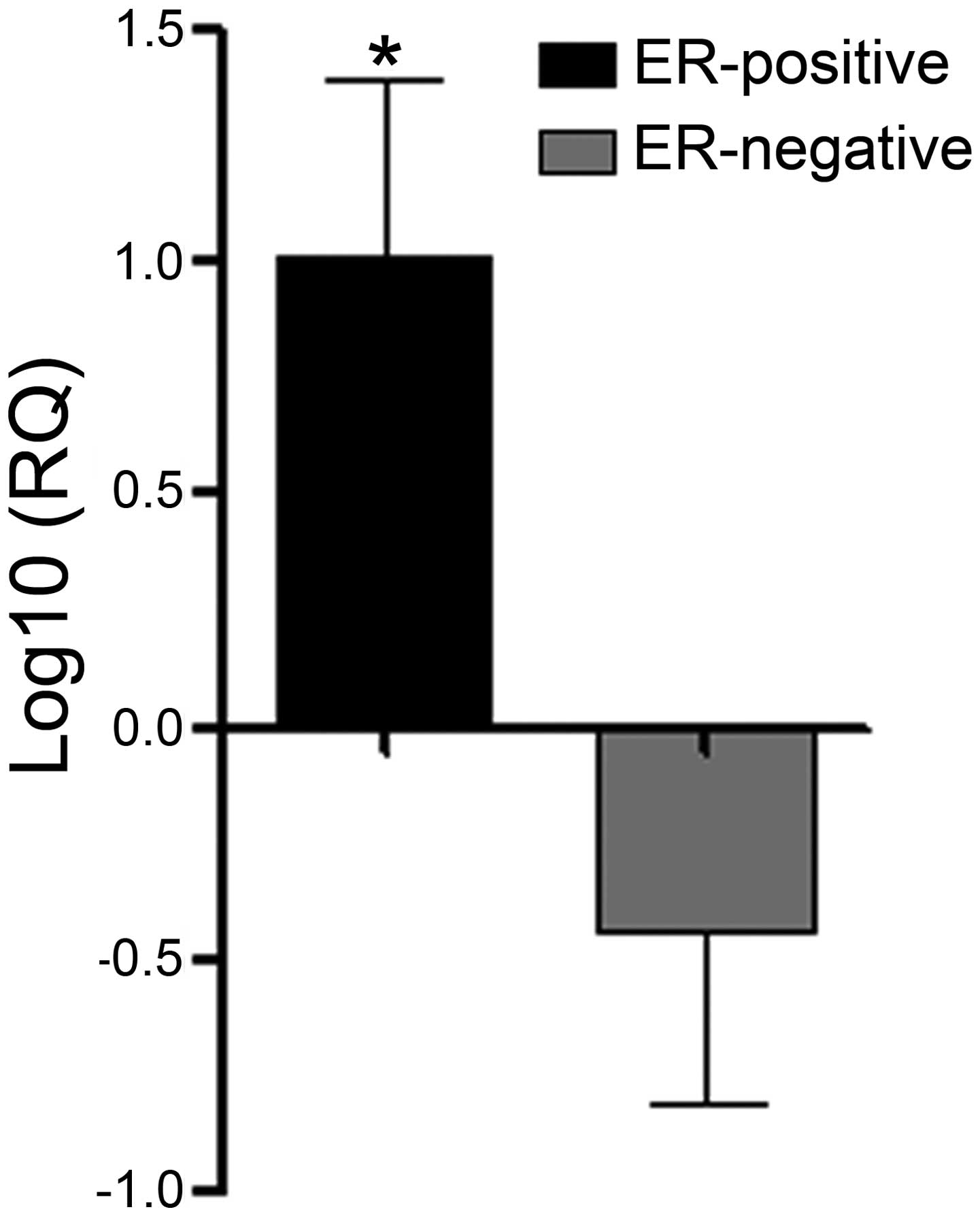
Results showed that the MT1 gene was overexpressed
in the ER-positive breast tumors compared to the ER-negative tumors
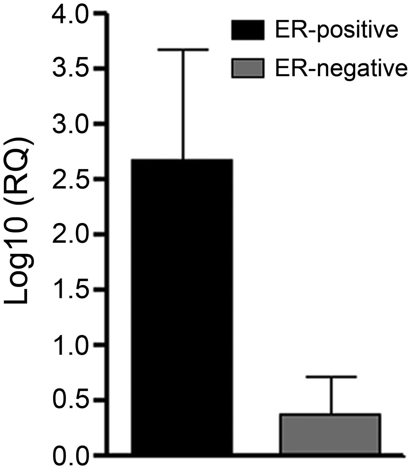
(p<0.05; Fig. 5). Although the
MT2 receptor was not detectable in the mammary tumors, it was
expressed but apparently not fully functional (p>0.05; Fig. 6). Furthermore, melatonin treatment
in the ER-positive tumors that overexpressed MT1 showed efficient
an oncostatic effect by inhibiting cell viability.
Discussion
In the present study, we used an
immunohistochemistry technique to examine the expression of the
estrogen receptor in 10 breast tumor samples. We categorized these
samples into two subgroup and compared the cellular response to
melatonin by MTT assay and immunocytochemistry for the cell
proliferation Ki-67 and apoptosis caspase-3 markers. We found that
treatment with a pharmacological dose (1 mM) of melatonin inhibited
cell proliferation of ER-positive tumors. For ER-negative tumors
the reduction in cell proliferation was observed at the highest
dose of melatonin evaluated (10 mM). However, the 10 mM dose has
been described as cytotoxic (32).
According to Jung et al (32) treatment at pharmacological levels of
melatonin (8 and 16 mM) in ER-negative (MDA-MB-231) cells showed
weak cytotoxicity. Melatonin has been described as able to inhibit
the precursors of estradiol production, reducing the mitogenic
response of ER-positive breast cancer cells (15). Rato et al (33) found that MCF-7 cells stimulated with
estradiol had a high binding activity to the ER, which was
prevented by treatment with melatonin suggesting their role in
inhibition of the estrogen signaling pathway. Other authors also
showed that melatonin inhibits proliferation of ER-positive breast
cancer (MCF-7) cells (11,14,23,34),
but not ER-negative (MDA-MB-231) cells (11,23).
These studies indicate the positive relationship between the
estrogen receptor and melatonin.
Furthermore, Eck et al (23) found that combined in vitro
treatment of melatonin and all-trans-retinoic acid inhibited
the growth of MCF-7 cells, but was ineffective against MDA-MB-231
cells, also suggesting that this treatment be used only for
estrogen-dependent mammary tumors. Consistent with our results,
Leman et al (35) showed
that melatonin at a pharmacological level (1 mM) significantly
decreased the proliferation of ER-positive breast cancer MCF-7
cells.
Cellular responses to melatonin in our study were
related to ER-positive tumors and is in agreement with the
literature.
Furthermore, additional routes of action of
melatonin have been described. Some authors showed that melatonin
induced apoptosis in MCF-7 cells, leading to decreased expression
of the ER and the anti-apoptotic Bcl-2 protein, and increased
expression of the pro-apoptotic Bax protein (23,35).
Our results showed that treatment with melatonin in ER-positive
canine mammary (CMT-U229) cells increased the expression of
caspase-3 protein. In the same way, Sanchez-Hidalgo et al
(36) demonstrated that the
treatment with melatonin in a panel of human cell lines increased
the expression of caspase-3. Similarly, the results of Wang et
al (37) showed that treatment
with melatonin significantly increased the activities and the
levels of caspase-3 protein, confirming the involvement of the
caspase-3 pathway in melatonin-induced cell apoptosis indicating
that caspase inhibitors can prevent melatonin-induced cell
apoptosis in breast cancer cells. It was also suggested that the
oncostatic actions of melatonin are mediated by MT1 melatonin
receptor; melatonin binds to the high affinity G-protein linked,
MT1 and MT2, reducing proliferation and inducing cell
differentiation (27,38). Furthermore, the expression of MT1
receptor was found to be positively correlated with the expression
of the ER (26). Through activation
of its receptor MT1, melatonin can suppress the development of
cancer by a broad spectrum of mechanisms with and without the
involvement of ER (38). Thus, in
this study, the expression of MT1 and MT2 receptors in breast
tumors of dogs was evaluated by real-time PCR. Furthermore, the
expression of these receptors was compared to the expression of ER
and cellular responses to melatonin. Our results showed that
ER-positive tumors had a high expression of melatonin receptor MT1
and these tumors responded better to treatment with melatonin.
However, these tumors did not express the MT2 receptor. Consistent
with our results, other authors also showed that treatment with
melatonin of ER-positive cancer cells that overexpress the
melatonin receptor MT1 significantly decreased cell proliferation
(11,39–41).
Moreover, Lai et al (26)
verified high expression of the MT1 receptor in ER-positive MCF-7
cells and low expression in an ER-negative MDA-MB-231 cell line;
however, both strains did not express the MT2 receptor. Similarly,
Ram et al. (42) found a
positive correlation between the MT1 receptor and the expression of
ER-α in women with breast cancer. However, Yuan et al
(11) indicated that both MCF-7 and
MDA-MB-231 cells have a high expression of the MT1 receptor.
Breast cancer is one of the cancers with the highest
mortality rate, thus new therapeutic approaches are needed to make
the treatment more effective. Through this study it was possible to
verify the action of melatonin in primary cell culture of canine
mammary tumors. Thus, the results found in this study in canine
mammary tumors were similar to those observed in strains of human
breast cancer described in the literature, verifying that the
canine mammary tumors are an excellent model for the study of
breast cancer. Our results demonstrated that melatonin is able to
reduce mammary tumor cell viability and proliferation and this
reduction was most pronounced in ER-positive cells that overexpress
the MT1 receptor. Thus, ER-positive tumors with high expression of
MT1 respond better to treatment with melatonin. Furthermore,
treatment with melatonin increased the apoptosis of canine mammary
tumor cells. Taken together, our results showed that melatonin
treatment inhibits cell viability, proliferation and induces
apoptosis in canine mammary tumors. In this way, the therapeutic
response of melatonin and the expression of MT1 receptor in
estrogen-dependent breast tumor will greatly aid the potential of
its use as a therapeutic agent in the treatment of this tumor
type.
Acknowledgements
We thank Dr Cassali for the support in the reading
and analyzing the slides, the veterinary practitioners from São
José do Rio Preto and region and the owners of the dogs who
cooperated with this study. The study was made possible by
financial support received from FAPESP and CNPq.
References
|
1
|
Manuali E, De Giuseppe A, Feliziani F, et
al: CA 15-3 cell lines and tissue expression in canine mammary
cancer and the correlation between serum levels and tumour
histological grade. BMC Vet Res. 8:862012. View Article : Google Scholar : PubMed/NCBI
|
|
2
|
Schneider R: Comparison of age, sex, and
incidence rates in human and canine breast cancer. Cancer.
26:419–426. 1970. View Article : Google Scholar : PubMed/NCBI
|
|
3
|
Mottolese M, Morelli L, Agrimi U, et al:
Spontaneous canine mammary tumors. A model for monoclonal antibody
diagnosis and treatment of human breast cancer. Lab Invest.
71:182–187. 1994.PubMed/NCBI
|
|
4
|
Phillips JC, Lembcke L and Chamberlin T: A
novel locus for canine osteosarcoma (OSA1) maps to CFA34, the
canine orthologue of human 3q26. Genomics. 96:220–227. 2010.
View Article : Google Scholar : PubMed/NCBI
|
|
5
|
MacEwen EG: Spontaneous tumors in dogs and
cats: models for the study of cancer biology and treatment. Cancer
Metastasis Rev. 9:125–136. 1990. View Article : Google Scholar : PubMed/NCBI
|
|
6
|
Peleteiro M: Tumores mamários na cadela e
na gata. Revista Portuguesa de Ciências Veterinárias. 89:10–29.
1994.
|
|
7
|
Andrade FH, Figueiroa FC, Bersano PR,
Bissacot DZ and Rocha NS: Malignant mammary tumor in female dogs:
environmental contaminants. Diagn Pathol. 5:452010. View Article : Google Scholar : PubMed/NCBI
|
|
8
|
Hamilton A and Hortobagyi G: Chemotherapy:
what progress in the last 5 years? J Clin Oncol. 23:1760–1775.
2005. View Article : Google Scholar : PubMed/NCBI
|
|
9
|
Winer E, Gralow J, Diller L, et al:
Clinical cancer advances 2008: major research advances in cancer
treatment, prevention, and screening - a report from the American
Society of Clinical Oncology. J Clin Oncol. 27:812–826. 2009.
View Article : Google Scholar :
|
|
10
|
Hsiao YH, Chou MC, Fowler C, Mason JT and
Man YG: Breast cancer heterogeneity: mechanisms, proofs, and
implications. J Cancer. 1:6–13. 2010. View
Article : Google Scholar : PubMed/NCBI
|
|
11
|
Yuan L, Collins AR, Dai J, Dubocovich ML
and Hill SM: MT(1) melatonin receptor overexpression enhances the
growth suppressive effect of melatonin in human breast cancer
cells. Mol Cell Endocrinol. 192:147–156. 2002. View Article : Google Scholar : PubMed/NCBI
|
|
12
|
Maganhin CC1, Carbonel AA, Hatty JH, et
al: Melatonin effects on the female genital system: a brief review.
Rev Assoc Med Bras. 54:267–271. 2008.(In Portuguese). View Article : Google Scholar : PubMed/NCBI
|
|
13
|
Luchetti F, Canonico B, Betti M, et al:
Melatonin signaling and cell protection function. FASEB J.
24:3603–3624. 2010. View Article : Google Scholar : PubMed/NCBI
|
|
14
|
Cos S and Sánchez-Barceló EJ: Melatonin,
experimental basis for a possible application in breast cancer
prevention and treatment. Histol Histopathol. 15:637–647.
2000.PubMed/NCBI
|
|
15
|
Neto J and Scaldaferri P: Melatonina e
câncer - revisão da literatura. Revista Brasileira Cancerol.
51:49–58. 2005.(In Portuguese).
|
|
16
|
Jablonska K, Pula B, Zemla A, et al:
Expression of melatonin receptor MT1 in cells of human invasive
ductal breast carcinoma. J Pineal Res. 54:334–345. 2013. View Article : Google Scholar : PubMed/NCBI
|
|
17
|
Mediavilla MD, Sanchez-Barcelo EJ, Tan DX,
Manchester L and Reiter RJ: Basic mechanisms involved in the
anti-cancer effects of melatonin. Curr Med Chem. 17:4462–4481.
2010. View Article : Google Scholar : PubMed/NCBI
|
|
18
|
Sturgeon SR, Doherty A, Reeves KW, et al:
Urinary levels of melatonin and risk of postmenopausal breast
cancer: Women’s health initiative observational cohort. Cancer
Epidemiol Biomarkers Prev. 23:629–637. 2014. View Article : Google Scholar : PubMed/NCBI
|
|
19
|
Sanchez-Barcelo EJ, Mediavilla MD,
Alonso-Gonzalez C and Rueda N: Breast cancer therapy based on
melatonin. Recent Pat Endocr Metab Immune Drug Discov. 6:108–116.
2012. View Article : Google Scholar : PubMed/NCBI
|
|
20
|
Bejarano I, Espino J, Barriga C, Reiter
RJ, Pariente JA and Rodríguez AB: Pro-oxidant effect of melatonin
in tumour leucocytes: relation with its cytotoxic and pro-apoptotic
effects. Basic Clin Pharmacol Toxicol. 108:14–20. 2011. View Article : Google Scholar
|
|
21
|
Rodriguez C, Martín V, Herrera F, et al:
Mechanisms involved in the pro-apoptotic effect of melatonin in
cancer cells. Int J Mol Sci. 14:6597–6613. 2013. View Article : Google Scholar : PubMed/NCBI
|
|
22
|
Cucina A, Proietti S, D’Anselmi F, et al:
Evidence for a biphasic apoptotic pathway induced by melatonin in
MCF-7 breast cancer cells. J Pineal Res. 46:172–180. 2009.
View Article : Google Scholar : PubMed/NCBI
|
|
23
|
Eck KM, Yuan L, Duffy L, et al: A
sequential treatment regimen with melatonin and all-trans retinoic
acid induces apoptosis in MCF-7 tumour cells. Br J Cancer.
77:2129–2137. 1998. View Article : Google Scholar : PubMed/NCBI
|
|
24
|
Blask DE, Sauer LA and Dauchy RT:
Melatonin as a chronobiotic/anticancer agent: cellular,
biochemical, and molecular mechanisms of action and their
implications for circadian-based cancer therapy. Curr Top Med Chem.
2:113–132. 2002. View Article : Google Scholar : PubMed/NCBI
|
|
25
|
Hardeland R, Pandi-Perumal SR and
Cardinali DP: Melatonin. Int J Biochem Cell Biol. 38:313–316. 2006.
View Article : Google Scholar
|
|
26
|
Lai L, Yuan L, Cheng Q, Dong C, Mao L and
Hill SM: Alteration of the MT1 melatonin receptor gene and its
expression in primary human breast tumors and breast cancer cell
lines. Breast Cancer Res Treat. 118:293–305. 2009. View Article : Google Scholar
|
|
27
|
Grant SG, Melan MA, Latimer JJ and
Witt-Enderby PA: Melatonin and breast cancer: cellular mechanisms,
clinical studies and future perspectives. Expert Rev Mol Med.
11:e52009. View Article : Google Scholar : PubMed/NCBI
|
|
28
|
Misdorp W, Else R, Hellmen E and Lipscomb
E: Definitions and explanatory notes. Who Histological
Classification of Mammary Tumors of the Dog and Cat. Armed Forces
Institute of Pathology; Washington: pp. 18–27. 1999
|
|
29
|
Cassali G, Bertagnolli A, Lavalle G, et
al: Perpectives for diagnosis, prognosis and treatment of mammary
neoplasms in dogs. In: 34th World Small Animal Veterinary Congress
- WSAVA; São Paulo. 2009
|
|
30
|
Li XR, Liu M, Zhang YJ, et al: ER, PgR,
HER-2, Ki-67, topoisomerase IIα, and nm23-H1 proteins expression as
predictors of pathological complete response to neoadjuvant
chemotherapy for locally advanced breast cancer. Med Oncol.
28(Suppl 1): S48–S54. 2011. View Article : Google Scholar
|
|
31
|
Livak K and Schimittgen T: Analyzing
real-time PCR data by the comparative C(T) method. Nature
Protocols. 3:1101–1108. 2008. View Article : Google Scholar : PubMed/NCBI
|
|
32
|
Jung JW, Park SB, Lee SJ, Seo MS, Trosko
JE and Kang KS: Metformin represses self-renewal of the human
breast carcinoma stem cells via inhibition of estrogen
receptor-mediated OCT4 expression. PLoS One. 6:e280682011.
View Article : Google Scholar : PubMed/NCBI
|
|
33
|
Rato AG, Pedrero JG, Martinez MA, del Rio
B, Lazo PS and Ramos S: Melatonin blocks the activation of estrogen
receptor for DNA binding. FASEB J. 13:857–868. 1999.PubMed/NCBI
|
|
34
|
Mao L, Cheng Q, Guardiola-Lemaître B, et
al: In vitro and in vivo antitumor activity of melatonin receptor
agonists. J Pineal Res. 49:210–221. 2010. View Article : Google Scholar : PubMed/NCBI
|
|
35
|
Leman ES, Sisken BF, Zimmer S and Anderson
KW: Studies of the interactions between melatonin and 2 Hz, 0.3 mT
PEMF on the proliferation and invasion of human breast cancer
cells. Bioelectromagnetics. 22:178–184. 2001. View Article : Google Scholar : PubMed/NCBI
|
|
36
|
Sánchez-Hidalgo M, Guerrero JM, Villegas
I, Packham G and de la Lastra CA: Melatonin, a natural programmed
cell death inducer in cancer. Curr Med Chem. 19:3805–3821. 2012.
View Article : Google Scholar : PubMed/NCBI
|
|
37
|
Wang J, Xiao X, Zhang Y, et al:
Simultaneous modulation of COX-2, p300, Akt, and Apaf-1 signaling
by melatonin to inhibit proliferation and induce apoptosis in
breast cancer cells. J Pineal Res. 53:77–90. 2012. View Article : Google Scholar : PubMed/NCBI
|
|
38
|
Oprea-Ilies G, Haus E, Sackett-Lundeen L,
et al: Expression of melatonin receptors in triple negative breast
cancer (TNBC) in African American and Caucasian women: relation to
survival. Breast Cancer Res Treat. 137:677–687. 2013. View Article : Google Scholar :
|
|
39
|
Collins A, Yuan L, Kiefer TL, Cheng Q, Lai
L and Hill SM: Overexpression of the MT1 melatonin receptor in
MCF-7 human breast cancer cells inhibits mammary tumor formation in
nude mice. Cancer Lett. 189:49–57. 2003. View Article : Google Scholar
|
|
40
|
Hill SM, Blask DE, Xiang S, et al:
Melatonin and associated signaling pathways that control normal
breast epithelium and breast cancer. J Mammary Gland Biol
Neoplasia. 16:235–245. 2011. View Article : Google Scholar : PubMed/NCBI
|
|
41
|
Gonzalez-Angulo AM, Morales-Vasquez F and
Hortobagyi GN: Overview of resistance to systemic therapy in
patients with breast cancer. Adv Exp Med Biol. 608:1–22. 2007.
View Article : Google Scholar : PubMed/NCBI
|
|
42
|
Ram PT, Kiefer T, Silverman M, Song Y,
Brown GM and Hill SM: Estrogen receptor transactivation in MCF-7
breast cancer cells by melatonin and growth factors. Mol Cell
Endocrinol. 141:53–64. 1998. View Article : Google Scholar : PubMed/NCBI
|