Cancer treatments have markedly improved in recent
decades. Surgery is considered the main treatment for primary
tumors. Yet, surgery has limitations since it can be highly
invasive, sometimes causing the loss of function of organs.
Improvements in chemoradiotherapy are promising as this modality is
less invasive and may preserve organ function. Utilizing a
combination of various cancer therapies, such as surgery,
radiotherapy, systemic chemotherapy and molecular targeted therapy,
locoregional cancer control rates have greatly improved (1). However, relapse and metastasis, which
worsen patient outcomes, are of concern. It is important to prevent
cancer recurrence and metastasis as the resultant tumors are often
more viable and resistant to chemoradiotherapy. The reasons for
therapeutic resistant of cancer cells are controversial. However,
the cancer stem cell (CSC) theory may provide an accurate
explanation.
The CSC theory explains that a small population of
cells in a tumor has stem cell properties, such as self-renewal,
multiple differentiation and tumor initiation capacities. The idea
of CSCs has attacted interest recently, but it was conceptualized
in 1971 with the discovery of mouse myeloma tumor stem cells
(2). When injected into mice, not
all leukemia cells but only a small population of leukemia cells
initiated tumors, and these tumor-initiating cells were termed
CSCs. In humans, CSCs were first discovered in leukemia by Bonnet
and Dick in 1997 (3). The
recurrence rates of patients with residual tumors after surgery and
the rates of distant metastases of patients with circulating cancer
cells after successful treatment of the primary tumor were not
100%, although these patients were part of a high-risk group
(4–9). These data suggested that not all
cancer cells have the potential to cause recurrence and metastasis
(10). Recent studies have revealed
that CSCs play an important role in maintaining tumor growth
(11). Growing evidence suggests
that CSCs are resistant to chemoradiotherapy and cause tumor
metastasis and recurrence (12).
Overall, CSCs are a stem cell-like population in cancer with tumor
initiation, self-renewal and multiple differentiation capacities
which cause metastasis and recurrence, and it appears that the
targeting of CSCs during cancer treatment is a promising strategy
for a permanent cancer cure.
In this review, we discuss how the idea of CSCs can
be used in future clinical practice. We focus on radiotherapy,
particularly the potential of carbon ion beam therapy in relation
to CSCs.
To date, in order to target CSCs, markers of CSCs
have been studied. CSCs have been identified in several types of
tumors, and biomarkers have been established, such as those
identified by SP assay (13–15):
CD44 in breast CSCs (16); and
CD133 in hematopoietic CSCs, (17)
neural stem cells (18) and colon
cancer cells (19,20). These biomarkers are sometimes
related with the prognosis of tumors.
CD133 is a major marker of CSCs in various types of
cancers, including glioblastoma, rectal cancer and lung cancer
(21–24). CD133 is also expressed in
differentiated epithelial cells, and some CD133-expressing cancer
cells have CSC properties (16–25).
MicroRNA expression in CSCs can also be used for the
identification of CSCs. MicroRNAs are endogenous RNAs, which
contribute to oncogenic transformation, tumor suppression, and cell
differentiation as well as pluripotency (26–28).
Several microRNAs are known as CSC regulators.
miR-181 was found to be highly expressed in CSCs in HCC, in
embryonic liver tissues, and pluripotent hepatic stem cells in the
human liver (29). The expression
of miR-130b in HCC positively regulates CD133+ CSCs with
respect to self-renewal, tumor initiation and chemoresistance
properties (30). miR-34a was found
to inhibit the growth of CSCs and metastasis in prostate cancer by
directly repressing CD44 expression (31). miR-200b negatively regulates CSCs in
breast cancer by reducing CSC formation (32). These microRNAs regulate CSCs
positively or negatively, and identification of their mechanism of
action may make it possible to target or eliminate CSCs
specifically.
CSCs can be identified by measuring expression
levels of those biomarkers, although these markers are not
specific. A combination of these markers may be more useful for
identification of CSCs.
CSCs are a small population present in cancer cells
with unique properties: i) self-renewal capability, ii) cell
differentiation ability and iii) cancer initiation potential
(33,34). These properties of CSCs contribute
to chemoradiotherapy resistance and cause tumor recurrence.
Enhanced DNA repair capacity and reduced reactive oxygen species
(ROS) levels may be responsible for the radioresistance of CSCs
(35–38).
Several pathways such as OCT4, WNT, NOTCH, Sonic
Hedgehog (SHH), B lymphoma Mo-MLV insertion region 1 homolog
(BMI1), and SNAIL1/SLUG are known to be linked to the radiation
resistance of CSCs (39). Wang
et al reported that inhibition of the NOTCH pathway with
γ-secretase inhibitors sensitized glioma CSCs to radiation
(40). Chen et al observed
that the survival rate of CD133+ cells was higher than
that of CD133− cells after radiation in lung cancer. In
the same study, OCT4 knockdown improved the treatment effects of
chemoradiotherapy on CD133+ cells (41). CD133 is expressed in
radiation-resistant CSCs of glioma (36) and colon cancer (42). The DNA repair- and cell
cycle-regulating proteins Chk1/Chk2 have been proven to be related
to such resistance. It has also been demonstrated that the
inhibition of these kinases improves treatment sensitivity
(36). Zhang et al reported
that the EMT-inducing transcription factor zinc finger E-box
binding homeobox 1 (ZEB1) is a regulator of radiosensitivity and
DNA damage response in breast cancer cells (43). In the same study, downregulation of
ZEB1 was found to radiosensitize breast cancer cells, indicating
that ZEB1-targeting agents could be used as tumor sensitizers.
Recent studies have revealed that certain
CSC-related mechanisms are epigenetic. Suvà et al observed
that CSCs can be reprogrammed from normal cancer cells in
glioblastoma. They also showed that inhibition of the core
transcription factors can suppress cancer stem cell properties, and
these proteins can be used as therapeutic targets (44). Seguin et al reported that
integrin β3 expression and the related KRAS-RalB-NF-κB pathway are
both necessary and sufficient for CSC formation and erlotinib
resistance. Bortezomib reverses both tumor stemness and erlotinib
resistance by inhibiting this pathway (45). These findings suggest that the
therapeutic resistance of tumors is related to genetic alterations,
and epigenetic removal of CSC properties would enhance the
therapeutic sensitivity of tumors.
MicroRNAs (miRNAs) are small non-coding RNAs that
regulate epigenetic gene expression (26). A growing body of evidence suggests
that miRNAs are associated with tumor initiation and progression
(46–49). Cui et al reported that
nanoparticle-delivered miR-200c serves as an effective
radio-sensitizer of gastric cancer cells and suppresses CSC-like
properties (50). CSC-targeted
therapies including the miR-200 family tend to damage normal stem
cells. Damage to normal cells must be considered in clinical
practice, and it is difficult to selectively damage cancer cells
while sparing normal cells. By exploiting cellular uptake
differences depending on gelatinase levels, Cui et al showed
that it possible to spare normal cells via the gelatinase strategy.
Gelatinases are overexpressed in numerous cancers but are
undetectable in normal cells (50).
Yao et al (52) demonstrated
that miR-205 is related with radiosensitivity in breast cancer.
Downregulation of miR-205 is observed in radio-resistant cancer
cells, and it is highly associated with poor distant relapse-free
survival in breast cancer patients. Such findings suggest that
miRNAs could be utilized as promising radiosensitizers with minimal
side effects, while further in vivo studies and clinical
trials are required for clinical utilization.
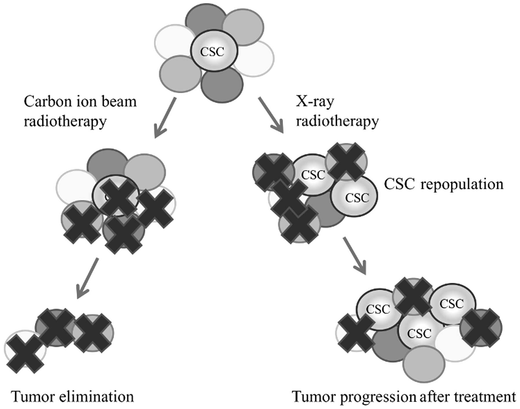
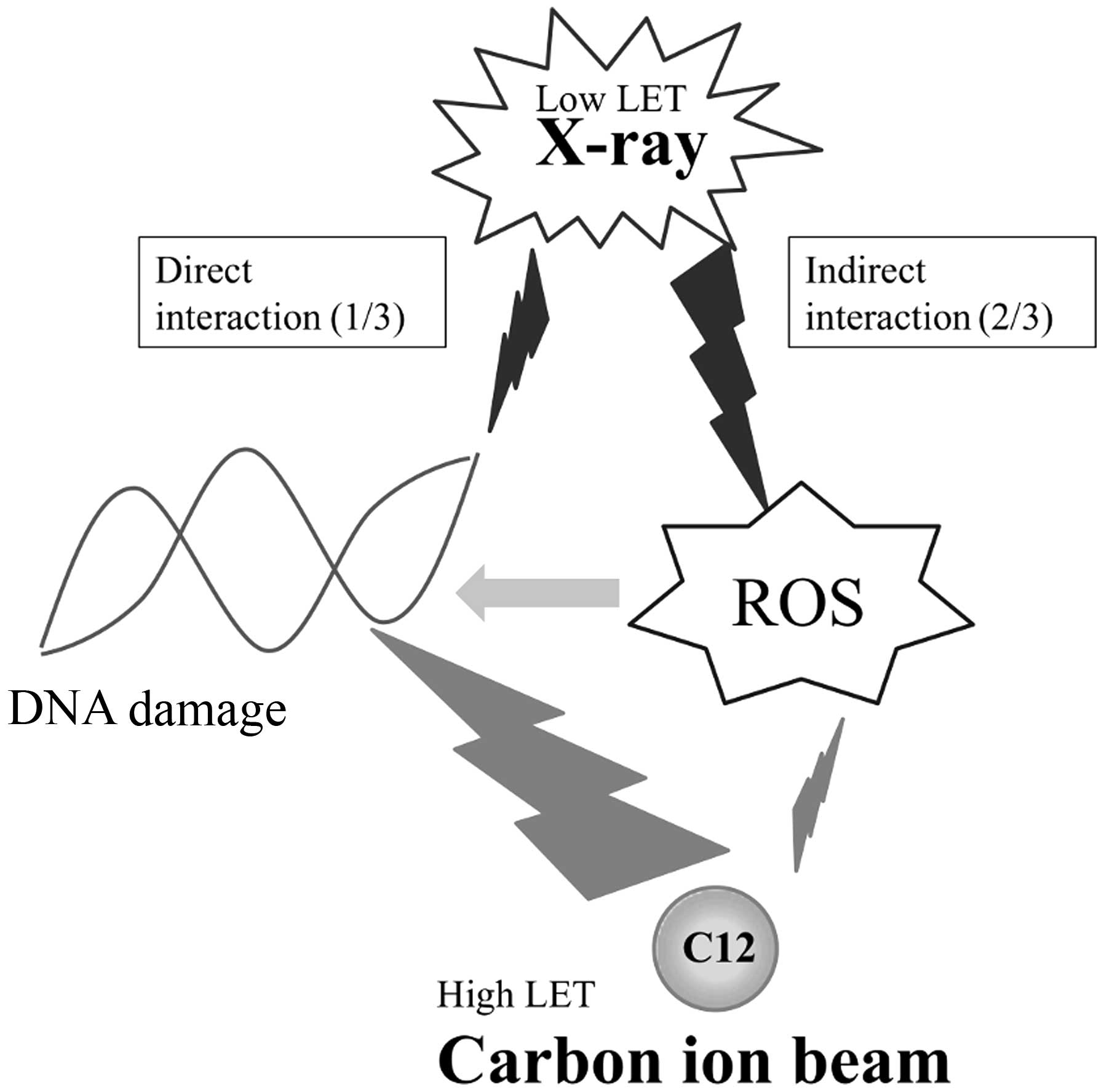
X-ray-induced DNA damage mostly occurs through ROS
interaction. CSCs have lower ROS levels and enhanced protection
from oxidative damage and therefore exhibit better radiation
resistance compared with normal cancer cells (35,53,54).
Nevertheless, fractionated radiotherapy may result in the
repopulation of CSCs (55).
Although normal cancer cells are killed by radiotherapy,
radioresistant CSCs can survive and proliferate during such
treatment. Thus, an increased proportion of CSCs makes a tumor more
aggressive (Figs. 1 and 2).
High-LET heavy ions may be the key to treating the
therapeutic resistance of CSCs. Heavy-ion radiotherapy such as
carbon ion radiotherapy has higher relative biological
effectiveness (RBE), ranging between 3 and 5 (56,57),
and therefore is more effective against hypoxic radioresistant
cells than conventional X-ray radiotherapy. Carbon ion radiotherapy
for malignant tumors has yielded favorable results in several
clinical trials (58–63). It was reported that
Bcl-2-overexpressing HeLa cells are more resistant to γ-rays (0.2
keV/µm) and helium ions (16.2 keV/µm) than neomycin
resistance gene-expressing HeLa cells, whereas heavy ions
(76.3–1610 keV/µm) yield similar survival regardless of
Bcl-2 overexpression. This implies that heavy-ion radiotherapy may
be equally effective against CSCs (64) and that carbon ion beam radiotherapy
may not induce CSC repopulation in contrast to X-ray
radiotherapy.
Notably, when cancer cells are irradiated by X-rays
and a carbon ion beam, genomic expression patterns are changed
differentially. Carbon ion beam-specific gene expression patterns
may be promising therapeutic targets, which have not been well
studied to date.
CSCs are responsible for tumor recurrence,
metastasis and treatment resistance; thus, they are key targets by
which to ensure the permanent elimination of cancer cells. Several
CSC markers have been recently discovered. Although these markers
are not specific, CSCs can be reliably identified using
combinations of these markers. Furthermore, specific epigenetic
alterations, which are believed to be responsible for the
therapeutic resistance of CSCs, are now better understood. These
markers and alterations are promising therapeutic targets. By
inhibiting the expression of the responsible genes, CSCs can be
radiosensitized. Targeting CSCs while sparing non-CSCs is a
challenging task as these cell populations exhibit similar
expression patterns and biological properties. Recent studies and
insights into epigenetic processes and cellular metabolism are
providing clues regarding novel promising targeted therapeutic
agents that specifically inhibit the growth of CSCs;
nanoparticle-delivered miR-200c is a good example. Nevertheless,
most studies are currently at the in vitro level, and
translational research is needed to use these ideas in clinical
practice.
Carbon ion beam radiotherapy is a promising method
for the elimination of CSCs, with strong effects on CSCs. The tumor
control rates of carbon ion beam radiotherapy may be further
improved by the use of CSC-targeting drugs such as microRNAs as
radiosensitizers. Such combination therapies for carbon ion beam
radiotherapy have not yet been adequately researched, and further
studies are warranted to establish an optimal combination.
We thank each laboratory staff member for the
fruitful discussion and technical assistance. This study was
supported in part by a Grant-in-Aid for Scientific research from
the Ministry of Education, Culture, Sports, Science, and
Technology; a Grant-in-Aid from the Ministry of Health, Labor and
Welfare; a grant from the National Institute of Biomedical
Innovation; and a grant from the Osaka University Drug Discovery
Funds. Partial support was received from Taiho Pharmaceutical Co.,
Ltd., Evidence Based Medical Research Center, Chugai Co., Ltd.,
Yakult Honsha Co., Ltd., Merck Co., Ltd., and the Takeda Science
and Medical Research Foundation through the institutional
endowments; the funders had no role in study design, data
collection and analysis, decision to publish, or preparation of the
manuscript.
|
1
|
Koch U, Krause M and Baumann M: Cancer
stem cells at the crossroads of current cancer therapy failures -
radiation oncology perspective. Semin Cancer Biol. 20:116–124.
2010. View Article : Google Scholar : PubMed/NCBI
|
|
2
|
Park CH, Bergsagel DE and McCulloch EA:
Mouse myeloma tumor stem cells: A primary cell culture assay. J
Natl Cancer Inst. 46:411–422. 1971.PubMed/NCBI
|
|
3
|
Bonnet D and Dick J: Human acute myeloid
leukemia is organized as a hierarchy that originates from a
primitive hematopoietic cell. Nat Med. 3:730–737. 1997. View Article : Google Scholar : PubMed/NCBI
|
|
4
|
Riethdorf S, Wikman H and Pantel K:
Review: Biological relevance of disseminated tumor cells in cancer
patients. Int J Cancer. 123:1991–2006. 2008. View Article : Google Scholar : PubMed/NCBI
|
|
5
|
Fusi A, Collette S, Busse A, Suciu S,
Rietz A, Santinami M, Kruit WH, Testori A, Punt CJ, Dalgleish AG,
et al: Circulating melanoma cells and distant metastasis-free
survival in stage III melanoma patients with or without adjuvant
interferon treatment (EORTC 18991 side study). Eur J Cancer.
45:3189–3197. 2009. View Article : Google Scholar : PubMed/NCBI
|
|
6
|
Swindle P, Eastham JA, Ohori M, Kattan MW,
Wheeler T, Maru N, Slawin K and Scardino PT: Do margins matter? The
prognostic significance of positive surgical margins in radical
prostatectomy specimens. J Urol. 179(Suppl): S47–S51. 2008.
View Article : Google Scholar : PubMed/NCBI
|
|
7
|
Loree TR and Strong EW: Significance of
positive margins in oral cavity squamous carcinoma. Am J Surg.
160:410–414. 1990. View Article : Google Scholar : PubMed/NCBI
|
|
8
|
Barrow BJ, Janjan NA, Gutman H, Benjamin
RS, Allen P, Romsdahl MM, Ross MI and Pollock RE: Role of
radiotherapy in sarcoma of the breast - a retrospective review of
the M.D. Anderson experience. Radiother Oncol. 52:173–178. 1999.
View Article : Google Scholar : PubMed/NCBI
|
|
9
|
Alektiar KM, Velasco J, Zelefsky MJ,
Woodruff JM, Lewis JJ and Brennan MF: Adjuvant radiotherapy for
margin-positive high-grade soft tissue sarcoma of the extremity.
Int J Radiat Oncol Biol Phys. 48:1051–1058. 2000. View Article : Google Scholar : PubMed/NCBI
|
|
10
|
Patel P and Chen EI: Cancer stem cells,
tumor dormancy, and metastasis. Front Endocrinol (Lausanne).
3:1252012.
|
|
11
|
Schepers AG, Snippert HJ, Stange DE, van
den Born M, van Es JH, van de Wetering M and Clevers H: Lineage
tracing reveals Lgr5+ stem cell activity in mouse
intestinal adenomas. Science. 337:730–735. 2012. View Article : Google Scholar : PubMed/NCBI
|
|
12
|
Singh A and Settleman J: EMT, cancer stem
cells and drug resistance: An emerging axis of evil in the war on
cancer. Oncogene. 29:4741–4751. 2010. View Article : Google Scholar : PubMed/NCBI
|
|
13
|
Chiba T, Kita K, Zheng YW, Yokosuka O,
Saisho H, Iwama A, Nakauchi H and Taniguchi H: Side population
purified from hepatocellular carcinoma cells harbors cancer stem
cell-like properties. Hepatology. 44:240–251. 2006. View Article : Google Scholar : PubMed/NCBI
|
|
14
|
Haraguchi N, Utsunomiya T, Inoue H, Tanaka
F, Mimori K, Barnard GF and Mori M: Characterization of a side
population of cancer cells from human gastrointestinal system. Stem
Cells. 24:506–513. 2006. View Article : Google Scholar
|
|
15
|
Ehata S, Johansson E, Katayama R, Koike S,
Watanabe A, Hoshino Y, Katsuno Y, Komuro A, Koinuma D, Kano MR, et
al: Transforming growth factor-β decreases the cancer-initiating
cell population within diffuse-type gastric carcinoma cells.
Oncogene. 30:1693–1705. 2011. View Article : Google Scholar
|
|
16
|
Al-Hajj M, Wicha MS, Benito-Hernandez A,
Morrison SJ and Clarke MF: Prospective identification of
tumorigenic breast cancer cells. Proc Natl Acad Sci USA.
100:3983–3988. 2003. View Article : Google Scholar : PubMed/NCBI
|
|
17
|
Yin AH, Miraglia S, Zanjani ED,
Almeida-Porada G, Ogawa M, Leary AG, Olweus J, Kearney J and Buck
DW: AC133, a novel marker for human hematopoietic stem and
progenitor cells. Blood. 90:5002–5012. 1997.
|
|
18
|
Uchida N, Buck DW, He D, Reitsma MJ, Masek
M, Phan TV, Tsukamoto AS, Gage FH and Weissman IL: Direct isolation
of human central nervous system stem cells. Proc Natl Acad Sci USA.
97:14720–14725. 2000. View Article : Google Scholar : PubMed/NCBI
|
|
19
|
O'Brien CA, Pollett A, Gallinger S and
Dick JE: A human colon cancer cell capable of initiating tumour
growth in immunodeficient mice. Nature. 445:106–110. 2007.
View Article : Google Scholar
|
|
20
|
Ricci-Vitiani L, Lombardi DG, Pilozzi E,
Biffoni M, Todaro M, Peschle C and De Maria R: Identification and
expansion of human colon-cancer-initiating cells. Nature.
445:111–115. 2007. View Article : Google Scholar
|
|
21
|
Murat A, Migliavacca E, Gorlia T, Lambiv
WL, Shay T, Hamou MF, de Tribolet N, Regli L, Wick W, Kouwenhoven
MC, et al: Stem cell-related 'self-renewal' signature and high
epidermal growth factor receptor expression associated with
resistance to concomitant chemoradiotherapy in glioblastoma. J Clin
Oncol. 26:3015–3024. 2008. View Article : Google Scholar : PubMed/NCBI
|
|
22
|
Tamura K, Aoyagi M, Wakimoto H, Ando N,
Nariai T, Yamamoto M and Ohno K: Accumulation of CD133-positive
glioma cells after high-dose irradiation by Gamma Knife surgery
plus external beam radiation. J Neurosurg. 113:310–318. 2010.
View Article : Google Scholar : PubMed/NCBI
|
|
23
|
Saigusa S, Tanaka K, Toiyama Y, Yokoe T,
Okugawa Y, Kawamoto A, Yasuda H, Morimoto Y, Fujikawa H, Inoue Y,
et al: Immunohistochemical features of CD133 expression:
Association with resistance to chemoradiotherapy in rectal cancer.
Oncol Rep. 24:345–350. 2010. View Article : Google Scholar : PubMed/NCBI
|
|
24
|
Shien K, Toyooka S, Ichimura K, Soh J,
Furukawa M, Maki Y, Muraoka T, Tanaka N, Ueno T, Asano H, et al:
Prognostic impact of cancer stem cell-related markers in non-small
cell lung cancer patients treated with induction chemoradiotherapy.
Lung Cancer. 77:162–167. 2012. View Article : Google Scholar : PubMed/NCBI
|
|
25
|
Shmelkov SV, Butler JM, Hooper AT, Hormigo
A, Kushner J, Milde T, St Clair R, Baljevic M, White I, Jin DK, et
al: CD133 expression is not restricted to stem cells, and both
CD133+ and CD133− metastatic colon cancer
cells initiate tumors. J Clin Invest. 118:2111–2120.
2008.PubMed/NCBI
|
|
26
|
Bartel DP: MicroRNAs: Genomics,
biogenesis, mechanism, and function. Cell. 116:281–297. 2004.
View Article : Google Scholar : PubMed/NCBI
|
|
27
|
O'Donnell KA, Wentzel EA, Zeller KI, Dang
CV and Mendell JT: c-Myc-regulated microRNAs modulate E2F1
expression. Nature. 435:839–843. 2005. View Article : Google Scholar : PubMed/NCBI
|
|
28
|
Johnson SM, Grosshans H, Shingara J, Byrom
M, Jarvis R, Cheng A, Labourier E, Reinert KL, Brown D and Slack
FJ: RAS is regulated by the let-7 microRNA family. Cell.
120:635–647. 2005. View Article : Google Scholar : PubMed/NCBI
|
|
29
|
Ji J, Yamashita T, Budhu A, Forgues M, Jia
HL, Li C, Deng C, Wauthier E, Reid LM, Ye QH, et al: Identification
of microRNA-181 by genome-wide screening as a critical player in
EpCAM-positive hepatic cancer stem cells. Hepatology. 50:472–480.
2009. View Article : Google Scholar : PubMed/NCBI
|
|
30
|
Ma S, Tang KH, Chan YP, Lee TK, Kwan PS,
Castilho A, Ng I, Man K, Wong N, To KF, et al: miR-130b promotes
CD133(+) liver tumor-initiating cell growth and self-renewal via
tumor protein 53-induced nuclear protein 1. Cell Stem Cell.
7:694–707. 2010. View Article : Google Scholar : PubMed/NCBI
|
|
31
|
Liu C, Kelnar K, Liu B, Chen X,
Calhoun-Davis T, Li H, Patrawala L, Yan H, Jeter C, Honorio S, et
al: The microRNA miR-34a inhibits prostate cancer stem cells and
metastasis by directly repressing CD44. Nat Med. 17:211–215. 2011.
View Article : Google Scholar : PubMed/NCBI
|
|
32
|
Iliopoulos D, Lindahl-Allen M, Polytarchou
C, Hirsch HA, Tsichlis PN and Struhl K: Loss of miR-200 inhibition
of Suz12 leads to polycomb-mediated repression required for the
formation and maintenance of cancer stem cells. Mol Cell.
39:761–772. 2010. View Article : Google Scholar : PubMed/NCBI
|
|
33
|
Sagar J, Chaib B, Sales K, Winslet M and
Seifalian A: Role of stem cells in cancer therapy and cancer stem
cells: A review. Cancer Cell Int. 7:92007. View Article : Google Scholar : PubMed/NCBI
|
|
34
|
Bixby S, Kruger GM, Mosher JT, Joseph NM
and Morrison SJ: Cell-intrinsic differences between stem cells from
different regions of the peripheral nervous system regulate the
generation of neural diversity. Neuron. 35:643–656. 2002.
View Article : Google Scholar : PubMed/NCBI
|
|
35
|
Diehn M, Cho RW, Lobo NA, Kalisky T, Dorie
MJ, Kulp AN, Qian D, Lam JS, Ailles LE, Wong M, et al: Association
of reactive oxygen species levels and radioresistance in cancer
stem cells. Nature. 458:780–783. 2009. View Article : Google Scholar : PubMed/NCBI
|
|
36
|
Bao S, Wu Q, McLendon RE, Hao Y, Shi Q,
Hjelmeland AB, Dewhirst MW, Bigner DD and Rich JN: Glioma stem
cells promote radioresistance by preferential activation of the DNA
damage response. Nature. 444:756–760. 2006. View Article : Google Scholar : PubMed/NCBI
|
|
37
|
Phillips TM, McBride WH and Pajonk F: The
response of CD24−/low/CD44+ breast
cancer-initiating cells to radiation. J Natl Cancer Inst.
98:1777–1785. 2006. View Article : Google Scholar : PubMed/NCBI
|
|
38
|
Chiou SH, Kao CL, Chen YW, Chien CS, Hung
SC, Lo JF, Chen YJ, Ku HH, Hsu MT and Wong TT: Identification of
CD133-positive radioresistant cells in atypical teratoid/rhabdoid
tumor. PLoS One. 3:e20902008. View Article : Google Scholar : PubMed/NCBI
|
|
39
|
Ogawa K, Yoshioka Y, Isohashi F, Seo Y,
Yoshida K and Yamazaki H: Radiotherapy targeting cancer stem cells:
Current views and future perspectives. Anticancer Res. 33:747–754.
2013.PubMed/NCBI
|
|
40
|
Wang J, Wakeman TP, Lathia JD, Hjelmeland
AB, Wang XF, White RR, Rich JN and Sullenger BA: Notch promotes
radioresistance of glioma stem cells. Stem Cells. 28:17–28.
2010.
|
|
41
|
Chen YC, Hsu HS, Chen YW, Tsai TH, How CK,
Wang CY, Hung SC, Chang YL, Tsai ML, Lee YY, et al: Oct-4
expression maintained cancer stem-like properties in lung
cancer-derived CD133-positive cells. PLoS One. 3:e26372008.
View Article : Google Scholar : PubMed/NCBI
|
|
42
|
Ieta K, Tanaka F, Haraguchi N, Kita Y,
Sakashita H, Mimori K, Matsumoto T, Inoue H, Kuwano H and Mori M:
Biological and genetic characteristics of tumor-initiating cells in
colon cancer. Ann Surg Oncol. 15:638–648. 2008. View Article : Google Scholar
|
|
43
|
Zhang P, Wei Y, Wang L, Debeb BG, Yuan Y,
Zhang J, Yuan J, Wang M, Chen D, Sun Y, et al: ATM-mediated
stabilization of ZEB1 promotes DNA damage response and
radioresistance through CHK1. Nat Cell Biol. 16:864–875. 2014.
View Article : Google Scholar : PubMed/NCBI
|
|
44
|
Suvà ML, Rheinbay E, Gillespie SM, Patel
AP, Wakimoto H, Rabkin SD, Riggi N, Chi AS, Cahill DP, Nahed BV, et
al: Reconstructing and reprogramming the tumor-propagating
potential of glioblastoma stem-like cells. Cell. 157:580–594. 2014.
View Article : Google Scholar : PubMed/NCBI
|
|
45
|
Seguin L, Kato S, Franovic A, Camargo MF,
Lesperance J, Elliott KC, Yebra M, Mielgo A, Lowy AM, Husain H, et
al: An integrin β3-KRAS-RalB complex drives tumour
stemness and resistance to EGFR inhibition. Nat Cell Biol.
16:457–468. 2014. View Article : Google Scholar : PubMed/NCBI
|
|
46
|
Calin GA, Dumitru CD, Shimizu M, Bichi R,
Zupo S, Noch E, Aldler H, Rattan S, Keating M, Rai K, et al:
Frequent deletions and down-regulation of micro-RNA genes miR15 and
miR16 at 13q14 in chronic lymphocytic leukemia. Proc Natl Acad Sci
USA. 99:15524–15529. 2002. View Article : Google Scholar
|
|
47
|
He L, Thomson JM, Hemann MT,
Hernando-Monge E, Mu D, Goodson S, Powers S, Cordon-Cardo C, Lowe
SW, Hannon GJ, et al: A microRNA polycistron as a potential human
oncogene. Nature. 435:828–833. 2005. View Article : Google Scholar : PubMed/NCBI
|
|
48
|
Ma L, Teruya-Feldstein J and Weinberg RA:
Tumour invasion and metastasis initiated by microRNA-10b in breast
cancer. Nature. 449:682–688. 2007. View Article : Google Scholar : PubMed/NCBI
|
|
49
|
Iorio MV and Croce CM: MicroRNAs in
cancer: Small molecules with a huge impact. J Clin Oncol.
27:5848–5856. 2009. View Article : Google Scholar : PubMed/NCBI
|
|
50
|
Cui FB, Liu Q, Li RT, Shen J, Wu PY, Yu
LX, Hu WJ, Wu FL, Jiang CP, Yue GF, et al: Enhancement of
radiotherapy efficacy by miR-200c-loaded gelatinase-stimuli
PEG-Pep-PCL nanoparticles in gastric cancer cells. Int J Nanomed.
13:2345–2358. 2014.
|
|
51
|
Gupta GP and Massagué J: Cancer
metastasis: Building a framework. Cell. 127:679–695. 2006.
View Article : Google Scholar : PubMed/NCBI
|
|
52
|
Yao HP, Zhou YQ, Zhang R and Wang MH:
MSP-RON signalling in cancer: Pathogenesis and therapeutic
potential. Nat Rev Cancer. 13:466–481. 2013. View Article : Google Scholar : PubMed/NCBI
|
|
53
|
Blazek ER, Foutch JL and Maki G: Daoy
medulloblastoma cells that express CD133 are radioresistant
relative to CD133− cells, and the CD133+
sector is enlarged by hypoxia. Int J Radiat Oncol Biol Phys.
67:1–5. 2007. View Article : Google Scholar
|
|
54
|
Kim HM, Haraguchi N, Ishii H, Ohkuma M,
Okano M, Mimori K, Eguchi H, Yamamoto H, Nagano H, Sekimoto M, et
al: Increased CD13 expression reduces reactive oxygen species,
promoting survival of liver cancer stem cells via an
epithelial-mesenchymal transition-like phenomenon. Ann Surg Oncol.
19(Suppl 3): S539–S548. 2012. View Article : Google Scholar
|
|
55
|
Kim JJ and Tannock IF: Repopulation of
cancer cells during therapy: An important cause of treatment
failure. Nat Rev Cancer. 5:516–525. 2005. View Article : Google Scholar : PubMed/NCBI
|
|
56
|
Grün R, Friedrich T, Elsässer T, Krämer M,
Zink K, Karger CP, Durante M, Engenhart-Cabillic R and Scholz M:
Impact of enhancements in the local effect model (LEM) on the
predicted RBE-weighted target dose distribution in carbon ion
therapy. Phys Med Biol. 57:7261–7274. 2012. View Article : Google Scholar : PubMed/NCBI
|
|
57
|
Durante M and Loeffler JS: Charged
particles in radiation oncology. Nat Rev Clin Oncol. 7:37–43. 2010.
View Article : Google Scholar
|
|
58
|
Jingu K, Tsujii H, Mizoe JE, Hasegawa A,
Bessho H, Takagi R, Morikawa T, Tonogi M, Tsuji H, Kamada T, et al
Organizing Committee for the Working group for head-and-neck
Cancer: Carbon ion radiation therapy improves the prognosis of
unresectable adult bone and soft-tissue sarcoma of the head and
neck. Int J Radiat Oncol Biol Phys. 82:2125–2131. 2012. View Article : Google Scholar
|
|
59
|
Ishikawa H, Tsuji H, Kamada T, Akakura K,
Suzuki H, Shimazaki J and Tsujii H; Working Group for Genitourinary
Tumors: Carbon-ion radiation therapy for prostate cancer. Int J
Urol. 19:296–305. 2012. View Article : Google Scholar : PubMed/NCBI
|
|
60
|
Kato S, Ohno T, Tsujii H, Nakano T, Mizoe
JE, Kamada T, Miyamoto T, Tsuji H, Kato H, Yamada S, et al Working
group of the Gynecological Tumor: Dose escalation study of carbon
ion radiotherapy for locally advanced carcinoma of the uterine
cervix. Int J Radiat Oncol Biol Phys. 65:388–397. 2006. View Article : Google Scholar : PubMed/NCBI
|
|
61
|
Kato H, Tsujii H, Miyamoto T, Mizoe JE,
Kamada T, Tsuji H, Yamada S, Kandatsu S, Yoshikawa K, Obata T, et
al Liver Cancer Working Group: Results of the first prospective
study of carbon ion radiotherapy for hepatocellular carcinoma with
liver cirrhosis. Int J Radiat Oncol Biol Phys. 59:1468–1476. 2004.
View Article : Google Scholar : PubMed/NCBI
|
|
62
|
Miyamoto T, Baba M, Sugane T, Nakajima M,
Yashiro T, Kagei K, Hirasawa N, Sugawara T, Yamamoto N, Koto M, et
al Working group for Lung Cancer: Carbon ion radiotherapy for stage
I non-small cell lung cancer using a regimen of four fractions
during 1 week. J Thorac Oncol. 2:916–926. 2007. View Article : Google Scholar : PubMed/NCBI
|
|
63
|
Mizoe JE, Hasegawa A, Jingu K, Takagi R,
Bessyo H, Morikawa T, Tonoki M, Tsuji H, Kamada T, Tsujii H, et al
Organizing Committee for the Working Group for Head Neck Cancer:
Results of carbon ion radiotherapy for head and neck cancer.
Radiother Oncol. 103:32–37. 2012. View Article : Google Scholar : PubMed/NCBI
|
|
64
|
Nakano T, Suzuki Y, Ohno T, Kato S, Suzuki
M, Morita S, Sato S, Oka K and Tsujii H: Carbon beam therapy
overcomes the radiation resistance of uterine cervical cancer
originating from hypoxia. Clin Cancer Res. 12:2185–2190. 2006.
View Article : Google Scholar : PubMed/NCBI
|