Introduction
Gastric cancer is one of the most common malignant
cancers. According to incomplete statistics, there are
approximately one million new cases of gastric cancer diagnosed
worldwide each year, and gastric cancer is the second leading cause
of death, accounting for ~8% of cancer-related deaths (1). Since the early symptoms of gastric
cancer are not obvious, the cancer is typically in the middle and
advanced stages by diagnosis, and its five-year survival rate is
less than 20% (2). In 2006 alone,
there were 950,000 new cases of gastric cancer, which accounted for
9% of new cancer cases, behind only lung, breast and colon cancer
(3). Therefore, gastric cancer is a
malignant tumor that seriously endangers human health.
Approximately 70% of gastric cancers occur in developing countries,
which have less medical resources than developed countries
(4). More than 40% of patients with
gastric cancer are Chinese (5).
Although research is committed to the development of emerging
fields such as nano-medicine (6–8) and
stem cell technology (9–11), surgical resection, radiotherapy and
chemotherapy are still the main treatment methods for malignant
tumors at present. However, current chemotherapy drugs have many
issues. Although these drugs are able to delay tumor growth and
extend survival, they are highly controversial in tumor treatment
since of the lack of an ideal therapeutic effect, the frequent
occurrence of drug resistance, and strong toxic side-effects
(12–14). Therefore, the extraction of highly
effective, low-toxicity active ingredients from natural products to
replace or combine with existing chemotherapy drugs has become a
new research trend.
Proanthocyanidins are flavonoids that are widely
present in the skin and seeds of various plants, with the highest
content in grape seeds (15,16).
Their molecular formula is
C30H26O12, and their molecular
weight is 578.52 Da. Currently, proanthocyanidins can be extracted
from many natural compounds and are also a major component of many
Chinese medicines (17–19). Long-term studies have shown that
proanthocyanidins possess anti-inflammatory properties, decrease
blood pressure, and inhibit platelet aggregation, atherosclerosis
and oxidation, among other functions (20–24).
Since they are widely available and exhibit low toxicity and few
side-effects, proanthocyanidins have received wide attention. In
recent years, many experiments have shown that proanthocyanidins
have antitumor activity both in vivo and in vitro,
and proanthocyanidins were shown to inhibit or kill many types of
tumors (25–29).
Autophagy and apoptosis of tumor cells induced by
drugs are two of the major causes of tumor cell death. Autophagy is
an important cellular metabolic process that is highly conserved
throughout evolution and is widely present in eukaryotic cells.
Autophagy is a programmed cell death that is different from
apoptosis and is termed type II programmed cell death (30,31).
Autophagy is characterized by the presence of a large number of
autophagosomes in the cytoplasm, and various digestive enzymes in
the lysosome digest and degrade the contents in the vacuoles to
convert them into substances required by the body for energy
(32,33). Several studies have confirmed that
autophagy plays an important role in tumorigenesis and therapy
(34–36). However, reports on the effect of
autophagy induced by drugs in tumor cells are not consistent and
suggest that autophagy can have synergistic or antagonistic effects
with apoptosis (37,38). In the present study, we treated the
human gastric cancer cell line MGC-803 with proanthocyanidins to
determine whether proanthocyanidins induced autophagy and apoptosis
in these cells and to identify the mechanism of proanthocyanidins
action to further determine the effect of proanthocyanidin-induced
autophagy on apoptosis.
Materials and methods
Reagents
Proanthocyanidins were bought from Nanjing Zelang
Medical Technology Co. Ltd. (Nanjing, China), with a purity of over
98% as testified by high-performance liquid chromatography.
SDS-proanthocyanidins GE Gel Preparation kit
glyceraldehyde-3-phosphate dehydrogenase (GAPDH) were purchased
from Nanjing KeyGen Biology China (Nanjing, China),
monodansylcadaverine (MDC) autophagy detection kit, Annexin
V-APC/7-AAD apoptosis detection kit, First Strand cDNA Synthesis
kit and Taq DNA polymerase were purchased from Sigma-Aldrich (St.
Louis, MO, USA). Cytotoxicity assay kit, 3-methyladenine (3-MA),
Hoechst 33342/propidium iodide (PI) double staining kit and MTT
cell proliferation were purchased from Thermo Fisher Scientific
(Shanghai, China).
Cell culture
The human gastric cancer cell line MGC-803 was
purchased from Nanjing KeyGen Biology China. The cells were
cultured in RPMI-1640 complete medium containing 10% calf serum
(CS) at 37°C in a 5% CO2 incubator. Cells in the
logarithmic growth phase were used for the experiments.
MTT assay for cell proliferation
(IC50)
A cell suspension with a concentration of
5×10⁴ cells/ml was prepared, and 100 µl of the
cell suspension was added to each well of a 96-well culture plate,
incubated at 37°C in a 5% CO2 incubator (Sanyo XD-101;
Sanyo, Osaka, Japan) for 24 h. Complete medium was used to dilute
the drug to the desired concentrations (400, 200, 100, 50, 25,
12.5, 6.25, 3.125, 1.5625 and 0.78125 µg/ml), and 100
µl of the corresponding drug-containing medium was added to
each well. A negative control and a positive control group were
also included. The 96-well plate was incubated at 37°C in a 5%
CO2 incubator (Sanyo XD-101) for 48 h. The plate was
then subjected to MTT staining, and the OD value was measured at
λ=490 nm. The inhibition rate and drug IC50 value of
each group were calculated.
Annexin V APC/7-AAD double staining to
detect apoptosis
Cells growing in the logarithmic phase were
trypsinized and seeded into a 6-well plate. The corresponding
drug-containing medium was added (100, 20 or 4 µg/ml) after
the cells were attached to the plate and negative control group was
included at the same time. After treatment with the drug for 48 h,
0.25% trypsin (without EDTA) was used to trypsinize and gather the
cells. The cells were washed twice with phosphate-buffered saline
(PBS) (centrifugation at 2,000 rpm, 5 min), and 5×10⁵
cells were collected. The cells were then resuspended in 500
µl of binding buffer. After 5 µl of Annexin V-APC was
added and mixed well, 5 µl of 7-AAD was added and mixed
well. The reaction was performed at room temperature for 5–15 min
in the dark, and a flow cytometer (FACSCalibur; Becton-Dickinson,
USA) was used to detect apoptosis.
Transmission electron microscopy
MGC-803 cells in the logarithmic growth phase were
incubated in drug-containing medium (100, 20 and 4 µg/ml). A
negative control group was included at the same time. All the cells
were harvested 24 h later. Trypsin (0.25%) was used to remove the
cells from the plate. The cells were then centrifuged at 1,000 rpm
for 10 min. After the supernatant was discarded, the cells were
washed twice with PBS, and 2.5% glutaric acid was added. The cells
were fixed for 90 min at 4°C. After the cells were embedded,
sectioned and stained with uranyl acetate and lead citrate, the
autophagosomes were observed under a transmission electronic
microscope (JEM-1011, Japan).
MDC staining to detect autophagy
Cells in the logarithmic growth phase were
trypsinized and seeded into a 6-well plate. The next day, after the
cells attached to the walls, drug-containing medium was added (100,
20 and 4 µg/ml). A negative control group was included at
the same time. After treatment with the drug for 48 h, 0.25%
trypsin (without EDTA) was used to gather the cells. Wash buffer
(1X; 300 µl) was used to wash the cells once, and an
appropriate amount of 1X wash buffer was added to resuspend the
cells, with the cell concentration adjusted to 106
cells/ml. A total of 90 µl of cell suspension was
transferred to a new microfuge tube and 10 µl of MDC
staining solution was added and gently mixed. After staining at
room temperature for 15–45 min in the dark, the cells were gathered
by centrifugation at 800 × g for 5 min. Wash buffer was used to
wash the cells three times, and the cells were resuspended in 100
µl of gatherion buffer. The cell suspension was dropped onto
a slide and covered with a coverslip. The slide was then observed
under a fluorescence microscope (Olympus IX51; Olympus, Japan).
Western blotting to determine protein
expression
Cells in the logarithmic growth phase were
trypsinized and seeded onto a 6-well plate. The next day, after the
cells attached, drug-containing medium was added (100, 20 and 4
µg/ml). A negative control group was included at the same
time. Pre-chilled lysis buffer (200 µl) was added to each
group. After mixing, the lysate was incubated on ice for 30 min.
After vortexing, the lysate was centrifuged at 13,000 × g for 10
min at 4°C. The supernatant was saved, and the BCA method was used
to measure the protein concentration of the samples. The proteins
were resolved on a 10% SDS-PAGE gel and transferred to a PVDF
membrane. After the membrane was blocked overnight with 5% non-fat
milk, the primary antibody (1:200) was added and incubated
overnight at 4°C in a sealed bag. TBST was used to wash the
membrane three times for 10 min, and the membrane was then
incubated with the secondary antibody (1:4,000) for 1 h. Finally,
the membrane was incubated with chemiluminescence solution and
exposed to film.
Hoechst 33342/PI double staining to
detect apoptosis
Cells in the logarithmic growth phase were
trypsinized and seeded into a 6-well plate. The next day, after the
cells attached, drug-containing medium was added. A negative
control group was included at the same time. After treatment with
the drug for 48 h, 0.25% trypsin (without EDTA) was used to gather
the cells. A total of 105–106 cells was
resuspended in 1 ml of medium, 10 µl of Hoechst 33342
staining solution was added to the cells and mixed well, and the
suspension was incubated at 37°C for 5–15 min. The cells were
centrifuged at 500–1,000 rpm for 5 min at 4°C, and the supernatant
was discarded. Buffer A (1 ml) was used to resuspend the cells, and
5 µl of PI staining solution was added, and incubated at
room temperature for 5–15 min in the dark. The suspension was mixed
well and observed under a fluorescence microscope Olympus IX51.
Fluorescence quantitative PCR to detect
gene expression
Total RNA was isolated from logarithmically growing
MGC-803 cells, and the purity of the RNA was determined. The
isolated RNA was reverse transcribed into cDNA using a kit from
Thermo Fisher. Fluorescent staining and a quantitative PCR were
used to perform real-time quantitative PCR (ABI StepOne Plus, USA).
The primers were synthesized by Nanjing GenScript Technology Co.,
Ltd. with the sequences: GAPDH (101-bp product)
[5′-ACAACTTTGGTATCGTGGAAGG-3′ (sense), and
5′-GCCATCACGCCACAGTTTC-3′ (antisense)]; Beclin1 [(140-bp product)
(5′-ATGTCCACAGAAAGTGCCAA-3′ (sense), and 5′-GGGTGATCCACATCTGTCTG-3′
(antisense)]; and BCL-2 (114-bp product) [(5′-AAATC
CGACCACTAATTGCC-3′ (sense), and 5′-TGCTCTTCAGATGGTGATCC-3′
(antisense)]. The amplification conditions were 95°C
pre-denaturation for 5 min followed by 95°C denaturation for 15
sec, 60°C annealing for 20 sec, and 72°C extension for 40 sec for a
total of 40 cycles. The specificity of the amplified products was
monitored by melting curves. Software was used to calculate the
relative expression of the target genes in each group, and GAPDH
was used as an internal reference to assess the expression of
target genes.
Statistical methods
The data are presented as the mean ± standard
deviation. The SPSS 16.0 statistical software was used for data
analysis. Analysis of variance (ANOVA) was used to compare the
difference between groups under different conditions, and p<0.05
was considered to indicate a statistically significant result.
Results
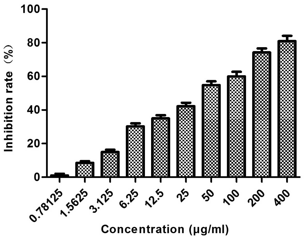
The inhibitory effect of
proanthocyanidins on the proliferation of MGC-803 cells
As shown in Fig. 1,
a range of concentrations of proanthocyanidins (400, 200, 10, 50,
25, 12.5, 6.25, 3.125, 1.5625 and 0.78125 µg/ml) was used to
treat MGC-803 cells for 48 h, and the results showed that
proanthocyanidins inhibited MGC-803 cell proliferation in a
dose-dependent manner. At 48 h, the IC50 value was
40.654 µg/ml.
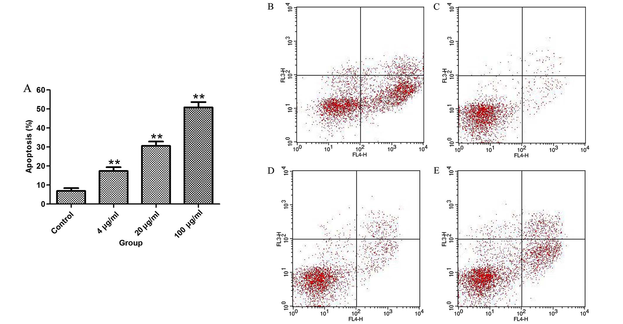
The effect of proanthocyanidins on the
apoptosis of MGC-803 cells
To test whether the inhibition of cell proliferation
by proanthocyanidins was associated with apoptosis, MGC-803 cells
were treated with proanthocyanidins for 48 h, and then, flow
cytometry was used to analyze the percentage of apoptotic cells.
The results in Fig. 2 show that
proanthocyanidins induced apoptosis in MGC-803 cells in a
dose-dependent manner.
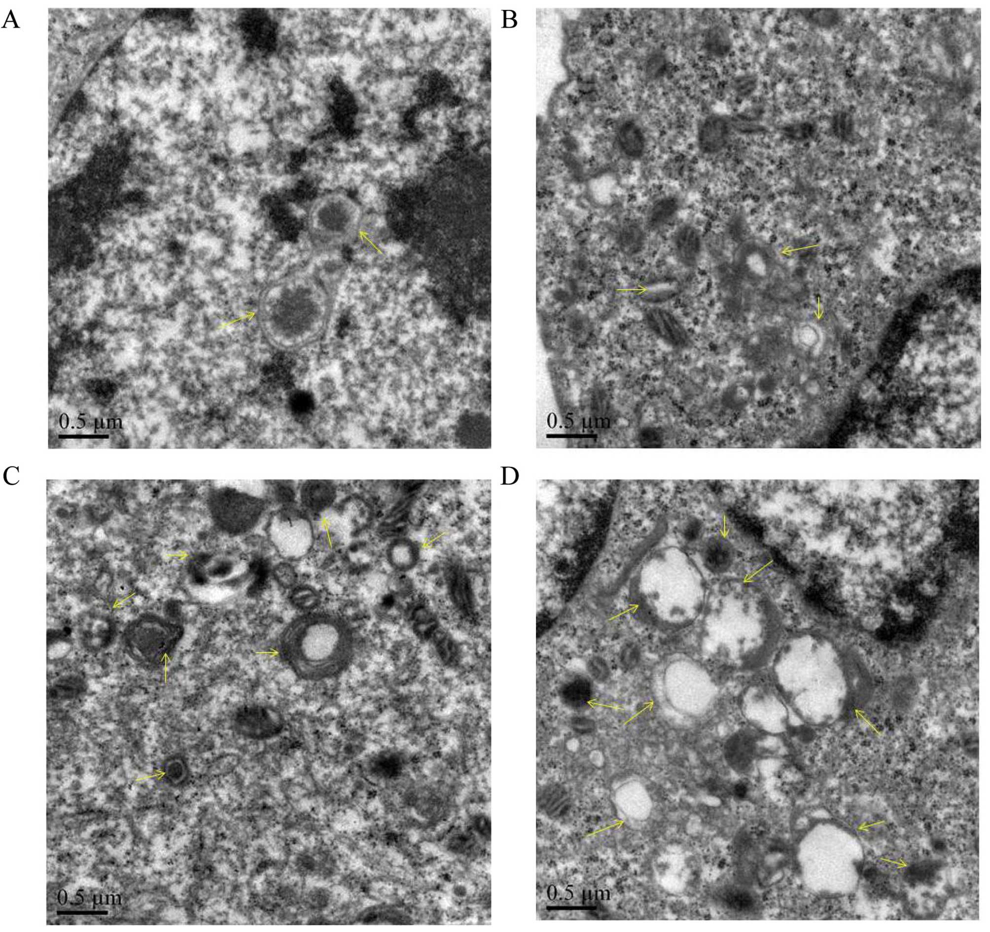
The effect of proanthocyanidins on the
microstructural morphology of MGC-803 cells
To verify whether the cytoplasmic vacuoles observed
by inverted microscopy were related to autophagy, transmission
electron microscopy was used to observe autophagosomes in MGC-803
cells treated with proanthocyanidins. As shown in Fig. 3, untreated cells had normal nuclei,
cytoplasm and organelles, whereas proanthocyanidin-treated cells
showed a high number of autophagosomes of various sizes, and
autophagosomes containing mitochondria were also observed by
electron microscopy. This suggests that autophagy occurred in the
cells after proanthocyanidin treatment.
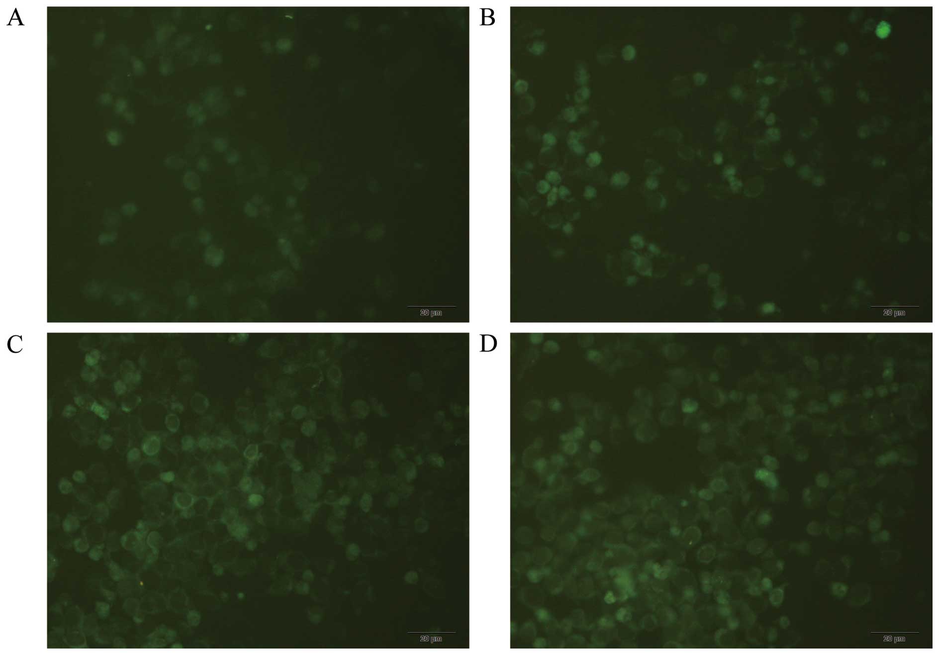
MDC staining for autophagosome
labeling
An inverted fluorescence microscope was used to
observe MDC-labeled autophagic vacuoles, clear punctate structures
were observed in the cytoplasm and perinuclear region, and the
changes of the particles inside the cell were used to determine the
level of autophagy. As shown in Fig.
4, compared with the control group, proanthocyanidins-treated
cells showed stronger fluorescence intensity and more autophagic
vacuoles labeled with MDC, and the number and staining intensity of
the vacuoles increased with increasing proanthocyanidins dose. This
suggests that proanthocyanidins induced autophagy.
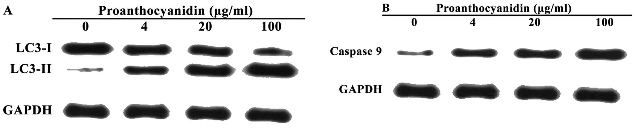
Proanthocyanidins induces LC3 and caspase
9 expression in MGC-803 cells
Western blot analyses were used to detect changes in
the levels of LC3 and caspase 9 after MGC-803 cells were treated
with proanthocyanidins. On SDS-PAGE gels, LC3-II ran faster than
LC3-I, producing two bands by western blotting. Fig. 5A shows that the untreated cells
exhibited only a faint LC3-I band, whereas the LC3-II band was not
detected. In contrast, after treatment with proanthocyanidins, the
level of LC3-II increased significantly in a dose-dependent manner.
Fig. 5B shows that treatment with
proanthocyanidins significantly increased the expression of caspase
9 compared with the control group in a dose-dependent manner.
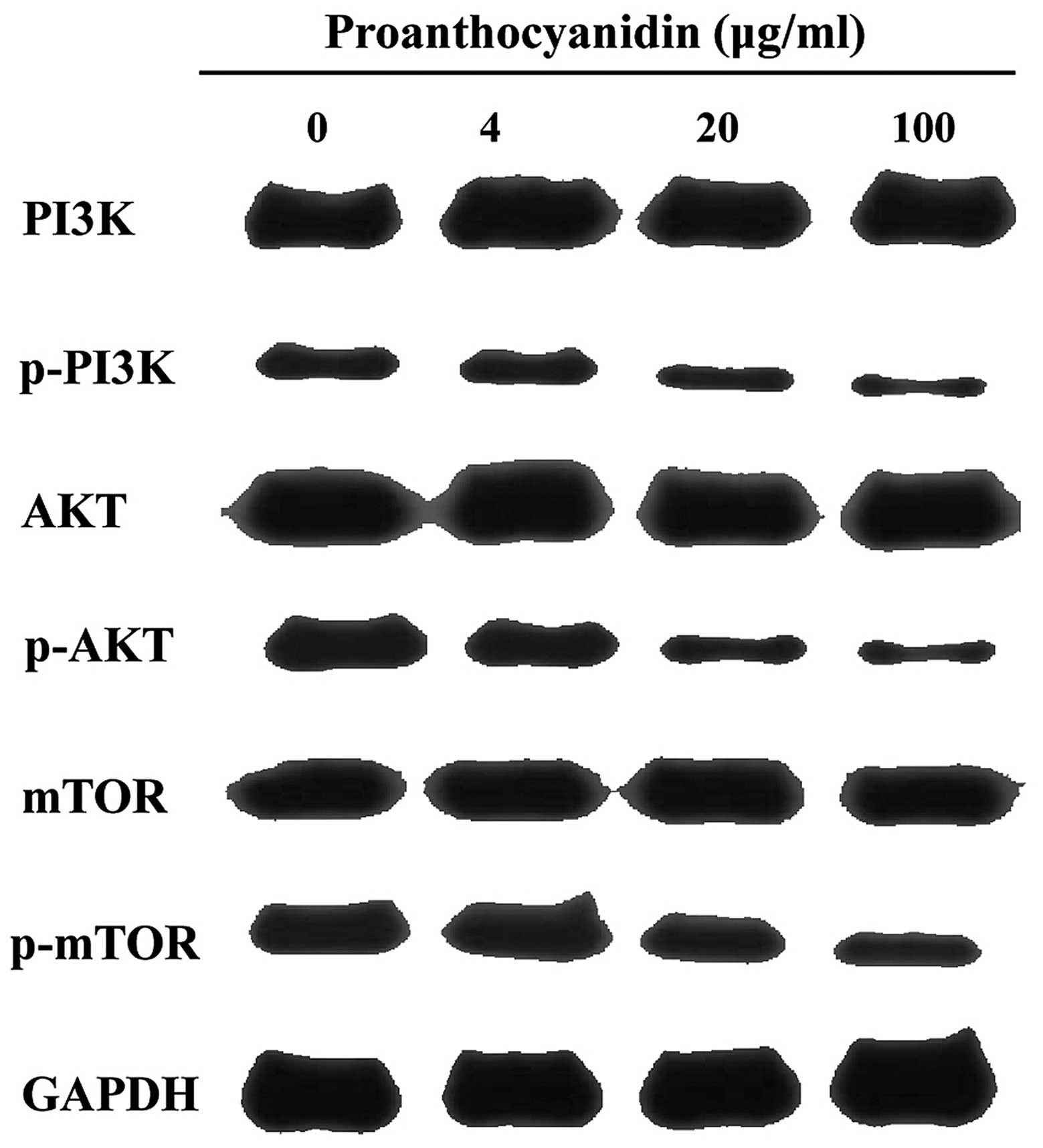
Effect of proanthocyanidins on the
phosphatidylinositol 3 kinase (PI3K)/protein kinase B
(PKB/AKT)/mammalian target of rapamycin (mTOR) signaling
pathway
The PI3K/AKT/mTOR signaling pathway is the canonical
pathway that negatively regulates the initiation of autophagy. It
has been reported that inhibition of this pathway induces cell
autophagy. As shown in Fig. 6,
western blot analyses showed that proanthocyanidins inhibited the
phosphorylation of PI3K, AKT and mTOR in the PI3K/AKT/mTOR
signaling pathway.
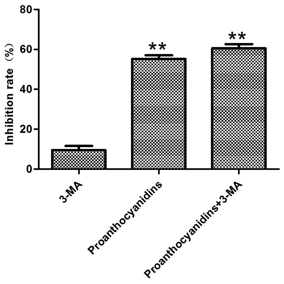
Inhibition of autophagy increased the
cytotoxicity of proanthocyanidins
Preliminary experiments determined that
proanthocyanidin treatment of MGC-803 cells activated both
autophagy and apoptosis and that the inhibition of MGC-803 cell
proliferation by proanthocyanidins occurred in a dose-dependent
manner. To understand whether the cytotoxicity exhibited by
proanthocyanidins was mediated by autophagy, the autophagy
inhibitor 3-MA was added, and MTT assays were used to examine its
cytotoxicity. The results showed that compared with cells treated
with only proanthocyanidins, the addition of 3-MA significantly
increased the percentage of apoptotic MGC-803 cells (Fig. 7).
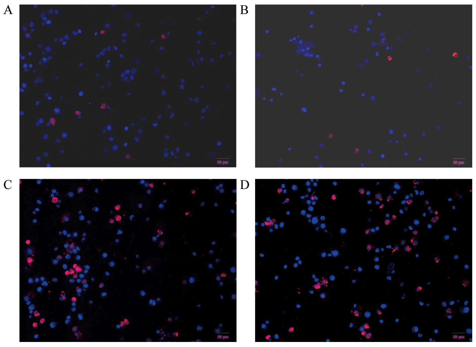
Hochest 33342 and PI double fluorescence
staining of live cells
Hochest 33342 and PI double staining is able to
distinguish live and dead cells. When cells are in the late
apoptotic stage or in the early necrotic stage, the nuclei are red
in color, whereas the nuclei of live cells are blue. As shown in
Fig. 8, MGC-803 cells treated with
proanthocyanidins for 48 h exhibited nuclei with a bead-like shape,
forming apoptotic bodies. There was no significant difference
between treatment with 3-MA alone (Fig.
8B) and control cells (Fig.
8A). However, cells treated with proanthocyanidins (Fig. 8C) exhibited an increased percentage
of apoptotic cells compared with the control cells (Fig. 8A) (p<0.001). Cells treated with
proanthocyanidins + 3-MA (Fig. 8D)
showed an increased percentage of apoptotic cells compared with
cells treated with proanthocyanidins alone (Fig. 8C) (p<0.01). These results showed
that apoptosis increased significantly in response to the
inhibition of autophagy induced by proanthocyanidins.
The effect of proanthocyanidins on
apoptosis following inhibition of autophagy
Since we found that the cytotoxicity of
proanthocyanidins was increased after the inhibition of autophagy,
we speculated whether autophagy inhibited the effect of
proanthocyanidins on apoptosis. Real-time quantitative PCR was used
to investigate MGC-803 cells treated with proanthocyanidins. Using
Beclin1 and BCL-2 as indicators, we investigated the effect of
proanthocyanidins on autophagy and apoptosis when autophagy was
inhibited. As shown in Fig. 9,
after the addition of 5 mM 3-MA, the expression of Beclin1
decreased compared with that in control cells. After treatment with
40.7 µg/ml proanthocyanidins, the expression of Beclin1
increased significantly compared with that in control cells.
However, when the proanthocyanidins were added after treatment with
3-MA, Beclin1 expression was significantly decreased compared with
the level in cells treated with proanthocyanidins alone. After
treatment with proanthocyanidins, BCL-2 decreased significantly
compared with expression in the control cells, whereas the addition
of 3-MA significantly decreased BCL-2 expression compared with that
in cells treated with proanthocyanidins alone. Thus, when autophagy
was inhibited, the apoptotic effect of proanthocyanidins was
increased.
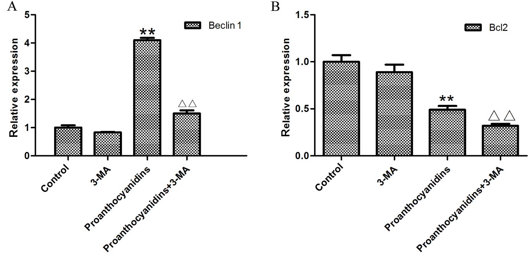 | Figure 9The effect of proanthocyanidins on
autophagy and apoptosis when autophagy was inhibited. After
treatment with 5 mM 3-MA, the expression of Beclin1 was decreased
compared with that in control cells. After treatment with 40.7
µg/ml proanthocyanidins, the expression of Beclin1 increased
significantly compared with that in control cells,
**p<0.01. However, when the proanthocyanidins were
added after treatment with 3-MA, Beclin1 expression was
significantly reduced compared with that in cells treated with
proanthocyanidins alone, ΔΔp<0.01. (A) Data are
expressed as the mean ± standard deviation (n=3). After treatment
with proanthocyanidins, the level of Bcl2 decreased significantly
compared with that in control cells, **p<0.01,
whereas simultaneous addition of 3-MA significantly decreased Bcl2
expression compared with that in cells treated with
proanthocyanidins alone, ΔΔp<0.01. (B) Data are
expressed as the mean ± standard deviation (n=3). |
Discussion
In the present study, MTT assays were used to
determine the effect of proanthocyanidins on the human gastric
cancer MGC-803 cells and to calculate the IC50. The
results showed that proanthocyanidins significantly inhibit MGC-803
cells in a dose-dependent manner.
To test whether the inhibition of MGC-803 cells by
proanthocyanidins is related to the induction of autophagy, we used
MDC staining to label autophagic vacuoles and transmission
electronic microscopy to observe autophagosomes, which are
published methods to confirm the presence of autophagy (39). MDC staining showed that
proanthocyanidin-treated cells exhibited increased autophagic
vacuoles, and transmission electronic microscopy confirmed the
existence of autophagosomes in the cells. Therefore, from the
morphology, we can preliminarily confirm that proanthocyanidins
induce autophagy in MGC-803 cells. Microtubule-associated protein 1
light chain 3 (LC3) plays a key role in autophagy in mammalian
cells. LC3 consists of soluble LC3-I and lipidated LC3-II. Under
various stresses, such as hypoxia and drug treatment, the cells
initiate autophagy, and LC3-I undergoes a ubiquitination-like
modification and processing to form LC3-II. Therefore, the level of
LC3-II is positively correlated with the number of autophagic
vacuoles. When autophagy occurs inside the cells, LC3-II increases
significantly, and the detection of the change in LC3-II levels can
accurately determine the amount of autophagy (40–43).
When western blot analyses were used to determine the levels of the
autophagy marker LC3, LC3-I levels decreased, whereas LC3-II
increased in proanthocyanidin-treated MGC-803 cells, and both of
these effects were dose-dependent. This confirmed that treatment
with proanthocyanidin-induced autophagy in MGC-803 cells.
We next used flow cytometry to observe apoptosis of
MGC-803 cells after treatment with proanthocyanidins. The
proanthocyanidins significantly induced apoptosis in MGC-803 cells.
According to published studies, proanthocyanidins induce apoptosis
in multiple tumor cell types, and the above result is consistent
with these reports (44,45). Apoptosis is initiated by p53, which
activates relevant proteins to form a channel on the mitochondria
to allow cytochrome c in the mitochondria to be released
into the cytoplasm, which eventually activates caspase family
proteases (46). Caspase family
proteases are a type of aspartate-specific cysteine-containing
proteases that are important for apoptosis. Caspases are divided
into apoptosis initiation factors and apoptosis effectors according
to their function (47). Caspase 9
is an important initiation factor and can be activated by other
proteins or by itself to activate a series of downstream effectors.
Eventually, the cells undergo biochemical and morphological changes
that lead to apoptosis (48). Our
experiments have shown that proanthocyanidins activate caspase 9
and induce apoptosis in MGC-803 cells.
Molecular signaling pathways are closely involved in
autophagy and apoptosis. Phosphatidylinositol 3 kinase
(PI3K)/protein kinase B (PKB/AKT)/mammalian target of rapamycin
(mTOR) is one of the currently most-studied pathways. This pathway
is well accepted as being associated with cell autophagy and
apoptosis. PI3K phosphorylates phosphatidylinositol (4,5)
bisphosphate [PtdIns (4,5)P2] in the cytoplasmic membrane to
generate phosphatidylinositol (3–5)
triphosphate [PtdIns (3,4,5) P3],
which recruits AKT to the inner side of the cytoplasmic membrane.
AKT is then phosphorylated and activated by another protein kinase,
3-phosphoinositide-dependent protein kinase 1 (PDK1). Activated AKT
further activates mTOR by inhibiting the tuberous sclerosis complex
(TSC1/2), which is an inhibitor of mTOR. The inhibition of TSC1/2
activity by phosphorylated AKT leads to the activation of mTOR.
mTOR is a serine/threonine kinase that inhibits autophagy when
activated (49). Similarly,
apoptosis is also affected by the PI3K/AKT pathway. Activated AKT
binds to Ser184 of the BCL-2 family member BAX. After
phosphorylation, BAX inactivates mitochondrial cytochrome c,
blocking the activation of caspases and inhibiting apoptosis
(50). Whereas, activated AKT also
phosphorylates Ser196 of caspase 9, inactivating it and thus
directly reducing apoptosis (51).
Multiple studies have shown that autophagy and apoptosis induced by
a variety of drugs are all associated with the PI3K/AKT/mTOR
pathway (52–55). Western blot analyses showed that
although proanthocyanidins did not affect the total amount of PI3K,
AKT, and mTOR, these compounds did significantly reduce the amount
of p-PI3K, p-AKT and p-mTOR; in other words, proanthocyanidins
reduced the activation of the PI3K/AKT/mTOR pathway. This may be
one of the reasons that proanthocyanidins induce autophagy and
apoptosis and is also consistent with other reports (56).
The results above showed that both autophagy and
apoptosis are activated when proanthocyanidins induce cell death in
the human gastric cancer MGC-803 cells and that the activation
mechanism is associated with the inhibition of the PI3K/AKT/mTOR
pathway.
To further explore the relationship between
proanthocyanidin-induced autophagy and apoptosis, we used the
classical autophagy inhibitor 3-methyladenine (3-MA) to determine
the effect of autophagy on apoptosis. MTT assays found that after
the addition of 3-MA, the effect of proanthocyanidins increased.
When Hoechst 33342/PI double fluorescence staining was used to
observe apoptosis after the inhibition of autophagy, apoptosis
increased significantly. These results suggest that the inhibition
of autophagy may increase apoptosis. To verify this hypothesis,
real-time quantitative PCR technology was used to determine the
mRNA level of Beclin1 and BCL-2. Beclin1 plays a key role in
autophagy of mammalian cells. Beclin1 is also the mammalian
homologue of the yeast protein ATG6/Vps30 and is located on human
chromosome 17q21 (57). Beclin1
promotes the formation of autophagosomes by forming a complex with
class III PI3K (58). The
expression of Beclin1 is positively correlated with autophagy in
multiple malignant tumor cell types (59–61).
BCL-2 is a key regulator of the known apoptosis proteins and is
negatively correlated with apoptosis induced by various signals
(62,63). When BCL-2 increases, the BCL-2/BAX
heterodimer interferes with the release of cytochrome c,
thereby blocking the activation of the upstream caspase protease
and in turn inhibiting apoptosis (64–66).
The inhibition of autophagy by 3-MA not only decreased Beclin1 mRNA
but also increased BCL-2 mRNA, which further confirmed that
apoptosis increases significantly when autophagy induced by
proanthocyanidins is blocked.
In summary, proanthocyanidins exhibit a significant
inhibitory effect on human gastric cancer cell (MGC-803)
proliferation in vitro and simultaneously activate autophagy
and apoptosis to promote cell death. The mechanism is associated
with interference of the PI3K/AKT pathway by proanthocyanidins and
a change in the amount of the downstream autophagy proteins LC3 and
Beclin1, as well as in the apoptosis proteins BCL-2 and caspase 9.
Furthermore, when proanthocyanidin-induced autophagy is inhibited,
apoptosis increases significantly and tumor cells undergo cell
death. Therefore, as an active ingredient of natural products with
low toxicity, proanthocyanidins can be used together with autophagy
inhibitors to enhance cytotoxicity.
Acknowledgments
The present study was supported by the State
Administration of Traditional Chinese Medicine of Jiangsu Province
(grant no. LZ13240).
References
|
1
|
Ren JS, Kamangar F, Qiao yL, Taylor PR,
Liang H, Dawsey SM, Liu B, Fan JH and Abnet CC: Serum pepsinogens
and risk of gastric and oesophageal cancers in the General
Population Nutrition Intervention Trial cohort. Gut. 58:636–642.
2009. View Article : Google Scholar : PubMed/NCBI
|
|
2
|
No authors listed. Survival of Cancer
Patients in Europe: The EUROCARE-2 study. IARC Sci Publ. 151:1–572.
1999.
|
|
3
|
Crew KD and Neugut AI: Epidemiology of
gastric cancer. World J Gastroenterol. 12:354–362. 2006.PubMed/NCBI
|
|
4
|
Jemal A, Bray F, Center MM, Ferlay J, Ward
E and Forman D: Global cancer statistics. CA Cancer J Clin.
61:69–90. 2011. View Article : Google Scholar : PubMed/NCBI
|
|
5
|
Zhang J, Li y, Chen X, Liu T, Chen y, He
W, Zhang Q and Liu S: Autophagy is involved in anticancer effects
of matrine on SGC-7901 human gastric cancer cells. Oncol Rep.
26:115–124. 2011.PubMed/NCBI
|
|
6
|
Kawasaki ES and Player A: Nanotechnology,
nanomedicine, and the development of new, effective therapies for
cancer. Nanomedicine. 1:101–109. 2005. View Article : Google Scholar
|
|
7
|
Valentini F, Carbone M and Palleschi G:
Carbon nanostructured materials for applications in nano-medicine,
cultural heritage, and electrochemical biosensors. Anal Bioanal
Chem. 405:451–465. 2013. View Article : Google Scholar
|
|
8
|
Alexis F, Rhee JW, Richie JP,
Radovic-Moreno AF, Langer R and Farokhzad OC: New frontiers in
nanotechnology for cancer treatment. Urol Oncol. 26:74–85. 2008.
View Article : Google Scholar : PubMed/NCBI
|
|
9
|
Chen SL, Fang WW, Ye F, Liu yH, Qian J,
Shan SJ, Zhang JJ, Chunhua RZ, Liao LM, Lin S, et al: Effect on
left ventricular function of intracoronary transplantation of
autologous bone marrow mesenchymal stem cell in patients with acute
myocardial infarction. Am J Cardiol. 94:92–95. 2004. View Article : Google Scholar : PubMed/NCBI
|
|
10
|
Bang OY, Lee JS, Lee PH and Lee G:
Autologous mesenchymal stem cell transplantation in stroke
patients. Ann Neurol. 57:874–882. 2005. View Article : Google Scholar : PubMed/NCBI
|
|
11
|
Kuo TK, Hung SP, Chuang CH, Chen CT, Shih
YR, Fang SC, Yang VW and Lee OK: Stem cell therapy for liver
disease: Parameters governing the success of using bone marrow
mesenchymal stem cells. Gastroenterology. 134:2111–2121.
2121.e1–2121.e3. 2008. View Article : Google Scholar : PubMed/NCBI
|
|
12
|
Shi Y, Moon M, Dawood S, McManus B and Liu
PP: Mechanisms and management of doxorubicin cardiotoxicity. Herz.
36:296–305. 2011. View Article : Google Scholar : PubMed/NCBI
|
|
13
|
Poletti V, Casoni GL, Cancellieri A,
Piciucchi S, Dubini A and Zompatori M: Diffuse alveolar damage.
Pathologica. 102:453–463. 2010.
|
|
14
|
Naranjo TW, Lopera DE, Diaz-Granados LR,
Duque JJ, Restrepo AM and Cano LE: Combined
itraconazole-pentoxifylline treatment promptly reduces lung
fibrosis induced by chronic pulmonary paracoccidioidomycosis in
mice. Pulm Pharmacol Ther. 24:81–91. 2011. View Article : Google Scholar
|
|
15
|
de Pascual-Teresa S, Santos-Buelga C and
Rivas-Gonzalo JC: Quantitative analysis of flavan-3-ols in Spanish
foodstuffs and beverages. J Agric Food Chem. 48:5331–5337. 2000.
View Article : Google Scholar : PubMed/NCBI
|
|
16
|
Li X, Chen D, Wang G and Lu y: Study of
interaction between human serum albumin and three antioxidants:
Ascorbic acid, α-tocopherol, and proanthocyanidins. Eur J Med Chem.
70:22–36. 2013. View Article : Google Scholar
|
|
17
|
Wiesneth S, Petereit F and Jürgenliemk G:
Salix daphnoides: A Screening for Oligomeric and Polymeric
Proanthocyanidins. Molecules. 20:13764–13779. 2015. View Article : Google Scholar : PubMed/NCBI
|
|
18
|
Fernández K, Vega M and Aspé E: An
enzymatic extraction of proanthocyanidins from País grape seeds and
skins. Food Chem. 168:7–13. 2015. View Article : Google Scholar
|
|
19
|
Roopchand DE, Krueger CG, Moskal K,
Fridlender B, Lila MA and Raskin I: Food-compatible method for the
efficient extraction and stabilization of cranberry pomace
polyphenols. Food Chem. 141:3664–3669. 2013. View Article : Google Scholar : PubMed/NCBI
|
|
20
|
Lombardo Bedran TB, Palomari Spolidorio D
and Grenier D: Green tea polyphenol epigallocatechin-3-gallate and
cranberry proanthocyanidins act in synergy with cathelicidin
(LL-37) to reduce the LPS-induced inflammatory response in a
three-dimensional co-culture model of gingival epithelial cells and
fibroblasts. Arch Oral Biol. 60:845–853. 2015. View Article : Google Scholar : PubMed/NCBI
|
|
21
|
Pons Z, Guerrero L, Margalef M, Arola L,
Arola-Arnal A and Muguerza B: Effect of low molecular grape seed
proanthocyani-dins on blood pressure and lipid homeostasis in
cafeteria diet-fed rats. J Physiol Biochem. 70:629–637. 2014.
View Article : Google Scholar : PubMed/NCBI
|
|
22
|
Zhang XY, Li WG, Zheng TZ and Li W:
Effects of proantho-cyanidins on contractile activity of aortic
smooth muscle and platelet aggregation in experimental animals.
Zhongguo ying yong Sheng Li Xue Za Zhi. 21:383–386. 2005.In
Chinese. PubMed/NCBI
|
|
23
|
Mohana T, Navin AV, Jamuna S, Sakeena
Sadullah MS and Niranjali Devaraj S: Inhibition of differentiation
of monocyte to macrophages in atherosclerosis by oligomeric
proanthocyanidins -In-vivo and in-vitro study. Food Chem Toxicol.
82:96–105. 2015. View Article : Google Scholar : PubMed/NCBI
|
|
24
|
de Sá M, Justino V, Spranger MI, Zhao yQ,
Han L and Sun BS: Extraction yields and anti-oxidant activity of
proanthocyanidins from different parts of grape pomace: Effect of
mechanical treatments. Phytochem Anal. 25:134–140. 2014. View Article : Google Scholar
|
|
25
|
Luan yy, Liu ZM, Zhong Jy, Yao Ry and Yu
HS: Effect of grape seed proanthocyanidins on tumor vasculogenic
mimicry in human triple-negative breast cancer cells. Asian Pac J
Cancer Prev. 16:531–535. 2015. View Article : Google Scholar : PubMed/NCBI
|
|
26
|
D'Angelo L, Piazzi G, Pacilli A,
Prossomariti A, Fazio C, Montanaro L, Graziani G, Fogliano V,
Munarini A, Bianchi F, et al: A combination of eicosapentaenoic
acid-free fatty acid, epigallocatechin-3-gallate and
proanthocyanidins has a strong effect on mTOR signaling in
colorectal cancer cells. Carcinogenesis. 35:2314–2320. 2014.
View Article : Google Scholar : PubMed/NCBI
|
|
27
|
Huang S, Yang N, Liu y, Gao J, Huang T, Hu
L, Zhao J, Li y, Li C and Zhang X: Grape seed proanthocyanidins
inhibit colon cancer-induced angiogenesis through suppressing the
expression of VEGF and Ang1. Int J Mol Med. 30:1410–1416.
2012.PubMed/NCBI
|
|
28
|
Sharma SD and Katiyar SK: Dietary grape
seed proanthocy-anidins inhibit UVB-induced cyclooxygenase-2
expression and other inflammatory mediators in UVB-exposed skin and
skin tumors of SKH-1 hairless mice. Pharm Res. 27:1092–1102. 2010.
View Article : Google Scholar : PubMed/NCBI
|
|
29
|
Zhang XY, Li WG, Wu YJ and Gao MT:
Amelioration of doxorubicin-induced myocardial oxidative stress and
immunosuppression by grape seed proanthocyanidins in tumour-bearing
mice. J Pharm Pharmacol. 57:1043–1052. 2005. View Article : Google Scholar : PubMed/NCBI
|
|
30
|
Shen S, Kepp O, Michaud M, Martins I,
Minoux H, Métivier D, Maiuri MC, Kroemer RT and Kroemer G:
Association and dissociation of autophagy, apoptosis and necrosis
by systematic chemical study. Oncogene. 30:4544–4556. 2011.
View Article : Google Scholar : PubMed/NCBI
|
|
31
|
Chiantore MV, Vannucchi S, Mangino G,
Percario ZA, Affabris E, Fiorucci G and Romeo G: Senescence and
cell death pathways and their role in cancer therapeutic outcome.
Curr Med Chem. 16:287–300. 2009. View Article : Google Scholar : PubMed/NCBI
|
|
32
|
Klionsky DJ: Autophagy: From phenomenology
to molecular understanding in less than a decade. Nat Rev Mol Cell
Biol. 8:931–937. 2007. View Article : Google Scholar : PubMed/NCBI
|
|
33
|
Levine B and Kroemer G: Autophagy in the
pathogenesis of disease. Cell. 132:27–42. 2008. View Article : Google Scholar : PubMed/NCBI
|
|
34
|
Wang MC, Wu AG, Huang YZ, Shao GL, Ji SF,
Wang RW, Yuan HJ, Fan XL, Zheng LH and Jiao QL: Autophagic
regulation of cell growth by altered expression of Beclin 1 in
triple-negative breast cancer. Int J Clin Exp Med. 8:7049–7058.
2015.PubMed/NCBI
|
|
35
|
Sui H, Shi C, Yan Z and Li H: Combination
of erlotinib and a PARP inhibitor inhibits growth of A2780 tumor
xenografts due to increased autophagy. Drug Des Devel Ther.
9:3183–3190. 2015. View Article : Google Scholar : PubMed/NCBI
|
|
36
|
Zheng JF, Li LL, Lu J, Yan K, Guo WH and
Zhang JX: XPD functions as a tumor suppressor and dysregulates
autophagy in cultured HepG2 cells. Med Sci Monit. 21:1562–1568.
2015. View Article : Google Scholar : PubMed/NCBI
|
|
37
|
Rosenfeldt MT and Ryan KM: The multiple
roles of autophagy in cancer. Carcinogenesis. 32:955–963. 2011.
View Article : Google Scholar : PubMed/NCBI
|
|
38
|
Duan Y, Ke J, Zhang H, He Y, Sun G and Sun
X: Autophagic cell death of human hepatoma G2 cells mediated by
procyanidins from Castanea mollissima Bl. Shell-induced reactive
oxygen species generation. Chem Biol Interact. 224:13–23. 2014.
View Article : Google Scholar : PubMed/NCBI
|
|
39
|
Mizushima N: Methods for monitoring
autophagy. Int J Biochem Cell Biol. 36:2491–2502. 2004. View Article : Google Scholar : PubMed/NCBI
|
|
40
|
Eskelinen EL and Saftig P: Autophagy: A
lysosomal degradation pathway with a central role in health and
disease. Biochim Biophys Acta. 1793:664–673. 2009. View Article : Google Scholar
|
|
41
|
He H, Dang Y, Dai F, Guo Z, Wu J, She X,
Pei Y, Chen Y, Ling W, Wu C, et al: Post-translational
modifications of three members of the human MAP1LC3 family and
detection of a novel type of modification for MAP1LC3B. J Biol
Chem. 278:29278–29287. 2003. View Article : Google Scholar : PubMed/NCBI
|
|
42
|
Zheng HY, Zhang XY, Wang XF and Sun BC:
Autophagy enhances the aggressiveness of human colorectal cancer
cells and their ability to adapt to apoptotic stimulus. Cancer Biol
Med. 9:105–110. 2012.
|
|
43
|
Gozuacik D and Kimchi A: Autophagy as a
cell death and tumor suppressor mechanism. Oncogene. 23:2891–2906.
2004. View Article : Google Scholar : PubMed/NCBI
|
|
44
|
Zhang Z, Zheng L, Zhao Z, Shi J, Wang X
and Huang J: Grape seed proanthocyanidins inhibit
H2O2-induced osteoblastic MC3T3-E1 cell
apoptosis via ameliorating H2O2-induced
mitochondrial dysfunction. J Toxicol Sci. 39:803–813. 2014.
View Article : Google Scholar
|
|
45
|
Chen Q, Liu XF and Zheng PS: Grape seed
proanthocyanidins (GSPs) inhibit the growth of cervical cancer by
inducing apoptosis mediated by the mitochondrial pathway. PLoS One.
9:e1070452014. View Article : Google Scholar : PubMed/NCBI
|
|
46
|
Prasad R, Vaid M and Katiyar SK: Grape
proanthocyanidin inhibit pancreatic cancer cell growth in vitro and
in vivo through induction of apoptosis and by targeting the
PI3K/Akt pathway. PLoS One. 7:e430642012. View Article : Google Scholar : PubMed/NCBI
|
|
47
|
Kim ME, Ha TK, Yoon JH and Lee JS:
Myricetin induces cell death of human colon cancer cells via
BAX/BCL2-dependent pathway. Anticancer Res. 34:701–706.
2014.PubMed/NCBI
|
|
48
|
Choi YJ, Saez B, Anders L, Hydbring P,
Stefano J, Bacon NA, Cook C, Kalaszczynska I, Signoretti S, Young
RA, et al: D-cyclins repress apoptosis in hematopoietic cells by
controlling death receptor Fas and its ligand FasL. Dev Cell.
30:255–267. 2014. View Article : Google Scholar : PubMed/NCBI
|
|
49
|
Yao W, Yue P, Zhang G, Owonikoko TK, Khuri
FR and Sun SY: Enhancing therapeutic efficacy of the MEK inhibitor,
MEK162, by blocking autophagy or inhibiting PI3K/Akt signaling in
human lung cancer cells. Cancer Lett. 364:70–78. 2015. View Article : Google Scholar : PubMed/NCBI
|
|
50
|
Xin M and Deng X: Nicotine inactivation of
the proapoptotic function of Bax through phosphorylation. J Biol
Chem. 280:10781–10789. 2005. View Article : Google Scholar : PubMed/NCBI
|
|
51
|
Song G, Ouyang G and Bao S: The activation
of Akt/PKB signaling pathway and cell survival. J Cell Mol Med.
9:59–71. 2005. View Article : Google Scholar : PubMed/NCBI
|
|
52
|
Pan ST, Qin Y, Zhou ZW, He ZX, Zhang X,
Yang T, Yang YX, Wang D, Qiu JX and Zhou SF: Plumbagin induces
G2/M arrest, apoptosis, and autophagy via p38 MAPK- and
PI3K/Akt/mTOR-mediated pathways in human tongue squamous cell
carcinoma cells. Drug Des Devel Ther. 9:1601–1626. 2015.
|
|
53
|
Zhou ZW, Li XX, He ZX, Pan ST, Yang Y,
Zhang X, Chow K, Yang T, Qiu JX, Zhou Q, et al: Induction of
apoptosis and autophagy via sirtuin1- and PI3K/Akt/mTOR-mediated
pathways by plumbagin in human prostate cancer cells. Drug Des
Devel Ther. 9:1511–1554. 2015. View Article : Google Scholar : PubMed/NCBI
|
|
54
|
Yuan L, Wei S, Wang J and Liu X:
Isoorientin induces apoptosis and autophagy simultaneously by
reactive oxygen species (ROS)-related p53, PI3K/Akt, JNK, and p38
signaling pathways in HepG2 cancer cells. J Agric Food Chem.
62:5390–5400. 2014. View Article : Google Scholar : PubMed/NCBI
|
|
55
|
Zhang H, Guo M, Chen JH, Wang Z, Du XF,
Liu PX and Li WH: Osteopontin knockdown inhibits αv,β3
integrin-induced cell migration and invasion and promotes apoptosis
of breast cancer cells by inducing autophagy and inactivating the
PI3K/Akt/mTOR pathway. Cell Physiol Biochem. 33:991–1002. 2014.
View Article : Google Scholar
|
|
56
|
Hu Y, Li L, Yin W, Shen L, You B and Gao
H: Protective effect of proanthocyanidins on anoxia-reoxygenation
injury of myocardial cells mediated by the PI3K/Akt/GSK-3β pathway
and mitochondrial ATP-sensitive potassium channel. Mol Med Rep.
10:2051–2058. 2014.PubMed/NCBI
|
|
57
|
Zhong Y, Wang QJ and Yue Z: Atg14L and
Rubicon: yin and yang of Beclin 1-mediated autophagy control.
Autophagy. 5:890–891. 2009. View Article : Google Scholar : PubMed/NCBI
|
|
58
|
Liang XH, Jackson S, Seaman M, Brown K,
Kempkes B, Hibshoosh H and Levine B: Induction of autophagy and
inhibition of tumorigenesis by beclin 1. Nature. 402:672–676. 1999.
View Article : Google Scholar : PubMed/NCBI
|
|
59
|
Baspinar S, Bircan S, Orhan H, Kapucuoglu
N and Bozkurt KK: The relation of beclin 1 and bcl-2 expressions in
high grade prostatic intraepithelial neoplasia and prostate
adenocarcinoma: A tissue microarray study. Pathol Res Pract.
210:412–418. 2014. View Article : Google Scholar : PubMed/NCBI
|
|
60
|
Fukui M, Yamabe N, Choi HJ, Polireddy K,
Chen Q and Zhu BT: Mechanism of ascorbate-induced cell death in
human pancreatic cancer cells: Role of Bcl-2, Beclin 1 and
autophagy. Planta Med. 81:838–846. 2015. View Article : Google Scholar : PubMed/NCBI
|
|
61
|
Tian PG, Jiang ZX, Li JH, Zhou Z and Zhang
QH: Spliced XBP1 promotes macrophage survival and autophagy by
interacting with Beclin-1. Biochem Biophys Res Commun. 463:518–523.
2015. View Article : Google Scholar : PubMed/NCBI
|
|
62
|
Patro SC, Pal S, Bi Y, Lynn K, Mounzer KC,
Kostman JR, Davuluri RV and Montaner LJ: Shift in monocyte
apoptosis with increasing viral load and change in
apoptosis-related ISG/Bcl2 family gene expression in chronically
HIV-1-infected subjects. J Virol. 89:799–810. 2015. View Article : Google Scholar :
|
|
63
|
Chaudhary P and Vishwanatha JK: c-Jun
NH2-terminal kinase-induced proteasomal degradation of c-FLIPL/S
and Bcl2 sensitize prostate cancer cells to Fas- and
mitochondria-mediated apoptosis by tetrandrine. Biochem Pharmacol.
91:457–473. 2014. View Article : Google Scholar : PubMed/NCBI
|
|
64
|
Lin W and Tongyi S: Role of Bax/Bcl-2
family members in green tea polyphenol induced necroptosis of
p53-deficient Hep3B cells. Tumour Biol. 35:8065–8075. 2014.
View Article : Google Scholar : PubMed/NCBI
|
|
65
|
Hua F, Cornejo MG, Cardone MH, Stokes CL
and Lauffenburger DA: Effects of Bcl-2 levels on Fas
signaling-induced caspase-3 activation: Molecular genetic tests of
computational model predictions. J Immunol. 175:985–995. 2005.
View Article : Google Scholar : PubMed/NCBI
|
|
66
|
Park SE, Shin WT, Park C, Hong SH, Kim Gy,
Kim SO, Ryu CH, Hong SH and Choi YH: Induction of apoptosis in
MDA-MB-231 human breast carcinoma cells with an ethanol extract of
Cyperus rotundus L. by activating caspases. Oncol Rep.
32:2461–2470. 2014.PubMed/NCBI
|