Introduction
Glioma, which is the most malignant tumor type,
accounts for more than 70% of all brain tumors (1). The most common subtype is glioblastoma
multiforme (GBM), with age-adjusted incidence rate ranging from
0.59 to 3.69/100,000 persons (2).
The biological properties of GBM primarily include high mortality
and recurrence rates, uncontrollable invasiveness, strong
angiogenesis (3) and widespread
hypoxia (4). Tumor angiogenesis is
an independent prognostic factor associated with poor survival
(4). In fact, there is increasing
evidence that hypoxia activates angiogenesis (5), metastasis (6) and many other cellular processes in
tumors. Although multitude of mechanisms have been proposed to
elucidate the hypoxia-induced angiogenesis of tumor cells, more
research is warranted to determine the role of a new way of
angiogenesis in mediating the effects of hypoxia.
Vasculogenic mimicry (VM) is a new tumor vascular
pattern different from angiogenesis (7). It describes a specific capacity of
aggressive tumor cells to form vessel-like networks without
vascular endothelial cells that provide adequate blood supply for
tumor growth (7,8). A variety of molecular mechanisms and
signal pathways participate in VM induction (8). Additionally, tumor stem cells are also
shown to be implicated in VM formation (9). As a unique vessel formation manner, VM
is associated with tumor invasion and poor patient prognosis in
various tumors (10). Due to its
important effects on tumor progression, VM-related target molecules
and strategies are being studied for anticancer treatment (8). However, the specific molecular
mechanisms of VM in glioma are still unclear.
miRNAs are a class of endogenous small non-coding
RNAs that have been identified as negative regulators of gene
expression at the post-transcriptional level (11,12).
These small molecules are incorporated into the RNA-induced
silencing complex and bind to the seed sequence in the
3′-untranslated regions (3′-UTRs) of their target mRNAs to silence
gene translation via mRNA degradation, translational repression
and/or miRNA-mediated mRNA decay (12). Therefore, additional research is
warranted to determine the important roles of numerous miRNAs in
diverse tumor-related cellular processes, such as proliferation
(13), angiogenesis (14) and metastasis (15). In gliomas, various tumor-promoting
and tumor-suppressing miRNAs (16–19)
have been identified. However, there are only few miRNAs targets
discovered regulating tumor VM (20–22).
Our observation suggest that miRNAs may be important for the tumor
VM and provide new insights into understanding the molecular
mechanism underlying tumor progression.
The value and novelty of the present study is to
indicate, for the first time in human glioma that microRNA-Let-7f
(miR-Let-7f) is a VM-negative regulator by targeting the periostin
(POSTN)-induced migration. Firstly, we demonstrated VM as an
indicator of poor prognosis by testing its positive rate in human
glioma tumor samples. Then, we investigated the expression of
miR-Let-7f in human glioma tumor samples, and we found that
miR-Let-7f functions as a clear VM suppressor. Further study using
the glioma cell line A172 revealed that miR-Let-7f knockdown
drastically enhanced the VM forming capacity of glioma cells and
the miR-Let-7f overexpressed glioma cells almost completely lost
the VM forming ability. Using specific small interfering RNA
(siRNA), POSTN was considered as the potential intermediary anti-VM
molecule of miR-Let-7f. These findings suggest an unexpected
fundamental tumor-suppressive role for miR-Let-7f in glioma due to
its anti-VM effect.
Materials and methods
Tissue samples and cell culture
The human glioma cell line A172 was purchased from
the Chinese Academy of Sciences Cell Bank. All cells were cultured
in Dulbecco's modified eagle's medium (DMEM) supplemented with 10%
fetal bovine serum (FBS) and maintained at 37°C with 5%
CO2 in a humidified chamber. Hypoxic conditions were
induced by incubating the cells in a modular incubator chamber with
a gas mixture containing 1% O2, 5% CO2 and
94% N2 at 37°C. Forty-five human glioma tissues
including 4 World Health Organization (WHO) grade I tumors, 8 grade
II tumors, 13 grade III tumors and 20 grade IV tumors, and 2 normal
brain tissues of decompression operation were obtained from the
Department of Neurosurgery of Qilu Hospital of Shandong University.
The glioma specimens were verified and classified according to the
WHO classification standard of tumors by two experienced clinical
pathologists. The present study was approved by the Institutional
Review Board of Shandong University. Written informed consent was
obtained from all patients, and the hospital ethics Committee
approved the experiments.
Cell transfection
Mature miR-Let-7f mimics, the scrambled mimic
control, the miR-Let-7f inhibitor, the scrambled inhibitor control,
the miR-584-5p mimic and POSTN siRNA were designed and synthesized
by RiboBio. Cell transfections and co-transfections were performed
using Lipofectamine 2000 when the cells reached 70% confluency
according to the manufacturer's instructions. Untransfected cells
were used as the blank control, while cells transfected with the
scrambled oligos were used as the negative controls. The
transfection efficiency was verified by quantitative real-time PCR.
Forty-eight hours after transfection, the glioma cells were
harvested for subsequent experiments.
Cell viability assay
A172 cells were seeded into 96-well culture plates
at a density of 3,000 cells/well. Cell proliferation was analyzed
at 24, 48, 72 and 96 h after transfection using a Cell Counting
Kit-8 (CCK-8). A volume of 10 µl of CCK-8 solution was added
to each well, and the cells were incubated for another 1 h in a
humidified incubator at 37°C. Optical density was measured at 450
nm using a microplate reader. Each assay was performed in
triplicate.
Wound healing and cell migration
assays
The transfected cells (1×10⁵) were seeded
in 6-well plates, incubated overnight, and at 90% confluency, the
cell monolayer was scratched with a sterile pipette tip. The
scratched plates were cultured in DMEM containing 1% FBS. Images
were captured at 0 and 12 h along the scrape line under a
microscope. Cell migration assays were performed using Transwell
chambers (8-µm; Corning). In total, 5×10⁴
transfected cells in FBS-free medium were seeded in the upper
chamber. Medium containing 10% FBS was added to the lower chamber.
After 12 h, the cells that did not migrate or invade were removed
using cotton buds. The cells migrating on the lower surface were
fixed and stained with crystal violet.
RNA extraction and real-time quantitative
PCR
Total RNA was extracted from the tissue samples
using TRIzol. Then, total RNA (50 ng) was reverse-transcribed with
miR-Let-7f stem-loop RT primers or with the U6 RT primers using a
ReverTra Ace qPCR RT kit to generate cDNA. Real-time PCR was
performed using a SYBR Premix Ex Taq™ kit with miR-Let-7f or U6 PCR
primers. The reactions were performed using a LightCycler 2.0
instrument. U6 expression was used as the endogenous control. The
absolute expression levels were calculated as concentration ratios
using a Roche LightCycler® 2.0 system.
Immunohistochemistry staining
Paraffin-embedded human glioma tissue samples were
sectioned and dewaxed. Endogenous peroxidase activity was quenched
by incubating the slides in methanol containing 3% hydrogen
peroxide for 30 min, after which the sections were incubated for 2
h at room temperature with normal goat serum and subsequently
incubated at 4°C overnight with primary antibody (1:300 CD34;
Abcam). Next, the sections were incubated with horseradish
peroxidase-linked second antibody, followed by reaction with
diaminobenzidine and counterstaining with PAS staining kit and then
Mayer's hematoxylin.
VM analysis
VM formation was evaluated using a commercial
Matrigel matrix (BD Biosciences, France). A172 glioma cells were
digested and resuspended at 5×04 cells/ml in DMEM
containing 1% FBS. Wells of 96-well tissue culture plates were
coated with Matrigel (50 µl/well; BD Biosciences) which was
allowed to polymerase at 37°C for 30 min. The glioma cell
suspension was then plated at 100 µl/well onto the surface
of Matrigel and incubated at 37°C. Cells were photographed using an
Olympus inverted microscope.
Statistical analysis
All experiments were performed three times. The
statistical analysis and experimental graphs were generated using
SPSS 17.0 and GraphPad Prism software. Descriptive statistics
including the means ± SD, Mann-Whitney test, Kaplan-Meier plots,
log-rank tests were used to analyze the significant differences.
p<0.05 and p<0.01 were considered to indicate a statistically
significant result.
Results
VM positively correlates with the WHO
grades of human glioma tissues
To examine whether the VM correlates with the WHO
grades of human glioma, we assessed the VM in 45 human glioma
specimens with different grades and 2 normal brain tissues by
immunochemical staining (Table I).
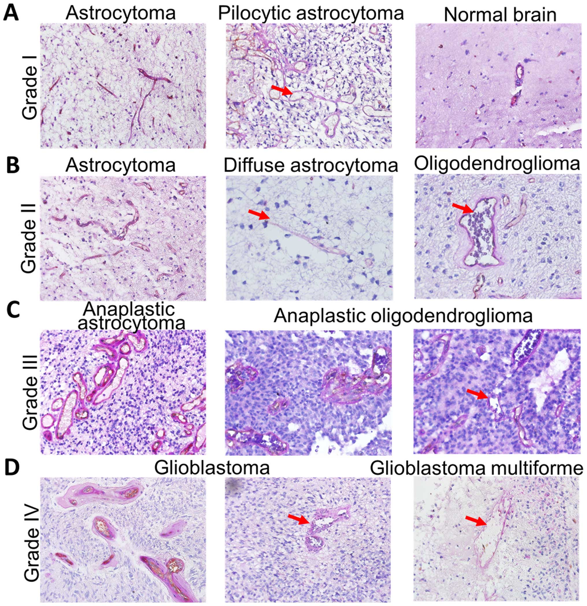
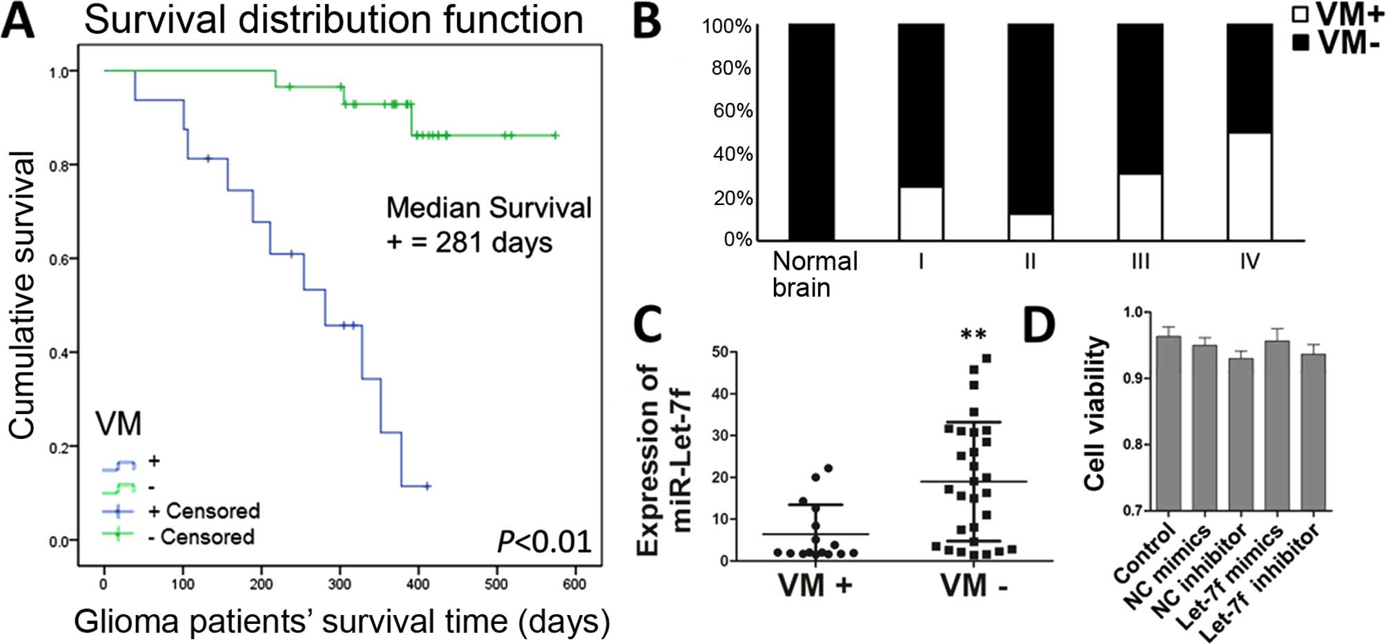
As shown in Fig. 1A, the VM was not
found in the normal brain tissues and WHO grade I astrocytoma
tissues, except pilocytic astrocytoma as its vascular hyperplasia.
We also found that the VM rarely existed in WHO grade II glioma
tissues (Figs. 1B and 2B). However, VM was very common in
high-grade (WHO III and IV) gliomas, particularly in GBM (Fig. 1C and D). Accordingly, the
VM-positive rate of human glioma was observably associated with its
WHO grades (Fig. 2B).
 | Table IDemographic parameters of patients
participating in the present study. |
Table I
Demographic parameters of patients
participating in the present study.
|
Characteristics | No. of
patients | N (%) |
|---|
| Assessable | | |
| Glioma | 45 | 95.74 |
| Normal brain
tissues | 2 | 4.26 |
| Gender | | |
| Male | 27 | 57.45 |
| Female | 20 | 42.55 |
| Age (years) | | |
| Median
(range) | 42.6 (4–69) | |
| Pathological
type | | |
| Astrocytoma | 6 | 12.77 |
| Anaplastic
astrocytoma | 10 | 21.28 |
| Pilocytic
astrocytoma | 2 | 4.26 |
|
Oligodendroglioma | 4 | 8.51 |
| Anaplastic
oligodendroglioma | 3 | 6.38 |
| Glioblastoma | 20 | 42.56 |
| Normal brain
tissues | 2 | 4.26 |
| WHO tumor grade at
diagnosis | | |
| I | 4 | 8.89 |
| II | 8 | 17.78 |
| III | 13 | 28.89 |
| IV | 20 | 44.44 |
VM indicates a poor prognosis of glioma
patients and negatively correlates with miR-Let-7f expression
levels
To analyze whether VM in clinical samples associated
with the clinical survival information of the 45 patients,
Kaplan-Meier estimates were used in the present study. As expected,
the subgroup of VM negative glioma patients presented a
significantly prolonged postoperative survival time (Fig. 2A). Since our previous research found
miR-Let-7f was identified as a potent anti-glioma miRNA (23), we hypothesized that miR-Let-7f was
probably involved in the regulation process of VM. To assess the
relationship between miR-Let-7f and VM, we verified the levels of
miR-Let-7f in these 45 human glioma specimens by quantitative
real-time PCR. The results showed that miR-Let-7f expression levels
were negatively correlated with VM (Fig. 2C). These data were particularly
strong for the VM suppressive effect of miR-Let-7f in glioma.
However, its underlying mechanism of anti-VM effects remains
unknown.
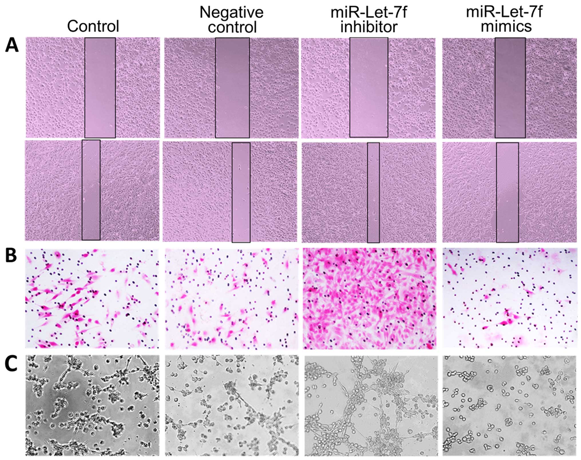
miR-Let-7f inhibits the migratory and VM
forming capacities of human glioma cells
Migration is known to play an important role in
tumor VM, and in an earlier study we showed that miR-Let-7f
regulated the glioma cell migration (23). To determine whether miR-Let-7f could
affect VM, we used the miR-Let-7f inhibitor and mimics. miRNA
inhibitors are synthetic, 2′-O-methyl-modified, single-stranded
molecules that interfere with miRNA function by sequestering them
via irreversible binding (24,25).
Mimics are synthetic, double-stranded, modified RNA molecules that
imitate the functions of endogenous miRNAs (26). As a proof of principle, the
transient transfection of 80 nM inhibitor or mimics did not affect
glioma cell viability (Fig. 2D). In
addition, we observed a significant pro-migratory effects of the
miR-Let-7f inhibitor and anti-migratory effects of its mimics in
wound healing and Transwell assays of A172 cells (Fig. 3A and B) in agreement with our
findings previously reported. Furthermore, VM tube formation assay
results confirmed that miR-Let-7f knockdown significantly promoted
the VM formation and overexpression of miR-Let-7f completely
blocked it (Fig. 3C). It suggests
that miR-Let-7f may hinder the VM forming capacity of glioma cells
by anti-migratory effects.
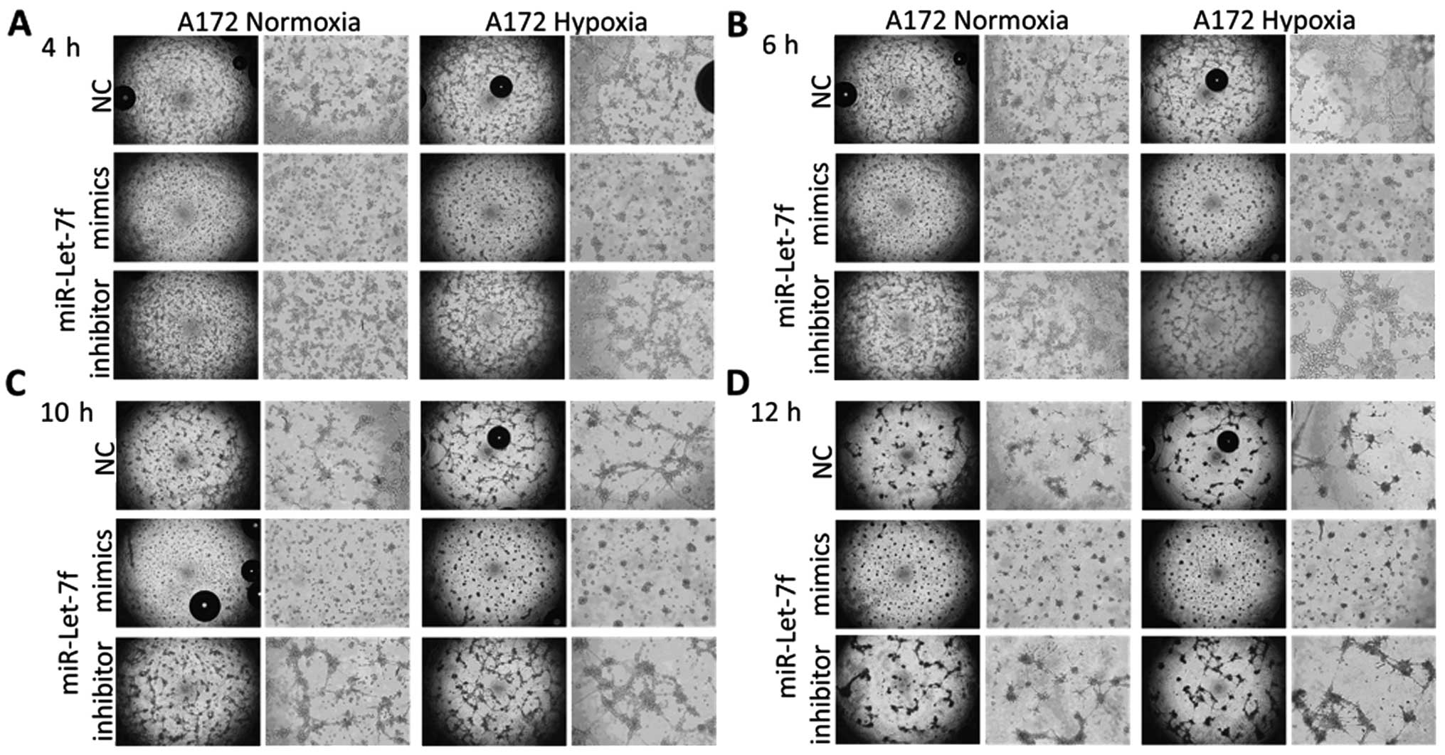
Overexpression of miR-Let-7f paralyses
hypoxia-induced VM formation
Several studies have demonstrated the
hypoxia-induced angiogenesis and VM, and the pro-migratory effects
of hypoxia. In this regard, we supposed that miR-Let-7f was
involved in the regulation of hypoxia-induced VM formation. After 6
h hypoxia treatment, A172 glioma cells revealed an excessively
enhanced VM formation (Fig. 4B,
upper).
To understand the mechanisms implicated in the VM
suppressive role under hypoxia condition of miR-Let-7f, we
investigated the time-dependent effects of miR-Let-7f knockdown and
overexpression in A172 glioma cells. First, we observed a
significant VM suppression effect of miR-Let-7f mimics,
particularly even under hypoxic conditions in A172 cells. While
miR-Let-7f inhibitor significantly promoted A172 glioma cell VM
under normoxic and hypoxia conditions (Fig. 4). Notably, the VM had a dynamic
process with increased observation time. The undisturbed VM
formation initiated at 4 h, peaked at 6 h and vanished after 12 h
in normoxia control A172 cells. In addition, miR-Let-7f inhibitor
further enhanced the VM structures, particularly revealing the
effect of hypoxia. In contrast, miR-Let-7f mimics completely
blocked the VM formation throughout the experiment whether with
hypoxia or not.
Taken together, our results clearly demonstrated
that miR-Let-7f knockdown markedly promoted the VM forming capacity
of human glioma cells and aggravate the hypoxia-induced VM
promoting effects. While miR-Let-7f overexpression antagonized the
VM formation, particularly the hypoxia-induced VM on human glioma
cells.
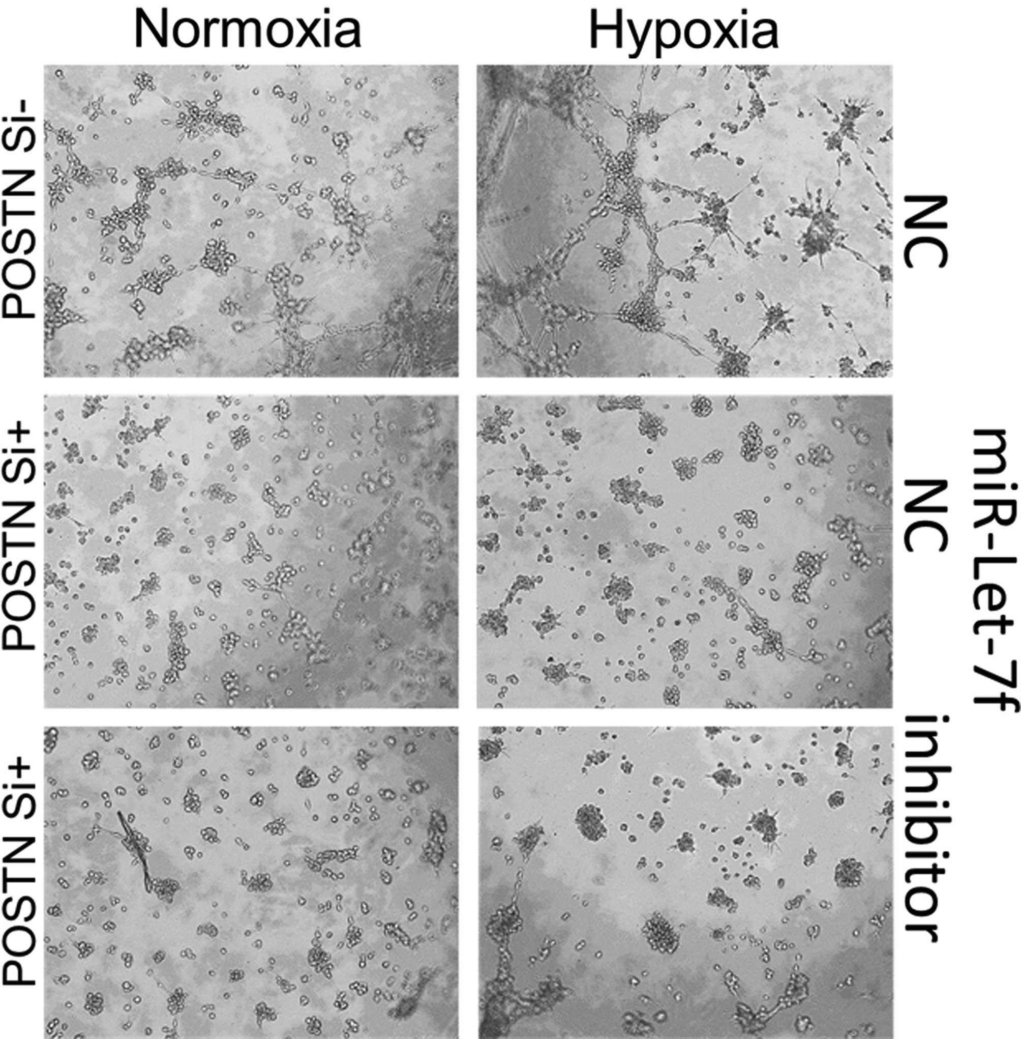
miR-Let-7f antagonizes the
hypoxia-induced VM promoting effects by targeting POSTN
As we previously reported, miR-Let-7f inhibited
glioma migratory ability by directly targeting POSTN (23). It suggested that miR-Let-7f may
block the VM forming capacity of glioma cells by regulating the
POSTN-dependent migration. To investigate whether miR-Let-7f and
POSTN are linked, we utilized a POSTN siRNA. siRNA transfected A172
glioma cells almost lost the migratory and VM forming ability as
miR-Let-7f overexpressed cells (Fig.
5, middle), and the VM promoting effect of miR-Let-7f inhibitor
was totally blocked (Fig. 5,
lower). In addition, the hypoxia effect was also blocked by POSTN
knockdown (Fig. 5, right). Hence we
could confirm that miR-Let-7f antagonizes the hypoxia-induced VM
promoting effects by targeting POSTN.
Discussion
There is growing interest in exploring the
tumor-suppressive miRNAs, and accumulating evidence demonstrates
their potential of prognostic and antitumor therapeutic prospects
(27,28). Several recent studies have confirmed
numerous highly expressed miRNA predictors (13,29–31)
for glioma prognosis. Moreover, an increasing number of
tumor-suppressive miRNAs has also been discovered (32). In the present study, we identified a
novel antitumor mechanism of miR-Let-7f by its anti-vasculogenic
mimicry (VM) effect in human glioma, and its possible regulatory
target in malignant glioma.
Cells undergo a variety of biological responses when
subjected to ischemic and hypoxic conditions, including the
activation of signaling pathways that regulate proliferation,
angiogenesis, metastasis and apoptosis. Tumor cells have adapted
these pathways to survive and even grow under ischemic and hypoxic
conditions. Tumor hypoxia is associated with poor prognosis and
resistance to antitumor therapy (33). Additionally, hypoxia-induced
angiogenesis is also a common feature in solid tumors due to their
overwhelming progression and relatively inadequate blood supply.
Cancer stem cells (CSCs) were identified as a VM-initiating cells
in many types of cancer (34). The
detailed VM phenomenon in non-GSC glioma cells has been reported
(35), but its mechanisms are still
unclear. Vascularization is crucial for the growth of hypoxic
tumors (36), and VM as a new
vascular type described as the non-endothelium-dependent vessels in
gliomas (37), represents an
important new tumor survival mechanism. VM may contribute to the
failure of current anti-angiogenic therapy (38), and the exact mechanism still
requires further study.
Then, we first investigated the effect of miR-Let-7f
as a new potential tumor suppressor based on its significant VM
inhibition in hypoxic glioma cells. However, the pathways
implicated in the tumor suppressive role of miR-Let-7f are poorly
defined. Our results suggest that miR-Let-7f may suppress VM by
inhibiting the migration capacity of glioma cells. Based on our
results and previous reports, we validated the role of potential
target POSTN in VM using the specific siRNA. This inhibitor
prevented the VM promoting effect of the miR-Let-7f inhibitor as
expected. These results demonstrated that POSTN is a direct target
of miR-Let-7f in VM forming regulation process.
In summary, the mechanism by which miR-Let-7f
functions is summarized as follows. First, miR-Let-7f downregulates
POSTN directly and inhibits POSTN-mediated migration. Consequently,
this decreased cell motility significantly induced the glioma cell
VM forming failure. Ultimately, reduced blood supply improved the
prognosis of glioma patients. However, a limitation of the present
study is the small number of samples, and the involvement of other
key invasion-associated proteins such as Rac1, Cdc42 (39) or MMPs (40) were not investigated. Therefore,
additional studies will be required to substantiate this
mechanism.
Acknowledgments
The present study was supported by grants from the
National Natural Science foundation of China (nos. 81101594,
81372719, 81172403, 81402077, 81571284 and 91542115). We thank the
senior editor of American Journal Experts for linguistic
advice.
References
|
1
|
Ohgaki H: Epidemiology of brain tumors.
Methods Mol Biol. 472:323–342. 2009. View Article : Google Scholar
|
|
2
|
Ostrom QT, Gittleman H, Stetson L, Virk SM
and Barnholtz-Sloan JS: Epidemiology of gliomas. Cancer Treat Res.
163:1–14. 2015. View Article : Google Scholar
|
|
3
|
McNamara MG and Mason WP: Antiangiogenic
therapies in glioblastoma multiforme. Expert Rev Anticancer Ther.
12:643–654. 2012. View Article : Google Scholar : PubMed/NCBI
|
|
4
|
Vaupel P: Hypoxia and aggressive tumor
phenotype: Implications for therapy and prognosis. Oncologist.
13(Suppl 3): S21–S26. 2008. View Article : Google Scholar
|
|
5
|
Mongiardi MP: Angiogenesis and hypoxia in
glioblastoma: A focus on cancer stem cells. CNS Neurol Disord Drug
Targets. 11:878–883. 2012. View Article : Google Scholar : PubMed/NCBI
|
|
6
|
Ji RC: Hypoxia and lymphangiogenesis in
tumor microenvironment and metastasis. Cancer Lett. 346:6–16. 2014.
View Article : Google Scholar
|
|
7
|
Seftor RE, Hess AR, Seftor EA, Kirschmann
DA, Hardy KM, Margaryan NV and Hendrix MJ: Tumor cell vasculogenic
mimicry: From controversy to therapeutic promise. Am J Pathol.
181:1115–1125. 2012. View Article : Google Scholar : PubMed/NCBI
|
|
8
|
Qiao L, Liang N, Zhang J, Xie J, Liu F, Xu
D, Yu X and Tian Y: Advanced research on vasculogenic mimicry in
cancer. J Cell Mol Med. 19:315–326. 2015. View Article : Google Scholar : PubMed/NCBI
|
|
9
|
Dong J, Zhao Y, Huang Q, Fei X, Diao Y,
Shen Y, Xiao H, Zhang T, Lan Q and Gu X: Glioma stem/progenitor
cells contribute to neovascularization via transdifferentiation.
Stem Cell Rev. 7:141–152. 2011. View Article : Google Scholar
|
|
10
|
Liu T, Sun B, Zhao X, Li Y, Gu Q, Dong X
and Liu F: OCT4 expression and vasculogenic mimicry formation
positively correlate with poor prognosis in human breast cancer.
Int J Mol Sci. 15:19634–19649. 2014. View Article : Google Scholar : PubMed/NCBI
|
|
11
|
Bartel DP: MicroRNAs: Genomics,
biogenesis, mechanism, and function. Cell. 116:281–297. 2004.
View Article : Google Scholar : PubMed/NCBI
|
|
12
|
Filipowicz W, Bhattacharyya SN and
Sonenberg N: Mechanisms of post-transcriptional regulation by
microRNAs: Are the answers in sight? Nat Rev Genet. 9:102–114.
2008. View
Article : Google Scholar : PubMed/NCBI
|
|
13
|
Zhang R, Pang B, Xin T, Guo H, Xing Y, Xu
S, Feng B, Liu B and Pang Q: Plasma miR-221/222 family as novel
descriptive and prognostic biomarkers for glioma. Mol Neurobiol.
Jan 31–2015.Epub ahead of print.
|
|
14
|
Dews M, Homayouni A, Yu D, Murphy D,
Sevignani C, Wentzel E, Furth EE, Lee WM, Enders GH, Mendell JT, et
al: Augmentation of tumor angiogenesis by a Myc-activated microRNA
cluster. Nat Genet. 38:1060–1065. 2006. View Article : Google Scholar : PubMed/NCBI
|
|
15
|
Ma L, Young J, Prabhala H, Pan E, Mestdagh
P, Muth D, Teruya-Feldstein J, Reinhardt F, Onder TT, Valastyan S,
et al: miR-9, a MYC/MYCN-activated microRNA, regulates E-cadherin
and cancer metastasis. Nat Cell Biol. 12:247–256. 2010.PubMed/NCBI
|
|
16
|
Zhao Z, Tan X, Zhao A, Zhu L, Yin B, Yuan
J, Qiang B and Peng X: microRNA-214-mediated UBC9 expression in
glioma. BMB Rep. 45:641–646. 2012. View Article : Google Scholar : PubMed/NCBI
|
|
17
|
Li X, Ling N, Bai Y, Dong W, Hui GZ, Liu
D, Zhao J and Hu J: MiR-16-1 plays a role in reducing migration and
invasion of glioma cells. Anat Rec. 296:427–432. 2013. View Article : Google Scholar
|
|
18
|
Ying Z, Li Y, Wu J, Zhu X, Yang Y, Tian H,
Li W, Hu B, Cheng SY and Li M: Loss of miR-204 expression enhances
glioma migration and stem cell-like phenotype. Cancer Res.
73:990–999. 2013. View Article : Google Scholar :
|
|
19
|
Lee HK, Bier A, Cazacu S, Finniss S, Xiang
C, Twito H, Poisson LM, Mikkelsen T, Slavin S, Jacoby E, et al:
MicroRNA-145 is downregulated in glial tumors and regulates glioma
cell migration by targeting connective tissue growth factor. PLoS
One. 8:e546522013. View Article : Google Scholar : PubMed/NCBI
|
|
20
|
Gao R, Cai C, Gan J, Yang X, Shuang Z, Liu
M, Li S and Tang H: miR-1236 down-regulates alpha-fetoprotein, thus
causing PTEN accumulation, which inhibits the PI3K/Akt pathway and
malignant phenotype in hepatoma cells. Oncotarget. 6:6014–6028.
2015. View Article : Google Scholar : PubMed/NCBI
|
|
21
|
Song Y, Mu L, Han X, Li Q, Dong B, Li H
and Liu X: MicroRNA-9 inhibits vasculogenic mimicry of glioma cell
lines by suppressing Stathmin expression. J Neurooncol.
115:381–390. 2013. View Article : Google Scholar : PubMed/NCBI
|
|
22
|
Wu N, Zhao X, Liu M, Liu H, Yao W, Zhang
Y, Cao S and Lin X: Role of microRNA-26b in glioma development and
its mediated regulation on EphA2. PLoS One. 6:e162642011.
View Article : Google Scholar : PubMed/NCBI
|
|
23
|
Yan S, Han X, Xue H, Zhang P, Guo X, Li T,
Guo X, Yuan G, Deng L and Li G: Let-7f inhibits glioma cell
proliferation, migration, and invasion by targeting periostin. J
Cell Biochem. 116:1680–1692. 2015. View Article : Google Scholar : PubMed/NCBI
|
|
24
|
Meister G, Landthaler M, Dorsett Y and
Tuschl T: Sequence- specific inhibition of microRNA- and
siRNA-induced RNA silencing. RNA. 10:544–550. 2004. View Article : Google Scholar : PubMed/NCBI
|
|
25
|
Hutvágner G, Simard MJ, Mello CC and
Zamore PD: Sequence-specific inhibition of small RNA function. PLoS
Biol. 2:e982004. View Article : Google Scholar : PubMed/NCBI
|
|
26
|
Kim DH and Rossi JJ: Strategies for
silencing human disease using RNA interference. Nat Rev Genet.
8:173–184. 2007. View
Article : Google Scholar : PubMed/NCBI
|
|
27
|
Yan W, Li R, Liu Y, Yang P, Wang Z, Zhang
C, Bao Z, Zhang W, You Y and Jiang T: MicroRNA expression patterns
in the malignant progression of gliomas and a 5-microRNA signature
for prognosis. Oncotarget. 5:12908–12915. 2014. View Article : Google Scholar : PubMed/NCBI
|
|
28
|
Auffinger B, Thaci B, Ahmed A, Ulasov I
and Lesniak MS: MicroRNA targeting as a therapeutic strategy
against glioma. Curr Mol Med. 13:535–542. 2013. View Article : Google Scholar
|
|
29
|
Yang CH, Yue J, Pfeffer SR, Fan M, Paulus
E, Hosni-Ahmed A, Sims M, Qayyum S, Davidoff AM, Handorf CR, et al:
MicroRNA-21 promotes glioblastoma tumorigenesis by down-regulating
insulin-like growth factor-binding protein-3 (IGFBP3). J Biol Chem.
289:25079–25087. 2014. View Article : Google Scholar : PubMed/NCBI
|
|
30
|
Barbano R, Palumbo O, Pasculli B, Galasso
M, Volinia S, D'Angelo V, Icolaro N, Coco M, Dimitri L, Graziano P,
et al: A miRNA signature for defining aggressive phenotype and
prognosis in gliomas. PLoS One. 9:e1089502014. View Article : Google Scholar : PubMed/NCBI
|
|
31
|
Lai NS, Wu DG, Fang XG, Lin YC, Chen SS,
Li ZB and Xu SS: Serum microRNA-210 as a potential noninvasive
biomarker for the diagnosis and prognosis of glioma. Br J Cancer.
112:1241–1246. 2015. View Article : Google Scholar : PubMed/NCBI
|
|
32
|
Que T, Song Y, Liu Z, Zheng S, Long H, Li
Z, Liu Y, Wang G, Liu Y, Zhou J, et al: Decreased miRNA-637 is an
unfavorable prognosis marker and promotes glioma cell growth,
migration and invasion via direct targeting Akt1. Oncogene.
34:4952–4963. 2015. View Article : Google Scholar : PubMed/NCBI
|
|
33
|
Harris AL: Hypoxia - a key regulatory
factor in tumour growth. Nat Rev Cancer. 2:38–47. 2002. View Article : Google Scholar : PubMed/NCBI
|
|
34
|
Fan YL, Zheng M, Tang YL and Liang XH: A
new perspective of vasculogenic mimicry: EMT and cancer stem cells
(Review). Oncol Lett. 6:1174–1180. 2013.PubMed/NCBI
|
|
35
|
Smith SJ, Ward JH, Tan C, Grundy RG and
Rahman R: Endothelial-like malignant glioma cells in dynamic three
dimensional culture identifies a role for VEGF and FGFR in a
tumor-derived angiogenic response. Oncotarget. 6:22191–22205. 2015.
View Article : Google Scholar : PubMed/NCBI
|
|
36
|
Jhaveri N, Chen TC and Hofman FM: Tumor
vasculature and glioma stem cells: Contributions to glioma
progression. Cancer Lett. Dec 16–2014.(epub ahead of print). pii:
S0304-3835(14)00783-6. View Article : Google Scholar : PubMed/NCBI
|
|
37
|
Yue WY and Chen ZP: Does vasculogenic
mimicry exist in astrocytoma? J Histochem Cytochem. 53:997–1002.
2005. View Article : Google Scholar : PubMed/NCBI
|
|
38
|
Mao JM, Liu J, Guo G, Mao XG and Li CX:
Glioblastoma vasculogenic mimicry: Signaling pathways progression
and potential anti-angiogenesis targets. Biomark Res. 3:2015.
View Article : Google Scholar : PubMed/NCBI
|
|
39
|
Cardama GA, Gonzalez N, Ciarlantini M,
Gandolfi Donadío L, Comin MJ, Alonso DF, Menna PL and Gomez DE:
Proapoptotic and antiinvasive activity of Rac1 small molecule
inhibitors on malignant glioma cells. Onco Targets Ther.
7:2021–2033. 2014.PubMed/NCBI
|
|
40
|
Könnecke H and Bechmann I: The role of
microglia and matrix metalloproteinases involvement in
neuroinflammation and gliomas. Clin Dev Immunol. 2013:9141042013.
View Article : Google Scholar : PubMed/NCBI
|