|
1
|
Barnes L, Eveson JW, Reichart P and
Sidransky D: World Health Organization Classification of Tumours:
Pathology and Genetics of Head and Neck Tumours. IARC Press; Lyon:
2005
|
|
2
|
Al-Swiahb JN, Chen CH, Chuang HC, Fang FM,
Tasi HT and Chien CY: Clinical, pathological and molecular
determinants in squamous cell carcinoma of the oral cavity. Future
Oncol. 6:837–850. 2010. View Article : Google Scholar : PubMed/NCBI
|
|
3
|
Stucken E, Weissman J and Spiegel JH: Oral
cavity risk factors: Experts' opinions and literature support. J
Otolaryngol Head Neck Surg. 39:76–89. 2010.PubMed/NCBI
|
|
4
|
Folz BJ, Silver CE, Rinaldo A, Fagan JJ,
Pratt LW, Weir N, Seitz D and Ferlito A: An outline of the history
of head and neck oncology. Oral Oncol. 44:2–9. 2008. View Article : Google Scholar
|
|
5
|
Scully C and Bagan JV: Recent advances in
Oral Oncology 2007: Imaging, treatment and treatment outcomes. Oral
Oncol. 44:211–215. 2008. View Article : Google Scholar : PubMed/NCBI
|
|
6
|
Jungbluth AA, Busam KJ, Kolb D, Iversen K,
Coplan K, Chen YT, Spagnoli GC and Old LJ: Expression of
MAGE-antigens in normal tissues and cancer. Int J Cancer.
85:460–465. 2000. View Article : Google Scholar : PubMed/NCBI
|
|
7
|
Katsura Y and Satta Y: Evolutionary
history of the cancer immunity antigen MAGE gene family. PLoS One.
6:e203652011. View Article : Google Scholar : PubMed/NCBI
|
|
8
|
van der Bruggen P, Traversari C, Chomez P,
Lurquin C, De Plaen E, Van den Eynde B, Knuth A and Boon T: A gene
encoding an antigen recognized by cytolytic T lymphocytes on a
human melanoma. Science. 254:1643–1647. 1991. View Article : Google Scholar : PubMed/NCBI
|
|
9
|
van der Bruggen P, Bastin J, Gajewski T,
Coulie PG, Boël P, De Smet C, Traversari C, Townsend A and Boon T:
A peptide encoded by human gene MAGE-3 and presented by HLA-A2
induces cytolytic T lymphocytes that recognize tumor cells
expressing MAGE-3. Eur J Immunol. 24:3038–3043. 1994. View Article : Google Scholar : PubMed/NCBI
|
|
10
|
Lee KD, Eura M, Ogi K, Nakano K,
Chikamatsu K, Masuyama K and Ishikawa T: Expression of the MAGE-1,
-2, -3, -4, and -6 genes in non-squamous cell carcinoma lesions of
the head and neck. Acta Otolaryngol. 116:633–639. 1996. View Article : Google Scholar : PubMed/NCBI
|
|
11
|
Eura M, Ogi K, Chikamatsu K, Lee KD,
Nakano K, Masuyama K, Itoh K and Ishikawa T: Expression of the MAGE
gene family in human head-and-neck squamous-cell carcinomas. Int J
Cancer. 64:304–308. 1995. View Article : Google Scholar : PubMed/NCBI
|
|
12
|
Barker PA and Salehi A: The MAGE proteins:
Emerging roles in cell cycle progression, apoptosis, and
neurogenetic disease. J Neurosci Res. 67:705–712. 2002. View Article : Google Scholar : PubMed/NCBI
|
|
13
|
Simpson AJ, Caballero OL, Jungbluth A,
Chen YT and Old LJ: Cancer/testis antigens, gametogenesis and
cancer. Nat Rev Cancer. 5:615–625. 2005. View Article : Google Scholar : PubMed/NCBI
|
|
14
|
Ries J, Schultze-Mosgau S, Neukam F,
Diebel E and Wiltfang J: Investigation of the expression of
melanoma antigen-encoding genes (MAGE-A1 to -A6) in oral squamous
cell carcinomas to determine potential targets for gene-based
cancer immunotherapy. Int J Oncol. 26:817–824. 2005.PubMed/NCBI
|
|
15
|
Ries J, Vairaktaris E, Mollaoglu N,
Wiltfang J, Neukam FW and nkenke E: Expression of
melanoma-associated antigens in oral squamous cell carcinoma. J
Oral Pathol Med. 37:88–93. 2008. View Article : Google Scholar : PubMed/NCBI
|
|
16
|
Kienstra MA, Neel HB, Strome SE and Roche
P: Identification of NY-ESO-1, MAGE-1, and MAGE-3 in head and neck
squamous cell carcinoma. Head Neck. 25:457–463. 2003. View Article : Google Scholar : PubMed/NCBI
|
|
17
|
Figueiredo DL, Mamede RC, Proto-Siqueira
R, Neder L, Silva WA Jr and Zago MA: Expression of cancer testis
antigens in head and neck squamous cell carcinomas. Head Neck.
28:614–619. 2006. View Article : Google Scholar : PubMed/NCBI
|
|
18
|
Montoro JR, Mamede RC, Neder Serafini L,
Saggioro FP, Figueiredo DL, Silva WA Jr, Jungbluth AA, Spagnoli GC
and Zago MA: Expression of cancer-testis antigens MAGE-A4 and
MAGE-C1 in oral squamous cell carcinoma. Head Neck. 34:1123–1128.
2012. View Article : Google Scholar
|
|
19
|
Cuffel C, Rivals JP, Zaugg Y, Salvi S,
Seelentag W, Speiser DE, Liénard D, Monnier P, Romero P, Bron L, et
al: Pattern and clinical significance of cancer-testis gene
expression in head and neck squamous cell carcinoma. Int J Cancer.
128:2625–2634. 2011. View Article : Google Scholar
|
|
20
|
Piffkò J, Bànkfalvi A, Tory K, Füzesi L,
Bryne M, Ofner D, Kusch F, Joos U and Schmid KW: Molecular
assessment of p53 abnormalities at the invasive front of oral
squamous cell carcinomas. Head Neck. 20:8–15. 1998. View Article : Google Scholar : PubMed/NCBI
|
|
21
|
Horta MC, de Assis LA, de Souza AF, de
Araújo VC, Gomez RS and Aguiar MC: p53 and p21WAF1/CIP1
overexpression at the invasive front of lower lip squamous cell
carcinoma. J Oral Pathol Med. 36:88–92. 2007. View Article : Google Scholar : PubMed/NCBI
|
|
22
|
Tumuluri V, Thomas GA and Fraser IS: The
relationship of proliferating cell density at the invasive tumour
front with prognostic and risk factors in human oral squamous cell
carcinoma. J Oral Pathol Med. 33:204–208. 2004. View Article : Google Scholar : PubMed/NCBI
|
|
23
|
Hartmann S, Kriegebaum U, Küchler N,
Brands RC, Linz C, Kübler AC and Müller-Richter UD: Correlation of
MAGE-A tumor antigens and the efficacy of various chemotherapeutic
agents in head and neck carcinoma cells. Clin Oral Investig.
18:189–197. 2014. View Article : Google Scholar
|
|
24
|
Hartmann S, Kriegebaum U, Küchler N,
Lessner G, Brands RC, Linz C, Schneider T, Kübler AC and
Müller-Richter UD: Efficacy of cetuximab and panitumumab in oral
squamous cell carcinoma cell lines: Prognostic value of MAGE-A
subgroups for treatment success. J Craniomaxillofac Surg.
41:623–629. 2013. View Article : Google Scholar : PubMed/NCBI
|
|
25
|
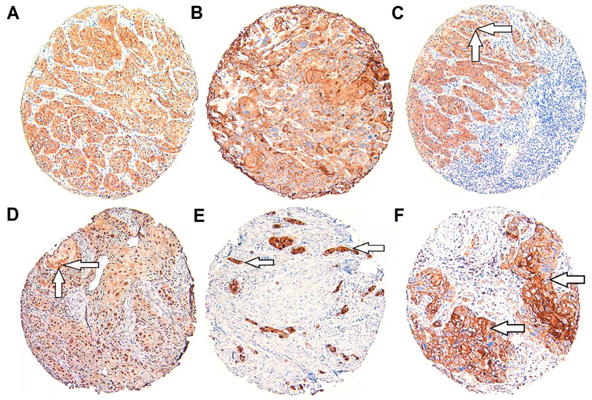
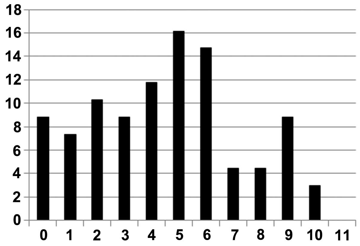
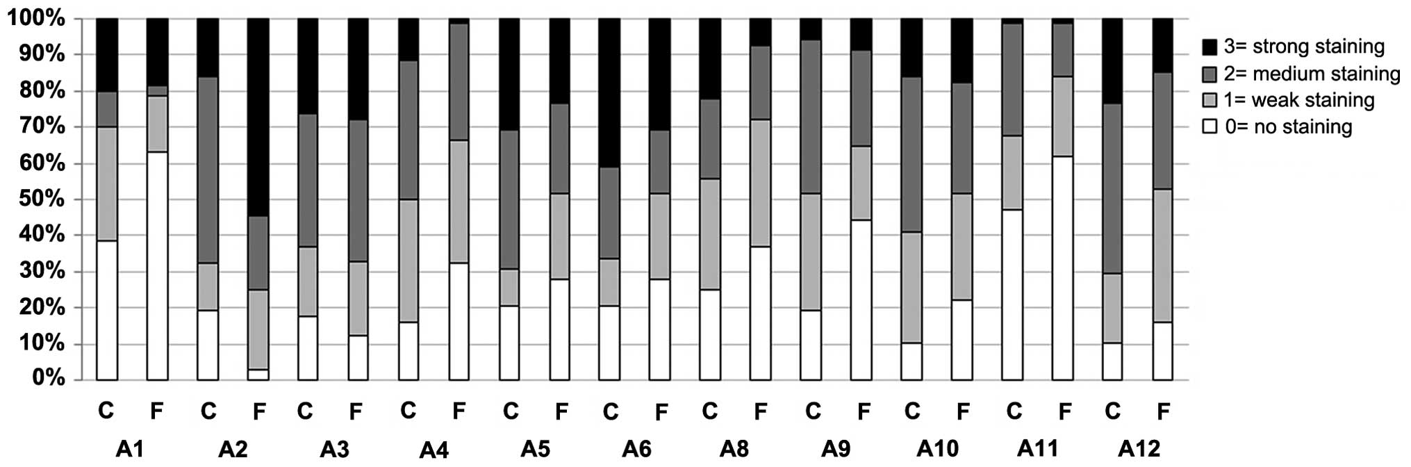
Remmele W and Stegner HE: Recommendation
for uniform definition of an immunoreactive score (IRS) for
immunohistochemical estrogen receptor detection (ER-ICA) in breast
cancer tissue. Pathologe. 8:138–140. 1987.In German. PubMed/NCBI
|
|
26
|
Thiery JP: Epithelial-mesenchymal
transitions in tumour progression. Nat Rev Cancer. 2:442–454. 2002.
View Article : Google Scholar : PubMed/NCBI
|
|
27
|
Yang CC, Zhu LF, Xu XH, Ning TY, Ye JH and
Liu LK: Membrane type 1 matrix metalloproteinase induces an
epithelial to mesenchymal transition and cancer stem cell-like
properties in SCC9 cells. BMC Cancer. 13:1712013. View Article : Google Scholar : PubMed/NCBI
|
|
28
|
Thiery JP: Epithelial-mesenchymal
transitions in development and pathologies. Curr Opin Cell biol.
15:740–746. 2003. View Article : Google Scholar : PubMed/NCBI
|
|
29
|
Kalluri R and Weinberg RA: The basics of
epithelial-mesenchymal transition. J Clin Invest. 119:1420–1428.
2009. View
Article : Google Scholar : PubMed/NCBI
|
|
30
|
Caballero OL and Chen YT: Cancer/testis
(CT) antigens: Potential targets for immunotherapy. Cancer Sci.
100:2014–2021. 2009. View Article : Google Scholar : PubMed/NCBI
|
|
31
|
Karpf AR, Bai S, James SR, Mohler JL and
Wilson EM: Increased expression of androgen receptor coregulator
MAGE-11 in prostate cancer by DNA hypomethylation and cyclic AMP.
Mol Cancer Res. 7:523–535. 2009. View Article : Google Scholar : PubMed/NCBI
|
|
32
|
Scanlan MJ, Gure AO, Jungbluth AA, Old LJ
and Chen YT: Cancer/testis antigens: An expanding family of targets
for cancer immunotherapy. Immunol Rev. 188:22–32. 2002. View Article : Google Scholar : PubMed/NCBI
|
|
33
|
Wischnewski F, Pantel K and Schwarzenbach
H: Promoter demethylation and histone acetylation mediate gene
expression of MAGE-A1, -A2, -A3, and -A12 in human cancer cells.
Mol Cancer Res. 4:339–349. 2006. View Article : Google Scholar : PubMed/NCBI
|
|
34
|
Wang Z, Zhang J, Zhang Y, Srivenugopal KS
and Lim SH: SPAN-XB core promoter sequence is regulated in myeloma
cells by specific CpG dinucleotides associated with the MeCP2
protein. Int J Cancer. 119:2878–2884. 2006. View Article : Google Scholar : PubMed/NCBI
|
|
35
|
Lim SH, Zhang Y and Zhang J: Cancer-testis
antigens: The current status on antigen regulation and potential
clinical use. Am J Blood Res. 2:29–35. 2012.PubMed/NCBI
|
|
36
|
Old LJ: Cancer/testis (CT) antigens - a
new link between gametogenesis and cancer. Cancer Immun.
1:12001.
|
|
37
|
Aprelikova O, Pandolfi S, Tackett S,
Ferreira M, Salnikow K, Ward Y, Risinger JI, Barrett JC and
Niederhuber J: Melanoma antigen-11 inhibits the hypoxia-inducible
factor prolyl hydroxylase 2 and activates hypoxic response. Cancer
Res. 69:616–624. 2009. View Article : Google Scholar : PubMed/NCBI
|
|
38
|
Patel SS, Shah KA, Shah MJ, Kothari KC and
Rawal RM: Cancer stem cells and stemness markers in oral squamous
cell carcinomas. Asian Pac J Cancer Prev. 15:8549–8556. 2014.
View Article : Google Scholar : PubMed/NCBI
|
|
39
|
Liu L, Wylie RC, Andrews LG and Tollefsbol
TO: Aging, cancer and nutrition: The DNA methylation connection.
Mech Ageing Dev. 124:989–998. 2003. View Article : Google Scholar : PubMed/NCBI
|
|
40
|
Müller-Richter UD, Dowejko A, Peters S,
Rauthe S, Reuther T, Gattenlöhner S, Reichert TE, Driemel O and
Kübler AC: MAGE-A antigens in patients with primary oral squamous
cell carcinoma. Clin Oral Investig. 14:291–296. 2010. View Article : Google Scholar
|
|
41
|
Napoletano C, Bellati F, Tarquini E, Tomao
F, Taurino F, Spagnoli G, Rughetti A, Muzii L, Nuti M and Benedetti
Panici P: MAGE-A and NY-ESO-1 expression in cervical cancer:
Prognostic factors and effects of chemotherapy. Am J Obstet
Gynecol. 198:99.e1–7. 2008. View Article : Google Scholar
|
|
42
|
Kreppel M, Drebber U, Eich HT, Dreiseidler
T, Zöller JE, Müller RP and Scheer M: Combined-modality treatment
in advanced oral squamous cell carcinoma: Primary surgery followed
by adjuvant concomitant radiochemotherapy. Strahlenther Onkol.
187:555–560. 2011. View Article : Google Scholar : PubMed/NCBI
|
|
43
|
Adelstein DJ, Lavertu P, Saxton JP, Secic
M, Wood BG, Wanamaker JR, Eliachar I, Strome M and Larto MA: Mature
results of a phase III randomized trial comparing concurrent
chemoradiotherapy with radiation therapy alone in patients with
stage III and IV squamous cell carcinoma of the head and neck.
Cancer. 88:876–883. 2000. View Article : Google Scholar : PubMed/NCBI
|
|
44
|
Kavalar R, Sarcevic B, Spagnoli GC,
Separovic V, Samija M, Terracciano L, Heberer M and Juretic A:
Expression of MAGE tumour-associated antigens is inversely
correlated with tumour differentiation in invasive ductal breast
cancers: An immunohistochemical study. Virchows Arch. 439:127–131.
2001. View Article : Google Scholar : PubMed/NCBI
|
|
45
|
Li M, Yuan YH, Han Y, Liu YX, Yan L, Wang
Y and Gu J: Expression profile of cancer-testis genes in 121 human
colorectal cancer tissue and adjacent normal tissue. Clin Cancer
Res. 11:1809–1814. 2005. View Article : Google Scholar : PubMed/NCBI
|
|
46
|
Jung EJ, Kim MA, Lee HS, Yang HK, Lee YM,
Lee BL and Kim WH: Expression of family A melanoma antigen in human
gastric carcinoma. Anticancer Res. 25:2105–2111. 2005.PubMed/NCBI
|
|
47
|
Forghanifard MM, Gholamin M, Farshchian M,
Moaven O, Memar B, Forghani MN, Dadkhah E, Naseh H, Moghbeli M,
Raeisossadati R, et al: Cancer-testis gene expression profiling in
esophageal squamous cell carcinoma: Identification of specific
tumor marker and potential targets for immunotherapy. Cancer Biol
Ther. 12:191–197. 2011. View Article : Google Scholar : PubMed/NCBI
|
|
48
|
Müller-Richter UD, Dowejko A, Driemel O,
Reuther T, Reichert TE and Kübler AC: Impact of MAGE-A antigens on
taxane response in oral squamous cell carcinoma. Oncol Lett.
1:181–185. 2010.PubMed/NCBI
|
|
49
|
Kim YD, Park HR, Song MH, Shin DH, Lee CH,
Lee MK and Lee SY: Pattern of cancer/testis antigen expression in
lung cancer patients. Int J Mol Med. 29:656–662. 2012.PubMed/NCBI
|
|
50
|
Zajicek G: neoplasia - a stem cell
pathology. Med Hypotheses. 13:125–136. 1984. View Article : Google Scholar : PubMed/NCBI
|
|
51
|
Costa FF, Le Blanc K and Brodin B: Concise
review: Cancer/testis antigens, stem cells, and cancer. Stem Cells.
25:707–711. 2007. View Article : Google Scholar
|
|
52
|
Gu X, Fu M, Ge Z, Zhan F, Ding Y, Ni H,
Zhang W, Zhu Y, Tang X, Xiong L, et al: High expression of MAGE-A9
correlates with unfavorable survival in hepatocellular carcinoma.
Sci Rep. 4:66252014. View Article : Google Scholar : PubMed/NCBI
|
|
53
|
Xu X, Tang X, Lu M, Tang Q, Zhang H, Zhu
H, Xu N, Zhang D, Xiong L, Mao Y, et al: Overexpression of MAGE-A9
predicts unfavorable outcome in breast cancer. Exp Mol Pathol.
97:579–584. 2014. View Article : Google Scholar : PubMed/NCBI
|
|
54
|
Shigematsu Y, Hanagiri T, Shiota H, Kuroda
K, Baba T, Mizukami M, So T, Ichiki Y, Yasuda M, So T, et al:
Clinical significance of cancer/testis antigens expression in
patients with non-small cell lung cancer. Lung Cancer. 68:105–110.
2010. View Article : Google Scholar
|
|
55
|
Bryne M, Koppang HS, Lilleng R and
Kjaerheim A: Malignancy grading of the deep invasive margins of
oral squamous cell carcinomas has high prognostic value. J Pathol.
166:375–381. 1992. View Article : Google Scholar : PubMed/NCBI
|
|
56
|
Piffkó J, Bánkfalvi A, Ofner D, Kusch F,
Böcker W, Joos U and Schmid KW: In situ assessment of cell
proliferation at the invasive front of oral squamous cell
carcinomas. Virchows Arch. 429:229–234. 1996.PubMed/NCBI
|
|
57
|
Pastorcic-Grgic M, Sarcevic B, Dosen D,
Juretic A, Spagnoli GC and Grgic M: Prognostic value of MAGE-A and
NY-ESO-1 expression in pharyngeal cancer. Head Neck. 32:1178–1184.
2010. View Article : Google Scholar
|
|
58
|
Suzuki S, Sasajima K, Sato Y, Watanabe H,
Matsutani T, Iida S, Hosone M, Tsukui T, Maeda S, Shimizu K, et al:
MAGE-A protein and MAGE-A10 gene expressions in liver metastasis in
patients with stomach cancer. Br J Cancer. 99:350–356. 2008.
View Article : Google Scholar : PubMed/NCBI
|
|
59
|
Metzler P, Mollaoglu N, Schwarz S, Neukam
FW, Nkenke E and Ries J: MAGE-A as a novel approach in the
diagnostic accuracy of oral squamous cell cancer: A case report.
Head Neck Oncol. 1:392009. View Article : Google Scholar : PubMed/NCBI
|