Introduction
Magnetic resonance imaging (MRI) is an important
tool used in the diagnosis of cancer (1). To improve the specificity and
sensitivity of MRI, contrast agents are used to increase the signal
intensity. Numerous different metallic contrast agents, based on
gadolinium (Gd) (Magnevist, ProHance), Fe (Feridex, Endorem), and
Mn (Teslascan), are currently available (2). Of these, paramagnetic contrast agents
based on Gd are better for tumor and vascular imaging, and
Gd-diethylene triamine pentaacetate (Gd-DTPA, Magnevist) is the
most commonly used MRI contrast agent. However, due to their low
molecular weights, conventional MRI contrast agents have short
imaging lifetime in vivo and lack specificity for target
organs. To overcome these drawbacks, nanoparticles were proposed to
be ideal as molecular probes and as MRI contrast agents, and
generally were able to overcome the drawbacks of small molecule
agents. Thus, some nanoparticles have been developed for molecular
imaging (3).
Besides nanoparticles, single-domain antibodies
(referred to as nanobodies) have attracted much interest for
molecular imaging investigations, using modalities such as
radionuclide-based, optical, and ultrasound imaging (4–8).
Nanobodies have many advantages owing to their small molecular
size, and can rapidly be distributed in the bloodstream and easily
reach target tissues within a short period of time following
injection, exhibiting great potential for tumor detection (9). Nanobodies bind tightly to targets on
the surfaces of cancer cells and can be internalized. Nanobodies
also have a low immunogenic potential and are rapidly cleared when
unbound, allowing for the acquisition of images with a high
tumor-to-background contrast at early time points after their
administration. They are also stable and specific (9).
Targeting tumors with nanobodies for cancer imaging
and therapy has emerged as a promising diagnostic and therapeutic
approach. Since epidermal growth factor receptor (EGFR) is highly
expressed in a variety of tumors, targeting with a contrast agent
using anti-EGFR nanobody has potential advantages. Single-photon
emission computed tomography (SPECT) imaging of EGFR expression
using an anti-EGFR nanobody as the targeting agent was first
reported by Huang et al (10). The radiolabelled nanobody
demonstrated high specificity and selectivity towards
EGFR-expressing cells. vosjan et al (4,11)
reported positron emission tomography (PET) imaging of EGFR
expression using the 7D12 nanobody. Biodistribution studies
(11) revealed high tumor uptake of
these nanobodies in EGFR-positive tumors and a high tumor-to-blood
ratio within 1 h post-injection.
The arginine-glycine-aspartic acid (RGD) peptide has
been used for tumor penetration in previous studies investigating
molecular imaging agents for tumors (5–8). These
peptides are known to have a relatively high and specific affinity
for the ανβ3-integrin receptor, which is highly expressed in tumor
vascular endothelial cells during angiogenesis in various tumor
types. Internalizing RGD (iRGD with a sequence of CRGDKGPDC)
differs from the RGD peptide in that it is tumor-specific, is
composed of nine amino acid residues, and has high cell
permeability. iRGD can target ανβ3-integrin receptor and
neuropilin-1 (NRP-1), which are highly expressed in a wide variety
of tumor cells (12–14). iRGD conjugated with radiolabels such
as 125I or 18F has been used to image
ανβ3-integrin receptor and NRP-1 expression using nuclear imaging
methods including SPECT and PET. This approach of nuclear imaging
with radiolabelled iRGD peptides has been shown to be effective and
sensitive (15,16).
In the present study, a previously described
recombinant protein with dual specificity and high permeability,
anti-EGFR-iRGD, was used. Recombinant anti-EGFR-iRGD protein
targeted the EGFR extracellular domain and integrin αvβ3/β5, had a
high penetration, and improved penetration of other drugs into the
deep zone of gastric cancer 3D multicellular spheroids (17).
Although nanobodies have shown potential as
molecular imaging contrast agents in several imaging techniques,
such as SPECT, PET, optical imaging, and ultrasound, the limited
spatial resolution of these imaging techniques prevents
ascertaining the exact location of the tumor. Compared with the
above methods, MRI has a better spatial resolution and can obtain
precise anatomical localization. Absence of radioactivity is
another important advantage. However, loading the fusion protein
with Gd to construct a targeting contrast agent for MRI is
challenging. Gd-chelates may be encapsulated inside a nanoparticle
core, absorbed on the surface, or covalently bound (18). However, the relaxivity of Gd-loaded
material for encapsulation and release/leakage of free Gd from the
Gd-nanoparticle complex was another clinical concern. Therefore,
chemical conjugation may be the most effective method to load Gd
with the targeting recombinant protein.
In the present study, we examined a reliable method
to construct a bispecific MRI contrast agent with high
permeability.
Materials and methods
Materials
Gd-DTPA (Magnevist) was purchased from Bayer
Schering Pharma AG (Berlin, Germany).
3-(4,5-dimethylthiazol-2-yl)-2,5-diphenyltetrazolium bromide (MTT)
for the cell viability assays and DTPA and
GdCl3·6H2O were obtained from Sigma-Aldrich
(St. Louis, Mo, USA). All other reagents and solvents of analytical
grade were obtained from different commercial sources. Human
gastric adenocarcinoma cells (BGC-823) were purchased from the Cell
Bank of Shanghai Institute of Biochemistry and Cell Biology
(Shanghai, China) and cultured in RPMI-1640 medium supplemented
with 10% foetal calf serum, 100 u/ml penicillin, and 100
µg/ml streptomycin, and incubated at 37°C and 5%
Co2.
Synthesis and characterization of
anti-EGFR-DTPA-Gd and anti-EGFR-iRGD-DTPA-Gd
Recombinant proteins anti-EGFR and anti-EGFR-iRGD
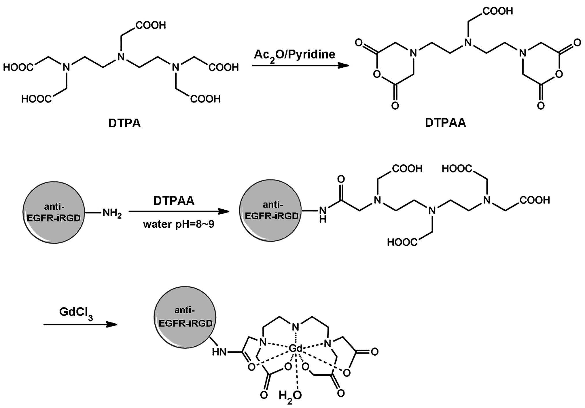
were prepared as reported previously (16). The synthesis of
anti-EGFR-iRGD-DTPA-Gd is shown in Fig.
1. DTPA anhydride (DTPAA) was synthesized as previously
reported (19). The fusion proteins
and DTPAA (2:1, mol/mol) were added gradually to NaHCo3
solution (0.1 M, pH 9) and stirred for 24 h at room temperature.
The reaction mixture was then dialyzed against water [molecular
weight cut-off (MwCo) 3500] for 24 h, during which time the water
was changed every 3 h, and the purified anti-EGFR-DTPA and
anti-EGFR-iRGD-DTPA were obtained. The anti-EGFR-DTPA or
anti-EGFR-iRGD-DTPA and GdCl3·6H2o were mixed
in an Eppendorf tube (Thermo Fisher Scientific, waltham, MA, USA)
at a molar ratio of 1:1, and the pH of the solution was adjusted to
7.0. The resulting mixture was agitated for 24 h at 60°C. The
reaction liquid was dialyzed (MwCo 3500) for 24 h, and the water
was changed every 3 h. The purified anti-EGFR-DTPA-Gd and
anti-EGFR-iRGD-DTPA-Gd were lyophilized to a powder and stored for
subsequent use. Gd content of the formed anti-EGFR-DTPA-Gd and
anti-EGFR-iRGD-DTPA-Gd were determined by inductively coupled
plasma-optical emission spectroscopy (ICP-OES).
In vitro cytotoxicity of
anti-EGFR-DTPA-Gd and anti-EGFR-iRGD-DTPA-Gd
Cell viability was determined using the MTT assay.
Briefly, BGC-823 cells in the logarithmic phase were seeded at
70–80% confluence per well in 96-well plates, incubated at 37°C
overnight, and treated with the indicated concentrations of
anti-EGFR-DTPA-Gd and anti-EGFR-iRGD-DTPA-Gd, or DTPA-Gd for 48 h.
Following treatment, 10 µl of 5 mg/ml MTT was added, and the
cells were incubated for 4 h at 37°C. The supernatant was
discarded, and 100 µl of dimethylsulphoxide (DMSo) was added
to each well. The absorbance in each well was measured by a
Multiskan Spectrum Microplate Reader (Thermo Fisher Scientific,
waltham, MA, USA) at 570 and 630 nm, and the net A570-A630 was
taken as the index of cell viability. The net absorbance from the
wells of the cells cultured with complete medium was taken as 100%
viability. The viability of the treated cells was calculated using
the formula: % viability = (A570-A630)
treated/(A570-A630) control x 100%.
In vitro cell targeting and competitive
binding assay
To examine whether anti-EGFR-DTPA-Gd and anti-EGFR-
iRGD-DTPA-Gd targeted BGC-823 cells, anti-EGFR- DTPA-Gd or
anti-EGFR-iRGD-DTPA-Gd were labelled with fluorescein
isothiocyanate (FITC). Briefly, purified proteins were suspended at
a concentration of 1 mg/ml, in conjugation buffer (National
Medicine Company, Shanghai, China) (7.56 g NaHCo3, 1.06
g Na2CO3, 7.36 g NaCl, in 1 l) at 4°C and the
pH was adjusted to 9.0. Freshly prepared FITC (1 mg/ml in DMSO) was
added to the antibody solution (at protein: FITC ratio of 1 mg:150
µg) gradually, agitating while adding to ensure proper
mixing and the solution was left at 4°C for conjugation reaction
for 8 h in the dark. NH4Cl was added to a final
concentration of 50 mM to terminate the reaction at 4°C. The
conjugate was then dialyzed against phosphate-buffered saline (PBS)
until the dialysate was clear.
BGC-823 cells in the logarithmic phase were seeded
in 24-well chamber slides at 50% confluence per well. After 16 h,
PBS supplemented with 5% bovine serum albumin was added to the
wells for blocking and incubated at 37°C with the appropriate
FITC-labelled anti-EGFR-DTPA-Gd or anti-EGFR-iRGD-DTPA-Gd for 1 h.
The cells were washed three times with cold PBS (pH 7.4), the
nucleus was labelled with Hoechst 33258, and the fixed cells were
observed with a fluorescence microscope (Zeiss LSM710, Carl Zeiss,
Germany). Competitive binding assays were performed. Briefly,
BGC-823 cells in the logarithmic phase were treated as described
above and incubated with anti-EGFR-DTPA-Gd-FITC or
anti-EGFR-iRGD-DTPA-Gd-FITC and an appropriate concentration of
competing EGFR rabbit monoclonal antibody (dilution, 1:500; cat.
no. 1114-1) (cetuximab or iRGD) at 37°C for 1 h. The cells were
washed in PBS and fixed, and the nucleus was stained with Hoechst
33258. The cells were then observed under a fluorescent
microscope.
MRI in vitro
In vitro MRI was performed on a 3.0 Tesla
Achieve scanner (Philips Medical Systems, Best, The Netherlands).
The T1-weighted MR images and T1-map images of
anti-EGFR-iRGD-DTPA-Gd and Gd-DTPA injections were obtained. MR
images were captured at different concentrations of Gd (77, 38.4,
19.2, 9.6, 4.8 and 2.4 µM). The samples were tested using
T1-weighted and T1-map pulse sequences. T1-weighted pulse sequences
held the time of echo constant at 15 msec while varying the time of
repetition to 200, 400, 700, 900, 1,200, 1,500, 2,000, 2,500 and
3,500 msec, respectively. Quantitative T1 relaxation maps were
reconstructed from the datasets. The T1 value of the samples was
measured for each of the contrast agents.
Evaluation of the targeting in vivo: MRI
in nude mouse tumor model
Animal procedures were carried out in compliance
with guidelines set by the Animal Care Committee at Drum Tower
Hospital (Nanjing, China). To prepare the xenograft mouse model,
athymic nude BALB/c mice (5–6 weeks, male; weighing, 18–22 g) were
purchased from Shanghai SLAC Laboratory Animal Co., Ltd. (Shanghai,
China). BGC-823 cells were collected by trypsin digestion, and five
million cells in 0.1 ml serum-free culture medium were injected
into the right axilla of each mouse on day 0. when the tumor volume
increased to 400 mm3 (approximately day 15), MRI was
performed on the mice. Tumor volumes were calculated from two
diameter measurements using a digital vernier calliper and the
formula: tumor volume = (length x width2)/2, where
length is the longest dimension and width is the widest
dimension.
MRI was performed using a 3.0T MR scanner (Achieve
3.0T, Philips Medical Systems). Tumor-bearing mice were randomly
divided into the anti-EGFR-iRGD-DTPA-Gd (n=5), anti-EGFR-DTPA-Gd
(n=5), and pure Gd-DTPA for MRI (n=5) groups. The mice were
anesthetized by intra-peritoneal injection of a mixture of ketamine
and xylazine. After anaesthesia, the tumor-bearing mice were placed
in a home-built cradle (22–26°C, relative humidity: 40–70%, food: 5
g/100 g and water: 6–7 ml/100 g). To collect baseline data, the
mice were scanned by a T2-weighted image and then scanned by
T1-weighted spin-echo sequence. Subsequently, the mice were
injected with the indicated paramagnetic contrast agents through
tail vein. T1 dynamic scans were taken at 15, 30 min, 1, 2 and 3 h
after injection using the same parameters as for pre-contrast
imaging. The signal intensity was measured on the contrasted
T1-weighted image. The mean areas under the curve (AUC) of the
anti-EGFR-iRGD-DTPA-Gd, anti-EGFR-DTPA-Gd, and DTPA-Gd groups of
different organs were calculated by the trapezoidal method
[AUC(0-t)].
Statistical analysis
Data are presented as the mean ± standard deviation.
Statistical tests were performed using SPSS 15.0 (SPSS, Inc.,
Chicago, IL, USA). Unpaired Student's t-tests were used to compare
the means of 2 groups. For multiple comparisons between groups, a
one-way ANOVA was performed to detect statistical differences.
Differences within the ANOVA were determined using a Tukey's post
hoc test. P<0.05 was considered to indicate a statistically
significant difference.
Results
Synthesis and characterization
Recombinant proteins anti-EGFR and anti-EGFR-iRGD
were purified successfully as reported previously (16). The synthesis of anti-EGFR-
iRGD-DTPA-Gd is shown in Fig. 1.
The Gd concentration of the Gd-conjugated anti-EGFR-iRGD was 78
µM, as determined by ICP-oES. According to the
pre-experiment, the protein and Gd were determined at a ratio of
1:2 to obtain optimal reaction conditions.
In vitro cytotoxicity of
anti-EGFR-DTPA-Gd and anti-EGFR- iRGD-DTPA-Gd
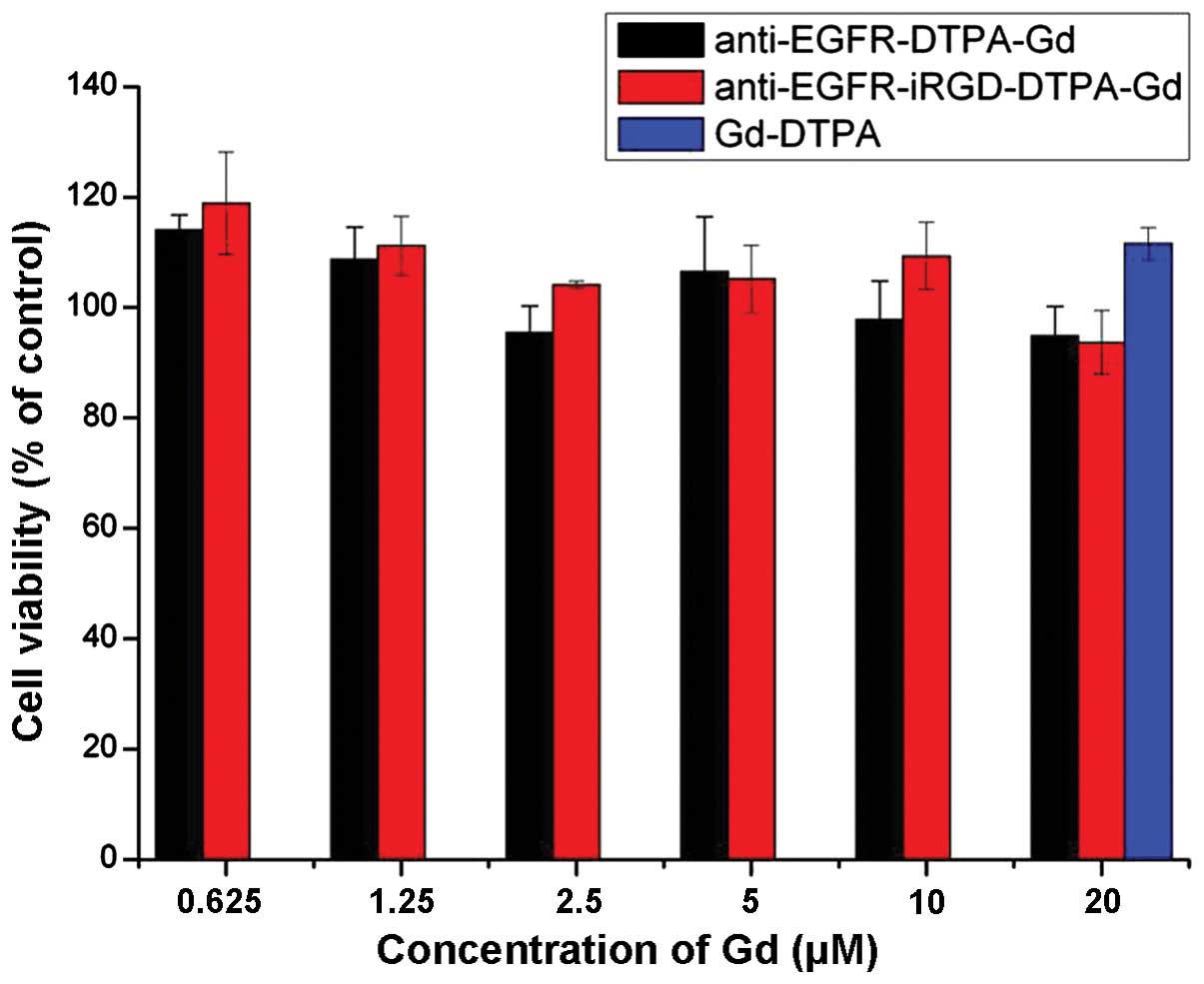
In vitro toxicity of anti-EGFR-DTPA-Gd and
anti-EGFR-iRGD-DTPA-Gd, and Gd-DTPA at different indicated molar
concentrations of Gd was evaluated using the MTT assay in BGC 823
cells. Magnevist at 20 µM was taken as the positive control.
As shown in Fig. 2, the cell
survival rates at different concentrations of the anti-EGFR-DTPA-Gd
and anti-EGFR-iRGD-DTPA-Gd were not significantly different
(P>0.05). Compared with the Magnevist group, the cell survival
rates for the anti-EGFR-DTPA-Gd- and anti-EGFR-iRGD-DTPA-Gd-treated
groups were slightly lower, although the differences were not
significant (P>0.05). The cell survival rates of all the groups
were >90%, suggesting that anti-EGFR-DTPA-Gd and
anti-EGFR-iRGD-DTPA-Gd did not influence the viability of BGC823
cells at the concentrations used in the present study.
In vitro cell targeting and competitive
binding assay
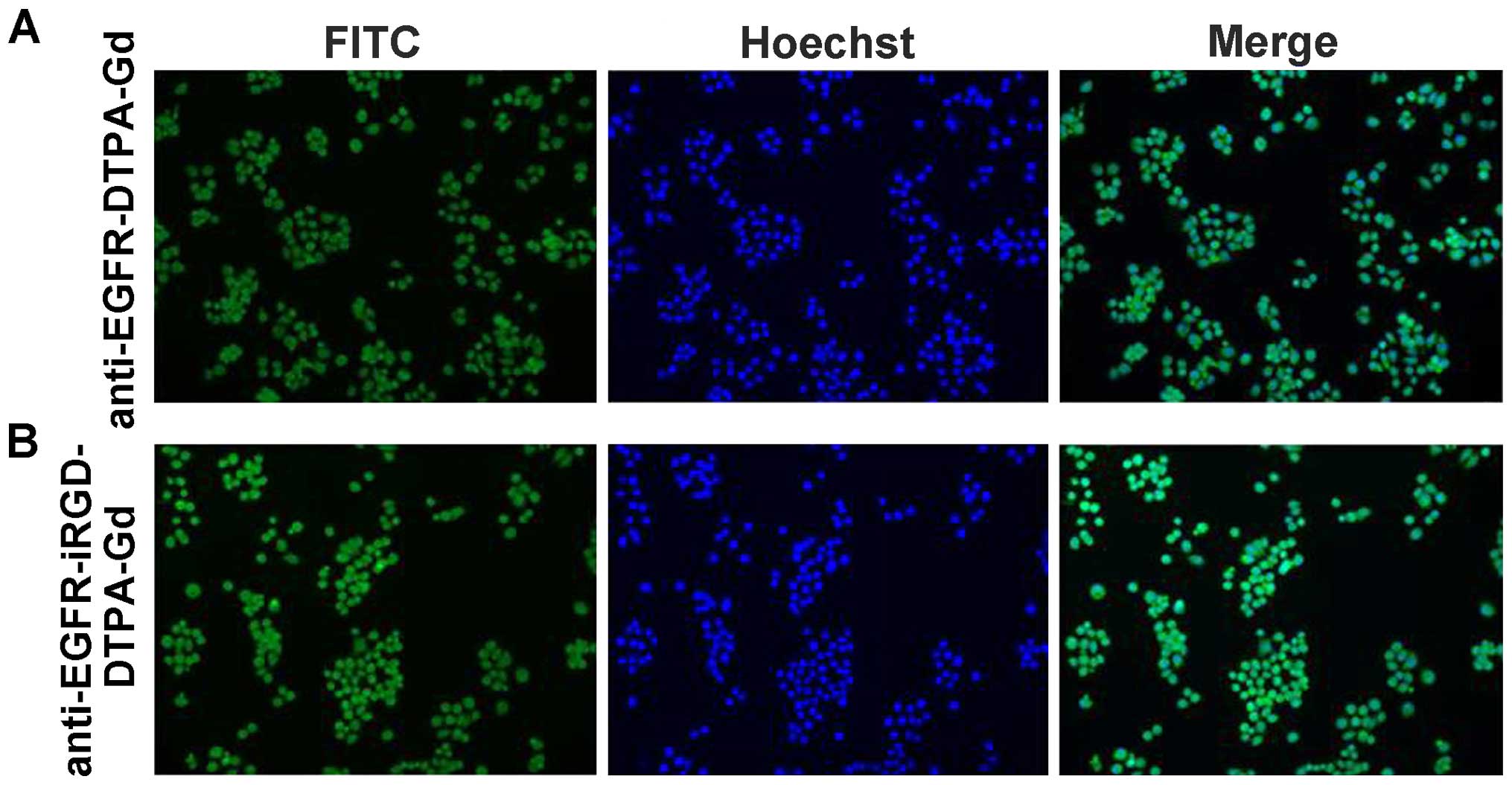
BGC-823 cells incubated with anti-EGFR-DTPA-Gd-FITC
or anti-EGFR-iRGD-DTPA-Gd-FITC, exhibited green fluorescence. Thus,
anti-EGFR-DTPA-Gd-FITC and anti-EGFR-iRGD-DTPA-Gd-FITC were able to
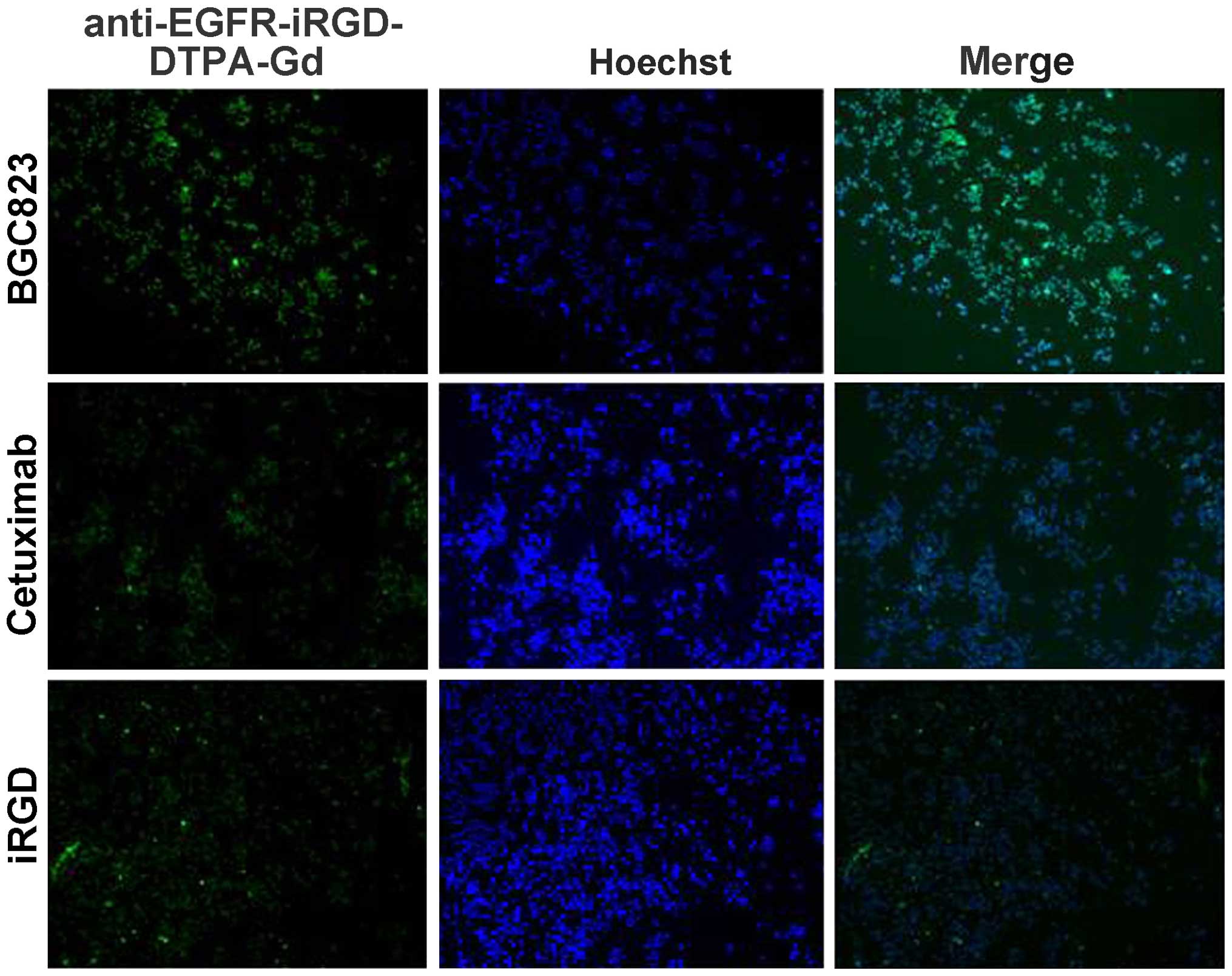
target gastric cancer BGC-823 cells (Fig. 3). when cetuximab (a monoclonal
anti-EGFR antibody acting as an inhibitor) or iRGD were added, the
uptake of anti-EGFR-DTPA-Gd-FITC and anti-EGFR-iRGD-DTPA-Gd-FITC in
BGC-823 cells decreased as proven by the decreased fluorescent
emission (Fig. 4).
As shown in Fig. 3,
the fluorescence intensity of anti-EGFR-iRGD-DTPA-Gd was stronger
after being mixed with 25 µg/ml cetuximab or 10 µg/ml
iRGD. This finding indicated that anti-EGFR-iRGD-DTPA-Gd binds the
same receptor as cetuximab and iRGD. The specificity and affinity
of anti-EGFR-iRGD-Gd binding to the target antigen were assessed
using a competitive binding assay. when fluorescence intensity for
anti-EGFR-iRGD-Gd-FITC taken up by BGC-823 was set as 100%, the
affinity of anti-EGFR-iRGD-Gd was decreased to 20.9 or 41.3% when
cetuximab or iRGD was added to compete with the antigen (Fig. 4). These results are semiquantitative
as calculated by the microscopy software. The binding of
anti-EGFR-iRGD-Gd to BGC-823 cells was specifically inhibited by
cetuximab and iRGD, indicating that anti-EGFR-iRGD-Gd binds to the
same receptor. These results indicated that anti-EGFR-iRGD-Gd
possesses specificity and affinity to EGFR and was internalized
through the same route as iRGD.
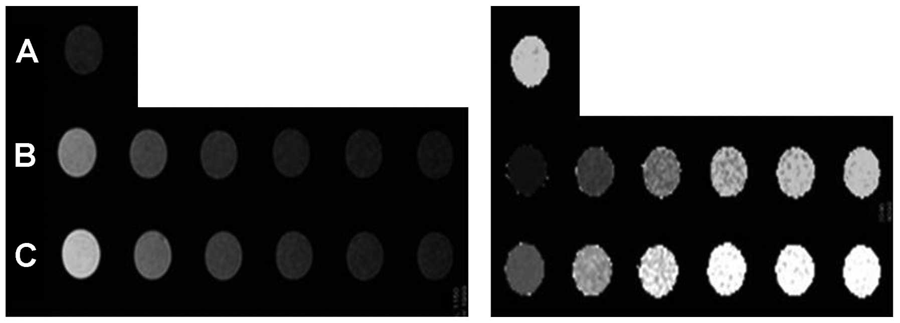
MRI in vitro
To test the ability of anti-EGFR-iRGD-DTPA-Gd as a
MRI contrast agent, the T1 longitudinal relaxation time of
H2o protons was evaluated. H2O,
anti-EGFR-DTPA-Gd, anti-EGFR-iRGD-DTPA-Gd, and Gd-DTPA injections
[(Gd)=2.4–77 µM] were evaluated at 3.0T at 24°C. As shown in
Fig. 5, in the
anti-EGFR-iRGD-DTPA-Gd group, higher Gd concentrations showed
higher signal intensities in the T1-weighted image and shorter T1
values in the T1-map image. The anti-EGFR-iRGD-DTPA-Gd group showed
a higher signal intensity in the T1-weighted image and a shorter T1
value in the T1-map image than those of the Gd-DTPA group at the
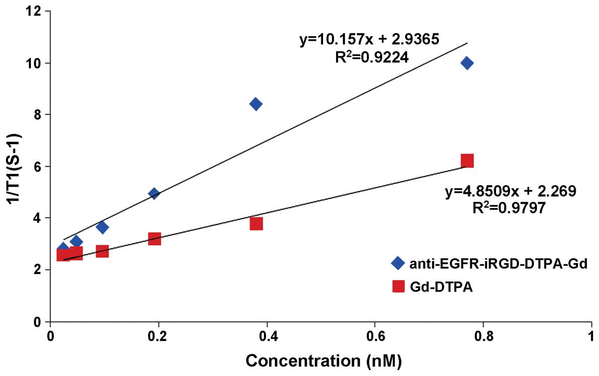
same Gd concentrations (P<0.05). The relaxivity 'r' was defined
as the slope of the curves 1/T1 with respect to the contrast agent
Gd concentration. The 'r' of anti-EGFR-iRGD-Gd was 10.157/mM/sec
and higher than Gd-DTPA (4.851/mM/sec) (Fig. 6). The results of the MRI in
vitro indicated that anti-EGFR-iRGD-DTPA-Gd is a better and
novel MRI molecular contrast agent, when compared with DTPA-Gd.
Evaluation of the targeting in vivo: MRI
in nude mouse tumor model
For MRI, anti-EGFR-iRGD-DTPA-Gd, anti-EGFR-DTPA-Gd,
and Gd-DTPA were injected into tumor-bearing mice via the vena
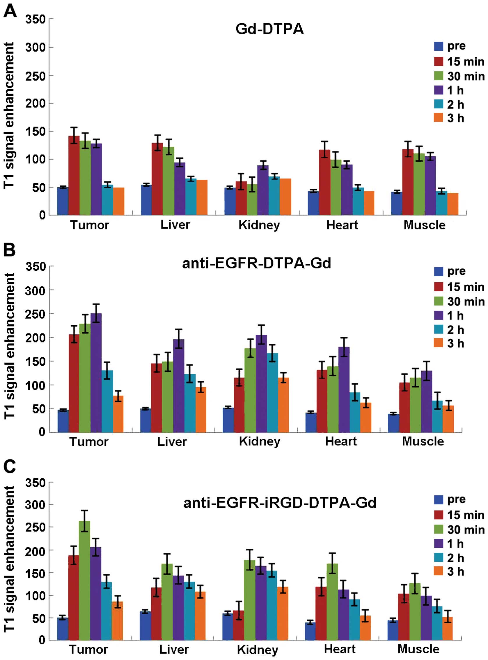
caudalis at 42 µmol Gd/kg. The mice were scanned at 15, 30
min, 1, 2 and 3 h following contrast injection. MRI and the results
of the enhanced signals in different tissues are summarized in
Figs. 7 and 8.
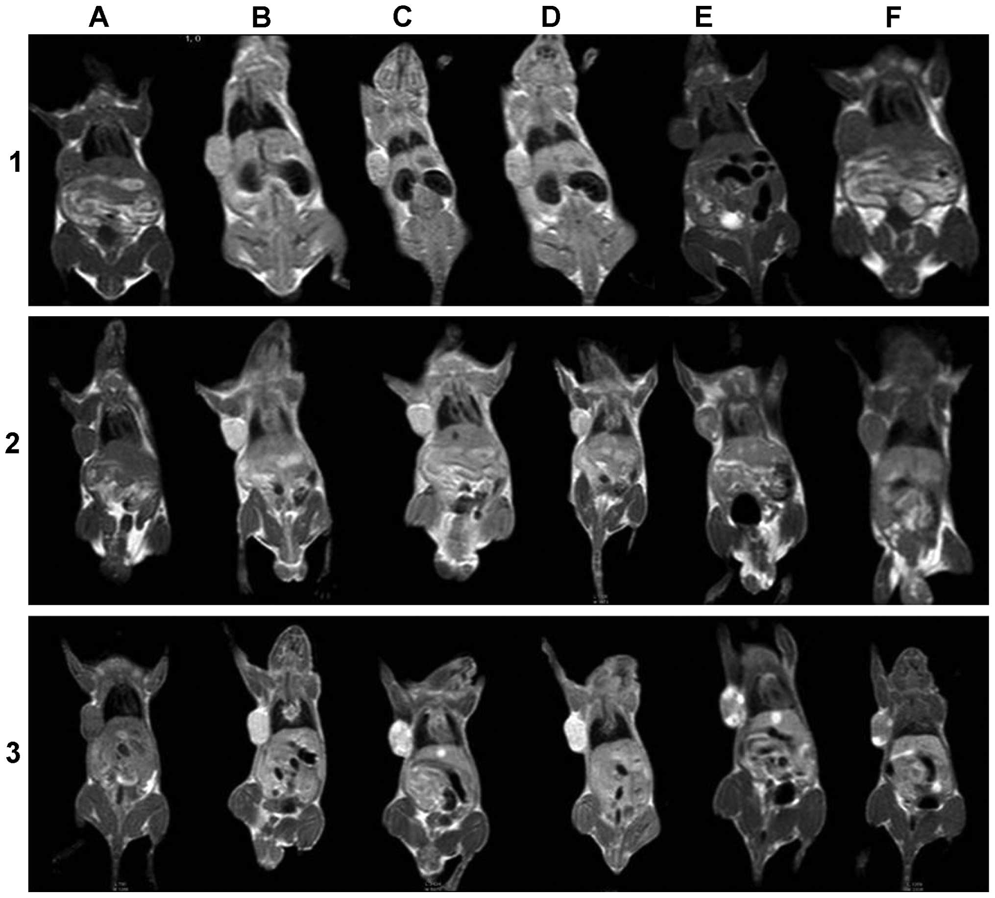
As shown in Figs.
7–1 and 8A, prior to the
injection of Gd-DTPA, the images of tumors and other organs were
dark (Fig. 7A). After the
administration of Magnevist, the contrast agents were
non-specifically distributed throughout the body in a short period
of time. The signal-to-noise tumor ratio increased from 33 to 50 by
15 min after injection of Gd-DTPA. Liver of this group also
brightened and the signal-to-noise ratio reached a maximum (from 36
to 46) by 15 min after injection and then rapidly decayed (Fig. 7–1B and C). Since the elimination of
Gd-DTPA was by renal clearance, an enhancement in the kidney
continued from 15 min to 1 h (from 32 to 42) (Fig. 7–1D), and the urinary bladder became
extremely bright compared to other tissues by 1 h after injection
of Gd-DTPA (Fig. 7–1E). The heart
and muscle also reached a maximum at 15 min after injection that
was then followed by a rapid decay. The entire body was as dark as
preinjection by 2 h after injection of Gd-DTPA (Fig. 7–1F).
After the injection of anti-EGFR-DTPA-Gd, the
contrast in the tumor was well distributed and the tumor was
significantly bright, with its boundary clearly distinct (Fig. 7–2). The signal-to-noise ratio of the
tumor increased significantly from 31 to 67 by 15 min and reached a
maximum at 1 h (from 31 to 100) (Fig.
8C). The liver and kidney were also enhanced and the signal
intensity signal-to-noise ratio reached a maximum (from -33 to -82
and from 34 to -111, respectively) by 1 h after the injection,
followed by rapid decay (Fig.
7–2D). Similar to the above result, the signal intensities of
the heart and muscle were enhanced to a maximum by 1 h after
injection and then gradually decreased. After 3 h, the enhanced
signal intensity was barely detectable.
After the injection of anti-EGFR-iRGD-DTPA-Gd, the
tumor signal was brighter than that of other tissues, and the
boundary was also clearly distinct (Fig. 7–3). The signal-to-noise ratio of the
tumor was maximal 30 min after injection (from -34 to -98) and then
gradually decreased (Fig. 8C).
After 3 h, an enhanced signal was present at the tumor margins. The
signal-to-noise ratio of the liver and kidney was also maximal at
30 min after injection (from -42 to 87 and from 40 to -89,
respectively) and decreased gradually. The signal intensity of
heart and muscle was maximal at 30 min after injection and then
decreased gradually. After 3 h, the enhanced signal intensity
remained higher than preinjection.
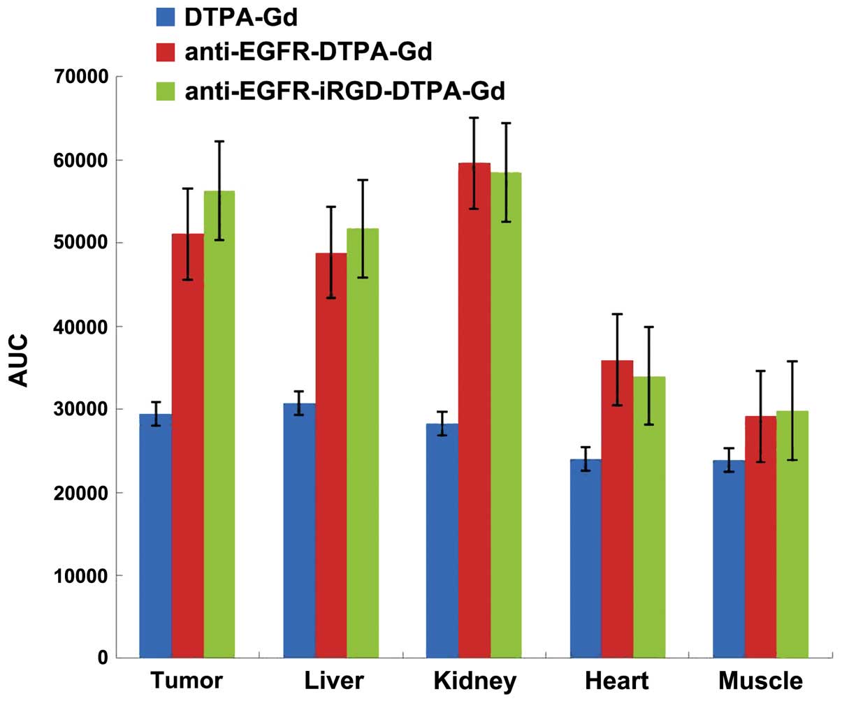
The enhanced AUCs of anti-EGFR-iRGD-DTPA-Gd,
anti-EGFR-DTPA-Gd, and Magnevist were calculated by the trapezoidal
method. The results are shown in Fig.
9. Compared to Gd-DTPA and anti-EGFR-DTPA-Gd,
anti-EGFR-iRGD-DTPA-Gd exhibited significant target enhancements in
the tumor and liver (P<0.01). In kidney, heart, and muscle,
anti-EGFR-DTPA-Gd had greater enhancements than
anti-EGFR-iRGD-DTPA-Gd and Gd-DTPA (P<0.01).
Discussion
In the present study, we designed and synthesized a
novel tumor-targeted molecular MRI contrast agent, which was
constructed by the conjugation of Gd-DTPA to a bispecific
recombinant protein of high permeability anti-EGFR-iRGD. This novel
agent has no cytotoxicity, and anti-EGFR-iRGD-DTPA-Gd was capable
of targeting the same receptor as cetuximab and iRGD in
vitro. Furthermore, in vivo MR images showed that signal
enhancements in tumors after injection of anti-EGFR-iRGD-DTPA-Gd
were significantly higher than those of anti-EGFR-DTPA-Gd or pure
Gd-DTPA.
Uppal et al reported that targeting with
Gd3+ probe mainly includes three types (20): i) discrete targeting peptide-
Gd-chelate: use of smaller molecules in combination with one or
more of the Gd chelate; ii) self-assembled nanoparticles including
Gd, which are usually emulsions, microcapsule or liposome
nanoparticles containing 10–1,000 Gd chelates prepared by
self-assembly method; and iii) 'smart' probes with triggering
activity, the product after biochemical reactions (such as enzyme
pyrolysis reaction and pH change) between probes and target
material can improve the effect of relaxivity. The synthetic probe
in this experiment belongs to the first category. Its advantage
lies in the small size of probes. The probe can freely pass through
the endothelial cell barrier into the imaging target and produce
higher target-background signal.
N-terminal cysteine containing tumor-homing peptide
(iRGD, CRGDK/EGPD/EC) has been identified as a highly efficient,
deep penetrating peptide (21,22).
when iRGD is chemically conjugated to or co-administered with an
anticancer drug, this peptide can carry the drug deep into
extravascular tumor tissue. iRGD also homes to tumors in a
tumor-specific and NRP-1-dependent manner. Thus, when the polymer
nanoparticles are modified or co-administered with iRGD, the tissue
penetrating and targeting abilities of drug-loaded nanoparticles
may be improved. In the present study, anti-EGFR-iRGD fusion
protein was confirmed to maintain the tumor tissue-targeting
abilities, which is consistent with previous results (17).
For the in vivo MRI study, a human gastric
cancer xenograft model with BGC-823 cells was established. The
images on the 3.0T MR scanner showed that the tumor was enhanced
clearly by Gd-DTPA, anti-EGFR-DTPA-Gd and anti-EGFR-iRGD-DTPA-Gd.
However, analysis of the MRI signal intensity revealed different
time-dependent enhancement patterns for the three groups. In the
pure Gd-DTPA group, the tumor signal intensity reached a maximum
more rapidly with mild enhancement and showed a rapid clearance
because the molecular weight of Gd-DTPA is small (~500 Da) and the
water-soluble Gd chelates diffuse from the tumor tissue easily. In
the anti-EGFR-DTPA-Gd group, the signal intensity of the tumor
reached a maximum at 1 h (from 47 to 221) and the signal intensity
of the tumor was higher than that of pure Gd-DTPA. we suggest that
the enhancement of anti-EGFR-DTPA-Gd is caused by the specific
binding to endothelial and tumor cells. In comparison to
anti-EGFR-DTPA-Gd, the signal intensity of the tumor of
anti-EGFR-iRGD-DTPA-Gd reached a maximum at 30 min (from 51 to 264)
and the signal intensity of tumor was higher than that of
anti-EGFR-DTPA-Gd. EGFR is overexpressed in BGC-823 cells, and
anti-EGFR-iRGD-DTPA-Gd can target tumor cells and be restricted to
the tumor area by the tumor cell surface EGFR-antibody reaction. On
the other hand, iRGD increased the penetration of
anti-EGFR-iRGD-DTPA-Gd. In addition, anti-EGFR-iRGD-Gd has a large
magnetic moment due to the covalent linkage of Gd-DTPA to
anti-EGFR-iRGD, and a larger molecular weight. we hypothesize that
the use of iRGD increases the penetrance of the contrast agent to
the cells and therefore, shortens the time required for reaching
the maximum signal.
In conclusion, a novel tumor-targeting molecular MRI
contrast agent was designed and synthesized, which was constructed
by the conjugation of Gd-DTPA to a bispecific recombinant protein
with high permeability. This novel agent without cytotoxicity
targeted gastric cancer cells and shows promise as an effective
novel MRI molecular contrast agent and for early diagnostic imaging
in clinical applications.
Acknowledgments
The present study was supported by the Science and
Technology Development fund of Nanjing (no. YKK12065), Health
Research projects in Jiangsu Province (no. Q201411), the National
Natural Science Foundation of China (no. 81502037), Fundamental
Research Funds for the Central Universities (no. 20610140698), and
the Scientific Research Foundation of the Graduate School of
Nanjing University (no. 2013CL15).
References
|
1
|
Weissleder R: Molecular imaging in cancer.
Science. 312:1168–1171. 2006. View Article : Google Scholar : PubMed/NCBI
|
|
2
|
Hu F, Joshi HM, Dravid VP and Meade TJ:
High-performance nanostructured MR contrast probes. Nanoscale.
2:1884–1891. 2010. View Article : Google Scholar : PubMed/NCBI
|
|
3
|
Moffat BA, Reddy GR, McConville P, Hall
DE, Chenevert TL, Kopelman RR, Philbert M, Weissleder R, Rehemtulla
A and Ross BD: A novel polyacrylamide magnetic nanoparticle
contrast agent for molecular imaging using MRI. Mol Imaging.
2:324–332. 2003. View Article : Google Scholar
|
|
4
|
Vosjan MJ, Vercammen J, Kolkman JA,
Stigter-van Walsum M, Revets H and van Dongen GA: Nanobodies
targeting the hepatocyte growth factor: Potential new drugs for
molecular cancer therapy. Mol Cancer Ther. 11:1017–1025. 2012.
View Article : Google Scholar : PubMed/NCBI
|
|
5
|
Garanger E, Boturyn D and Dumy P: Tumor
targeting with RGD peptide ligands-design of new molecular
conjugates for imaging and therapy of cancers. Anticancer Agents
Med Chem. 7:552–558. 2007. View Article : Google Scholar : PubMed/NCBI
|
|
6
|
Chen X, Liu S, Hou Y, Tohme M, Park R,
Bading JR and Conti PS: MicroPET imaging of breast cancer
alphav-integrin expression with 64Cu-labeled dimeric RGD peptides.
Mol Imaging Biol. 6:350–359. 2004. View Article : Google Scholar : PubMed/NCBI
|
|
7
|
Chen X, Tohme M, Park R, Hou Y, Bading JR
and Conti PS: Micro-PET imaging of alphavbeta3-integrin expression
with 18F-labeled dimeric RGD peptide. Mol Imaging. 3:96–104. 2004.
View Article : Google Scholar : PubMed/NCBI
|
|
8
|
Haubner R, Wester HJ, Weber WA, Mang C,
Ziegler SI, Goodman SL, Senekowitsch-Schmidtke R, Kessler H and
Schwaiger M: Noninvasive imaging of alpha(v)beta3 integrin
expression using 18F-labeled RGD-containing glycopeptide and
positron emission tomography. Cancer Res. 61:1781–1785.
2001.PubMed/NCBI
|
|
9
|
Oliveira S, Heukers R, Sornkom J, Kok RJ,
van Bergen En and Henegouwen PM: Targeting tumors with nanobodies
for cancer imaging and therapy. J Control Release. 172:607–617.
2013. View Article : Google Scholar : PubMed/NCBI
|
|
10
|
Huang L, Gainkam LO, Caveliers V, Vanhove
C, Keyaerts M, De Baetselier P, Bossuyt A, Revets H and Lahoutte T:
SPECT imaging with 99mTc-labeled EGFR-specific nanobody for in vivo
monitoring of EGFR expression. Mol Imaging Biol. 10:167–175. 2008.
View Article : Google Scholar : PubMed/NCBI
|
|
11
|
Vosjan MJ, Perk LR, Roovers RC, Visser GW,
Stigter-van Walsum M, van Bergen En, Henegouwen PM and van Dongen
GA: Facile labelling of an anti-epidermal growth factor receptor
Nanobody with 68Ga via a novel bifunctional desferal chelate for
immuno-PET. Eur J Nucl Med Mol Imaging. 38:753–763. 2011.
View Article : Google Scholar : PubMed/NCBI
|
|
12
|
Haspel N, Zanuy D, Nussinov R, Teesalu T,
Ruoslahti E and Aleman C: Binding of a C-end rule peptide to the
neuropilin-1 receptor: A molecular modeling approach. Biochemistry.
50:1755–1762. 2011. View Article : Google Scholar : PubMed/NCBI
|
|
13
|
Sugahara KN, Teesalu T, Karmali PP,
Kotamraju VR, Agemy L, Girard OM, Hanahan D, Mattrey RF and
Ruoslahti E: Tissue-penetrating delivery of compounds and
nanoparticles into tumors. Cancer Cell. 16:510–520. 2009.
View Article : Google Scholar : PubMed/NCBI
|
|
14
|
Hiki S and Kataoka K: Versatile and
selective synthesis of 'click chemistry' compatible
heterobifunctional poly(ethylene glycol)s possessing azide and
alkyne functionalities. Bioconjug Chem. 21:248–254. 2010.
View Article : Google Scholar : PubMed/NCBI
|
|
15
|
Hoang B, Lee H, Reilly RM and Allen C:
Noninvasive monitoring of the fate of 111In-labeled block copolymer
micelles by high resolution and high sensitivity microSPECT/CT
imaging. Mol Pharm. 6:581–592. 2009. View Article : Google Scholar : PubMed/NCBI
|
|
16
|
Ishihara T, Maeda T, Sakamoto H, Takasaki
N, Shigyo M, Ishida T, Kiwada H, Mizushima Y and Mizushima T:
Evasion of the accelerated blood clearance phenomenon by coating of
nanoparticles with various hydrophilic polymers. Biomacromolecules.
11:2700–2706. 2010. View Article : Google Scholar : PubMed/NCBI
|
|
17
|
Sha H, Zou Z, Xin K, Bian X, Cai X, Lu W,
Chen J, Chen G, Huang L, Blair AM, et al: Tumor-penetrating peptide
fused EGFR single-domain antibody enhances cancer drug penetration
into 3D multicellular spheroids and facilitates effective gastric
cancer therapy. J Control Release. 200:188–200. 2014. View Article : Google Scholar
|
|
18
|
Kielar F, Tei L, Terreno E and Botta M:
Large relaxivity enhancement of paramagnetic lipid nanoparticles by
restricting the local motions of the Gd(III) chelates. J Am Chem
Soc. 132:7836–7837. 2010. View Article : Google Scholar : PubMed/NCBI
|
|
19
|
Prudêncio M, Rohovec J, Peters JA, Tocheva
E, Boulanger MJ, Murphy ME, Hupkes HJ, Kosters W, Impagliazzo A and
Ubbink M: A caged lanthanide complex as a paramagnetic shift agent
for protein NMR. Chemistry. 10:3252–3260. 2004. View Article : Google Scholar : PubMed/NCBI
|
|
20
|
Uppal R and Caravan P: Targeted probes for
cardiovascular MRI. Future Med Chem. 2:451–470. 2010. View Article : Google Scholar : PubMed/NCBI
|
|
21
|
Sugahara KN, Teesalu T, Karmali PP,
Kotamraju VR, Agemy L, Greenwald DR and Ruoslahti E:
Coadministration of a tumor-penetrating peptide enhances the
efficacy of cancer drugs. Science. 328:1031–1035. 2010. View Article : Google Scholar : PubMed/NCBI
|
|
22
|
Sugahara KN, Braun GB, de Mendoza TH,
Kotamraju VR, French RP, Lowy AM, Teesalu T and Ruoslahti E: Tumor-
penetrating iRGD peptide inhibits metastasis. Mol Cancer Ther.
14:120–128. 2015. View Article : Google Scholar :
|