Introduction
Osteosarcoma is the most common non-hematological
primary bone tumor affecting children and adolescents (1), and patients with osteosarcoma have
unsatisfactory prognosis, especially for those with metastasis.
Currently available treatments have limited efficacy for
osteosarcoma with metastasis (2,3).
Therefore, exploring the molecular mechanism of the metastasis of
osteosarcoma is critical for identifying more effective therapeutic
targets that would significantly improve the prognosis of
osteosarcoma patients.
MicroRNAs (miRNAs) are small non-conding RNAs and
act as post-transcriptional regulators of gene expression by
interacting with 3′-UTR of targeted mRNAs and repressing the
stability or translation of targeted mRNAs (4). Numerous studies have demonstrated that
miRNAs played active roles in various cellular functions (5) including cell growth, differentiation
and motility. Abnormal expression and function of miRNAs has been
confirmed to play fundamental roles in human malignancies (6). In addition, miRNAs have emerged as
promising biomarkers for the diagnosis and prognosis of cancer
patients and attractive targets for cancer treatments (7).
MicroRNA-130a (miR-130a) has been described to
participate in the development and progression of various human
cancers. It was found to be overexpressed in gastric cancer
(8,9), esophageal cancer tissue (10), and basal cell carcinoma (11), suggesting an oncogenic role in human
cancer. However, studies of human hepatocellular carcinoma
(12,13), prostate carcinoma (14,15)
and breast cancer (16,17) showed that miR-130a was downregulated
and played tumor suppressive roles in these cancers. Therefore, the
expression status and biological role of miR-130a seems to be
dependent on the cancer type. However, the expression level,
clinical significance and biological function of miR-130a in
osteosarcoma remain unclear.
In this study, we confirmed that miR-130a expression
was significantly increased in osteosarcoma tissues. Higher
expression of miR-130a was observed in patients with metastasis.
Elevated miR-130a expression was closely associated with poor
clinicopathological features and prognosis of osteosarcoma
patients. Functional assays showed that miR-130a promoted the
migration and invasion of osteosarcoma cells by promoting
epithelial mesenchymal transition (EMT). Furthermore, we identified
phosphatase and tensin homolog (PTEN) as the downstream target of
miR-130a in osteosarcoma cells. miR-130a exerted its functional
influence on osteosarcoma cells by suppressing the expression of
PTEN.
Materials and methods
Clinical specimens
Tumor specimens and the adjacent non-cancer tissues
were obtained from 86 osteosarcoma patients who underwent surgery
in the Department of Orthopaedics, Children's Hospital of Zhejiang
University School of Medicine between January 2003 and January
2010. Written informed consents were obtained from all
participating patients. Clinical specimens were frozen and stored
at −80°C. The patients did not receive any chemo- or radiotherapy
before operation. The demographic and clinicopathological features
of all included patients are presented in Table I. All protocols involving clinical
samples were approved by the Ethics Review Committee of Children's
Hospital of Zhejiang University School of Medicine.
 | Table ICorrelation between the
clinicopathological characteristics and miR-130a expression in
osteosarcoma. |
Table I
Correlation between the
clinicopathological characteristics and miR-130a expression in
osteosarcoma.
| Parameter | Total no. of
patients
(n=86) | No. of patients
| P-value |
|---|
|
miR-130alow |
miR-130ahigh |
|---|
| Age | | | | 0.664 |
| >25 | 49 | 22 | 27 | |
| ≤25 | 37 | 19 | 18 | |
| Gender | | | | 0.827 |
| Male | 50 | 24 | 26 | |
| Female | 36 | 19 | 17 | |
| Location | | | | 0.658 |
| Proximal | 53 | 25 | 28 | |
| Distal | 33 | 18 | 15 | |
| Tumor size | | | | 0.490 |
| ≤5 cm | 58 | 31 | 27 | |
| >5 cm | 28 | 12 | 16 | |
|
Differentiation | | | | 0.635 |
| Well/moderate | 61 | 32 | 29 | |
| Poor | 25 | 11 | 14 | |
| Metastasis | | | | 0.002 |
| Absent | 55 | 35 | 20 | |
| Present | 31 | 8 | 23 | |
| TNM stage | | | | 0.023 |
| I + II | 51 | 25 | 26 | |
| III + IV | 35 | 8 | 27 | |
Reverse transcription-quantitative PCR
(RT-qPCR)
Total RNA was isolated from the osteosarcoma tissues
and cells using TRIzol (Life Technologies) according to the
manufacturer's instructions. The TaqMan Human MiRNA Assay kit
(Applied Biosystems, Foster City, CA, USA) and a SYBR®
Premix Ex Taq™ II (Perfect Real-time) kit (Takara Bio, Shiga,
Japan) were employed to perform the PCR amplification. The primer
of miR-130a (HmiRQP0156), PTEN (HQP015535), U6 (HmiRQP9001) and
GAPDH (HQP006940) were obtained from Genecopoeia (Guangzhou,
China). The relative expression of miR-130a is shown as fold
difference relative to U6 while the relative expression of PTEN is
shown as fold difference relative to GAPDH.
Cell culture and transfection
Human osteosarcoma cell lines HOS58, SaoS-2 and
MG63, and the human normal osteoblasts (hFOB1.19) were grown in
complete Dulbecco's modified Eagle's medium (DMEM; HyClone, Logan,
UT, USA) supplemented with 10% fetal bovine serum (FBS; HyClone),
100 U/ml penicillin, and 100 µg/ml streptomycin. Cell
cultures were incubated in a humidified incubator containing of 5%
CO2 at 37°C.
miR-130a expressing vector (HmiR0170-MR03), the
control vector (CmiR0001-MR03), miR-130a inhibitor
(HmiR-AN0156-AM03) and the negative control (CmiR-AN0001-AM03),
were purchased from GeneCopoeia. Plasmids carrying PTEN expressing
vector (#28298) and PTEN specific shRNA (#25638) were obtained from
Addgene (Cambridge, MA, USA). The vectors mentioned above were
transfected into the osteosarcoma cells using Lipofectamine 2000
(Invitrogen, Carlsbad, CA, USA) following the manufacturer's
protocol.
Transwell assays
The migration and invasion of osteosarcoma cells
were evaluated in Transwell chambers with 8-µm inserts
(Millipore Corp., Billerica, MA, USA) according to the
manufacturer's instructions. Osteosarcoma cells were resuspended in
serum-free medium and seeded into the top chamber of each insert.
Serum-containing medium (10% FBS) was used in the lower chamber as
the attractant. In the invasion assay, each upper chamber was
coated with mixture of DMEM and Matrigel (Becton-Dickinson Labware,
Bedford, MA, USA) at a ratio of 8:1, and 24 h after cell seeding,
the cells in the upper surface of the filter were removed with a
cotton swab, and the migrated or invaded cells were stained with
0.1% crystal violet. Cell numbers on the lower surface were
counted. Three independent experiments were performed.
Western blotting
Cells were collected and washed twice with
phosphate-buffered saline (PBS), and lysed with RIPA lysis buffer
(BioMed, Beijing, China). Lysate was centrifuged at 14,000 rpm for
20 min and the supernatants containing protein were collected.
Protein concentration was measured using the BCA kit (Pierce,
Rockford, IL, USA). Protein (30–40 µg) of each sample was
separated and transferred to PVDF membrane. The blots were
incubated with the following primary antibodies: PTEN (1:1,000,
Cell Signaling Technologies, Danvers, MA, USA), E-cadherin
(1:1,000, Cell Signaling Technologies), vimentin (1:1,000, Cell
Signaling Technologies) and GAPDH (1:1500, Santa Cruz
Biotechnology, Santa Cruz, CA, USA). After washing the membranes
with TBST, blots were incubated with anti-mouse or anti-rabbit
secondary antibodies (1:10,000; Bio-Rad, Hercules, CA, USA), and
the signals were detected using the Bio-Rad Gel imaging system.
Luciferase reporter assay
Wild-type 3′-UTR sequence of PTEN (wt PTEN-3′-UTR)
which was predicted to interact with miR-130a and the mutated
sequence (mt PTEN-3′-UTR) were synthesized and inserted into the
pGL3 control vector (Promega, Madison, WI, USA). For luciferase
reporter assay, cells were transfected with the wild-type construct
or mutant construct, and miR-130a expressing vector, miR-130a
inhibitor, control vector or negative control. At 48 h after
transfection, the luciferase activity was measured using the
Dual-Luciferase Reporter Assay system (Promega, Shanghai, China)
with a luminometer (Promega). Results were obtained from three
independent experiments performed in duplicate.
Statistical analysis
SPSS statistical package for Windows version 13
(SPSS, Inc., Chicago, IL, USA) and GraphPad Prism 5 software
(GraphPad Software, Inc., USA) were used to perform the statistical
analysis, including the Pearson's Chi-square test, the Spearman's
rank correlation coefficient, the Student's t-test, the
Kaplan-Meier plot, and the log-rank test or ANOVA was performed in
this study. P<0.05 was considered to indicate a statistically
significant difference.
Results
miR-130a expression is increased in
osteosarcoma and is associated with the metastasis of
osteosarcoma
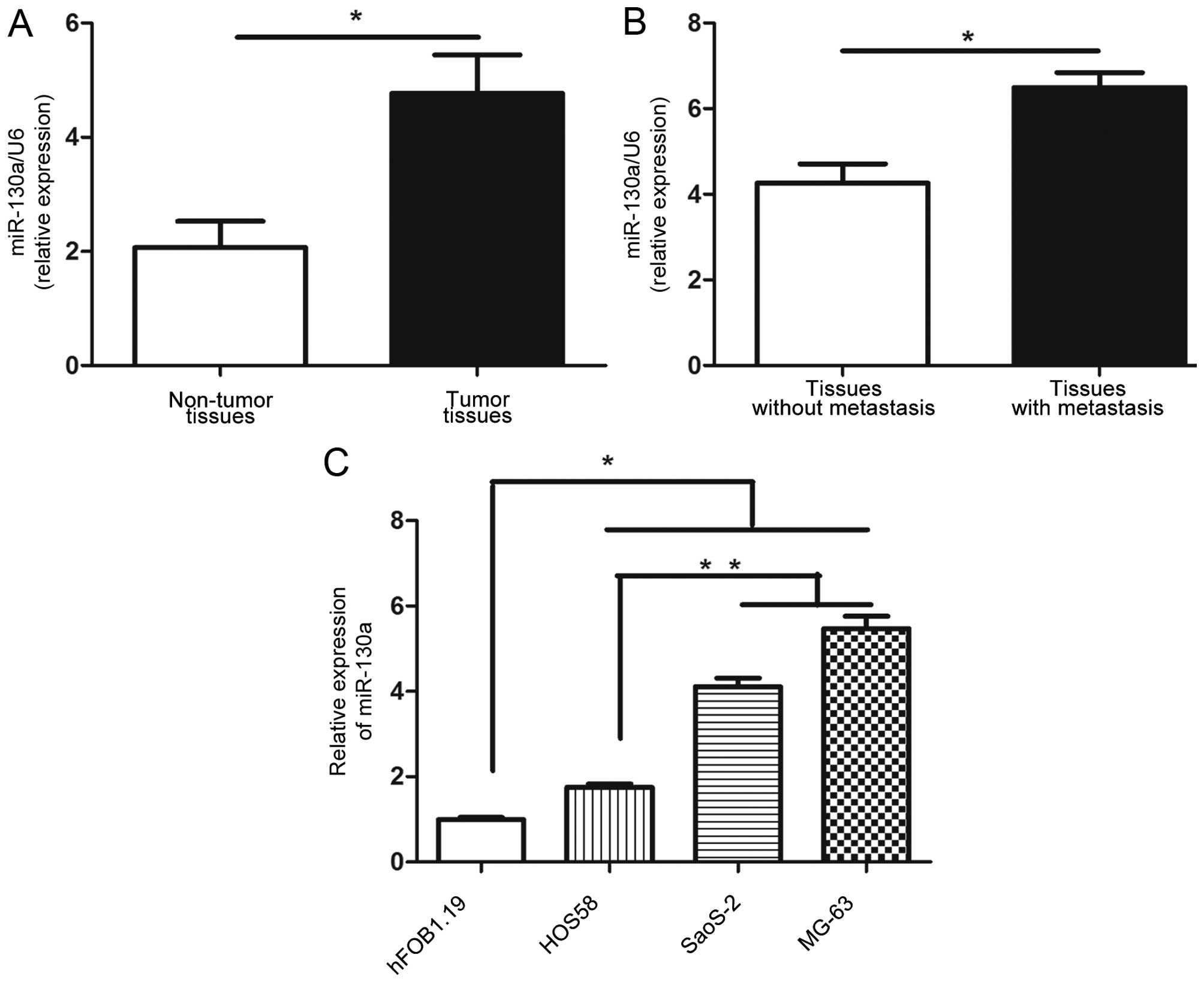
Quantitative RT-PCR assay was conducted to evaluate
the expression level of miR-130a in osteosarcoma tissues and
matched non-tumor tissues. miR-130a in osteosarcoma tissues was
increased significantly compared with that in the adjacent
non-tumor tissues (P<0.05, Fig.
1A). Moreover, in tissues with metastasis, the level of
miR-130a was significantly higher than that in those without
metastasis (P<0.05, Fig. 1B).
Furthermore, the expression level of miR-130a in osteosarcoma cell
lines was measured. Compared with normal human osteoblastic cell
line, hFOB1.19, osteosarcoma cell lines, including HOS58, SaoS-2
and MG63, had significantly higher level of miR-130a (P<0.05,
Fig. 1C). MG63 and SaoS-2 cells had
obviously higher expression level of miR-130a than HOS58 cells
(P<0.01, Fig. 1C). These results
indicate that miR-130a is significantly upregulated in osteosarcoma
and is associated with the metastasis of osteosarcoma.
High expression level of miR-130a is
correlated with adverse clinicopathological features and poor
prognosis of osteosarcoma patients
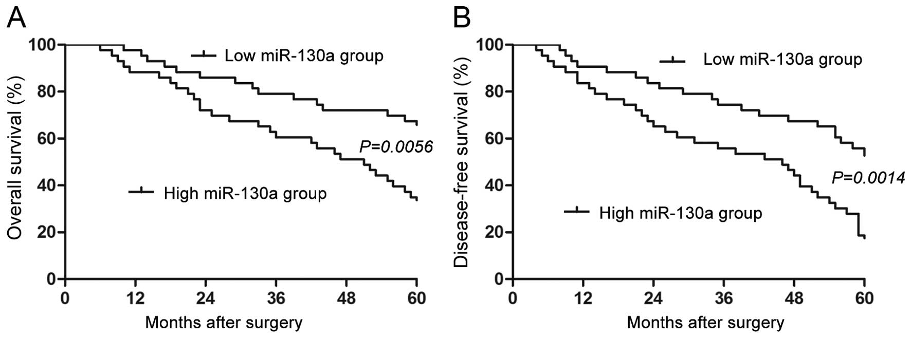
Next, the clinical significance of miR-130a
expression level in osteosarcoma tissues was examined. The
expression of miR-130a was assessed either low (n=43) or high
(n=43) based on the median level of miR-130a in the 86 patients
cohort. As presented in Table I,
high expression level of miR-130a in osteosarcoma patients was
closely associated with metastasis (P=0.002) and advanced TNM stage
(P=0.023). Importantly, Kaplan-Meier analysis further showed that
patients with high miR-130a expression level had significantly
shorter overall survival (P= 0.0056, Fig. 2A) and disease-free survival
(P=0.0014, Fig. 2B). These results
suggest miR-130a is a promising prognostic predictor for
osteosarcoma patients.
miR-130a promotes the migration, invasion
and EMT of osteosarcoma cells
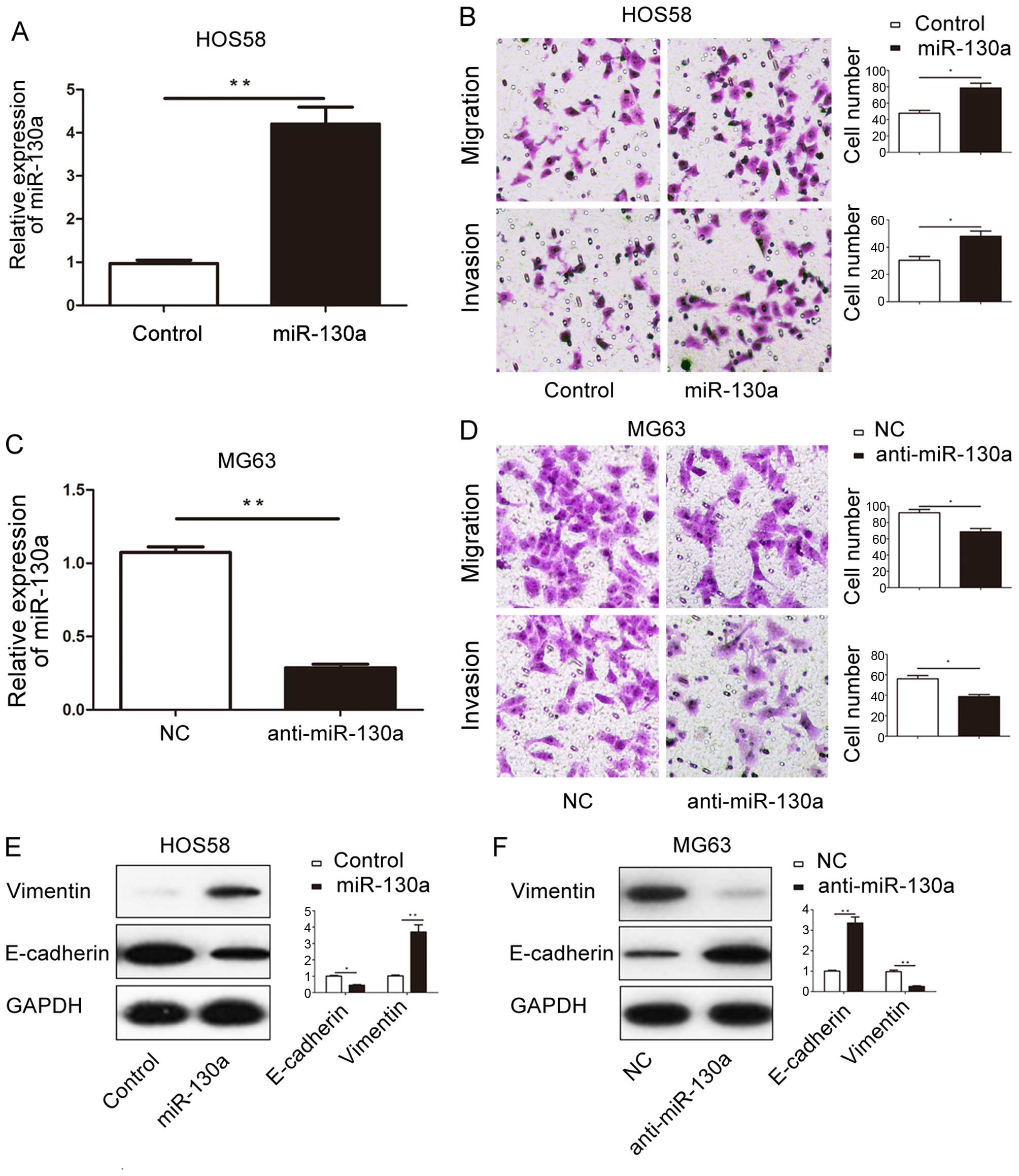
The functional role of miR-130a in osteosarcoma
cells was explored. HOS58 cells with miR-130a mimics, and the
expression level of miR-130a was significantly increased after
transfection (P<0.01, Fig. 3A).
Functionally, as suggested by the Transwell assay, HOS58 cells
overexpressing miR-130a (HOS58-miR-130a cells) showed increased
migratory (P<0.05, upper part in Fig. 3B) and invasive (P<0.05, lower
part in Fig. 3B) ability. In
contrast, miR-130a inhibitor significantly downregulated the
expression level of miR-130a in MG63 cells (P<0.01, Fig. 3C). The migration (P<0.05, upper
part in Fig. 3D) and invasion
(P<0.05, lower part in Fig. 3D)
of MG63 cells was significantly decreased after the expression of
miR-130a was inhibited. These data indicate that miR-130a
potentiate the metastatic behavior of osteosarcoma cells.
The EMT process, which has been regarded as a
fundamental process of the cancer metastasis (18–20),
can increase the migratory and invasive ability of osteosarcoma
cells (21). Therefore, we further
explored whether miR-130a promotes the migration and invasion of
osteosarcoma cells by promoting EMT. The results of western blot
showed that overexpression of miR-130a in HOS58 cells resulted in
decreased level of E-cadherin (P<0.05, Fig. 3E) and increased expression of
Vimentin (P<0.01, Fig. 3E). On
the contrary, downregulating miR-130a in MG63 cells led to
increased level of E-cadherin (P<0.01, Fig. 3F) and decreased expression of
Vimentin (P<0.01, Fig. 3F).
These results suggest that miR-130a can promote EMT phenotype of
osteosarcoma cells.
PTEN is a downstream target of miR-130a
in osteosarcoma cells
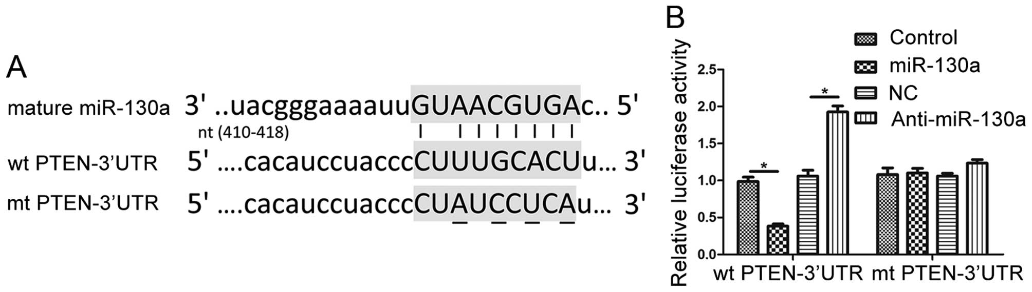
To further investigate the underlying mechanisms for
the functional influence of miR-130a in osteosarcoma cells, two
publicly available databases (TargetScan 6.2 and miRanda) were
searched for the potential downstream target of miR-130a. PTEN,
which is a well-recognized tumor suppressor and an important
participator in the development and progression of osteosarcoma,
was suggested to be a potential downstream target of miR-130a. As
shown in Fig. 4A, the 3′-UTR of
PTEN mRNA contained the complementary sequence of miR-130a. This
suggests that miR-130a can potentially bind to the 3′-UTR of PTEN.
To confirm this prediction, we performed dual-luciferase reporter
assays to elucidate whether miR-130a could directly bind to the
3′-UTR of PTEN. Forced expression miR-130a significantly decreased
the luciferase activity of PTEN with a wild-type (wt) 3′-UTR
(P<0.01 Fig. 4B), but had no
significant influence on that with a mutant (mt) 3′-UTR. On the
contrary, when miR-130a inhibitor was transfected, the luciferase
activity of wt PTEN 3′-UTR obviously increased (P<0.01, Fig. 4B) while that of mt PTEN 3′-UTR
remained unchanged.
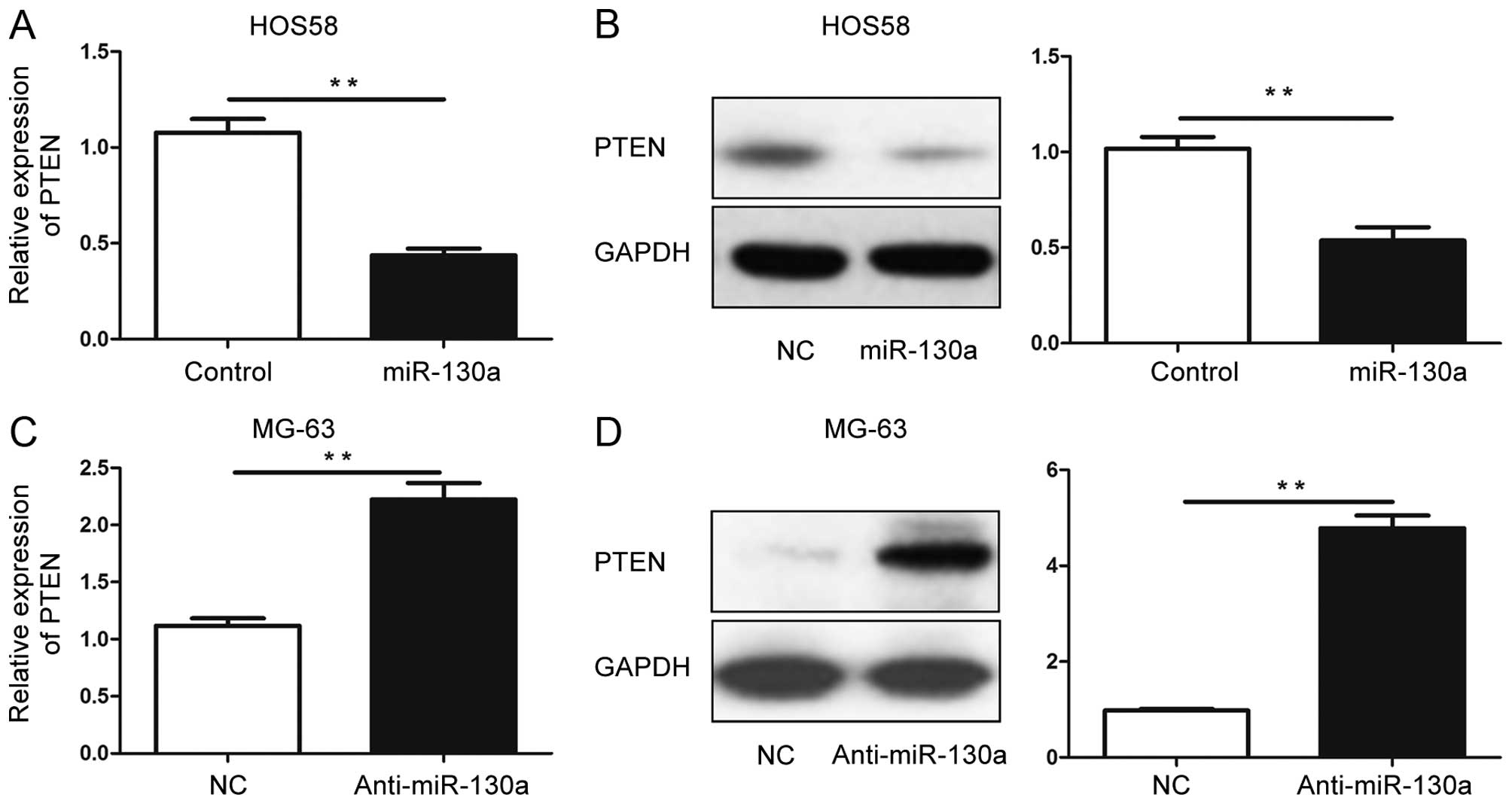
Moreover, we investigated whether alteration of
miR-130a could affect the expression level of PTEN mRNA and
protein. qRT-PCR and western blot were performed to examine the
expression level of PTEN after forced expression of miR-130a in
HOS58 cells. The result of qRT-PCR showed that PTEN mRAN level in
HOS58 cells was significantly decreased after overexpression of
miR-130a (P<0.01, Fig. 5A).
Consistently, the protein level of PTEN was significantly decreased
after overexpression of miR-130a (P<0.01, Fig. 5B). In contrast, inhibiting the
expression of miR-130a in MG63 cells resulted in significantly
increased level of PTEN mRNA (P<0.01, Fig. 5C) and protein (P<0.01, Fig. 5D). Taken together, these data
indicate the PTEN is a direct downstream target of miR-130a, and
miR-130a could regulate the expression of PTEN by interacting with
the 3′-UTR of PTEN.
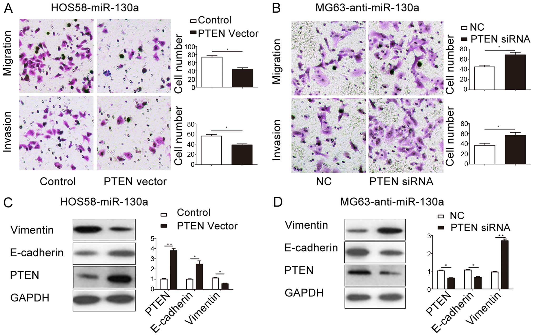
miR-130a promotes the metastasis and EMT
of osteosarcoma cells by inhibiting PTEN
To further clarify whether PTEN can serve as the
functional mediator of miR-130a in osteosarcoma cells, plasmids
containing PTEN expressing vector and PTEN shRNA were transfected
into HOS58 cells with overexpressed miR-130a (HOS58-miR-130a cells)
and MG63 cells with downregulated miR-130a (MG63-anti-miR-130a
cells), respectively. Functionally, overexpression of PTEN partly
abrogated the promoting effects of miR-130a on the migration
(P<0.05, upper part of Fig. 6A)
and invasion (P<0.05, lower part of Fig. 6A) of HOS58 cells. Inhibiting PTEN
expression in MG63-anti-miR-130a cells partly reversed the
inhibitory effects of miR-130a on the migration (P<0.05, upper
part of Fig. 6B) and invasion
(P<0.05, upper part of Fig. 6B)
of MG63 cells. Moreover, the results of western blot showed that
transfection of PTEN expressing vector in HOS58-miR-130a cells
resulted in significant increase of PTEN (P<0.01, Fig. 6C), and led to upregulation of
E-cadherin (P<0.05, Fig. 6C) and
downregulation of Vimentin (P<0.05, Fig. 6C). Transfection of PTEN shRNA in
MG63-anti-miR-130a cells significantly inhibited the expression of
PTEN (P<0.05, Fig. 6D), and
resulted in the downregulation of E-cadherin (P<0.05, Fig. 6D) and increase of Vimentin
(P<0.01, Fig. 6D).
Discussion
Metastasis is the culprit of the poor prognosis of
osteosarcoma patients, and few effective treatments are available
at present for these patients with metastasis (22). Metastasis of osteosarcoma cells is a
complex process in which numerous molecules and signaling pathways
are involved (23). Among them,
microRNAs have been found to exert significant influence on this
process (24). Identifying
metastasis-related microRNAs can potentially help clinicians to
find novel biomarkers and effective therapeutic targets for
osteosarcoma patients.
miR-130a, which was recently identified as a
cancer-related microRNA, has been found to be abnormally expressed
in various human malignancies, including gastric cancer (8,9),
esophageal cancer (10), basal cell
carcinoma (11), hepatocellular
carcinoma (12,13), prostate carcinoma (14,15)
and breast cancer (16,17). However, the results of these studies
showed miR-130a could play either oncogenic roles or tumor
suppressive roles in different human cancers. In this study, we
demonstrated for the first time that miR-130a was significantly
overexpressed in osteosarcoma tissues and cell lines. Patients with
metastasis had significantly higher expression level of miR-130a
than those without metastasis. Furthermore, correlation analysis
showed that increased level of miR-130a was closely associated with
adverse clinical features (metastasis and advanced TNM stage) and
poor prognosis (reduced OS and DFS) of osteosarcoma patients. These
data indicate miR-130a plays oncogenic role in osteosarcoma and can
probably contribute to the metastasis of osteosarcoma.
Functionally, miR-130a was found to promote the
proliferation and angiogenesis of gastric cancer cells by targeting
RUNX3 (9). miR-130a was confirmed
to inhibit cell proliferation, invasion and migration of breast
cancer cells by targeting the RAB5A (16). Moreover, a study of prostate cancer
suggested that miR-130a was involved in the paclitaxel-resistance
of prostate cancer cells (14).
These studies show that the exact functional role of miR-130a in
human cancers seems to be cancer-type specific. In this present
study, the results of transwell assays showed that overexpression
of miR-130a promoted the migration and invasion of HOS58 cells,
while suppression of miR-130a attenuated the metastatic behavior of
MG63 cells. These results indicate that miR-130a can promote the
metastasis behavior of osteosarcoma cells. Furthermore, we
confirmed that miR-130a could promote the EMT of osteosarcoma cells
by examining the expression of EMT markers after overexpression or
suppression of miR-130a. These data suggest that miR-130a can
probably promote the metastasis by regulating the EMT process of
osteosarcoma cells.
Phosphatase and tensin homolog (PTEN) is a widely
accepted tumor suppressor (25),
and has been found to regulate the proliferation, apoptosis and
invasive behavior of osteosarcoma cells (26–28).
Previous studies showed that PTEN was downstream target of miR-23
(29), miR-93 (30), miR-196a (31) and miR-214 (32) in osteosarcoma cells. In this study,
we confirmed that PTEN was a downstream target of miR-130a. First,
predicted binding sequences of miR-130a were found in the 3′-UTR of
PTEN based on the data from two publicly available databases.
Second, altering miR-130a level in osteosarcoma cells significantly
influenced the luciferase activity of wt 3′-UTR of PTEN, but had no
influence on that of mt 3′-UTR of PTEN, suggesting miR-130a could
interact with the 3′-UTR of PTEN. Third, overexpression of miR-130a
in HOS58 cells resulted in decreased, while suppression of miR-130a
in MG63 led to increased, expression level of PTEN mRNA and
protein, thus indicating that miR-130a can regulate the expression
of PTEN by binding to the 3′-UTR region of PTEN.
Furthermore, we performed rescue experiments in
HOS58-miR-130a cells and MG63-anti-miR-130a cells to confirm that
PTEN was not only a downstream target of miR-130a, but also a
functional mediator of miR-130a in osteosarcoma cells. We found
that restoring the expression of PTEN abrogated the promoting
effects of miR-130a overexpression on metastatic behavior and EMT
of HOS58 cells, while inhibiting the expression of PTEN reversed
the inhibitory effects of miR-130a knockdown on metastatic
behaviors and EMT of MG63 cells. These data suggest that miR-130a
exerts its functional effects on osteosarcoma cells by modulating
the expression of PTEN.
In conclusion, this study confirms for the first
time that miR-130a expression is significantly increased in
osteosarcoma tissues and cells. The increased expression of
miR-130a is associated with adverse prognostic features of
osteosarcoma patients. miR-130a was identified as a valuable
biomarker for the prognosis of osteosarcoma patients. Functionally,
miR-130a can promote the migration, invasion and EMT of
osteosarcoma cells. Mechanistically, this study demonstrates that
PTEN is a downstream target of miR-130a in osteosarcoma and
miR-130a exerts its functional significance on osteosarcoma cells
by suppressing PTEN.
Acknowledgments
This study was supported by Scientific Research
Foundation of Henan (no. 092102310090).
References
|
1
|
Torre LA, Bray F, Siegel RL, Ferlay J,
Lortet-Tieulent J and Jemal A: Global cancer statistics, 2012. CA
Cancer J Clin. 65:87–108. 2015. View Article : Google Scholar : PubMed/NCBI
|
|
2
|
Heymann D and Rédini F: Targeted therapies
for bone sarcomas. Bonekey Rep. 2:3782013. View Article : Google Scholar :
|
|
3
|
Yarber JL and Agulnik M: Targeted
therapies in bone sarcomas: Current approach and future directions.
Expert Opin Investig Drugs. 20:973–979. 2011. View Article : Google Scholar : PubMed/NCBI
|
|
4
|
Yates LA, Norbury CJ and Gilbert RJ: The
long and short of microRNA. Cell. 153:516–519. 2013. View Article : Google Scholar : PubMed/NCBI
|
|
5
|
Rosa A and Brivanlou AH: MicroRNAs in
early vertebrate development. Cell Cycle. 8:3513–3520. 2009.
View Article : Google Scholar : PubMed/NCBI
|
|
6
|
Calin GA and Croce CM: MicroRNA signatures
in human cancers. Nat Rev Cancer. 6:857–866. 2006. View Article : Google Scholar : PubMed/NCBI
|
|
7
|
Jansson MD and Lund AH: MicroRNA and
cancer. Mol Oncol. 6:590–610. 2012. View Article : Google Scholar : PubMed/NCBI
|
|
8
|
Jiang H, Yu W-W, Wang L-L and Peng Y:
miR-130a acts as a potential diagnostic biomarker and promotes
gastric cancer migration, invasion and proliferation by targeting
RUNX3. Oncol Rep. 34:1153–1161. 2015.PubMed/NCBI
|
|
9
|
Lee SH, Jung YD, Choi YS and Lee YM:
Targeting of RUNX3 by miR-130a and miR-495 cooperatively increases
cell proliferation and tumor angiogenesis in gastric cancer cells.
Oncotarget. 6:33269–33278. 2015.PubMed/NCBI
|
|
10
|
Liu SG, Qin XG, Zhao BS, Qi B, Yao WJ,
Wang TY, Li HC and Wu XN: Differential expression of miRNAs in
esophageal cancer tissue. Oncol Lett. 5:1639–1642. 2013.PubMed/NCBI
|
|
11
|
Sand M, Skrygan M, Sand D, Georgas D, Hahn
SA, Gambichler T, Altmeyer P and Bechara FG: Expression of
microRNAs in basal cell carcinoma. Br J Dermatol. 167:847–855.
2012. View Article : Google Scholar : PubMed/NCBI
|
|
12
|
Li B, Huang P, Qiu J, Liao Y, Hong J and
Yuan Y: MicroRNA-130a is down-regulated in hepatocellular carcinoma
and associates with poor prognosis. Med Oncol. 31:2302014.
View Article : Google Scholar : PubMed/NCBI
|
|
13
|
Yang J, Han S, Huang W, Chen T, Liu Y, Pan
S and Li S: A meta-analysis of microRNA expression in liver cancer.
PLoS One. 9:e1145332014. View Article : Google Scholar : PubMed/NCBI
|
|
14
|
Fujita Y, Kojima T, Kawakami K, Mizutani
K, Kato T, Deguchi T and Ito M: miR-130a activates apoptotic
signaling through activation of caspase-8 in taxane-resistant
prostate cancer cells. Prostate. 75:1568–1578. 2015. View Article : Google Scholar : PubMed/NCBI
|
|
15
|
Boll K, Reiche K, Kasack K, Mörbt N,
Kretzschmar AK, Tomm JM, Verhaegh G, Schalken J, von Bergen M, Horn
F, et al: MiR-130a, miR-203 and miR-205 jointly repress key
oncogenic pathways and are downregulated in prostate carcinoma.
Oncogene. 32:277–285. 2013. View Article : Google Scholar
|
|
16
|
Pan Y, Wang R, Zhang F, Chen Y, Lv Q, Long
G and Yang K: MicroRNA-130a inhibits cell proliferation, invasion
and migration in human breast cancer by targeting the RAB5A. Int J
Clin Exp Pathol. 8:384–393. 2015.PubMed/NCBI
|
|
17
|
Stückrath I, Rack B, Janni W, Jäger B,
Pantel K and Schwarzenbach H: Aberrant plasma levels of circulating
miR-16, miR-107, miR-130a and miR-146a are associated with lymph
node metastasis and receptor status of breast cancer patients.
Oncotarget. 6:13387–13401. 2015. View Article : Google Scholar : PubMed/NCBI
|
|
18
|
Tsai JH and Yang J: Epithelial-mesenchymal
plasticity in carcinoma metastasis. Genes Dev. 27:2192–2206. 2013.
View Article : Google Scholar : PubMed/NCBI
|
|
19
|
Wei SC, Fattet L and Yang J: The forces
behind EMT and tumor metastasis. Cell Cycle. 14:2387–2388. 2015.
View Article : Google Scholar : PubMed/NCBI
|
|
20
|
Heerboth S, Housman G, Leary M, Longacre
M, Byler S, Lapinska K, Willbanks A and Sarkar S: EMT and tumor
metastasis. Clin Transl Med. 4:62015. View Article : Google Scholar : PubMed/NCBI
|
|
21
|
Xu M, Jin H, Xu CX, Sun B, Song ZG, Bi WZ
and Wang Y: miR-382 inhibits osteosarcoma metastasis and relapse by
targeting Y box-binding protein 1. Mol Ther. 23:89–98. 2015.
View Article : Google Scholar :
|
|
22
|
Benjamin RS: Osteosarcoma: Better
treatment through better trial design. Lancet Oncol. 16:12–13.
2015. View Article : Google Scholar : PubMed/NCBI
|
|
23
|
Kansara M, Teng MW, Smyth MJ and Thomas
DM: Translational biology of osteosarcoma. Nat Rev Cancer.
14:722–735. 2014. View
Article : Google Scholar : PubMed/NCBI
|
|
24
|
Jones KB, Salah Z, Del Mare S, Galasso M,
Gaudio E, Nuovo GJ, Lovat F, LeBlanc K, Palatini J, Randall RL, et
al: miRNA signatures associate with pathogenesis and progression of
osteosarcoma. Cancer Res. 72:1865–1877. 2012. View Article : Google Scholar : PubMed/NCBI
|
|
25
|
Parsons R and Simpson L: PTEN and cancer.
Tumor Suppressor Genes. El-Deiry WS: Springer; New York: pp.
147–166. 2003, View Article : Google Scholar
|
|
26
|
Nielsen-Preiss SM, Silva SR and Gillette
JM: Role of PTEN and Akt in the regulation of growth and apoptosis
in human osteoblastic cells. J Cell Biochem. 90:964–975. 2003.
View Article : Google Scholar : PubMed/NCBI
|
|
27
|
Kun C, Zongsheng Y and Yong H: Effects of
PTEN gene transfection on apoptosis of human osteosarcom cell line
MG-63 [J]. Acta Universitatis Medicinalis Anhui. 5:0122007.
|
|
28
|
Hu Y, Xu S, Jin W, Yi Q and Wei W: Effect
of the PTEN gene on adhesion, invasion and metastasis of
osteosarcoma cells. Oncol Rep. 32:1741–1747. 2014.PubMed/NCBI
|
|
29
|
Tian K, Di R and Wang L: MicroRNA-23a
enhances migration and invasion through PTEN in osteosarcoma.
Cancer Gene Ther. 22:351–359. 2015. View Article : Google Scholar : PubMed/NCBI
|
|
30
|
Kawano M, Tanaka K, Itonaga I, Ikeda S,
Iwasaki T and Tsumura H: microRNA-93 promotes cell proliferation
via targeting of PTEN in Osteosarcoma cells. J Exp Clin Cancer Res.
34:762015. View Article : Google Scholar : PubMed/NCBI
|
|
31
|
Shang Y, Wang L-Q, Guo Q-Y and Shi T-L:
MicroRNA-196a overexpression promotes cell proliferation and
inhibits cell apoptosis through PTEN/Akt/FOXO1 pathway. Int J Clin
Exp Pathol. 8:2461–2472. 2015.PubMed/NCBI
|
|
32
|
Liu CJ, Yu KL, Liu GL and Tian DH: MiR-214
promotes osteosarcoma tumor growth and metastasis by decreasing the
expression of PTEN. Mol Med Rep. 12:6261–6266. 2015.PubMed/NCBI
|