Introduction
Pediatric malignancies are the leading cause of
disease-related death in children and the second overall cause of
death after accidents (1).
Pediatric head and neck cancers account for overall 12% of all
pediatric cancers (2). Recently,
pediatric head and neck cancers were shown to increase faster than
pediatric cancers overall. Lymphoma, particularly non-Hodgkin
lymphoma, rhabdomyosarcoma (RMS), and nasopharyngeal carcinoma
(NPC) are the most common pediatric cancers of the head and neck
region (3,4). Among them, RMS is the most common
soft-tissue sarcoma found in children and adolescents (5). The currently available treatments
include surgery, chemotherapy, and radiation. In view of their high
metastatic potential, chemotherapy has become one of the main
treatments recently. Despite recent advances in therapeutic
modalities, children with tumor metastasis have poor prognosis.
Therefore, there is an unmet need for new and effective treatment
modalities for pediatric head and neck cancers.
Platinum-based agents form the backbone of the
standard chemotherapeutic regimens for head and neck cancers of
both children and adults. Specifically, cisplatin (CDDP) is the
most commonly used drug in the treatment of RMS and NPC, the two
most common pediatric cancers of the head and neck region (6,7).
However, CDDP is nephrotoxic with the toxicity profile increasing
with increase in dose; development of drug resistance and several
dose-limiting toxicities have severely led to poor patient
compliance and failure of chemotherapy (8,9).
Therefore, any strategy that would reduce the dose and toxicity of
CDDP-based chemotherapy consequently is a welcome measure in
oncology practice.
The combination of CDDP with other therapeutic
methods, such as gene therapy, has become an attractive treatment
project for head and neck cancers (10,11).
Nanocarrier-based chemotherapeutic drugs, either alone or in
combination with gene anticancer therapies, are currently under
development with the goal of improving survival and clinical
outcome (12–14). Among the various non-viral gene
vectors, cationic lipids are ideal gene carriers because of their
superior transfection efficiency, excellent gene-incorporation
ability (15). Cationic
lipid-polymer hybrid nanoparticles (CLPNs), which may take
advantage of the unique strengths of both polymeric nanoparticles
(PNPs) and liposomes, are the ideal carriers for co-delivery of
chemotherapeutics and genes (16–19).
CLPNs are core-shell nanoparticle structures comprising polymer
cores and lipid layers (20).
Recently, LPNs of CDDP with high drug-loading capability were
investigated and showed excellent safety and antitumor efficacy in
mice (14,21). Nano-combination delivery systems of
CDDP with DNA may reverse drug resistance and hence enhance the
efficacy of CDDP to eventually improve overall treatment
outcomes.
The present study describes a simple and efficient
method for fabrication of CLPNs for co-delivery of CDDP and DNA
(CDDP/DNA CLPNs). Plasmid-enhanced green fluorescent protein was
selected as the model DNA. CDDP-loaded CLPNs (CDDP CLPNs),
CDDP-loaded PNPs (CDDP PNPs), and DNA-loaded
Lipofectamine® 2000 (DNA LIPO) were also prepared for
comparison. The influence of cationic lipid concentration on the
characteristics of CLPNs was evaluated. In vitro anticancer
effect of free drug and CLPNs, and in vitro transfection
efficiency were investigated on human RMS cell line, RD-4 cells.
Finally, in vivo antitumor efficacy study was performed in
the xenograft tumor model.
Materials and methods
Materials
CDDP, PLGA (50:50) [molecular weight (MW),
7,000–17,000], poly(vinyl alcohol) (PVA) (87–89% hydrolyzed; MW,
13,000–23,000),
3-(4,5-dimethylthiazol-2-yl)-2,5-diphenyltetrazolium bromide (MTT)
were purchased from Sigma-Aldrich (Shanghai, China). Plasmid EGFP
(pEGFP) was obtained from Clontech Laboratories, Inc. (Palo Alto,
CA, USA). Cationic lipid
1,2-di-(9Z-octadecenoyl)-3-trimethylammonium-propane (DOTAP) was
purchased from Avanti Polar Lipids (Alabaster, AL, USA).
Lipofectamine® 2000 Transfection Reagent was obtained
from Thermo Fisher Scientific (Waltham, MA, USA). Quant-iT™
PicoGreen® dsDNA quantitation reagent was obtained from
Invitrogen by Life Technologies (Carlsbad, CA, USA). Fetal bovine
serum (FBS) was the product of Thermo Fisher Scientific (Fairlawn,
NJ, USA). All other chemicals were of analytical grade and used as
obtained without further purification.
Preparation of CDDP/DNA CLPNs and CDDP
CLPNs
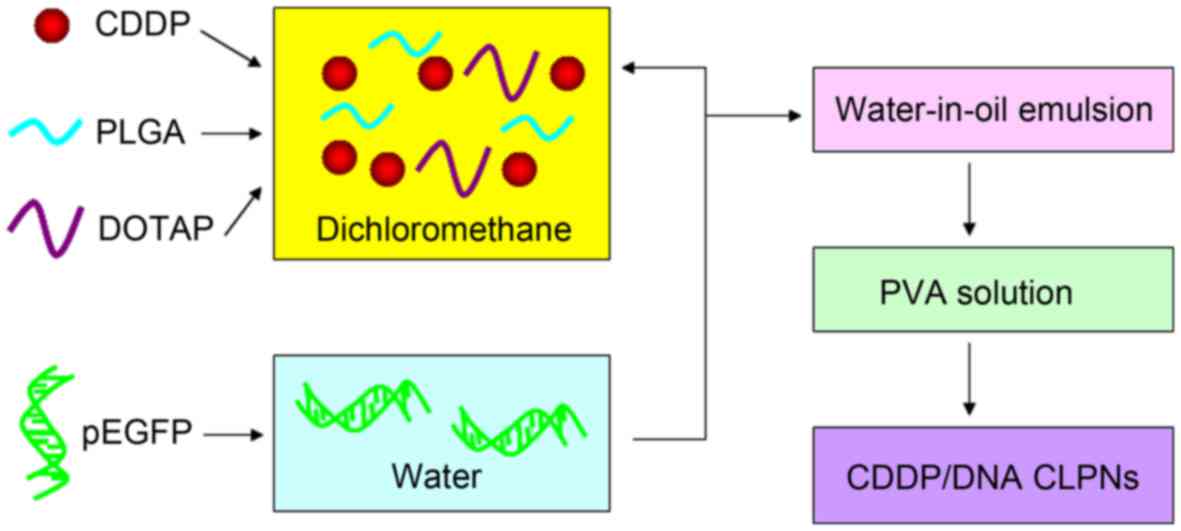
CDDP/DNA CLPNs were prepared by the modified double
emulsion solvent evaporation method with self-assembly (Fig. 1) (18). Briefly, CDDP (0.1 g), PLGA (0.3 g),
and DOTAP were dissolved in dichloromethane (10 ml) to form the
organic phase. pEGFP (0.1 g) was dissolved in an aqueous solution
(1 ml). The water-in-oil emulsion was formed by the addition of an
aqueous phase into an organic phase with sonication. The primary
emulsion was transferred to 1% w/v PVA aqueous solution and
sonicated in an ice bath. The resultant secondary
(water-in-oil-in-water) emulsion was stirred overnight at room
temperature until the evaporation of dichloromethane was complete.
The final formulated CDDP/DNA CLPNs were washed three times by a
repeating centrifugation step and freeze-dried at −20°C. To
investigate the influence of cationic lipid concentrations on size,
charge, and in vitro performance, five formulation groups of
CDDP/DNA CLPNs were prepared with different concentrations of
cationic lipid (DOTAP) to polymer (PLGA) ratio (5, 10, 15, 20 and
25%), named CDDP/DNA CLPNs 1 (5%), CDDP/DNA CLPNs 2 (10%), CDDP/DNA
CLPNs 3 (15%), CDDP/DNA CLPNs 4 (20%), CDDP/DNA CLPNs 5 (25%).
CDDP CLPNs were prepared by the same method without
the use of pEGFP.
Blank CLPNs were prepared by the same method without
the use of pEGFP and CDDP.
Preparation of CDDP PNPs
CDDP PNPs were prepared by the emulsion solvent
evaporation method (22). Briefly,
CDDP (0.1 g), PLGA (0.3 g) were dissolved in dichloromethane (10
ml) to form the organic phase. The organic phase was added
drop-wise into 1% w/v PVA aqueous solution being stirred at 400 rpm
at room temperature until the evaporation of dichloromethane was
complete. Then the resulting suspension was washed three times by
repeating the centrifugation step and freeze-dried at −20°C to get
the final formulated CDDP PNPs.
Blank PNPs were prepared by the same method without
the use of CDDP.
Preparation of DNA LIPO
For the comparison of the gene transfection efficacy
of CDDP/DNA CLPNs, DNA LIPO complexes were prepared by the
following method (12): DNA and
Lipofectamine® 2000 (1:2 w/w) was mixed for 30 sec using
a vortex mixer. Then, the DNA LIPO was obtained by incubating the
mixture for 30 min and freeze-dried at −20°C.
Characterization
Morphology
The morphology of CDDP/DNA CLPNs, and CDDP PNPs was
examined by transmission electron microscopy (TEM) (JEM-200CX; JEOL
Co., Ltd., Tokyo, Japan) (23).
Samples were stained at room temperature with freshly prepared and
sterile-filtered 1% (w/v) phosphotungstic acid aqueous solution.
The samples were then placed in a carbon-coated copper grid and
air-dried prior to imaging (24).
Size and surface charge
The mean particle diameter, size distribution
(polydispersity index), and surface charge (ζ potential) of
CDDP/DNA CLPNs and other nanoparticles were determined by dynamic
light scattering (DLS) using a Zetasizer Nano ZS (Malvern
Instruments, Malvern, UK) (25).
DNA-binding efficiency (DE)
The DE of CDDP/DNA CLPNs was determined by the
PicoGreen fluorometry method (26).
Briefly, free pEGFP was isolated from the CDDP/DNA CLPNs by
centrifugation at 15,000 rpm and 4°C for 30 min, the supernatants
were collected, and the concentration of pEGFP was assessed by a
fluorescence spectrophotometer (Ex/Em = 480/520 nm) (Hitachi
F-2500; Hitachi High-Technologies Corp., Tokyo, Japan). The amount
of DNA loaded in the CDDP/DNA CLPNs was calculated as:
DE(%)=weight of DNAtotally added–weight
of freeDNA testedweight of DNA totally added×100 (1)
CDDP encapsulation efficiency (CE)
The CE in the CDDP/DNA CLPNs and CDDP PNPs was
determined by inductively coupled plasma-optical emission
spectrometry (ICP-OES) (27). The
detection was carried out at λ=265.9 nm. The amount of CDDP was
calculated as:
DE(%)=weight of CDDPtotally added– weight
of non-encapsulated CDDPweight of CDDP totally added×100 (2)
Plasma stability
The plasma stability of the CDDP/DNA CLPNs and other
nanoparticles was determined in the presence of 50% plasma (v/v)
(28,29). The formulations were incubated in
phosphate-buffered saline (PBS) solution containing 50% FBS at 37°C
for 24 h, separately. At scheduled times (0, 2, 4, 8, 12 and 24 h),
1 ml of each sample was diluted with 2 ml THF and the mixture was
bath-sonicated for 5 min, followed by centrifugation at 10,000 rpm
for 5 min. The variation trends of the size, CE, and DE were
calculated by the method indicated above.
Cells
Human RMS cell line, RD-4 cells, were obtained from
the American Type Culture Collection (Manassas, VA, USA) and
cultured at 37°C and 5% CO2 in DMEM supplemented with
10% FBS and 1% penicillin (5,000 U/ml)-streptomycin (5,000
U/ml).
In vitro cytotoxicity
The in vitro cytotoxicity of CDDP/DNA CLPNs
and other nanoparticles was evaluated on RD-4 cells by MTT assay
(30). Cells cultured in the above
section were seeded in a 96-well plate at a seeding density of
104 cells/well. The culture medium was then replaced
with various doses of CDDP/DNA CLPNs and other nanoparticles.
Culture medium was used as a blank control. After 48 h of
incubation, the media were replaced with 90 µl of free-serum medium
and 10 µl of MTT solution (5 mg/ml in sterile PBS). After a 4-h
incubation at 37°C, the MTT solution in the wells was replaced with
100 µl dimethylsulfoxide (DMSO). The absorption at 570 nm (OD570)
was measured on a spectrophotometer. The cell viability was
converted and expressed as the percentage of the control, and the
IC50 values were calculated accordingly.
In vitro gene transfection
The in vitro transfection efficacy of
CDDP/DNA CLPNs and DNA LIPO was evaluated on RD-4 cells, using CDDP
CLPNs and blank CLPNs as control (24). Cells cultured in the above section
were seeded in a 96-well plate at a seeding density of
104 cells/well, 24 h prior to transfection. The media
were replaced with 200 µl serum-free media containing CDDP/DNA
CLPNs and other nanoparticles at 37°C. Naked DNA solution was used
as a negative control. DNA LIPO was used as a positive control. The
original incubation medium was replaced with 1 ml of complete
medium after incubation for 4 h at 37°C in a 5% CO2
incubator, and then cells were incubated sequentially until 48 h
post-transfection.
Flow cytometry was carried out for the quantitation
of the cells which were successfully transfected. At the end of the
incubation, cells were washed once with 1 ml of PBS and were
detached with trypsin/EDTA. Then the cells were centrifuged at
1,500 rpm, at 4°C for 5 min, the supernatant was discarded, and the
cells were washed once with 1 ml of PBS, centrifuged again (1,500
rpm, 4°C for 5 min), the supernatant was discarded, and the cells
were re-suspended in 300 µl of PBS and directly introduced to a BD
FACSCalibur flow cytometer (Becton, Dickinson and Co., Franklin
Lakes, NJ, USA).
Animals
BALB/c nude mice (5–6 week-old, 18–22 g) were
purchased from the Shanghai Slack Laboratory Animal Co., Ltd.
(Shanghai, China) and were maintained under specific pathogen-free
conditions. Ethics approval was received from the Medical Ethics
Committee of Kunming Medical University (no. KMMU20160316-1).
In vivo antitumor efficacy
The in vivo antitumor efficacy of CDDP/DNA
CLPNs and other nanoparticles was investigated in RD-4
tumor-bearing BALB/c nude mouse models, which were developed by
injection of RD-4 cells in the right armpit of BALB/c mice
(31). When tumor volume reached
~50 mm3, transplanted mice were randomly divided into
seven groups (8 mice/group) separately. Formulations for the seven
groups were as follows: group 1, 0.9% saline (control group); group
2, CDDP solution (8 mg/kg); group 3, blank PNPs; group 4, CDDP PNPs
(4 mg/kg); group 5, blank CLPNs; group 6, CDDP CLPNs (4 mg/kg);
group 7, CDDP/DNA CLPNs (4 mg/kg). The mice of each group were
given the above formulations by tail vein injection once every 3
days. Three weeks later, all the mice were sacrificed by cervical
dislocation and the tumor tissue samples were taken out. Tumor
volume of each mouse was measured with a digital caliper every 3
days, and was calculated according to the equation:
V(mm3)=(major axis)×(monor
axis)22 (3)
The antitumor efficacy of each formulation was
evaluated by tumor inhibition rate (TIR), which was calculated by
measuring the tumor weight (TW) using the following formula:
TIR(%)= TW ofcontrol group– TW oftreated
groupTW of control group (4)
In vivo gene delivery
The RD-4 tumor-bearing BALB/c nude mouse model was
designed by the method indicated in the above section. Then, six
groups of tumor-bearing mice received a 300-µl intravenous
injection: group 1, 0.9% saline (control group); group 2, naked DNA
solution; group 3, DNA LIPO; group 4, blank CLPNs; group 5, CDDP
CLPNs; group 6, CDDP/DNA CLPNs, respectively. After 48 h, mice were
sacrificed by cervical dislocation, and their tumor tissues were
taken and washed with cold saline twice. Tissues were homogenized
in lysis buffer (0.05% Triton X-100, 2 mM EDTA, 0.1 M Tris-HCl, pH
7.8). After several cycles of freezing and thawing, the homogenates
were centrifuged at 10,000 rpm for 5 min to obtain the cells. Cells
were cultured by the method of ‘In vitro cytotoxicity’
section.
For qualitative analysis, fluorescent cells were
observed using an inverted fluorescence microscope (Olympus ZX71;
Olympus Corp., Tokyo, Japan) and the picture was captured. The
transfection efficiency of the fluorescent cells was quantified
using the flow cytometry method described in the ‘In vitro
transfection’ section.
Statistical analysis
The data are presented as means ± standard deviation
(SD). Statistical analysis was performed by a one-tailed Student's
t-test in Excel. Statistical significance was established at the 5%
level (P<0.05) for all statistical analyses.
Results
Optimization of CDDP/DNA CLPNs
In order to optimize cationic lipid (DOTAP) to
polymer (PLGA) ratio, CDDP/DNA CLPNs with different concentrations
of lipids were prepared using different ratios of cationic lipid to
polymer: 5, 10, 15, 20 and 25%. The influence of the lipid to the
polymer ratio was determined in terms of particle size,
polydispersity index, surface charge, DE and CE (Table I).
 | Table I.Optimization and characterization of
nanoparticles. |
Table I.
Optimization and characterization of
nanoparticles.
| Nanoparticles | Particle diameter
(nm) | Polydispersity
index | Surface charge
(mV) | DE (%) | CE (%) |
|---|
| DNA LIPO | 139.4±4.8 | 0.10±0.015 | +43.1±4.7 | 91.5±3.9 | N/A |
| Blank PNPs | 78.3±2.9 | 0.08±0.009 | −26.3±2.8 | N/A | N/A |
| CDDP PNPs | 82.1±3.2 | 0.09±0.010 | −28.9±3.2 | N/A | 83.6±2.4 |
| Blank CLPNs | 118.3±3.8 | 0.12±0.013 | +41.6±2.5 | N/A | N/A |
| CDDP CLPNs | 117.6±4.4 | 0.16±0.012 | +39.8±3.3 | N/A | 85.3±2.8 |
| CDDP/DNA CLPNs
1 | 335.2±36.8 | 0.67±0.094 | +6.6±1.4 | 35.7±7.4 | 17.5±5.3 |
| CDDP/DNA CLPNs
2 | 243.1±21.9 | 0.36±0.062 | +12.8±2.7 | 53.8±8.9 | 63.8±6.5 |
| CDDP/DNA CLPNs
3 | 137.6±3.3 | 0.10±0.008 | +25.4±3.5 | 90.7±3.1 | 82.1±2.7 |
| CDDP/DNA CLPNs
4 | 133.6±3.7 | 0.12±0.010 | +31.1±4.4 | 88.6±4.2 | 62.5±4.9 |
| CDDP/DNA CLPNs
5 | 141.2±4.5 | 0.13±0.014 | +36.9±3.8 | 89.3±6.1 | 51.6±8.3 |
It was observed that the particle diameters of
CDDP/DNA CLPNs 1, CDDP/DNA CLPNs 2, CDDP/DNA CLPNs 3, CDDP/DNA
CLPNs 4, and CDDP/DNA CLPNs 5 were 335.2, 243.1, 137.6, 133.6 and
141.2 nm, respectively. The polydispersity index of CDDP/DNA CLPNs
1 (0.67) and CDDP/DNA CLPNs 2 (0.36) were significantly higher than
other formulations, suggesting that the ratios of 5 and 10% were
not suitable. The obviously lower DE and CE of CDDP/DNA CLPNs 1 and
CDDP/DNA CLPNs 2 also confirmed this conclusion. The CE of CDDP/DNA
CLPNs 4 (62.5) and CDDP/DNA CLPNs 5 (51.6) were significantly lower
than CDDP/DNA CLPNs 3 (82.1) (P<0.05). So the optimized lipid
(DOTAP) to polymer (PLGA) ratio was 15% (CDDP/DNA CLPNs 3).
Characterization
Morphology
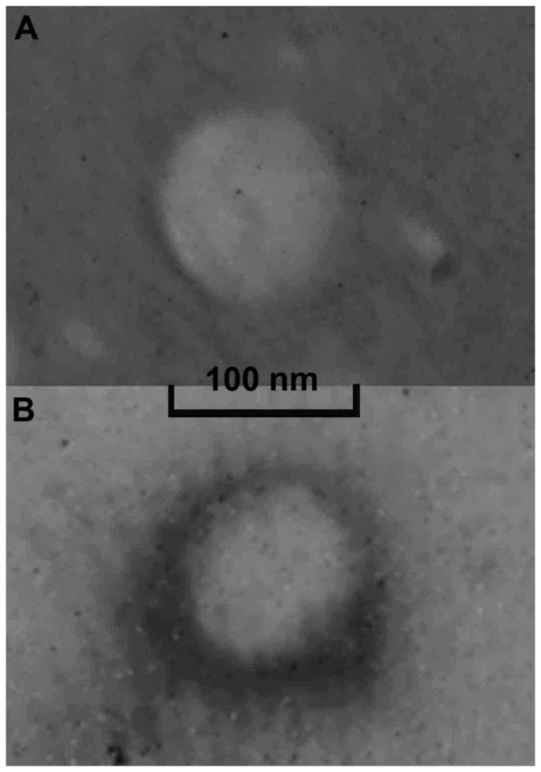
The overall morphology of the CDDP/DNA CLPNs was a
clear core-shell type spherical nanoparticle (Fig. 2A). The core-shell structure was
clearly visible with a white core and the shell is grey. In
comparison, the morphology of CDDP PNPs was white spherical
particle without outer layer (Fig.
2B). The size scales in the images show that the size of
CDDP/DNA CLPNs was slightly larger than 100 nm, while the diameter
of CDDP PNPs was smaller than 100 nm.
Size, surface charge, DE and CE
The size of CDDP/DNA CLPNs, CDDP CLPNs, blank CLPNs,
CDDP PNPs, and blank PNPs was 137.6±3.3, 117.6±4.4, 118.3±3.8,
82.1±3.2 and 78.3±2.9 nm, respectively. The size of CDDP CLPNs and
blank CLPNs, CDDP PNP and blank PNPs had no obvious difference
(P>0.05). The size of CDDP CLPNs was larger than CDDP PNPs
(P<0.05). The size of CDDP/DNA CLPNs was the largest.
The surface charge of CDDP/DNA CLPNs, CDDP CLPNs,
and CDDP PNPs was +25.4±3.5, +39.8±3.3 and −28.9±3.2 mV. CDDP PNPs
have negative ζ potential. In contrast, CLPNs have positive
charges. The surface charge of CDDP/DNA CLPNs was lower than CDDP
CLPNs.
The DE of CDDP/DNA CLPNs and DNA LIPO was 90.7±3.1
and 91.5±3.9%, respectively. The CE of CDDP/DNA CLPNs, CDDP CLPNs,
and CDDP PNPs was 82.1±2.7, 85.3±2.8 and 83.6±2.4, respectively.
These results illustrated that the loading capacity of CDDP and
binding ability of DNA was outstanding for all the drug-loaded
nanoparticles prepared in this study.
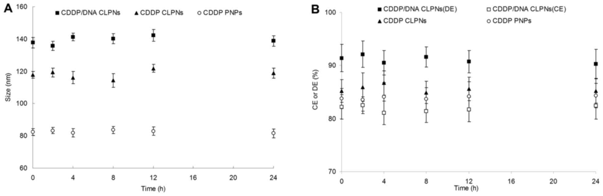
Plasma stability
Fig. 3 illustrated
the changes in size, drug and gene-loading efficiency of different
particles in the presence of serum. The particle diameter, CE or DE
had no significant variation up to 24 h. Due to these results,
CDDP/DNA CLPNs, and CDDP PNPs were considered very stable after
intravenous administration.
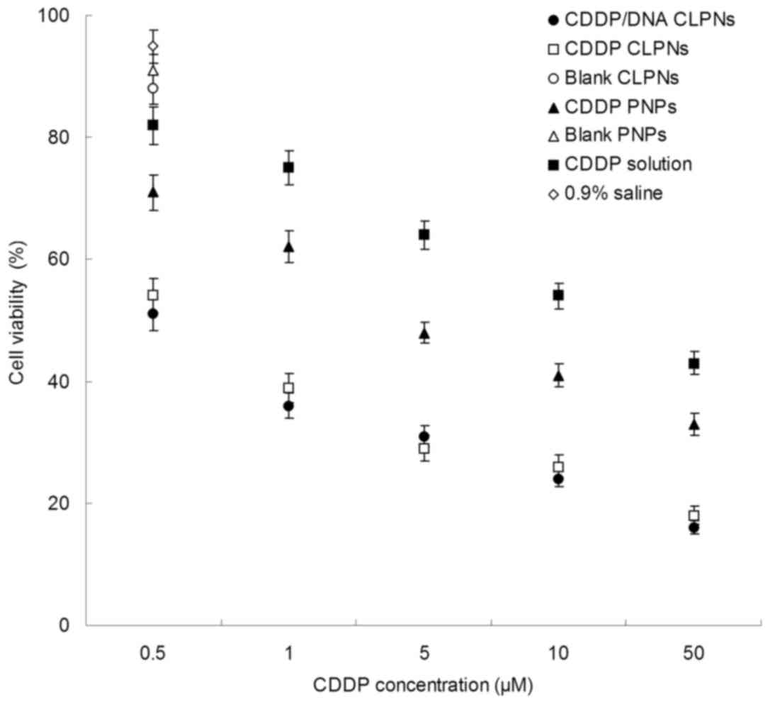
In vitro cytotoxicity
As shown in Fig. 4,
treatment with various concentrations of CDDP-loaded particles for
48 h caused various viability reductions in RMS cell lines.
IC50 of CDDP/DNA CLPNs (0.54±0.06 µM) and CDDP CLPNs
(0.58±0.07 µM) showed no significant difference in RD-4 cells
(Table II). The cytotoxicity of
CDDP/DNA CLPNs was significantly higher (IC50 was 8
times dose advantage) than CDDP PNPs (4.12±0.36 µM).
IC50 of CDDP solution was 20.36±3.85 µM.
| Table II.IC50 values of
samples. |
Table II.
IC50 values of
samples.
| Samples | CDDP/DNA CLPNs | CDDP CLPNs | CDDP PNPs | CDDP solution |
|---|
| IC50 of
CDDP (µM) | 0.54±0.06 | 0.58±0.07 | 4.12±0.36 | 20.36±3.85 |
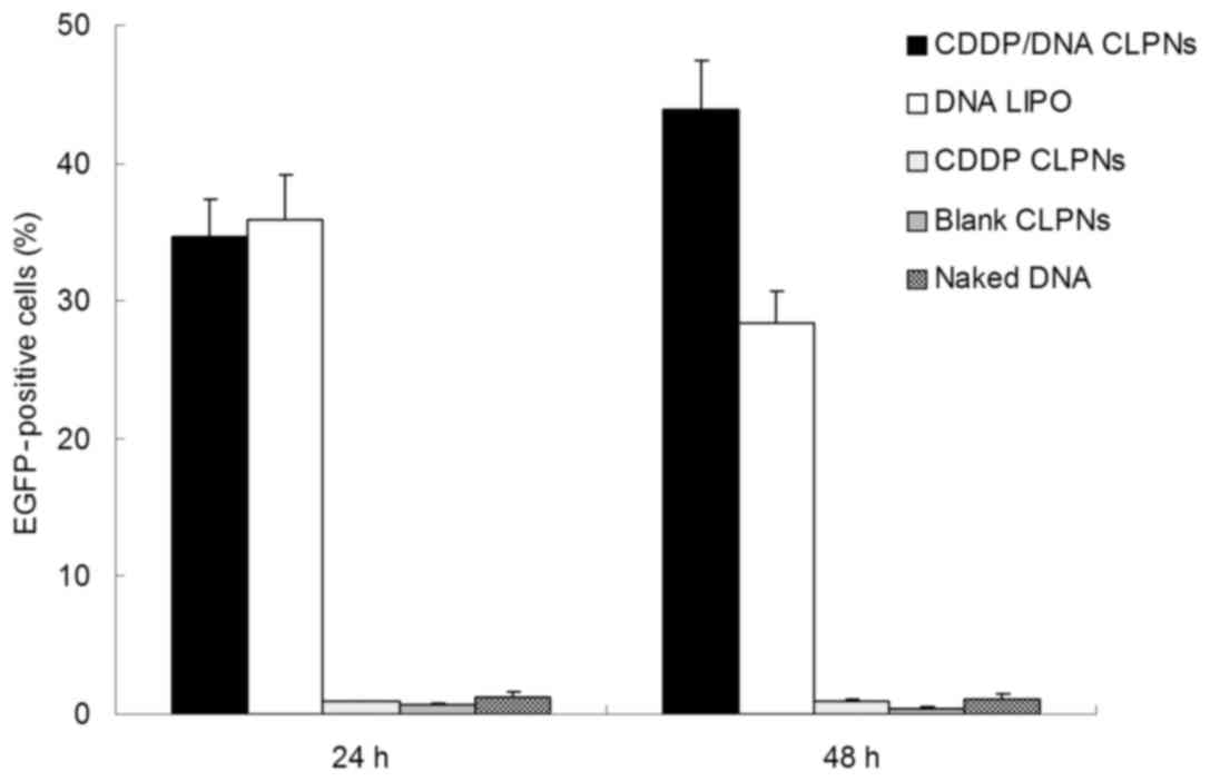
In vitro gene transfection
Fig. 5 exhibits the
in vitro transfection efficacy of CDDP/DNA CLPNs and DNA
LIPO evaluated on RD-4 cells. Gene transfection efficiency was
measured by analyzing the expression of the EGFP by flow cytometry.
When delivering genes into RD-4 cells for 24 h, CDDP/DNA CLPNs
showed the same transfection efficiency as DNA LIPO (P>0.05).
However, after 48-h post-transfection, the CDDP/DNA CLPNs was more
efficient than the DNA LIPO (P<0.05). Naked DNA solution group
got no obvious gene expression, indicating that DNA cannot
successfully transfect the cancer cells without carriers. CDDP CLPN
and blank CLPN groups showed no gene expression in RD-4 cells,
ruling out the fluorescent cells caused by the CLPNs and CDDP.
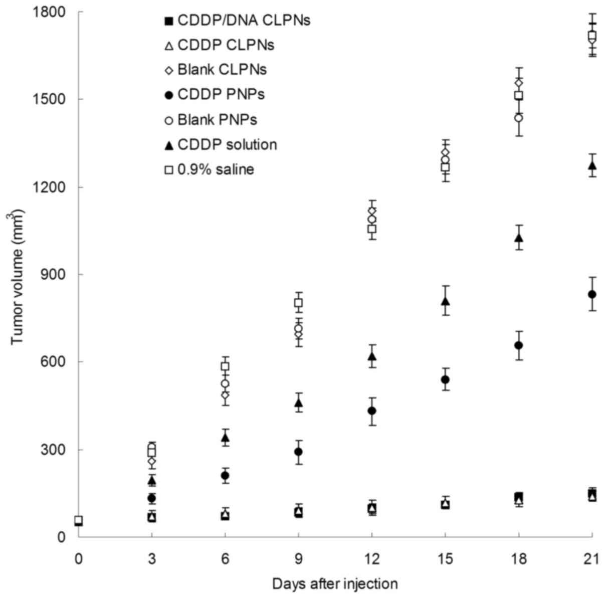
In vivo antitumor
Fig. 6 shows the
in vivo antitumor efficacy of CDDP/DNA CLPNs and other
nanoparticles was investigated in RD-4 tumor-bearing BALB/c nude
mouse models. CDDP/DNA CLPNs, CDDP CLPNs, CDDP PNPs and CDDP
solutions showed significant tumor regression in tumor-bearing
mice, with a reduction in tumor volume. Mice in the other three
groups shared similar tumor-growth pattern. At 21 days of
administration, the TW and TIR of tumor-bearing mice are given in
Table III. CDDP/DNA CLPN and CDDP
CLPN groups exhibited the highest TIR (90 and 91%), followed by
CDDP PNPs (52%) and CDDP solution (25%).
| Table III.TW and TIR of samples. |
Table III.
TW and TIR of samples.
| Samples | CDDP/DNA CLPNs | CDDP CLPNs | CDDP PNPs | CDDP solution | 0.9% Saline |
|---|
| TW (g) | 0.12±0.02 | 0.11±0.02 | 0.61±0.09 | 0.95±0.12 | 1.26±0.23 |
| TIR (%) | 90 | 91 | 52 | 25 | 100 |
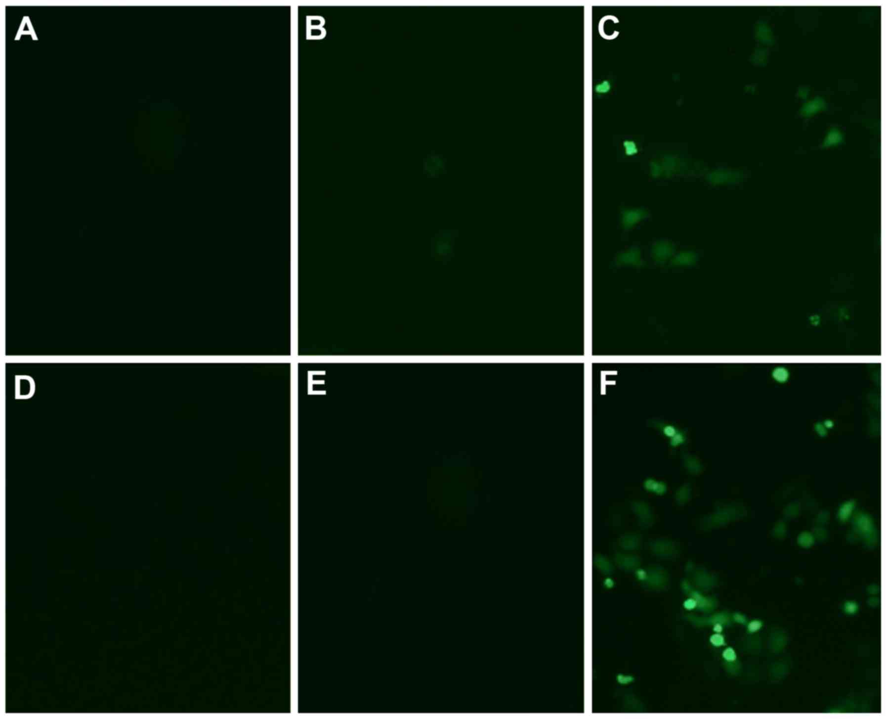
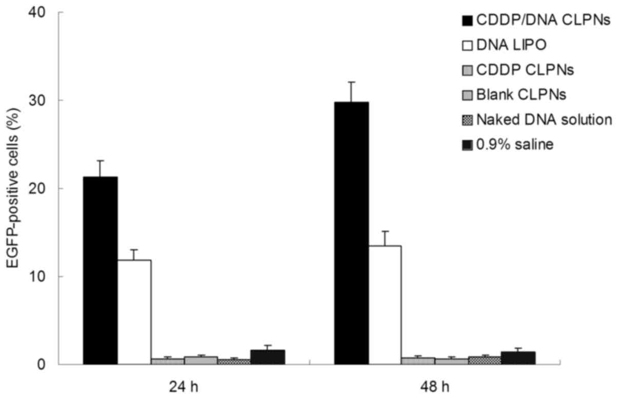
In vivo gene delivery
Fig. 7 illustrates
the images of the fluorescent cells after 48 h of administration.
The highest transfection efficiency appeared in the pictures is
CDDP/DNA CLPNs group. The image of DNA LIPO group came in the
second place. The naked DNA solution group has slightly weaker
fluorescence. Other groups do not have any fluorescence according
to the images. The transfection efficiency of the fluorescent cells
was quantified using a flow cytometry method (Fig. 8). Substantially higher transfection
efficiency was observed in CDDP/DNA CLPNs than DNA LIPO group at
both 24- and 48-h post-administration (P<0.05). One explanation
may be that as an in vitro transfection vector,
Lipofectamine® 2000 transfection reagent was not so
stable and might have some toxicity in vivo. The highest
in vivo gene transfection efficiency of CDDP/DNA CLPNs was
detected after 48 h, showing the stability and controlled release
of the CLPNs.
Discussion
The aim of the present study was to develop
nanoparticles for co-delivery of CDDP and DNA. The influence of
cationic lipid concentration on the characteristics of CLPNs was
evaluated. In order to optimize cationic lipid (DOTAP) to polymer
(PLGA) ratio, CDDP/DNA CLPNs with different concentrations of
lipids were prepared using different ratios of cationic lipid to
polymer: 5, 10, 15, 20 and 25%. The influence of the lipid to
polymer ratio was determined in terms of particle size,
polydispersity index, surface charge, DE and CE (Table I). It was observed that the particle
diameters of CDDP/DNA CLPNs 1, CDDP/DNA CLPNs 2, CDDP/DNA CLPNs 3,
CDDP/DNA CLPNs 4, and CDDP/DNA CLPNs 5 were 335.2, 243.1, 137.6,
133.6 and 141.2 nm, respectively. It was also found that there was
a decrease in particle size as the lipid concentrations increased.
The concentration of cationic lipids could play a significant role
in controlling the size of CDDP/DNA CLPNs, possibly reducing the
coalescence of particles (32). The
polydispersity index of CDDP/DNA CLPNs 1 (0.67) and CDDP/DNA CLPNs
2 (0.36) was significantly higher than other formulations,
suggesting that the ratios of 5 and 10% were not suitable. The
obviously lower DE and CE of CDDP/DNA CLPNs 1 and CDDP/DNA CLPNs 2
also confirmed this conclusion. The CE of CDDP/DNA CLPNs 4 (62.5)
and CDDP/DNA CLPNs 5 (51.6) were significantly lower than CDDP/DNA
CLPNs 3 (82.1) (P<0.05). So the optimized lipid (DOTAP) to
polymer (PLGA) ratio was 15%.
The core-shell structure of CDDP/DNA CLPNs was
clearly visible with a white core and the shell is grey (Fig. 2). In comparison, the morphology of
CDDP PNPs was white spherical particles without outer layer. The
TEM image illustrates the self-assembly process of the
lipid-polymer core-shell structure by forming a lipid layer on the
surface of the inner polymer core (33,34).
The size scales in the images show that the size of CDDP/DNA CLPNs
was slightly larger than 100 nm, while the diameter of CDDP PNPs
was smaller than 100 nm.
The size of CDDP CLPNs and blank CLPNs, CDDP PNP and
blank PNPs had no obvious difference (P>0.05). This means that
the loading of CDDP did not change the size of the particles. The
size of CDDP CLPNs was larger than CDDP PNPs (P<0.05). This
could be explained by the lipid shell on the polymer core enlarged
the size of the particles. The size of CDDP/DNA CLPNs was the
largest; indicating that the DNA could attribute to the increase of
the particle diameter. Surface charge is an important indication of
the stability of a colloidal system in a particular medium
(35). The surface charge of
CDDP/DNA CLPNs, CDDP CLPNs, and CDDP PNPs was +25.4±3.5, +39.8±3.3
and −28.9±3.2 mV. For CDDP PNPs, negatively charged PLGA gave the
PNPs negative ζ potential. In contrast, inclusion of cationic
lipids changed the surface charges of the particles; CLPNs have
positive charges. The surface charge of CDDP/DNA CLPNs was lower
than CDDP CLPNs, suggesting that the combination of anionic DNA
decreased the charge of the CLPNs. The DE of CDDP/DNA CLPNs and DNA
LIPO was ~90%. The CE of CDDP/DNA CLPNs, CDDP CLPNs, and CDDP PNPs
was >80%. These results illustrated that the loading capacity of
CDDP and binding ability of DNA was outstanding for all the
drug-loaded nanoparticles prepared in this study.
The plasma stability of the CDDP/DNA CLPNs and other
nanoparticles was determined in the presence of plasma. The
particle diameter, CE or DE had no significant variation up to 24 h
(Fig. 3). This stability would be
advantageous when the complexes are administered in vivo.
One of the major concerns for cationic non-viral vectors is the
disassociation of DNA from the vectors due to electrostatic
interaction between the vectors and the plasma composition such as
proteins (29). The dissociated DNA
would be quickly degraded by enzymolysis. Thus, the stability of
the cationic non-viral vectors in human plasma is one of the most
essential properties. Also the stability in the plasma is required
for the drug-loaded nanoparticles. These would increase the
stability of the loaded drug and DNA during the circulation after
intravenous administration and enhance the targeting proportion of
the vector to the target organ, and thus would be favorable to
improve the therapeutic effects.
In vitro cytotoxicity of CDDP/DNA CLPNs was
evaluated against RD-4 cells, and was compared with the
cytotoxicity induced by free-CDDP solution, blank nanoparticles,
and 0.9% saline solution. As showed in Fig. 4, treatment with various
concentrations of CDDP-loaded particles for 48 h caused various
viability reductions in RMS cell lines. IC50 of CDDP/DNA
CLPNs (0.54±0.06 µM) and CDDP CLPNs (0.58±0.07 µM) showed no
significant difference in RD-4 cells, indicating that DNA does not
affect the ability of the CDDP loaded in the CLPNs (Table I) (P>0.05). The cytotoxicity of
CDDP/DNA CLPNs was significantly higher (IC50 was 8
times dose advantage) than CDDP PNPs (4.12±0.36 µM). This result
may suggest that the in vitro tumor cell inhibition of
drug-loaded CLPNs was more efficient than drug-loaded PNPs
(P<0.05). IC50 of CDDP solution was 20.36±3.85 µM.
These results indicate that CDDP/DNA CLPNs exhibited superior
cytotoxicity to RD-4 cells as evidenced by its significantly
decreased CDDP IC50 values.
In vitro transfection efficacy of CDDP/DNA
CLPNs and DNA LIPO was evaluated on RD-4 cells, using CDDP CLPNs
and blank CLPNs as control. Gene transfection efficiency was
measured by analyzing the expression of the EGFP by flow cytometry.
When delivering genes into RD-4 cells for 24 h, CDDP/DNA CLPNs
showed the same transfection efficiency as DNA LIPO (P>0.05).
However, after 48-h post-transfection, the CDDP/DNA CLPNs were more
efficient than the DNA LIPO (P<0.05). This could be explained by
the polymer core and the lipid shell controlling the release of the
DNA thus gaing the long-lasting efficiency (36). Naked DNA solution group had no
obvious gene expression; indicating that DNA cannot successfully
transfect the cancer cells without carriers. CDDP CLPNs and blank
CLPN groups showed no gene expression in RD-4 cells, ruling out the
fluorescent cells caused by the CLPNs and CDDP. The results
illustrated that CLPNs are able to condense DNA efficiently,
deliver DNA into the cells by ionic interactions with the cell
membrane and release DNA into the cytoplasm from endocytic vesicles
(37). Better transfection
efficiency than commercial Lipofectamine 2000 and DNA complex
indicates that the CLPNs can be a good transfection reagent
(38).
In vivo antitumor efficacy of CDDP/DNA CLPNs
and other nanoparticles was investigated in RD-4 tumor-bearing
BALB/c nude mouse models (Fig. 5).
CDDP/DNA CLPNs, CDDP CLPNs, CDDP PNPs and CDDP solution showed
significant tumor regression in tumor-bearing mice, with a
reduction of tumor volume. Mice in the other three groups shared
similar tumor-growth pattern, suggesting that blank carriers were
not capable of inhibiting tumor growth (19). At 21 days of administration, the TW
and TIR of tumor-bearing mice are shown in Table III. CDDP/DNA CLPN and CDDP CLPN
groups exhibited the highest TIR (90 and 91%), followed by CDDP
PNPs (52%) and CDDP solution (25%). Fig. 7 illustrates the images of the
fluorescent cells. The highest transfection efficiency appearing in
the images is in CDDP/DNA CLPN group. The image of DNA LIPO group
comes in the second place. The naked DNA solution group has
slightly weaker fluorescence. Other groups have no fluorescence
according to the images. The transfection efficiency of the
fluorescent cells was quantified using a flow cytometry method
(Fig. 8). Significantly higher
transfection efficiency was observed in CDDP/DNA CLPNs than DNA
LIPO group at both 24- and 48-h post-administration (P<0.05).
One explanation may be that as an in vitro transfection
vector, Lipofectamine® 2000 Transfection Reagent was not
so stable and might have some toxicity in vivo. The highest
in vivo gene transfection efficiency of CDDP/DNA CLPNs was
detected after 48 h, showing the stability and controlled release
of the CLPNs. These results could be explained as the lipid shell
of CLPNs has high affinity to the lipid-structured cell surface,
the cationic surface charge could absorb onto the negatively
charged cell surface, promote the fusion of the nanocarriers to the
cell membrane and deliver more drug and gene into the tumor cells
(39). The structure of CLPNs may
delay the drug/gene release more than other vectors, bring about
the long-lasting drug/gene delivery effect in tumor tissues
(40). Outstanding delivery ability
of CLPNs for both CDDP and DNA could combine the therapeutic
efficiency of both drug and gene for the treatment of pediatric
RMS.
In the present study, we described a simple and
efficient method for the fabrication of CLPNs for co-delivery of
CDDP and DNA for the therapy of childhood head and neck cancers.
The results illustrated that the concentration of the cationic
lipid has influence on the characteristics of CLPNs. In
vitro anticancer effect, in vitro transfection
efficiency, in vivo antitumor and gene delivery efficacy of
CDDP/DNA CLPNs have advantages over other formulations tested.
Excellent ability of CLPNs for co-delivery of CDDP and DNA could
combine the therapeutic efficiency of both drug and gene for the
treatment of pediatric RMS.
References
|
1
|
Ward E, DeSantis C, Robbins A, Kohler B
and Jemal A: Childhood and adolescent cancer statistics, 2014. CA
Cancer J Clin. 64:83–103. 2014. View Article : Google Scholar : PubMed/NCBI
|
|
2
|
Choi DK and Schmidt ML: Chemotherapy in
children with head and neck cancers: Perspectives and review of
current therapies. Oral Maxillofacial Surg Clin N Am. 28:127–138.
2016. View Article : Google Scholar
|
|
3
|
Marcus KJ and Tishler RB: Head and neck
carcinomas across the age spectrum: Epidemiology, therapy, and late
effects. Semin Radiat Oncol. 20:52–57. 2010. View Article : Google Scholar : PubMed/NCBI
|
|
4
|
Wang Y, Lipari P, Wang X, Hailey J, Liang
L, Ramos R, Liu M, Pachter JA, Bishop WR and Wang Y: A fully human
insulin-like growth factor-I receptor antibody SCH 717454
(Robatumumab) has antitumor activity as a single agent and in
combination with cytotoxics in pediatric tumor xenografts. Mol
Cancer Ther. 9:410–418. 2010. View Article : Google Scholar : PubMed/NCBI
|
|
5
|
Morrison R, Gardiner C, Evidente A, Kiss R
and Townley H: Incorporation of ophiobolin a into novel
chemoembolization particles for cancer cell treatment. Pharm Res.
31:2904–2917. 2014. View Article : Google Scholar : PubMed/NCBI
|
|
6
|
Xie M, Zhang H, Xu Y, Liu T, Chen S, Wang
J and Zhang T: Expression of folate receptors in nasopharyngeal and
laryngeal carcinoma and folate receptor-mediated endocytosis by
molecular targeted nanomedicine. Int J Nanomed. 8:2443–2451. 2013.
View Article : Google Scholar
|
|
7
|
Chen Y, Xu G, Zheng Y, Yan M, Li Z, Zhou
Y, Mei L and Li X: Nanoformulation of D-α-tocopheryl polyethylene
glycol 1000 succinate-b-poly(ε-caprolactone-ran-glycolide) diblock
copolymer for siRNA targeting HIF-1α for nasopharyngeal carcinoma
therapy. Int J Nanomed. 10:1375–1386. 2015.
|
|
8
|
Koganti S, Jagani HV, Palanimuthu VR,
Mathew JA, Rao MC and Rao JV: In vitro and in vivo evaluation of
the efficacy of nanoformulation of siRNA as an adjuvant to improve
the anticancer potential of cisplatin. Exp Mol Pathol. 94:137–147.
2013. View Article : Google Scholar : PubMed/NCBI
|
|
9
|
Oberoi HS, Nukolova NV, Kabanov AV and
Bronich TK: Nanocarriers for delivery of platinum anticancer drugs.
Adv Drug Deliv Rev. 65:1667–1685. 2013. View Article : Google Scholar : PubMed/NCBI
|
|
10
|
Kojima H, Iida M, Yaguchi Y, Suzuki R,
Hayashi N, Moriyama H and Manome Y: Enhancement of cisplatin
sensitivity in squamous cell carcinoma of the head and neck
transfected with a survivin antisense gene. Arch Otolaryngol Head
Neck Surg. 132:682–685. 2006. View Article : Google Scholar : PubMed/NCBI
|
|
11
|
Jiang M, Liu Z, Xiang Y, Ma H, Liu S, Liu
Y and Zheng D: Synergistic antitumor effect of AAV-mediated TRAIL
expression combined with cisplatin on head and neck squamous cell
carcinoma. BMC Cancer. 11:542011. View Article : Google Scholar : PubMed/NCBI
|
|
12
|
Shao Z, Shao J, Tan B, Guan S, Liu Z, Zhao
Z, He F and Zhao J: Targeted lung cancer therapy: Preparation and
optimization of transferrin-decorated nanostructured lipid carriers
as novel nanomedicine for co-delivery of anticancer drugs and DNA.
Int J Nanomed. 10:1223–1233. 2015. View Article : Google Scholar
|
|
13
|
Teo PY, Cheng W, Hedrick JL and Yang YY:
Co-delivery of drugs and plasmid DNA for cancer therapy. Adv Drug
Deliv Rev. 98:41–63. 2016. View Article : Google Scholar : PubMed/NCBI
|
|
14
|
Yang T, Zhao P, Rong Z, Li B, Xue H, You
J, He C, Li W, He X, Lee RJ, et al: Anti-tumor efficiency of
lipid-coated cisplatin nanoparticles co-loaded with microRNA-375.
Theranostics. 6:142–154. 2016. View Article : Google Scholar : PubMed/NCBI
|
|
15
|
Wasungu L and Hoekstra D: Cationic lipids,
lipoplexes and intracellular delivery of genes. J Control Release.
116:255–264. 2006. View Article : Google Scholar : PubMed/NCBI
|
|
16
|
Zhang L, Chan JM, Gu FX, Rhee JW, Wang AZ,
Radovic-Moreno AF, Alexis F, Langer R and Farokhzad OC:
Self-assembled lipid - polymer hybrid nanoparticles: A robust drug
delivery platform. ACS Nano. 2:1696–1702. 2008. View Article : Google Scholar : PubMed/NCBI
|
|
17
|
Yang XZ, Dou S, Wang YC, Long HY, Xiong
MH, Mao CQ, Yao YD and Wang J: Single-step assembly of cationic
lipid-polymer hybrid nanoparticles for systemic delivery of siRNA.
ACS Nano. 6:4955–4965. 2012. View Article : Google Scholar : PubMed/NCBI
|
|
18
|
Bose RJ, Arai Y, Ahn JC, Park H and Lee
SH: Influence of cationic lipid concentration on properties of
lipid-polymer hybrid nanospheres for gene delivery. Int J Nanomed.
10:5367–5382. 2015.
|
|
19
|
He C, Lu J and Lin W: Hybrid nanoparticles
for combination therapy of cancer. J Control Release. 219:224–236.
2015. View Article : Google Scholar : PubMed/NCBI
|
|
20
|
Hadinoto K, Sundaresan A and Cheow WS:
Lipid-polymer hybrid nanoparticles as a new generation therapeutic
delivery platform: A review. Eur J Pharm Biopharm. 85(3 Pt A):
427–443. 2013. View Article : Google Scholar : PubMed/NCBI
|
|
21
|
Guo S, Wang Y, Miao L, Xu Z, Lin CM, Zhang
Y and Huang L: Lipid-coated Cisplatin nanoparticles induce
neighboring effect and exhibit enhanced anticancer efficacy. ACS
Nano. 7:9896–9904. 2013. View Article : Google Scholar : PubMed/NCBI
|
|
22
|
Han L, Ren Y, Long L, Zhong Y, Shen C, Pu
P, Yuan X and Kang C: Inhibition of C6 glioma in vivo by
combination chemotherapy of implantation of polymer wafer and
intracarotid perfusion of transferrin-decorated nanoparticles.
Oncol Rep. 27:121–128. 2012.PubMed/NCBI
|
|
23
|
Nakata H, Miyazaki T, Iwasaki T, Nakamura
A, Kidani T, Sakayama K, Masumoto J and Miura H: Development of
tumor-specific caffeine-potentiated chemotherapy using a novel drug
delivery system with Span 80 nano-vesicles. Oncol Rep.
33:1593–1598. 2015.PubMed/NCBI
|
|
24
|
Yu W, Liu C, Ye J, Zou W, Zhang N and Xu
W: Novel cationic SLN containing a synthesized single-tailed lipid
as a modifier for gene delivery. Nanotechnology. 20:2151022009.
View Article : Google Scholar : PubMed/NCBI
|
|
25
|
Wang RH, Cao HM, Tian ZJ, Jin B, Wang Q,
Ma H and Wu J: Efficacy of dual-functional liposomes containing
paclitaxel for treatment of lung cancer. Oncol Rep. 33:783–791.
2015.PubMed/NCBI
|
|
26
|
Yu W, Liu C, Liu Y, Zhang N and Xu W:
Mannan-modified solid lipid nanoparticles for targeted gene
delivery to alveolar macrophages. Pharm Res. 27:1584–1596. 2010.
View Article : Google Scholar : PubMed/NCBI
|
|
27
|
Yang Q, Zhang S, Kang M, Dong R and Zhao
J: Synergistic growth inhibition by sorafenib and cisplatin in
human osteosarcoma cells. Oncol Rep. 33:2537–2544. 2015.PubMed/NCBI
|
|
28
|
Almeida PV, Shahbazi MA, Mäkilä E,
Kaasalainen M, Salonen J, Hirvonen J and Santos HA: Amine-modified
hyaluronic acid-functionalized porous silicon nanoparticles for
targeting breast cancer tumors. Nanoscale. 6:10377–10387. 2014.
View Article : Google Scholar : PubMed/NCBI
|
|
29
|
Ye Y, Zhang X, Zhang T, Wang H and Wu B:
Design and evaluation of injectable niclosamide nanocrystals
prepared by wet media milling technique. Drug Dev Ind Pharm.
41:1416–1424. 2015. View Article : Google Scholar : PubMed/NCBI
|
|
30
|
Gao L, Xu Z, Wang Y, Sun B, Song Z, Yang
B, Liu X, Lin Y, Peng J, Han G, et al: Anticancer effect of SZC017,
a novel derivative of oleanolic acid, on human gastric cancer
cells. Oncol Rep. 35:1101–1108. 2016.PubMed/NCBI
|
|
31
|
Lv X, Liu F, Shang Y and Chen SZ: Honokiol
exhibits enhanced antitumor effects with chloroquine by inducing
cell death and inhibiting autophagy in human non-small cell lung
cancer cells. Oncol Rep. 34:1289–1300. 2015.PubMed/NCBI
|
|
32
|
Mainardes RM and Evangelista RC: PLGA
nanoparticles containing praziquantel: Effect of formulation
variables on size distribution. Int J Pharm. 290:137–144. 2005.
View Article : Google Scholar : PubMed/NCBI
|
|
33
|
Liu P, Sun L, Zhou DS, Zhang P, Wang YH,
Li D, Li QH and Feng RJ: Development of alendronate-conjugated poly
(lactic-co-glycolic acid)-dextran nanoparticles for active
targeting of cisplatin in osteosarcoma. Sci Rep. 5:173872015.
View Article : Google Scholar : PubMed/NCBI
|
|
34
|
Asthana S, Jaiswal AK, Gupta PK, Dube A
and Chourasia MK: Th-1 biased immunomodulation and synergistic
antileishmanial activity of stable cationic lipid-polymer hybrid
nanoparticle: Biodistribution and toxicity assessment of
encapsulated amphotericin B. Eur J Pharm Biopharm. 89:62–73. 2015.
View Article : Google Scholar : PubMed/NCBI
|
|
35
|
Valencia PM, Basto PA, Zhang L, Rhee M,
Langer R, Farokhzad OC and Karnik R: Single-step assembly of
homogenous lipid-polymeric and lipid-quantum dot nanoparticles
enabled by microfluidic rapid mixing. ACS Nano. 4:1671–1679. 2010.
View Article : Google Scholar : PubMed/NCBI
|
|
36
|
Seedat N, Kalhapure RS, Mocktar C, Vepuri
S, Jadhav M, Soliman M and Govender T: Co-encapsulation of
multi-lipids and polymers enhances the performance of vancomycin in
lipid-polymer hybrid nanoparticles: In vitro and in silico studies.
Mater Sci Eng C. 61:616–630. 2016. View Article : Google Scholar
|
|
37
|
D'Mello S, Salem AK, Hong L and Elangovan
S: Characterization and evaluation of the efficacy of cationic
complex mediated plasmid DNA delivery in human embryonic palatal
mesenchyme cells. J Tissue Eng Regen Med. 10:927–937. 2016.
View Article : Google Scholar : PubMed/NCBI
|
|
38
|
Dong W, Jin GH, Li SF, Sun QM, Ma DY and
Hua ZC: Cross-linked polyethylenimine as potential DNA vector for
gene delivery with high efficiency and low cytotoxicity. Acta
Biochim Biophys Sin (Shanghai). 38:780–787. 2006. View Article : Google Scholar : PubMed/NCBI
|
|
39
|
Khurana B, Goyal AK, Budhiraja A, Aora D
and Vyas SP: Lipoplexes versus nanoparticles: pDNA/siRNA delivery.
Drug Deliv. 20:57–64. 2013. View Article : Google Scholar : PubMed/NCBI
|
|
40
|
Krishnamurthy S, Vaiyapuri R, Zhang L and
Chan JM: Lipid-coated polymeric nanoparticles for cancer drug
delivery. Biomater Sci. 3:923–936. 2015. View Article : Google Scholar : PubMed/NCBI
|