Introduction
It is well known that sex hormones (SexHs) regulate
the growth and function of the reproductive organs and are
responsible for the development of secondary sex characteristics.
While peptide-based sex hormones, including follicle-stimulating
hormone (FSH), luteinizing hormone (LH), and prolactin (PRL), are
secreted by the pituitary gland, steroid SexHs, such as estrogens,
androgens, and progesterone, are released from the ovaries or
testes. Notably, an important source of SexHs during development is
the placenta (1,2). In parallel, as previously postulated,
maternally-derived SexHs may also affect the developing embryo
(3,4).
Evidence has accumulated which revealed, that as
potent mitogens, SexHs play an important role in the development
and progression of cancers arising from tissues that are sensitive
to SexH stimulation, such as the gonads, uterus, prostate, and
breast (5–8). However, it has been recently
demonstrated that both pituitary- and gonad-derived SexHs also play
a role in the pathogenesis of other malignancies, such as lung
cancer (9), rhabdomyosarcoma
(10) and leukemia (11), and direct migration and adhesion of
cells derived from these malignancies.
It has been previously postulated by us and other
researchers that the most primitive stem cells residing in adult
tissues share several characteristics with primordial germ cells
(PGCs), which are precursors of gametes and most likely precursors
of stem cells in other tissues (12–14).
This tempting hypothesis suggests that in postnatal tissues there
are stem cells endowed with embryonic/epiblast/germline potential.
In fact, in adult tissues stem cells, now known as very small
embryonic-like stem cells (VSELs), have been identified to fulfill
some of these criteria (15,16).
These small cells have been found to be involved in physiological
tissue and organ rejuvenation, but it is hypothesized that in some
situations they also give rise to malignancies (17).
The notion that a population of stem cells involved
in early development resides in adult tissues may corroborate the
150-year-old ‘embryonic rest hypothesis’ of cancer development. In
the middle of the XIX century, two German pathologists, Rudolf
Virchow and Julius Cohnheim, proposed this intriguing hypothesis,
in which cancer may develop from embryonic cell remnants that
remain in the developing organs following embryogenesis (18,19).
This hypothesis was popular among pathologists in the 19th and 20th
centuries but later was largely abandoned. In fact, the morphology
of most primitive tumors often mimics tissues in early development,
and such tumors may express markers that are characteristic of
embryonic, epiblast, and/or germline cells. Recently, we
demonstrated that as postulated the most primitive stem cells
residing in adult tissues, VSELs express functional SexH receptors
(20–25).
To pursue this idea we investigated whether SexHs
play a role in regulating the biology of the murine embryonic ES-D3
cell line as well as the murine P19 teratocarcinoma cell line and
the human embryonal carcinoma NTera2 cell line. The results
revealed that these cells derived in early development expressed
SexH receptors at the mRNA and protein levels, and stimulation of
these receptors induced phosphorylation of p42/44 MAPK, p38 MAPK,
and AKT. Moreover, ES-D3, P19, and NTera2 cells responded with
increased migration and adhesion to physiological concentrations of
FSH, LH, and PRL. With these results in mind we proposed that
pituitary SexHs regulate the biology of stem cells involved in
early development.
Materials and methods
Cell lines
All cell lines were obtained from the American Type
Culture Collection (ATCC; Manassas, VA, USA). The human multipotent
embryonal carcinoma cell line NTera2 was cultured in Dulbecco's
modified Eagle's medium (DMEM) supplemented with 10% fetal bovine
serum (FBS). The murine teratocarcinoma cell line P19 was cultured
in minimum essential medium Eagle-α modification (MEM-α) with
ribonucleosides and deoxyribonucleosides, supplemented with 7.5%
bovine calf serum (BCS) and 2.5% FBS. The murine embryonic stem
cell line ES-D3 was cultured in DMEM supplemented with 15%
heat-inactivated FBS and 10 ng/ml leukemia inhibitory factor (LIF).
All media contained 100 U/ml penicillin and 10 µg/ml streptomycin.
All cells were cultured in a humidified atmosphere of 5%
CO2 at 37°C. The media was changed every 48 h, and the
cells were split upon reaching confluency as previously described
(26).
RT-PCR analysis
Total RNA was isolated using the RNeasy Mini kit,
including treatment with DNase I (both from Qiagen Inc.,
Germantown, MD, USA). The purified mRNA (2,500 ng) was afterwards
reverse-transcribed into cDNA using the First Strand cDNA Synthesis
kit (Thermo Scientific, Waltham, MA, USA) according to the
manufacturer's instructions and using a mixture of oligo(dT) and
random hexamers. Amplification of the synthesized cDNA fragments
was carried out using AmpliTaq Gold® Polymerase (Applied
Biosystems, Foster City, CA, USA) and sequence-specific primers
(Table I) with 1 cycle of 8 min at
95°C; 2 cycles of 2 min at 95°C, 1 min at 60°C, and 1 min at 72°C;
40 cycles of 30 sec at 95°C, 1 min at 60°C, 1 min at 72°C; and 1
cycle of 10 min at 72°C.
 | Table I.List of primers used in this
study. |
Table I.
List of primers used in this
study.
|
| Murine primers |
|---|
|
|
|
|---|
|
| Forward | Reverse |
|---|
| FSHR |
TCAACGGAACCCAGCTAGATG |
GTCTAAAACGACTGGCCCAGAG |
| LHR |
ATCTGTAACACAGGCATCCGG |
CGTTCCCTGGTATGGTGGTTAT |
| PRLR |
TGCTTGCTGGGAAGTACGG |
GGTGACGGAGATAGTTGGGG |
| ERα |
GCCAAGGAGACTCGCTACTGTG |
TGTCAATGGTGCATTGGTTTGT |
| ERβ |
TACCCCTTGGCTACCGCAA |
GCATCAGGAGGTTGGCCAG |
| ProgR |
TCGACAGCTTGCATGATCTTG |
CCAGTGTCCGGGATTGGAT |
| AR |
GACTGCATGTACGCGTCGC |
GGCGTAACCTCCCTTGAAAGAG |
| BMG |
CATACGCCTGCAGAGTTAAGC |
GATCACATGTCTCGATCCCAGTAG |
|
|
| Human
primers |
|
|
|
|
| Forward | Reverse |
|
| FSHR |
GCTTCTGAGATCTGTGGAGGTT |
GGACAAACCTCAGTTCAATGGC |
| LHR |
CCGGTCTCACTCGACTATCAC |
TGAGGAGGTTGTCAAAGGCA |
| PRLR |
CTGGGCTTTCTGCCTTACTCA |
TTCTTTAGTTTTGCCAGGGAGCA |
| ERα |
AGGTGCCCTACTACCTGGAG |
CGGTCTTTTCGTATCCCACCT |
| ERβ |
TTTTTGGACACCCACTCCCC |
CACCTGTTGAGGAAAGCGAG |
| ProgR |
CGGACACCTTGCCTGAAGTT |
CGGACACCTTGCCTGAAGTT |
| AR |
CGACTTCACCGCACCTGATG |
CTTCTGTTTCCCTTCAGCGG |
| β-actin |
GGATGCAGAAGGAGATCACTG |
GGATGCAGAAGGAGATCACTG |
Flow cytometric analysis
Cells were detached using non-enzymatic reagent
CellStripper® (Corning Costar, Lowell, MA, USA),
followed by a 3-h incubation in appropriate medium with 0.5% BSA.
Then, the cells were washed with phosphate-buffered saline (PBS),
fixed by a 20-min incubation at RT in 3.7% paraformaldehyde, washed
again, and incubated for 30 min in 2.5% BSA in PBS. The cells were
then stained with primary rabbit polyclonal anti-FSHR antibody
(1:50, cat. no. sc-13935) or rabbit polyclonal anti-LHR antibody
(1:50, cat. no. sc-25828) (both from Santa Cruz Biotechnology Inc.,
Santa Cruz, CA, USA) for 1.5 h at 37°C. The cells were then washed
and incubated with FITC-conjugated secondary anti-rabbit antibody
(1:100, cat. no. 12154D; BD Biosciences, San Jose, CA, USA).
Subsequently, the cells were analyzed using an LSR cell cytometer
(BD Biosciences). Analysis of the data was performed using FlowJo
7.2.5 software (FlowJo, Ashland, OR, USA), and the cells incubated
with secondary antibody only were used as controls (1:100, cat. no.
12154D; BD Biosciences).
Phosphorylation of intracellular
pathway proteins
The P19 and ES-D3 cell lines were incubated
overnight and the NTera2 cell line for 9 h in appropriate medium
containing 0.5% BSA to render the cells quiescent. The cells were
then stimulated with pituitary hormones (all from ProSpec-Tany
TechnoGene Ltd., Ness-Ziona, Israel): FSH (10 U/ml), LH (10 U/ml),
and PRL (0.5 µg/ml) at 37°C for 5 min, then lysed for 20 min on ice
in RIPA lysis buffer (Santa Cruz Biotechnology Inc.) containing
protease and phosphatase inhibitors (Sigma-Aldrich, St. Louis, MO,
USA). The extracted proteins were separated on a 12% sodium dodecyl
sulfate-polyacrylamide gel electrophoresis (SDS-PAGE) gel and
transferred to a polyvinylidene difluoride (PVDF) membrane.
Phosphorylation of the serine/threonine kinase AKT (phospho-AKT473,
cat. no. 9271, dilution 1:1,000) and p44/42 mitogen-activated
kinase (phospho-p44/42 MAPK, cat. no. 9101, dilution 1:2,000) was
detected by rabbit antibodies with HRP-conjugated goat anti-rabbit
secondary antibodies (cat. no. 7074S, dilution 1:5,000).
Phosphorylation of the p38 mitogen-activated kinase (phospho-p38
MAPK, cat. no. 9216, dilution 1:1,000) was detected by mouse
antibody with HRP-conjugated goat anti-mouse secondary antibody
(cat. no. 7076, dilution 1:5,000). Equal loading in the lanes was
evaluated by stripping the blots and reprobing with anti-AKT (cat.
no. 9272, dilution 1:1,000) and anti-p38 MAPK (cat. no. 9212,
dilution 1:1,000) rabbit antibodies followed by incubation with
HRP-conjugated goat anti-rabbit secondary antibody (cat. no. 7074S,
dilution 1:5,000) or with anti-p42/44 MAPK mouse monoclonal
antibody followed by incubation with HRP-conjugated goat anti-mouse
secondary antibody (cat. no. 7076, dilution 1:5,000). All
antibodies were purchased from Cell Signaling (Danvers, MA, USA).
The membranes were developed with enhanced chemiluminescence (ECL)
reagent (Amersham Life Sciences, Arlington Heights, IL, USA) and
subsequently exposed to film (Hyperfilm; Amersham Life Sciences,
Arlington Heights, IL, USA).
Chemotaxis
Chemotaxis assays were performed in a modified
Boyden's chamber with 8-µm polycarbonate membrane inserts (Costar
Transwell; Corning Costar) as previously described (24). Cell suspensions were quiescent for
0.5–2 h in medium with 0.2% BSA. Prior to experiments the inserts
were covered with 1% gelatin in PBS for 1 h at 37°C. Immediately
after gelatin removal, the cells were seeded into the upper chamber
of an insert at a density of 8–12×104 cells/100 µl. The
lower chamber was filled with pre-warmed DMEM (for NTera2 and
ES-D3) or MEM-α (for P19) containing test reagents. The medium
supplemented with vehicle was used as a negative control. In some
experiments, ES-D3 cells were pretreated with inhibitors UO126 (1
µM; Promega, Madison, WI, USA), MK2206 (1 µM; Selleckchem, Houston,
TX, USA), SB203580 (10 µM; Tocris, Minneapolis, MN, USA) for 10 min
and loaded to the upper chamber. For these experiments, inhibitors
were also present in the lower chambers throughout the duration of
the experiment. After 36 h, the inserts were removed from the
Transwell supports. The cells that had not migrated were scraped
off with a cotton swab from the upper membrane, and the cells that
had migrated to the lower side of the membrane were fixed and
stained with HEMA 3 (following the manufacurer's protocol; Fisher
Scientific, Pittsburgh, PA, USA) and counted on the lower side of
the membrane using an inverted microscope.
Adhesion assay to fibronectin
Plates (96-wells) were coated with fibronectin (10
µg/ml) overnight at 4°C and blocked with 0.5% BSA for 1 h before
the experiment. The cells were made quiescent for 2 h with the
appropriate medium (DMEM or MEM-α) containing 0.5% BSA, followed by
incubation with pituitary hormones for 10 min. Subsequently, cell
suspensions (2×10³ cells/100 µl) were added directly to
the fibronectin-coated wells and incubated for 5 min at 37°C.
Following incubation, non-adherent cells were removed, and fresh
medium with BSA was added. The cells were incubated for an
additional 10 min, washed twice, and the number of adherent cells
counted using an inverted microscope was performed as previously
described (27).
Cell proliferation
Cells were seeded at an initial density of
1×10⁴ cells/cm2 (P19 and NTera2) or
0.5×10⁴ cells/cm2 (ES-D3). The medium was
changed after 24 h to new medium supplemented with 0.5% BSA with or
without hormones. For a positive control, new medium supplemented
with 10% FBS was employed. At 24, 48 and 72 h after the medium was
changed, the cells were trypsinized, stained with Trypan blue, and
counted using a Neubauer chamber.
Statistical analysis
Statistical analysis was performed using the t-test
(for data having a normal distribution) or the Mann-Whitney test
(for data not having a normal distribution), with P≤0.05 considered
significant.
Results
Murine and human cell lines involved
in early development express functional sex hormone (SexH)
receptors
First, we employed RT-PCR to assess the expression
of peptide- and steroid-based SexH receptors, and we determined
that the analyzed cell lines expressed a majority of these
receptors, including peptide-based SexH receptors (Fig. 1A). We additionally confirmed the
presence of FSH and LH receptors on the surface of the analyzed
cell lines by FACS (Fig. 1B) and
found that their expression was detectable on the surface of ES-D3,
P19, and NTera2 cells.
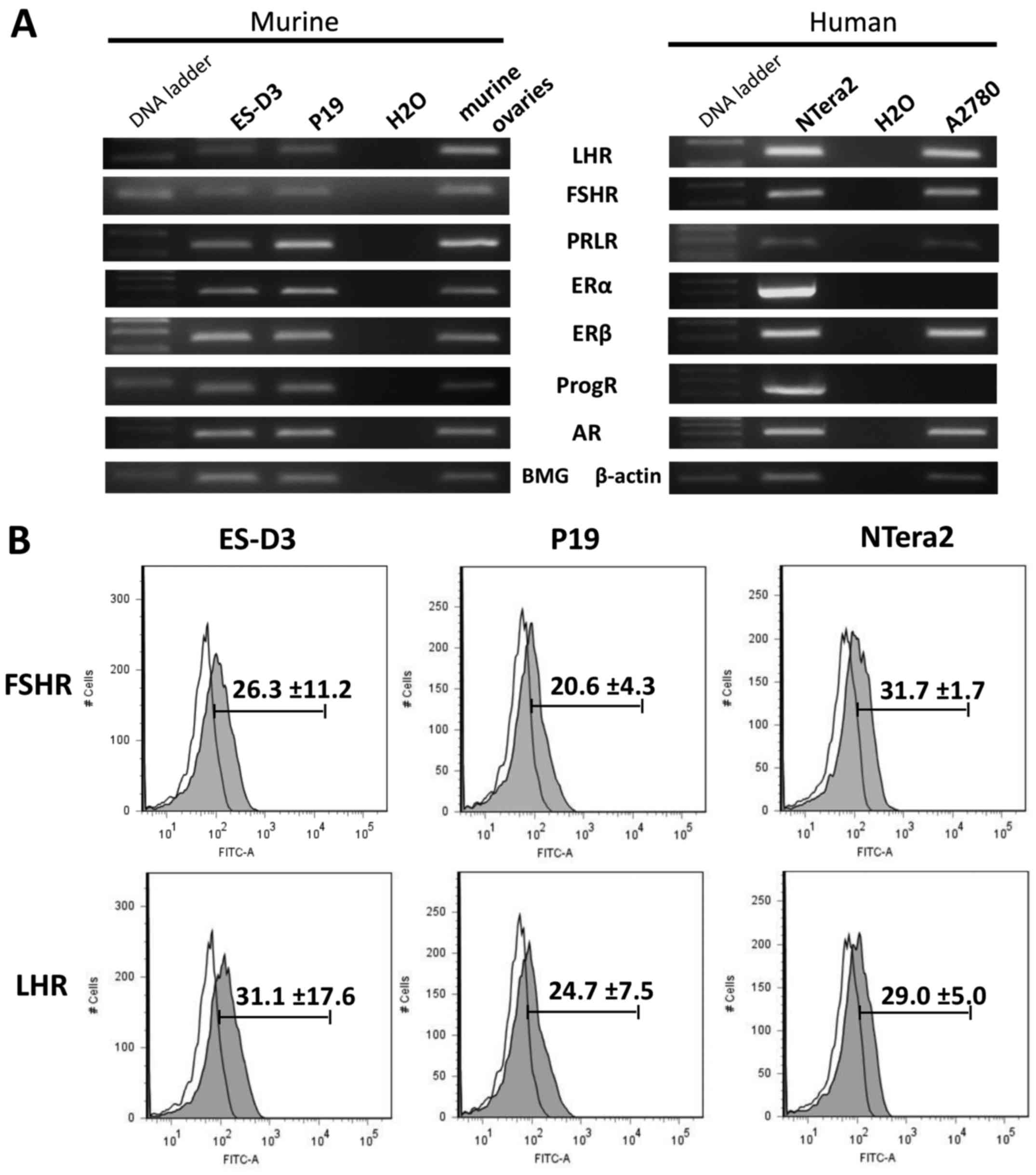 | Figure 1.Murine and human
early-developmental-stage cell lines express several sex hormone
receptors. (A) RT-PCR results for expression of LHR, FSHR, PRLR,
ERα, ERβ, ProgR, and AR in the murine stem cell line ES-D3, the
teratocarcinoma cell line P19, and the human embryonal carcinoma
cell line NTera2. cDNA from murine ovaries and from the human
ovarian cancer cell line A2780 were used as controls. (B) Flow
cytometric analysis of FSHR and LHR expression in the ES-D3, P19,
and NTera2 cell lines. Representative results from two independent
experiments are shown. LHR, luteinizing hormone receptor; FSHR,
follicle stimulating hormone receptor; PRLR, prolactin receptor;
ERα, estrogen α receptor; ERβ, estrogen β receptor; ProgR,
progesterone receptor; AR, androgen receptor. |
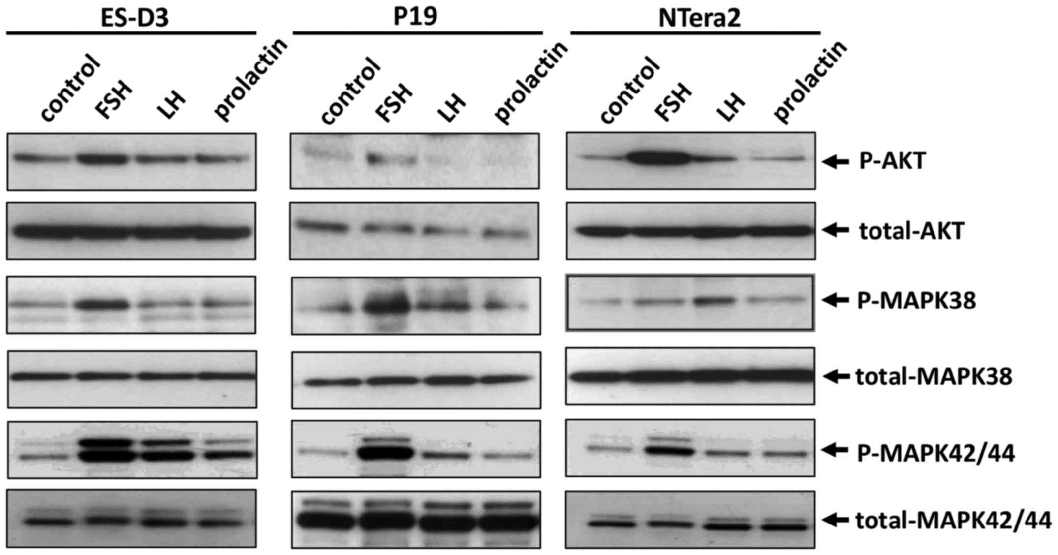
To address whether FSH, LH, and PRL receptors were
functional, we employed signal transduction experiments to examine
whether stimulation with peptide-based SexHs activated signaling
pathways involved in cell migration and adhesion. As shown in
Fig. 2, all of the SexHs studied
induced p42/44 MAPK, p38 MAPK and AKT phosphorylation; however, the
pattern of activation depended on the cell type. While ES-D3
responded robustly to stimulation by FSH, LH, and PRL by p42/44
MAPK and AKT stimulation, P19 and NTera2 cells exhibited a strong
response to FSH and a weaker response to LH. Robust activation of
p38 MAPK was observed in all cell lines in response to FSH, and in
a less extent to LH and prolactin. The response of AKT to
stimulation by pituitary SexHs was observed in ES-D3 and NTera2
cells after stimulation by FSH and LH and in P19 cells after
exposure to FSH.
Peptide-based SexH receptors modulate
the chemotactic responsiveness and adhesion of murine and human
cell lines in the early developmental stage
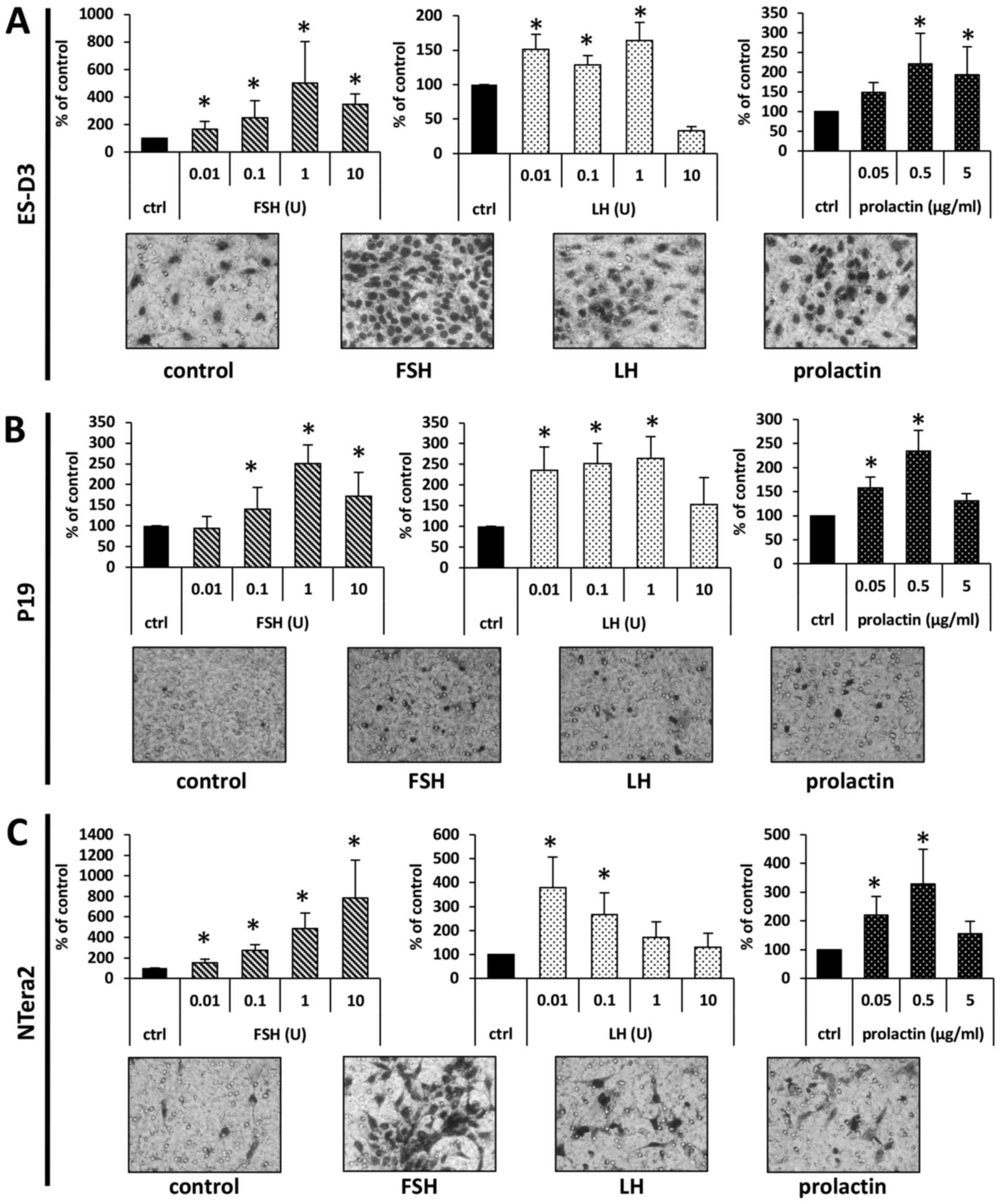
To address whether SexHs modulated migration of the
investigated murine and human cell lines, we employed the Transwell
system, in which two chambers were separated with a porous
membrane. The lower chamber was prefilled with warm medium
supplemented with the studied SexHs, and the cells were seeded into
the upper chamber. After 24 h the inserts were removed, and the
cells that had migrated from the upper to the lower side of the
membrane were stained and counted under a microscope. As shown in
Fig. 3, all studied SexHs
stimulated migration of ES-D3, P19, and NTera2 cells. Notably, all
cell lines responded not only to supraphysiological concentrations
of FSH and LH but also to lower concentrations detected in human
plasma. By contrast, while ES-D3 cells responded to a steep
gradient of PRL only (Fig. 3A), P19
and NTera2 cells were chemoattracted to a lower physiological dose
of this hormone (Fig. 3B and
C).
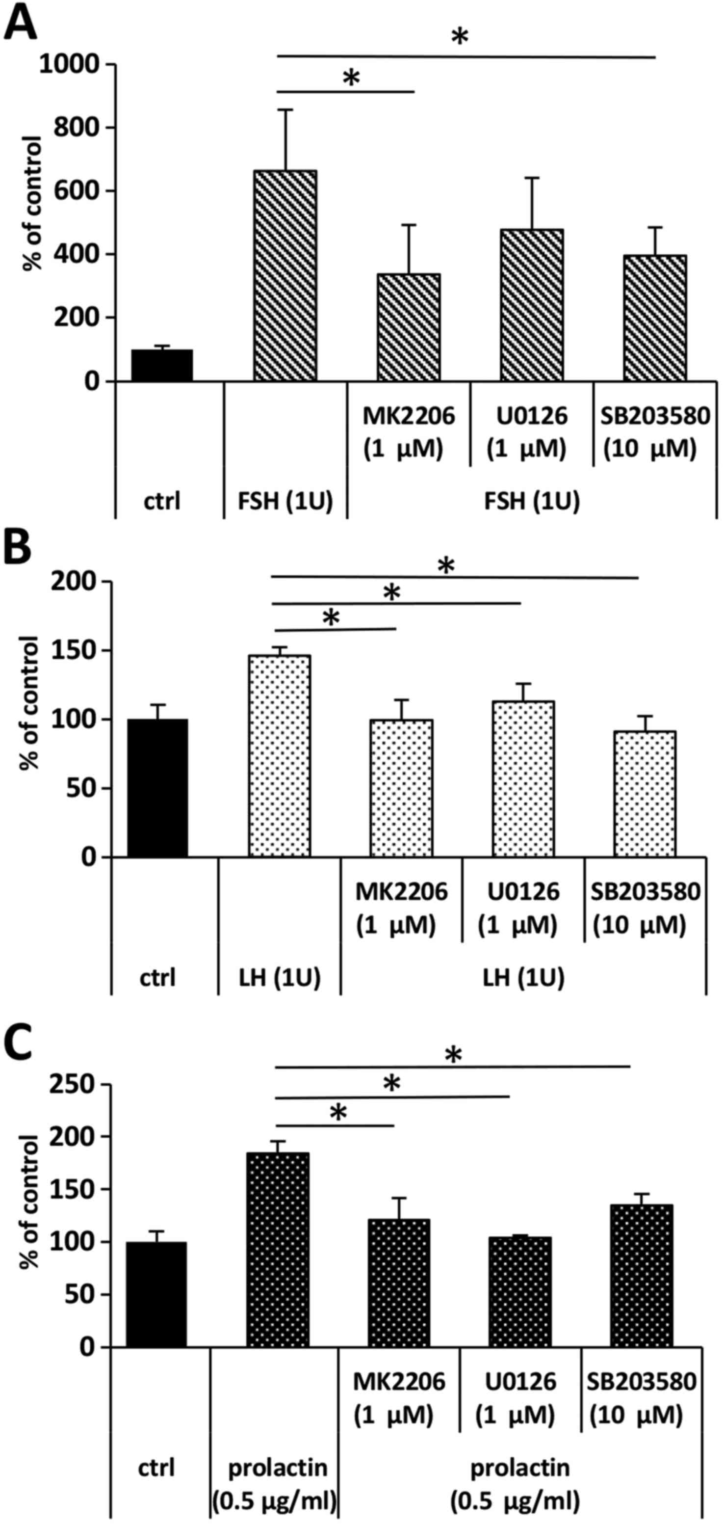
To address whether FSH-, LH-, and PRL-stimulated
migration dependend on the activation of AKT, p42/44, and p38 MAPK
signaling pathways, we assessed the migration of ES-D3 upon
stimulation with LH, FSH and prolactin in the presence of the
appropriate inhibitors. We determined that, while the response of
ES-D3 cells to FSH stimulation was decreased only in the presence
of AKT (MK2206) and p38 MAPK (SB203580) (Fig. 4A), their response to LH and
prolactin was inhibited by MEK, AKT, and p38 MAPK inhibitors,
UO126, MK2206, and SB203580, respectively (Fig. 4B and C).
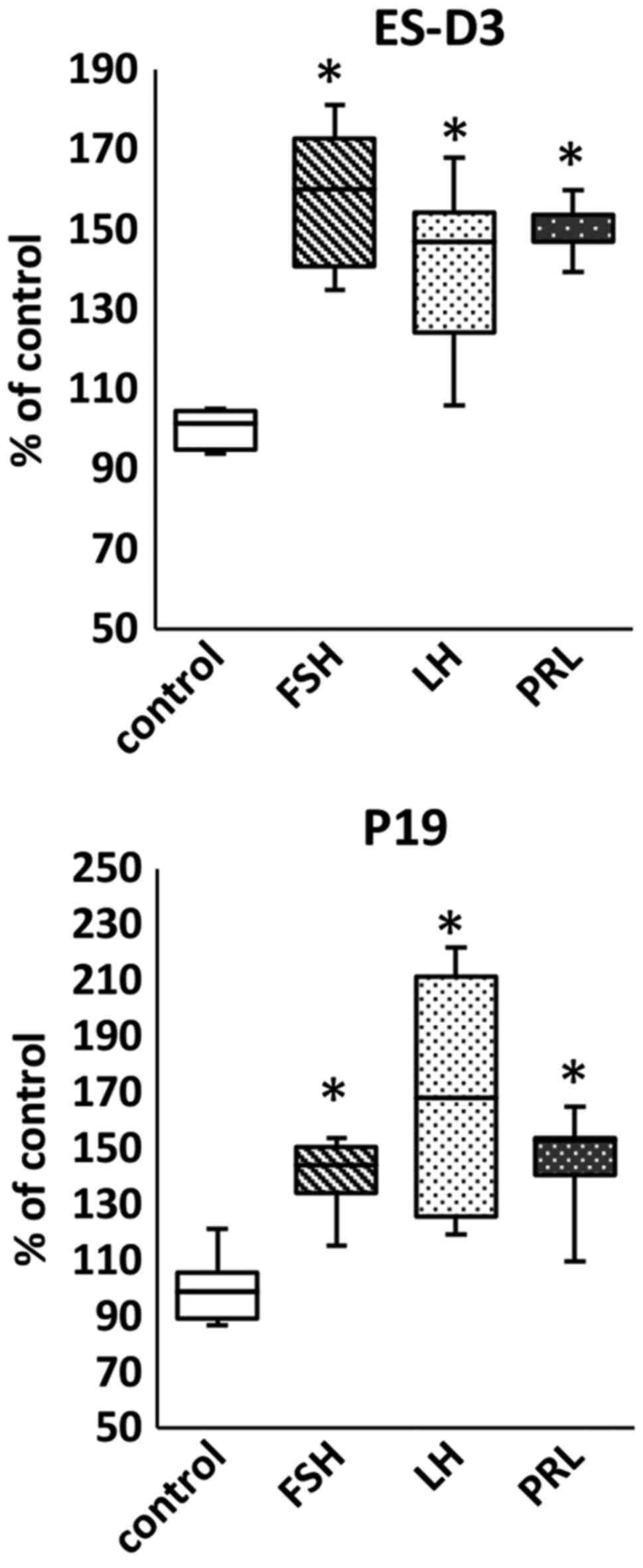
We also analyzed whether pituitary SexHs increased
adhesion of the cell lines investigated in our study and determined
that stimulation of ES-D3, P19, and NTera2 cells with FSH, LH, and
PRL induced adhesion of these cells to fibronectin (Fig. 5).
The effect of pituitary SexHs on the
proliferation of murine and human cell lines
early-developmental-stage
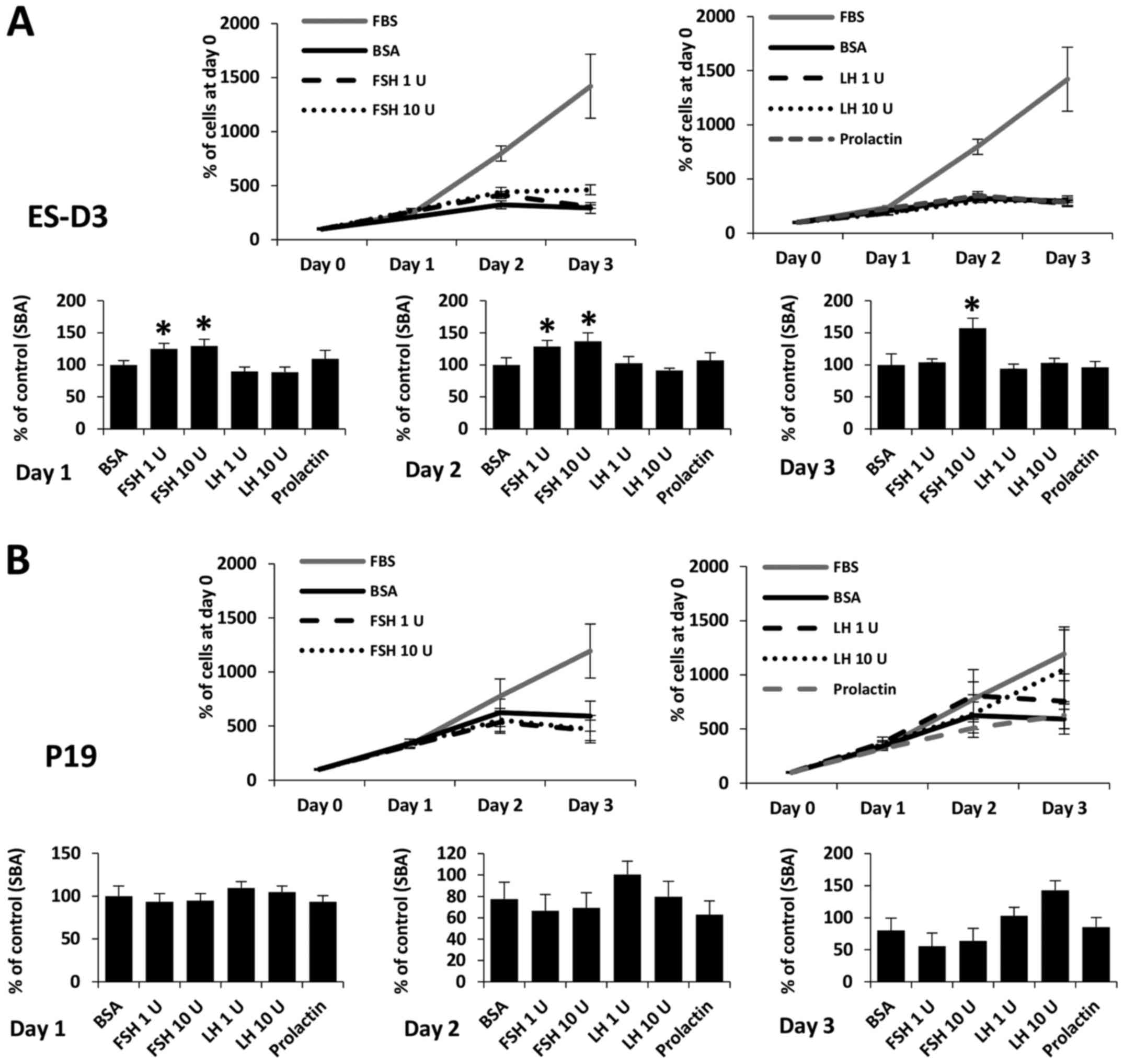
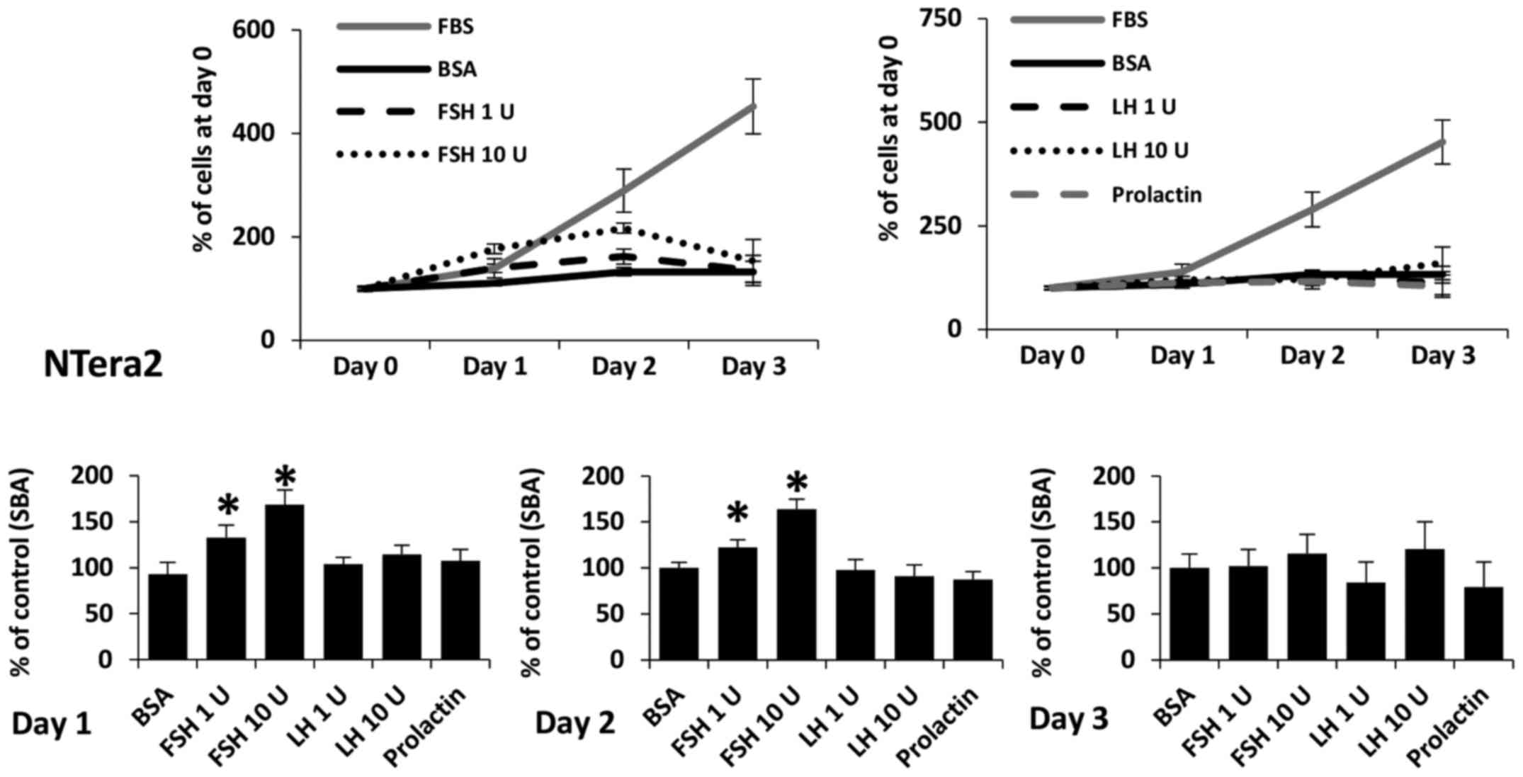
FSH, LH, and PRL are known to be potent mitogens for
some types of cells. As shown in Figs.
6 and 7, we added SexHs at the
indicated doses on day 0 and analyzed the number of cells 24, 48
and 72 h later. We observed that FSH slightly stimulated
proliferation of ES-D3 (Fig. 6A)
and NTera2 (Fig. 7) cells if added
to medium containing 0.5% BSA. By contrast, no effect of LH or PRL
was observed.
Discussion
The salient observation of this study is that
established cell lines derived in early development, including the
murine embryonic ES-D3, the murine teratocarcinoma P19, and the
human embryonal carcinoma NTera2 cell lines, express functional
SexH receptors. Thus, our results indicated that pituitary SexHs
may affect the biology of murine and human stem cells during the
earliest stages of embryogenesis. Moreover, placenta-derived
PRL-like hormones (known as lactogens), which bind with high
affinity to the PRL-receptor and thereby mimic the actions of PRL,
may also be involved in this process (28).
Recently, it was demonstrated that pituitary SexHs
were involved in metastatic lung cancer (9), certain sarcomas (10), and leukemia (11). In the present study we investigated
whether FSH, LH, and PRL stimulated cells in the early
developmental stage.
It is well known that pituitary SexHs affect
development and regulate biological processes in gonadal, prostate,
and breast tissue (5–8). We recently demonstrated that pituitary
SexHs were also involved in regulating normal hematopoiesis
(20,21), which supports their having direct
pleiotropic effects on extra-gonadal tissues in the adult
organism.
On the other hand, evidence has accumulated that
pituitary SexHs are involved in gonadal, breast, and prostate
malignancies (5–8). Recent evidence revealed as
aforementioned that these hormones were also involved in the
pathogenesis of lung cancer, pediatric sarcomas, and leukemia. In
our current study we demonstrated for the first time that certain
malignancies derived from cells involved in early development, such
as murine P19 teratocarcinoma cells and human embryonal carcinoma
NTera2 cells, were highly sensitive to pituitary SexH stimulation,
even when employed at physiological concentrations in peripheral
blood (29). Notably, the
concentration of some SexHs, such as FSH, increase with advancing
age as a result of age-dependent gonadal dysfunction (30,31).
Our results are important for another reason. It has
been proposed in the past that some cells from early embryonic
development may continue to reside in adult tissues (18,19,32–34).
This hypothesis was the basis for a theory proposed in the XIX
century by Recamier (1829) (32),
Remak (1854) (33), Virchow (1858)
(18) and later elaborated by
Durante (1874) (34) and Cohnheim
(1875) (19) that some malignancies
are derived from such embryonic remnants. This theory was known as
the embryonic rest hypothesis for cancer development and was
popular in older pathology textbooks (18,19,32–34).
The presence of certain populations of primitive
stem cells in adult tissues that are endowed with the ability to
differentiate into all three germ layers has been proposed by
several researchers. Examples of such cells described in the
literature include i) multipotent adult progenitor cells (MAPCs)
(35); ii) marrow-isolated adult
multilineage-inducible (MIAMI) cells (36); iii) multipotent adult stem cells
(MASCs) (37); iv)
elutriation-derived (Fr25/Lin−) stem cells (ELH SCs)
(38); v) spore-like stem cells
(39); vi) pluripotent
Sca-1+CD45−c-kit− cells (40); and vii) multilineage-differentiating
stress-enduring stem cells (Muse SCs) (41). An interesting population of stem
cells endowed with pan-germ-layer differentiation potential has
been identified by our team and confirmed subsequently by several
independent researchers as very small embryonic-like stem cells
(VSELs) (15,20–25,42–45).
The similarity of all the stem cells listed above with respect to
the expression of certain genes involved in early development
suggests that they are related to each other, that they represent
similar and overlapping populations of primitive SCs that reside in
adult tissues, and that they are endowed with broader
pan-germ-layer differentiation potential (46). These stem cell types were identified
by employing different direct or indirect isolation protocols and
identification techniques, and based on this they were given
different names. We propose that, due to their primitive embryonic
morphology and gene expression for embryonic and primordial
germline markers, VSELs are at the top of the hierarchy of these
cells, which is consistent with an old concept that germline stem
cells are not only developmental in origin but also remain as a
‘skeleton’ of the stem cell compartment in adult tissues (46).
We demonstrated in the past that VSELs express
several receptors for SexHs, and our results reported in this study
support the finding that the most primitive stem cells originating
during development are responsive to SexHs (20–25).
Since the pituitary gland develops later in embryogenesis, several
of these hormones are derived from maternal blood, and in fact the
potential effects of pituitary and placental SexHs on the
developing embryo are not very well addressed in literature.
Cell migration and adhesion are regulated by
different factors, such as chemokines, growth factors, cytokines,
bioactive lipids, and extracellular nucleotides (27,47–49).
However our recent and already published results indicate that
pituitary SexHs also play an important role, which has been
observed in both normal and malignant cells (9–11,20–23).
While pituitary SexHs were found to increase the proliferation of
several tumors, we observed only a very weak effect of FSH on
proliferation of ES-D3 and NTera-2 cells. The role of pituitary
SexHs, in particular, in the pathogenesis of cancer requires
further study, since, as aforementioned, the concentration of FSH
in peripheral blood increases with age due to gonadal dysfunction
(30,31).
In conclusion, we found that pituitary SexHs
stimulated migration and adhesion of established immortalized cell
lines derived from the inner cell mass of the blastocyst (ES-D3) as
well as from early germline tumors (P19, NTera2). Therefore,
pituitary SexHs derived from maternal blood may affect the early
stages of embryonic development. Moreover, lactogens, which can
bind with high affinity to the PRL-receptor and thereby mimic the
actions PRL, may also be involved in this process (28). This possibility, however, requires
further study.
Our observations also shed new light on the
potential role of these hormones in early embryonic development and
cancerogenesis, particularly in light of the embryonic rest
hypothesis proposed by Virchow and Connheim (18,19).
However, we are aware that this concept also warrants more
experimental investigation.
Acknowledgements
This study was supported by NIH grants 2R01 DK074720
and R01HL112788, the Stella and Henry Endowment, and NCN grant:
OPUS 2016/21/B/NZ4/00201 by the National Science Center in Poland
to MK.
References
|
1
|
Costa MA: The endocrine function of human
placenta: An overview. Reprod Biomed Online. 32:14–43. 2016.
View Article : Google Scholar : PubMed/NCBI
|
|
2
|
Pepe GJ and Albrecht ED: Actions of
placental and fetal adrenal steroid hormones in primate pregnancy.
Endocr Rev. 16:608–648. 1995. View Article : Google Scholar : PubMed/NCBI
|
|
3
|
Whitaker-Azmitia PM, Lobel M and Moyer A:
Low maternal progesterone may contribute to both obstetrical
complications and autism. Med Hypotheses. 82:313–318. 2014.
View Article : Google Scholar : PubMed/NCBI
|
|
4
|
Gürbüz B, Yalti S, Ozden S and Ficicioglu
C: High basal estradiol level and FSH/LH ratio in unexplained
recurrent pregnancy loss. Arch Gynecol Obstet. 270:37–39. 2004.
View Article : Google Scholar : PubMed/NCBI
|
|
5
|
van Kruchten M, van der Marel P, de Munck
L, Hollema H, Arts H, Timmer-Bosscha H, de Vries E, Hospers G and
Reyners A: Hormone receptors as a marker of poor survival in
epithelial ovarian cancer. Gynecol Oncol. 138:634–639. 2015.
View Article : Google Scholar : PubMed/NCBI
|
|
6
|
Lønning PE: Poor-prognosis estrogen
receptor-positive disease: Present and future clinical solutions.
Ther Adv Med Oncol. 4:127–137. 2012. View Article : Google Scholar : PubMed/NCBI
|
|
7
|
García-Cruz E, Piqueras M, Huguet J, Peri
L, Izquierdo L, Musquera M, Franco A, Alvarez-Vijande R, Ribal MJ
and Alcaraz A: Low testosterone levels are related to poor
prognosis factors in men with prostate cancer prior to treatment.
BJU Int. 110:E541–E546. 2012. View Article : Google Scholar : PubMed/NCBI
|
|
8
|
Ma CX, Bose R and Ellis MJ: Prognostic and
predictive biomarkers of endocrine responsiveness for estrogen
receptor positive breast cancer. Adv Exp Med Biol. 882:125–154.
2016. View Article : Google Scholar : PubMed/NCBI
|
|
9
|
Abdelbaset-Ismail A, Pedziwiatr D,
Schneider G, Niklinski J, Charkiewicz R, Moniuszko M, Kucia M and
Ratajczak MZ: Pituitary sex hormones enhance the pro metastatic
potential of human lung cancer cells by downregulating the
intracellular expression of heme oxygenase 1. Int J Oncol.
50:317–328. 2017. View Article : Google Scholar : PubMed/NCBI
|
|
10
|
Poniewierska-Baran A, Schneider G, Sun W,
Abdelbaset-Ismail A, Barr FG and Ratajczak MZ: Human
rhabdomyosarcoma cells express functional pituitary and gonadal sex
hormone receptors: Therapeutic implications. Int J Oncol.
48:1815–1824. 2016. View Article : Google Scholar : PubMed/NCBI
|
|
11
|
Abdelbaset-Ismail A, Borkowska S,
Janowska-Wieczorek A, Tonn T, Rodriguez C, Moniuszko M, Bolkun L,
Koloczko J, Eljaszewicz A, Ratajczak J, et al: Novel evidence that
pituitary gonadotropins directly stimulate human leukemic
cells-studies of myeloid cell lines and primary patient AML and CML
cells. Oncotarget. 7:3033–3046. 2016. View Article : Google Scholar : PubMed/NCBI
|
|
12
|
Donovan PJ and Gearhart J: The end of the
beginning for pluripotent stem cells. Nature. 414:92–97. 2001.
View Article : Google Scholar : PubMed/NCBI
|
|
13
|
Otte J, Wruck W and Adjaye J: New insights
into human primordial germ cells and early embryonic development
from single-cell analysis. FEBS Lett. 591:2226–2240. 2017.
View Article : Google Scholar : PubMed/NCBI
|
|
14
|
Aflatoonian B and Moore H: Germ cells from
mouse and human embryonic stem cells. Reproduction. 132:699–707.
2006. View Article : Google Scholar : PubMed/NCBI
|
|
15
|
Kucia M, Halasa M, Wysoczynski M,
Baskiewicz-Masiuk M, Moldenhawer S, Zuba-Surma E, Czajka R,
Wojakowski W, Machalinski B and Ratajczak MZ: Morphological and
molecular characterization of novel population of CXCR4+ SSEA-4+
Oct-4+ very small embryonic-like cells purified from human cord
blood: Preliminary report. Leukemia. 21:297–303. 2007. View Article : Google Scholar : PubMed/NCBI
|
|
16
|
Ratajczak MZ, Shin DM, Liu R, Marlicz W,
Tarnowski M, Ratajczak J and Kucia M: Epiblast/germ line hypothesis
of cancer development revisited: Lesson from the presence of Oct-4+
cells in adult tissues. Stem Cell Rev. 6:307–316. 2010. View Article : Google Scholar : PubMed/NCBI
|
|
17
|
Ratajczak MZ, Schneider G, Sellers ZP,
Kucia M and Kakar SS: The embryonic rest hypothesis of cancer
development - an old XIX century theory revisited. J Cancer Stem
Cell Res. 2:e10012014.
|
|
18
|
Virchow R: Editorial Archive fuer
pathologische. Anatomie und Physiologie fuer klinische Medizin.
8:23–54. 1855.(In German).
|
|
19
|
Conheim J: Congenitales, quergestreiftes
muskelsarkon der nireren. Virchows Arch. 65:64–69. 1875.(In
German). View Article : Google Scholar
|
|
20
|
Mierzejewska K, Borkowska S, Suszynska E,
Suszynska M, Poniewierska-Baran A, Maj M, Pedziwiatr D, Adamiak M,
Abdel-Latif A, Kakar SS, et al: Hematopoietic stem/progenitor cells
express several functional sex hormone receptors-novel evidence for
a potential developmental link between hematopoiesis and primordial
germ cells. Stem Cells Dev. 24:927–937. 2015. View Article : Google Scholar : PubMed/NCBI
|
|
21
|
Abdelbaset-Ismail A, Suszynska M,
Borkowska S, Adamiak M, Ratajczak J, Kucia M and Ratajczak MZ:
Human haematopoietic stem/progenitor cells express several
functional sex hormone receptors. J Cell Mol Med. 20:134–146. 2016.
View Article : Google Scholar : PubMed/NCBI
|
|
22
|
Patel H and Bhartiya D: Testicular stem
cells express follicle-stimulating hormone receptors and are
directly modulated by FSH. Reprod Sci. 23:1493–1508. 2016.
View Article : Google Scholar : PubMed/NCBI
|
|
23
|
Patel H, Bhartiya D, Parte S, Gunjal P,
Yedurkar S and Bhatt M: Follicle stimulating hormone modulates
ovarian stem cells through alternately spliced receptor variant
FSH-R3. J Ovarian Res. 6:522013. View Article : Google Scholar : PubMed/NCBI
|
|
24
|
Shaikh A, Anand S, Kapoor S, Ganguly R and
Bhartiya D: Mouse bone marrow VSELs exhibit differentiation into
three embryonic germ lineages and germ & hematopoietic cells in
culture. Stem Cell Rev. 13:202–216. 2017. View Article : Google Scholar : PubMed/NCBI
|
|
25
|
Ratajczak MZ, Bartke A and Darzynkiewicz
Z: Prolonged growth hormone/insulin/insulin-like growth factor
nutrient response signaling pathway as a silent killer of stem
cells and a culprit in aging. Stem Cell Rev. 13:443–453. 2017.
View Article : Google Scholar : PubMed/NCBI
|
|
26
|
Sellers ZP, Schneider G, Bujko K,
Suszynska M and Pedziwiatr D: Do cancer cell lines have fixed or
fluctuating stem cell phenotypes? - studies with the NTera2 cell
line. Stem Cell Rev. 13:603–610. 2017. View Article : Google Scholar : PubMed/NCBI
|
|
27
|
Schneider G, Glaser T, Lameu C,
Abdelbaset-Ismail A, Sellers ZP, Moniuszko M, Ulrich H and
Ratajczak MZ: Extracellular nucleotides as novel, underappreciated
pro-metastatic factors that stimulate purinergic signaling in human
lung cancer cells. Mol Cancer. 14:2012015. View Article : Google Scholar : PubMed/NCBI
|
|
28
|
Gertler A, Bignon C, Staten NR, Sakal E,
Tchelet A, Krivi GG and Djiane J: Proceedings of the Society for
Experimental Biology and Medicine: Interaction of lactogenic
hormones with the soluble extracellular domain of prolactin
receptors. Proc Soc Exp Biol Med. 206:pp. 273–279. 1994; View Article : Google Scholar : PubMed/NCBI
|
|
29
|
Fischbach F and Dunning MB: A Manual of
Laboratory and Diagnostic Tests. 9th. Wolters Kluwer
Health/Lippincott Williams & Wilkins; Philadelphia: 2014
|
|
30
|
Araujo AB and Wittert GA: Endocrinology of
the aging male. Best Pract Res Clin Endocrinol Metab. 25:303–319.
2011. View Article : Google Scholar : PubMed/NCBI
|
|
31
|
Limpaphayom K, Taechakraichana N and
Kittinunvorakoon C: Serum estradiol and gonadotropins level in
postmenopausal women with or without hormone replacement. J Med
Assoc Thai. 80:147–152. 1997.PubMed/NCBI
|
|
32
|
Androutsos G, Karamanou M, Stamboulis E,
Tsoucalas G, Kousoulis AA and Mandelenaki D: Joseph-Claude-Anthelme
Récamier (1774–1852): Forerunner in surgical oncology. J BUON.
16:572–576. 2011.PubMed/NCBI
|
|
33
|
Remak R: Ein beitrag zur
entwickelungsgeschichte der krebshaften geschwulste Deut Klin.
6:70–174. 1854.(In German).
|
|
34
|
Durante F: Arch. Memori ed Osservazioni di
Chirugia Practica. 11:217–226. 1874.(In Italian).
|
|
35
|
Jiang Y, Jahagirdar BN, Reinhardt RL,
Schwartz RE, Keene CD, Ortiz-Gonzalez XR, Reyes M, Lenvik T, Lund
T, Blackstad M, et al: Pluripotency of mesenchymal stem cells
derived from adult marrow. Nature. 418:41–49. 2002. View Article : Google Scholar : PubMed/NCBI
|
|
36
|
D'Ippolito G, Diabira S, Howard GA, Menei
P, Roos BA and Schiller PC: Marrow-isolated adult multilineage
inducible (MIAMI) cells, a unique population of postnatal young and
old human cells with extensive expansion and differentiation
potential. J Cell Sci. 117:2971–2981. 2004. View Article : Google Scholar : PubMed/NCBI
|
|
37
|
Beltrami AP, Cesselli D, Bergamin N,
Marcon P, Rigo S, Puppato E, D'Aurizio F, Verardo R, Piazza S,
Pignatelli A, et al: Multipotent cells can be generated in vitro
from several adult human organs (heart, liver, and bone marrow).
Blood. 110:3438–3446. 2007. View Article : Google Scholar : PubMed/NCBI
|
|
38
|
Goldenberg-Cohen N, Avraham-Lubin BC,
Sadikov T, Goldstein RS and Askenasy N: Primitive stem cells
derived from bone marrow express glial and neuronal markers and
support revascularization in injured retina exposed to ischemic and
mechanical damage. Stem Cells Dev. 21:1488–1500. 2012. View Article : Google Scholar : PubMed/NCBI
|
|
39
|
Vacanti MP, Roy A, Cortiella J, Bonassar L
and Vacanti CA: Identification and initial characterization of
spore-like cells in adult mammals. J Cell Biochem. 80:455–460.
2001. View Article : Google Scholar : PubMed/NCBI
|
|
40
|
Howell JC, Lee WH, Morrison P, Zhong J,
Yoder MC and Srour EF: Pluripotent stem cells identified in
multiple murine tissues. Ann NY Acad Sci. 996:158–173. 2003.
View Article : Google Scholar : PubMed/NCBI
|
|
41
|
Wakao S, Kitada M, Kuroda Y, Shigemoto T,
Matsuse D, Akashi H, Tanimura Y, Tsuchiyama K, Kikuchi T, Goda M,
et al: Multilineage-differentiating stress-enduring (Muse) cells
are a primary source of induced pluripotent stem cells in human
fibroblasts. Proc Natl Acad Sci USA. 108:pp. 9875–9880. 2011;
View Article : Google Scholar : PubMed/NCBI
|
|
42
|
Guerin CL, Rossi E, Saubamea B, Cras A,
Mignon V, Silvestre JS and Smadja DM: Human very small
embryonic-like cells support vascular maturation and therapeutic
revascularization induced by endothelial progenitor cells. Stem
Cell Rev. 13:552–560. 2017. View Article : Google Scholar : PubMed/NCBI
|
|
43
|
Golipoor Z, Mehraein F, Zafari F, Alizadeh
A, Ababzadeh S and Baazm M: Migration of bone marrow-derived very
small embryonic-like stem cells toward an injured spinal cord. Cell
J. 17:639–647. 2016.PubMed/NCBI
|
|
44
|
Bhartiya D, Shaikh A, Anand S, Patel H,
Kapoor S, Sriraman K, Parte S and Unni S: Endogenous, very small
embryonic-like stem cells: Critical review, therapeutic potential
and a look ahead. Hum Reprod Update. 23:41–76. 2016. View Article : Google Scholar : PubMed/NCBI
|
|
45
|
Kassmer SH, Jin H, Zhang PX, Bruscia EM,
Heydari K, Lee JH, Kim CF, Kassmer SH and Krause DS: Very small
embryonic-like stem cells from the murine bone marrow differentiate
into epithelial cells of the lung. Stem Cells. 31:2759–2766. 2013.
View Article : Google Scholar : PubMed/NCBI
|
|
46
|
Ratajczak MZ, Ratajczak J, Suszynska M,
Miller DM, Kucia M and Shin DM: A novel view of the adult stem cell
compartment from the perspective of a quiescent population of very
small embryonic-like stem cells. Circ Res. 120:166–178. 2017.
View Article : Google Scholar : PubMed/NCBI
|
|
47
|
Majka M, Drukala J, Lesko E, Wysoczynski
M, Jenson AB and Ratajczak MZ: SDF-1 alone and in co-operation with
HGF regulates biology of human cervical carcinoma cells. Folia
Histochem Cytobiol. 44:155–164. 2006.PubMed/NCBI
|
|
48
|
Schneider G, Sellers ZP, Bujko K, Kakar
SS, Kucia M and Ratajczak MZ: Novel pleiotropic effects of
bioactive phospholipids in human lung cancer metastasis.
Oncotarget. 8:58247–58263. 2017.PubMed/NCBI
|
|
49
|
Schneider G, Bryndza E, Poniewierska-Baran
A, Serwin K, Suszynska M, Sellers ZP, Merchant ML, Kaliappan A,
Ratajczak J, Kucia M, et al: Evidence that vitronectin is a potent
migration-enhancing factor for cancer cells chaperoned by
fibrinogen: A novel view of the metastasis of cancer cells to
low-fibrinogen lymphatics and body cavities. Oncotarget.
7:69829–69843. 2016.PubMed/NCBI
|