Introduction
Dermatitis and oral mucositis are the frequent
side-effects of chemotherapy and radiotherapy treatment in cancer
patients including head and neck cancer. Dermatitis appears as the
onset of erythema, swelling, an acneiform rash, severe pruritus,
xerosis cutis, blisters and ulceration in cancer patients, which
can lead to chronic inflammation, necrosis, fibrosis, hair and nail
alterations, stomatitis and lymphedema (1–3).
Mucositis causes acute oral pain and difficulty with swallowing,
which can result in reduced nutrient intake, significant
malnutrition and weight loss, as well as poor oral hygiene in head
and neck cancer patients (1,4).
Dermatitis as well as mucositis can cause interruption, sometimes
even termination of cancer treatments, which adversely affects the
prognosis and eventually reduces the survival rates of the patients
(1,5,6). There
could be multiple factors involved in triggering dermatitis and
mucositis, although the detailed mechanism of chemotherapy and/or
radiotherapy induced dermatitis and mucositis in cancer patients is
still unclear.
Chemotherapeutic agents including 5-fluorouracil
(5-FU) may harm rapidly dividing immature keratinocytes, as well as
dividing stem cells (7–9). Moreover, the basal cell layer of
epithelium can be directly damaged leading to the loss of the
renewal capacity of the epithelium, which may result in ulceration.
It has been reported that the secretions of pro-inflammatory
cytokines by epidermal keratinocytes play a key role in various
types of inflammations in the skin (10). Among these pro-inflammatory
cytokines, TNF-α has been implicated in the promotion of
inflammatory reactions via the activation of cytokines IL-6 and
IL-1β (10,11). Nuclear transcription factor-κB
(NF-κB) is the most important transcriptional regulator of
inflammatory pathways, and it directly controls the cellular
expression of these pro-inflammatory cytokines (10,12).
Activation of NF-κB induces the transcription of many
inflammation-related genes including the ‘classic cachectic
cytokines’ TNF-α, IL-6 and IL-1β (10,11,13).
Therefore, inhibiting or limiting the production of these
pro-inflammatory cytokines may help in the treatment of
inflammation and dermatitis in cancer patients. Although various
types of therapies have been introduced for the prevention and
treatment of chemotherapy-induced mucositis and dermatitis, the
efficacy of these treatments remains limited (8,15–21).
Elental® (EA Pharma Co., Ltd., Tokyo,
Japan), an elemental diet (ED) with L-glutamine which has an easily
digestible nutrition formula that combines amino acids,
carbohydrates, vitamins, minerals and with minimal fat content has
been used in Japan as a treatment for malnourished patients
(22,23). Elental® has been reported
to be useful in the treatment of Crohn's disease (24–27),
as well as in the management of chemotherapy-induced mucositis in
cancer patients (28,29). We have been using
Elental® for the treatment of malnutrition in patients
undergoing chemotherapy and/or radiotherapy in our hospital since
2011, and our clinical study revealed the efficacy of
Elental® in ameliorating chemotherapy-induced oral
mucositis and dermatitis in head and neck cancer patients (30). Recently, we reported that
Elental® may accelerate the recovery from 5-FU induced
oral mucositis and dermatitis through the induction of fibroblast
growth factor (31). However, the
detailed mechanism of its action against inflammations is still
unclear.
The aim of this study was to clarify the efficacy of
ED (Elental®) in reducing pro-inflammatory cytokine
production in keratinocytes in vivo and in vitro.
Additionally, we investigated the effect of Elental® on
the inhibition of NF-κB activation.
Materials and methods
Animals
Ten 4-week-old female athymic nude mice with a
CAnN.Cg-Foxn1nu/CrlCrlj genetic background were
purchased from CLEA Japan, Inc. (Tokyo, Japan). They were housed in
a temperature-controlled room with a 12 h light/dark cycle, under
sterile conditions in a pathogen-free environment, and received
water and food ad libitum. All procedures concerning animal
handling and treatment were conducted in accordance with the
Guidelines for Animal Experimentation of Yamaguchi University.
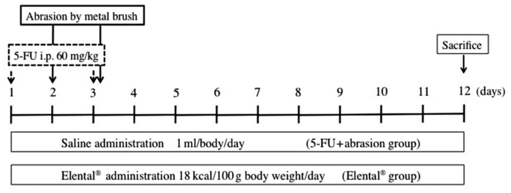
Induction of experimental
dermatitis
Dermatitis was induced in all nude mice by 2
intraperitoneal (i.p.) administrations of 60 mg/kg 5-FU (Wako,
Osaka, Japan) on the first and third day of the experiment
accompanied by superficial scratching on the dorsal skin with a
metal brush on the second and third day of the experiment under
anesthesia (pentobarbital sodium, 30 mg/kg, i.p.;
Somunopentyl®; Kyoritsu Seiyaku Co., Ltd., Tokyo, Japan)
(Fig. 1). The metal brush was
dragged 3 times in a linear fashion across the dorsum skin of nude
mice until erythematous changes of the skin were noted.
In vivo experimental groups
Fig. 1 shows the
experimental design of our in vivo study. We set up the
following two groups of nude mice (5 mice/group) with dermatitis
induced by 5-FU + abrasion: The 5-FU + abrasion group was the
untreated control in this experiment, and they received saline (1
ml/body/day) only, whereas the Elental® group received
Elental® (18 kcal/100 g body weight/day), which was
orally administered daily until the wounded area was totally healed
at the end of the experiment. The healing process of the dermatitis
of each mouse was examined every day and the affected area was
assessed. Each lesion was calculated by multiplying the major axis
by the minor axis. We purchased Elental® from EA Pharma
Co., Ltd. Tissue samples of the wounded area were collected after
the mice were sacrificed on the 12th day of the experiment.
Immunohistochemical staining
Tissue samples obtained from inflammatory lesions
were examined by immunohistochemical analyses using the universal
EnVision™ kit (Dako, Glostrup, Denmark).
Paraffin-embedded 4 µm-thick tissue sections were immersed in
xylene, and then in graded alcohols (100–70%). Endogenous
peroxidase activity was quenched with a 0.3% hydrogen
peroxide/methanol mixture for 20 min. Then, the sections were
rinsed in PBS and incubated with 2% blocking serum for 30 min,
followed by incubation with the anti-TNF-α-rabbit polyclonal
antibody (1:200; no. ab6671; Abcam, Cambridge, UK), the anti-IL-1β
rabbit polyclonal antibody (1:200; no. ab9787; Abcam), the
anti-IL-6 mouse monoclonal antibody (1:50; no. ab9324; Abcam), the
anti-NF-κB p65 rabbit polyclonal antibody (1:200; no. sc-109; Santa
Cruz Biotechnology, Inc., Santa Cruz, CA, USA) or the
anti-α-tubulin mouse monoclonal antibody (1:200; no. sc-5286; Santa
Cruz Biotechnology, Inc) for 8 h at 4°C. After rinsing the tissue
sections in phosphate-buffered saline (PBS) for 10 min, the
antibody was detected using the universal EnVision™ kit
according to the manufacturer's instructions. Tissues were finally
rinsed in tap water, and then counterstained with hematoxylin for
1–2 min. The tissue sections were subsequently dehydrated in graded
ethanol, cleared in Histo-Clear® (National Diagnostics,
Atlanta, GA, USA), and mounted with glass coverslips using DPX. At
least 1,000 cells were counted under a microscope in several random
fields of each section. The number of positive cells was divided by
the total number of counted cells and each labeling index was
expressed as a percentage.
Cell lines and cell culture
Immortalized human keratinocyte cell line, HaCaT was
purchased from RIKEN BioResource Center Cell Bank (Ibaraki, Japan).
Cells were cultured in Dulbecco's modified Eagle's medium
(D-MEM)/Ham's F-12 (Sigma-Aldrich, St. Louis, MO, USA) supplemented
with 10% fetal bovine serum (FBS) (Thermo Fisher Scientific, Inc.,
Waltham, MA, USA), 100 µg/ml streptomycin/100 U/ml penicillin
(Thermo Fisher Scientific, Inc.) at 37°C in a humidified atmosphere
containing 5% CO2.
Cell culture with Elental®
for western blotting and enzyme-linked immunosorbent assay
(ELISA)
HaCaT cells (2×10⁶ cells) were cultured
in 100 mm plates (BD Biosciences, Franklin Lakes, NJ, USA) with
D-MEM/Ham's F-12 medium containing 10% FBS for 48 h. Then, the
cells were cultured in D-MEM/Ham's F-12 medium but without FBS and
5-FU (2 µg/ml) for 24 h to induce cellular injury and apoptosis.
Subsequently, the cells were cultured with Elental® (0,
0.1, 0.5, 1, 5, 10, 50 and 100 µg/ml) dissolved in D-MEM/Ham's F-12
medium without FBS. After 12 h, the cell medium was collected for
ELISA, and the cells were collected by scraping for western
blotting. We used this culture method for the preparation of
samples for western blotting and ELISA.
Western blotting
HaCaT cells were cultured as aforementioned, and
then cells were lysed with RIPA Buffer (Thermo Fisher Scientific,
Inc.) to extract cell proteins. These whole cell lysates were used
as samples for the detection of TNF-α, IL-1β, IL-6 and α-tubulin
expression. For the detection of the expression of nuclear and
cytoplasmic NF-κB (p65), we extracted and separated cytoplasmic and
nuclear protein fractions from cells using NE-PER™ Nuclear and
Cytoplasmic Extraction Reagents (Thermo Fisher Scientific, Inc.)
according to the manufacturer's instructions. Protein samples
containing 50 µg of protein were subjected to electrophoresis on
NuPAGE® Novex® Bis-Tris precast gels (Thermo
Fisher Scientific, Inc.), and then transferred to a polyvinylidene
difluoride (PVDF) membrane using iBlot™ PVDF Transfer Stack and
iBlot™ Dry Blotting System (Thermo Fisher Scientific, Inc.)
according to the manufacturer's instructions. After blocking the
membrane with a blocking solution prepared from WesternBreeze™
Blocker/Diluent Part A and B (Thermo Fisher Scientific, Inc.), the
membranes were incubated with the anti-TNF-α rabbit polyclonal
antibody (1:500; no. ab6671; Abcam), anti-IL-1β rabbit polyclonal
antibody (1:500; no. ab9787; Abcam), anti-IL-6 mouse monoclonal
antibody (1:250; no. ab9324; Abcam), anti-NF-κB p65 rabbit
polyclonal antibody (1:500; no. sc-109; Santa Cruz Biotechnology,
Inc.), or anti-α-tubulin monoclonal antibody (1:500; no. sc-5286;
Santa Cruz Biotechnology, Inc.) overnight; followed by
Novex® alkaline-phosphatase conjugated goat anti-rabbit
(cat. no. WP2007; Thermo Fisher Scientific, Inc.) or (goat)
anti-mouse immunoglobulin G (IgG) secondary antibody (cat. no.
WP20006; Thermo Fisher Scientific, Inc.). The antibodies were
detected using a Novex™ AP Chromogenic Substrate (BCIP⁄NBT) (Thermo
Fisher Scientific, Inc.) according to the manufacturer's
instructions.
ELISA for quantitative determination
of TNF-α, IL-1β or IL-6
HaCaT cells were cultured as aforementioned, and
then we assessed TNF-α, IL-1β and IL-6 proteins that were released
into cultured medium from the Elental®-treated or
untreated control cells. These proteins were assessed using a
microtiter-based sandwich enzyme immunoassay system. We used
commercially available ELISA kits (Abcam) according to the
manufacturer's protocol and estimated the total amount of TNF-α,
IL-1β and IL-6 in the culture medium. Each sample was examined in
triplicate.
Immunocytochemical staining
Cells (2.0×10⁵ cells/well) were cultured
on cover glasses in 6-well plates (BD Biosciences) with 10% FBS
D-MEM/Ham's F-12 for 48 h. Then, the cells were cultured in
D-MEM/Ham's F-12 medium but without FBS and 5-FU (2 µg/ml). After
24 h, the cells were cultured with Elental® (0 and 100
µg/ml) and dissolved in D-MEM/Ham's F-12 medium without FBS. Twelve
hours later, the cells were washed with PBS, fixed with 4%
paraformaldehyde and incubated for 60 min at 37 °C with anti-NF-κB
p65 rabbit polyclonal antibody (1:250; no. sc-109; Santa Cruz
Biotechnology, Inc.). After applying the Envision+ System HRP
(Dako) for 60 min at room temperature, immunostaining was
visualized with diaminobenzidine. The sections were lightly
counterstained with hematoxylin.
Statistical analysis
All data are expressed as the mean ± SD. The
significance of the experimental results was determined by
Student's t-test or Mann-Whitney U test. The differences were
considered statistically significant when P<0.05.
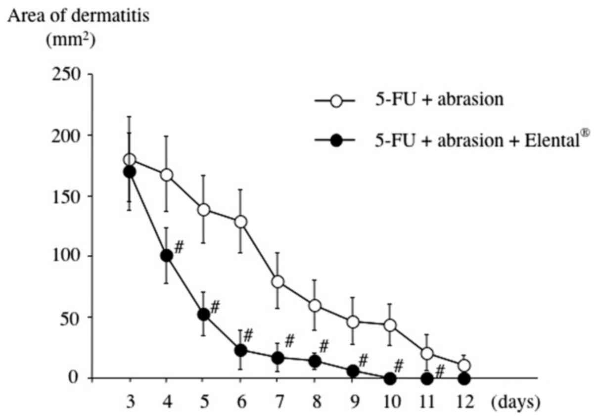
Results
Effect of Elental® on mouse
dorsal skin dermatitis
To induce dermatitis on the dorsal skin of mice,
5-FU administration and mechanical trauma were used. Ulcerated skin
tissue was observed after the second mechanical irritation (on day
3). As shown in Fig. 2, the
Elental® group exhibited a better healing rate than the
untreated control (5-FU + abrasion group). The affected dorsal area
was completely healed on days 9–10 in the Elental®
group, however it was healed on day 12 in the case of the 5-FU +
abrasion group.
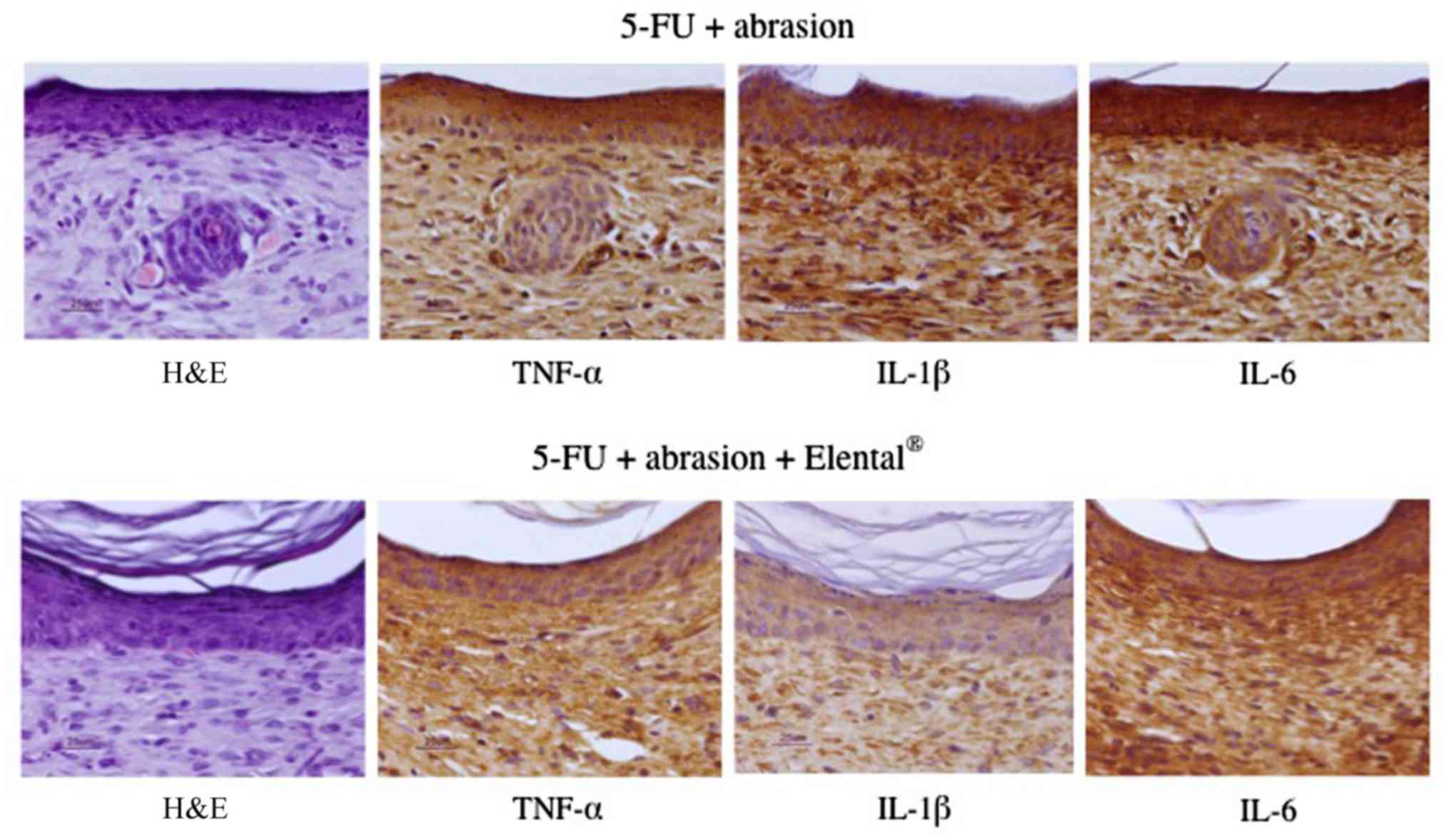
Effect of Elental® on the
expression of TNF-α, IL-1β and IL-6 in mouse dorsal skin
dermatitis
We then focused on pro-inflammatory cytokines,
TNF-α, IL-1β and IL-6 as the dermatitis-induced factors in this
study. Reduced expression of TNF-α, IL-1β and IL-6 was detected in
the dorsal skin tissue of the Elental® group compared to
the 5-FU + abrasion group (Fig. 3).
Particularly, the expression of IL-1β expression was greatly
reduced in the Elental® group compared to the untreated
control. This data indicated that Elental® may reduce
the expression of TNF-α, IL-1β and IL-6 in areas affected with
dermatitis.
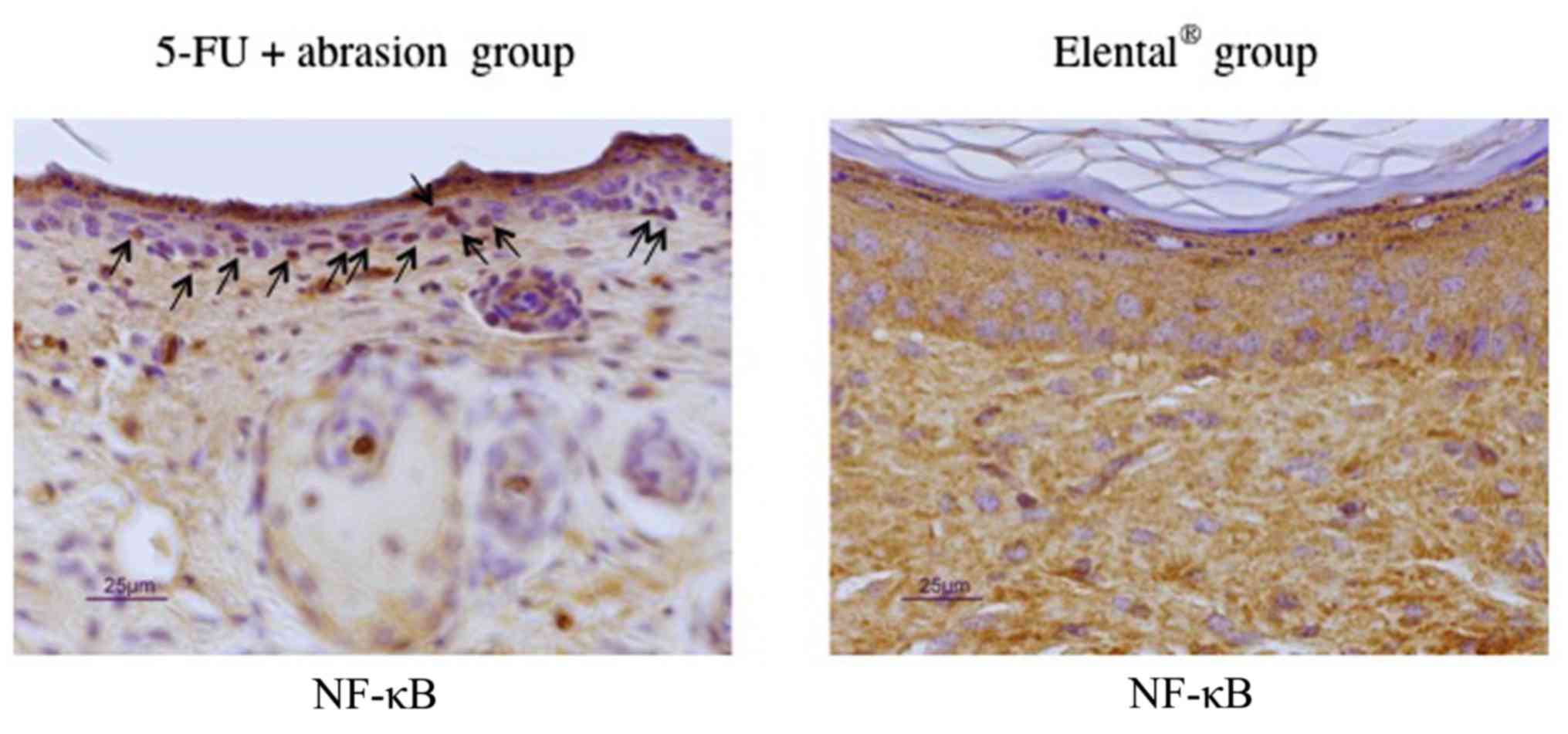
Effect of Elental® on the
expression of p65 in mouse dorsal skin dermatitis
Next, we tried to examine the expression of
transcription factor NF-κB in the dermatitis area of mouse dorsal
skin by immunohistochemical staining, as NF-κB plays an important
role in the regulation of TNF-α, IL-1β and IL-6. The expression of
p65 was mainly evident in the nucleus of the 5-FU + abrasion group,
but was mostly detected in the cytoplasm of the Elental®
group (Fig. 4). From this data we
can assume that, Elental® may attenuate 5-FU plus
abrasion-induced transcriptional activation of NF-κB.
Expression of TNF-α, IL-1β and IL-6 in
Elental®-treated cells
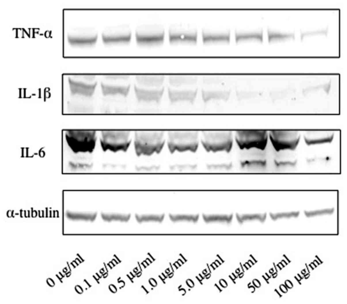
To clarify the mechanism involved in the healing
accelerating effect of Elental® for dermatitis, we
examined the expression of TNF-α, IL-1β and IL-6 in cells by
western blotting. Fig. 5 revealed
that Elental® (1.0–100 µg/ml) suppressed the expression
of TNF-α in cells compared to the untreated cells, and 100 µg/ml
Elental® exhibited the best result. Moreover,
Elental® (0.1–100 µg/ml) dose dependently suppressed the
expression of IL-1β, and moderately suppressed IL-6 expression in
treated cells compared to the untreated cells. IL-1β expression was
lowest in the10 and 50 µg/ml Elental® concentrations,
however, these doses slightly increased the expression of IL-6.
Additionally, IL-6 expression was the lowest in the 100 µg/ml
Elental®-treated cells compared to the untreated cells
(Fig. 5).
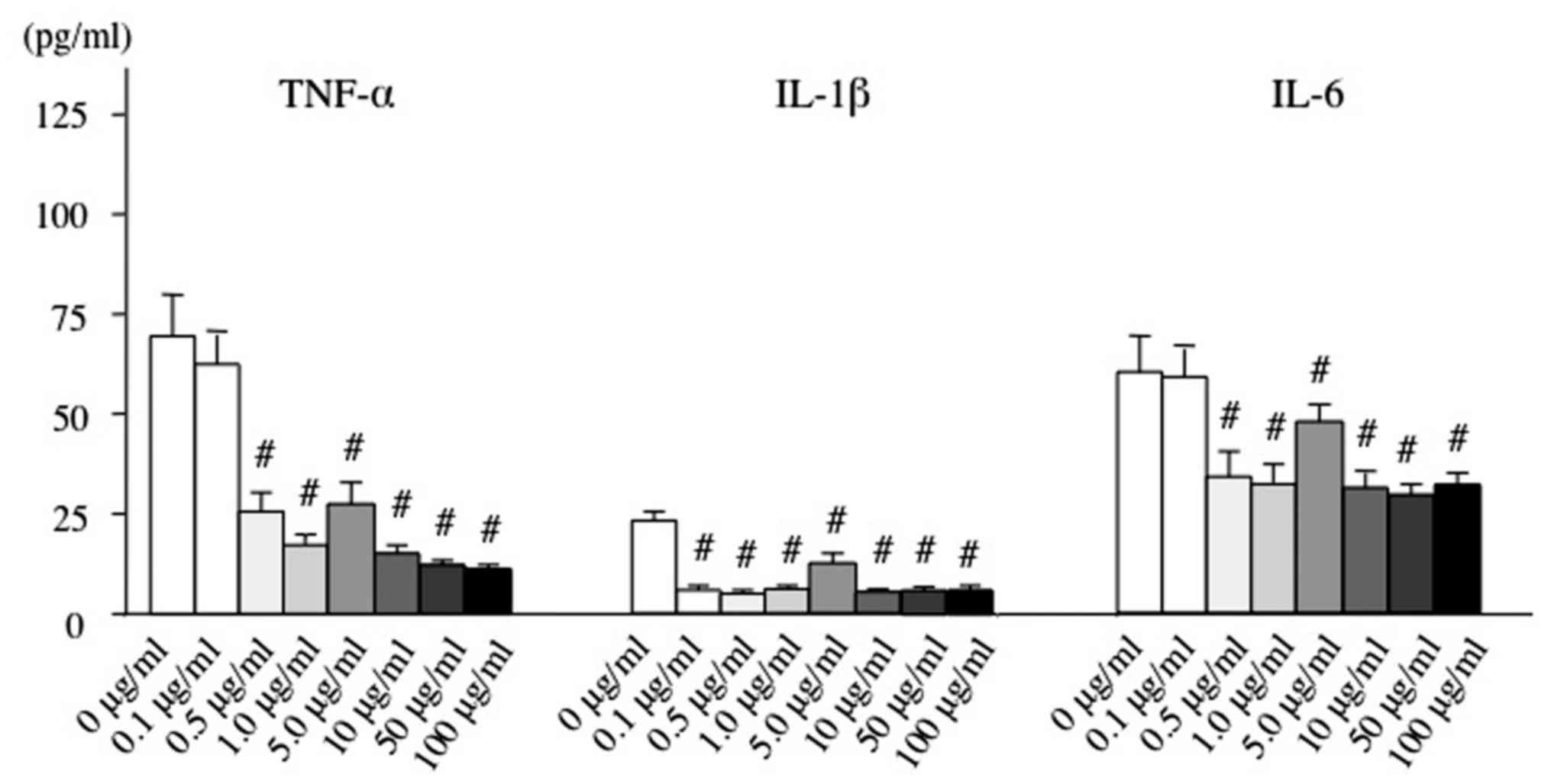
We also assessed the amount of TNF-α, IL-1β and IL-6
secreted into the culture medium by ELISA. As shown in Fig. 6, the amount of TNF-α, IL-1β and IL-6
secreted from Elental®-treated HaCaT cells was
significantly lower than that from untreated HaCaT cells. The
lowest expression of TNF-α was detected with the 100 µg/ml
Elental® treatment. In addition, all Elental®
concentrations decreased the expression of IL-1β almost at the same
rate except for the 5.0 µg/ml Elental® treatment.
Moreover, IL-6 expression was lowest with the 50 µg/ml
Elental® treatment.
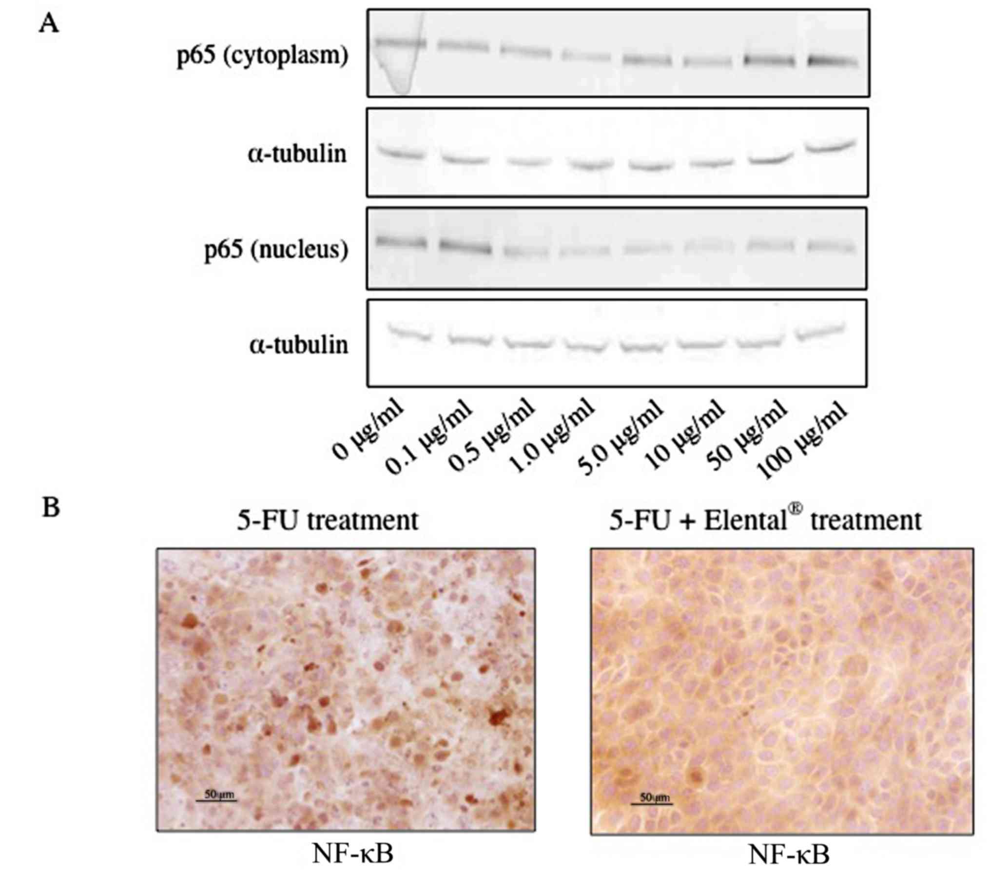
Effect of Elental® on the
expression of p65 in cultured HaCaT cells
We examined the expression of p65 NF-κB in cells by
western blotting and immunocytochemistry. Some Elental®
concentrations (5.0, 50 and 100 µg/ml) increased p65 expression in
the cytoplasm; however, 1.0–100 µg/ml Elental® decreased
p65 expression in nucleus of HaCaT cells (Fig. 7A). In addition, the expression of
p65 was detected in both the nucleus and cytoplasm of 5-FU-treated
HaCaT cells. However, p65 expression was mainly detected in the
cytoplasm of Elental®-treated HaCaT cells (Fig. 7B). This data supports our hypothesis
that Elental® may have an attenuating effect on
5-FU-induced transcriptional activation of NF-κB.
Discussion
The major adverse effects of chemotherapy and/or
radiotherapy including mucositis, dermatitis, dysphagia, xerostomia
and hematological toxicities often hamper cancer treatment, as it
can compromise the quality of life of patients (2,5,6). These
lead to a higher rate of unplanned breaks and delays in cancer
treatments, which always result in a poorer outcome (1,13,32).
However, effective treatments for chemotherapy-induced mucositis
and dermatitis have not been established, yet (33–37).
Elental® is a good source of nitrogen and
amino acids but low in fat. It has an easily digestible nutrition
formula that rarely requires a fully functional digestive system
(22). This ED is inexpensive, safe
and has been approved and covered by public insurance as a
prescription treatment for malnutrition in Japan.
Elental® has been reported to be effective in reducing
the severity of chemotherapy-induced mucositis and dermatitis in
colorectal cancer and esophageal cancer patients, as well as in
acute Crohn's disease (19–22). Elental® contains
L-glutamine (2.4 g/100 g), which helps in the treatment of cellular
injuries, chemotherapy-induced cell toxicities and mucositis
(22,32–37).
Our previous clinical study revealed the effectiveness of
Elental® for the treatment of chemotherapy-induced oral
mucositis and dermatitis without causing any adverse effects
(30). In the present study, we
examined the efficacy of Elental® in reducing
pro-inflammatory cytokine production in keratinocytes, and tried to
elucidate the detailed mechanisms of its action.
Elental® promoted the healing of
chemotherapy-induced dermatitis in our animal models as shown in
Fig. 2. Yamamoto et al
reported that Elental® reduced mucosal inflammation in
acute Crohn's disease by lowering the mucosal proinflammatory
cytokine production (23).
Therefore, we focused on pro-inflammatory cytokines including
TNF-α, IL-1β and IL-6 as the dermatitis-inducing factors in this
study. In addition, we examined NF-κB p65 which plays an important
role as a transcription factor in regulating many of the
pro-inflammatory cytokine genes including TNF-α, IL-1β and IL-6
(13).
In our animal model of dermatitis, we observed that
Elental® reduced the expression of TNF-α, IL-1β and
IL-6, and inhibited the nuclear transition of p65 NF-κB in the
keratinocytes of 5-FU-induced dermatitis regions (Figs. 3 and 4). Next, examined whether
Elental® reduced the production of pro-inflammatory
cytokines in vitro. Our data revealed that
Elental® had the desired effects on reducing
pro-inflammatory cytokine expression in HaCaT cells damaged by
5-FU-pretreatment. Briefly, Elental® decreased the
expression of TNF-α, IL-1β and IL-6 in HaCaT cells (Fig. 5), and reduced the production of
TNF-α, IL-1β and IL-6 released in the cultured medium of these
cells (Fig. 6). Moreover,
Elental® may have functioned as an inhibitor of NF-κB
p65 nuclear transition in 5-FU-pretreated HaCaT cells (Fig. 7).
Our findings revealed that Elental®
possibly attenuated 5-FU-induced transcriptional activation of
NF-κB, thereby reducing the expression of pro-inflammatory
cytokines. Several authors have reported that other agents, such as
caffeic acid, saikosaponin A and palmitic acid, can reduce
inflammation by inhibiting NF-κB, TNF-α, IL-1β and IL-6 (10,11,38),
and possibly Elental® works in the same way. However,
Elental® could be more beneficial for cancer patients
than those agents, since it is an ED and has other nutritional
benefits, but no side-effects. Moreover, Elental® has
been used clinically for several years in Japan, therefore, it is
already available for the treatment of cancer patients suffering
from chemotherapy-induced dermatitis and mucositis.
NF-κB is considered to be a good target for cancer
treatment as its activation directly affects cancer cell
proliferation, angiogenesis, metastasis, inflammation and apoptosis
(12,39), hence down-regulation of NF-κB
activity by Elental® could be overall useful for cancer
patients (40). It is also probable
that Elental® has regulatory effects on other
oncotargets besides NF-κB, therefore, extensive research and
clinical trials are necessary to identify its additional beneficial
effects. We suggest that, ED including Elental® may have
great potential for wide clinical application.
Acknowledgements
We thank Dr Dan Cui (Department of Pathology,
Yamaguchi University Graduate School of Medicine) for her valuable
suggestions and technical support in immunohistochemical
analysis.
Funding
The present study was supported in part by a
Grant-in-Aid from the Japanese Ministry of Education, Science and
Culture (grant no. 15K11292).
Availability of data and materials
The datasets used during the present study are
available from the corresponding author upon reasonable
request.
Authors' contributions
KH was involved in the study design, data analysis,
and writing of the manuscript. TF carried out all the experiments,
collected and evaluated data; also assisted in manuscript writing
and revision. YM and KM revised and edited the manuscript. All
authors read and approved the manuscript and agree to be
accountable for all aspects of the research in ensuring that the
accuracy or integrity of any part of the work are appropriately
investigated and resolved.
Ethics approval and consent to
participate
All procedures concerning animal handling and
treatment were conducted in accordance with the Guidelines for
Animal Experimentation of Yamaguchi University.
Consent for publication
Not applicable.
Competing interests
The authors have declared that they have no
competing interest.
References
|
1
|
Burdelya LG, Gleiberman AS, Toshkov I,
Aygun-Sunar S, Bapardekar M, Manderscheid-Kern P, Bellnier D,
Krivokrysenko VI, Feinstein E, Feinstein E and Gudkov AV: Toll-like
receptor 5 agonist protects mice from dermatitis and oral mucositis
caused by local radiation: Implications for head-and-neck cancer
radiotherapy. Int J Radiat Oncol Biol Phys. 83:228–234. 2012.
View Article : Google Scholar : PubMed/NCBI
|
|
2
|
Imai T, Matsuura K, Asada Y, Sagai S,
Katagiri K, Ishida E, Saito D, Sadayasu R, Wada H and Saijo S:
Effect of HMB/Arg/Gln on the prevention of radiation dermatitis in
head and neck cancer patients treated with concurrent
chemoradiotherapy. Jpn J Clin Oncol. 44:422–427. 2014. View Article : Google Scholar : PubMed/NCBI
|
|
3
|
Habl G, Potthoff K, Haefner MF, Abdollahi
A, Hassel JC, Boller E, Indorf M and Debus J: Differentiation of
irradiation and cetuximab induced skin reactions in patients with
locally advanced head and neck cancer undergoing
radioimmunotherapy: the HICARE protocol (head and neck cancer:
Immunochemo and radiotherapy with erbitux)-a multicenter phase IV
trial. BMC Cancer. 13:3452013. View Article : Google Scholar : PubMed/NCBI
|
|
4
|
Sonis ST: Oral mucositis. Anticancer
Drugs. 22:607–612. 2011. View Article : Google Scholar : PubMed/NCBI
|
|
5
|
Miyano K, Ueno T, Yatsuoka W and Uezono Y:
Treatment for Cancer Patients with Oral Mucositis: Assessment based
on the mucositis study group of the multinational association of
supportive care in cancer in international society of oral oncology
(MASCC/ISOO) in 2013 and proposal of possible novel treatment with
a Japanese herbal medicine. Curr Pharm Des. 22:2270–2278. 2016.
View Article : Google Scholar : PubMed/NCBI
|
|
6
|
Lalla RV, Sonis ST and Peterson DE:
Management of oral mucositis in patients who have cancer. Dent Clin
North Am. 52(61–77): viii2008.
|
|
7
|
Skubitz KM: Glutamine as a potential
treatment for the prevention of chemotherapy induced mucositis. J
Infusional Chemotherapy. 4:64–67. 1994.
|
|
8
|
Kyllo RL and Anadkat MJ: Dermatologic
adverse events to chemotherapeutic agents, part 1: Cytotoxics,
epidermal growth factor receptors, multikinase inhibitors, and
proteasome inhibitors. Semin Cutan Med Surg. 33:28–39. 2014.
View Article : Google Scholar : PubMed/NCBI
|
|
9
|
Shou J, Lieberman MD, Hofmann K, Leon P,
Redmond HP, Davies H and Daly JM: Dietary manipulation of
methotrexate-induced enterocolitis. J Parenter Enteral Nutr.
15:307–312. 1991. View Article : Google Scholar
|
|
10
|
Zhang M, Zhou J, Wang L, Li B, Guo J, Guan
X, Han Q and Zhang H: Caffeic acid reduces cutaneous tumor necrosis
factor alpha (TNF-α), IL-6 and IL-1β levels and ameliorates skin
edema in acute and chronic model of cutaneous inflammation in mice.
Biol Pharm Bull. 37:347–354. 2014. View Article : Google Scholar : PubMed/NCBI
|
|
11
|
Zhu J, Luo C, Wang P, He Q, Zhou J and
Peng H: Saikosaponin A mediates the inflammatory response by
inhibiting the MAPK and NF-κB pathways in LPS-stimulated RAW 264.7
cells. Exp Ther Med. 5:1345–1350. 2013. View Article : Google Scholar : PubMed/NCBI
|
|
12
|
Barnes PJ and Karin M: Nuclear
factor-kappaB: A pivotal transcription factor in chronic
inflammatory diseases. N Engl J Med. 336:1066–1071. 1997.
View Article : Google Scholar : PubMed/NCBI
|
|
13
|
McCarthy GM, Awde JD, Ghandi H, Vincent M
and Kocha WI: Risk factors associated with mucositis in cancer
patients receiving 5-fluorouracil. Oral Oncol. 34:484–490. 1998.
View Article : Google Scholar : PubMed/NCBI
|
|
14
|
Keefe DM, Schubert MM, Elting LS, Sonis
ST, Epstein JB, Raber-Durlacher JE, Migliorati CA, McGuire DB,
Hutchins RD and Peterson DE: Mucositis Study Section of the
Multinational Association of Supportive Care in Cancer and the
International Society for Oral Oncology: Updated clinical practice
guidelines for the prevention and treatment of mucositis. Cancer.
109:820–831. 2007. View Article : Google Scholar : PubMed/NCBI
|
|
15
|
Peterson DE, Bensadoun RJ and Roila F:
ESMO Guidelines Working Group: Management of oral and
gastrointestinal mucositis: ESMO clinical recommendations. Ann
Oncol. 20 Suppl 4:S174–S177. 2009. View Article : Google Scholar
|
|
16
|
Quinn B, Potting CM, Stone R, Blijlevens
NM, Fliedner M, Margulies A and Sharp L: Guidelines for the
assessment of oral mucositis in adult chemotherapy, radiotherapy
and haematopoietic stem cell transplantation patients. Eur J
Cancer. 44:61–72. 2008. View Article : Google Scholar : PubMed/NCBI
|
|
17
|
Henke M, Alfonsi M, Foa P, Giralt J,
Bardet E, Cerezo L, Salzwimmer M, Lizambri R, Emmerson L, Chen MG
and Berger D: Palifermin decreases severe oral mucositis of
patients undergoing postoperative radiochemotherapy for head and
neck cancer: A randomized, placebo-controlled trial. J Clin Oncol.
29:2815–2820. 2011. View Article : Google Scholar : PubMed/NCBI
|
|
18
|
Bensinger W, Schubert M, Ang KK, Brizel D,
Brown E, Eilers JG, Elting L, Mittal BB, Schattner MA, Spielberger
R, et al: NCCN Task Force Report. Prevention and management of
mucositis in cancer care. J Natl Compr Canc Netw. 6 Suppl 1:S1–S21;
quiz S22-S24. 2008.PubMed/NCBI
|
|
19
|
Svanberg A, Ohrn K and Birgegard G: Oral
cryotherapy reduces mucositis and improves nutrition-a randomised
controlled trial. J Clin Nurs. 19:2146–2151. 2010. View Article : Google Scholar : PubMed/NCBI
|
|
20
|
Scully C, Epstein J and Sonis S: Oral
mucositis: A challenging complication of radiotherapy,
chemotherapy, and radiochemotherapy. Part 2: Diagnosis and
management of mucositis. Head Neck. 26:77–84. 2004. View Article : Google Scholar : PubMed/NCBI
|
|
21
|
Cowen D, Tardieu C, Schubert M, Peterson
D, Resbeut M, Faucher C and Franquin JC: Low energy Helium-Neon
laser in the prevention of oral mucositis in patients undergoing
bone marrow transplant: Results of a double blind randomized trial.
Int J Radiat Oncol Biol Phys. 38:697–703. 1997. View Article : Google Scholar : PubMed/NCBI
|
|
22
|
Online EA Pharma Co., . Ltd Products
Information, Elental®. http://www.eapharma.co.jp/medicalexpert/product/elental/elental_information.htmlWebpage
in Japanese. 19–August. 2017
|
|
23
|
Yamamoto T, Nakahigashi M, Umegae S,
Kitagawa T and Matsumoto K: Impact of elemental diet on mucosal
inflammation in patients with active Crohn's disease: Cytokine
production and endoscopic and histological findings. Inflamm Bowel
Dis. 11:580–588. 2005. View Article : Google Scholar : PubMed/NCBI
|
|
24
|
Hanai H, Iida T, Takeuchi K, Arai H, Arai
O, Abe J, Tanaka T, Maruyama Y, Ikeya K, Sugimoto K, et al:
Nutritional therapy versus 6-mercaptopurine as maintenance therapy
in patients with Crohn's disease. Dig Liver Dis. 44:649–654. 2012.
View Article : Google Scholar : PubMed/NCBI
|
|
25
|
Johtatsu T, Andoh A, Kurihara M, Iwakawa
H, Tsujikawa T, Kashiwagi A, Fujiyama Y and Sasaki M: Serum
concentrations of trace elements in patients with Crohn's disease
receiving enteral nutrition. J Clin Biochem Nutr. 41:197–201. 2007.
View Article : Google Scholar : PubMed/NCBI
|
|
26
|
Yamamoto T, Nakahigashi M, Saniabadi AR,
Iwata T, Maruyama Y, Umegae S and Matsumoto K: Impacts of long-term
enteral nutrition on clinical and endoscopic disease activities and
mucosal cytokines during remission in patients with Crohn's
disease: A prospective study. Inflamm Bowel Dis. 13:1493–1501.
2007. View Article : Google Scholar : PubMed/NCBI
|
|
27
|
Yamamoto T, Nakahigashi M, Umegae S,
Kitagawa T and Matsumoto K: Impact of long-term enteral nutrition
on clinical and endoscopic recurrence after resection for Crohn's
disease: A prospective, non-randomized, parallel, controlled study.
Aliment Pharmacol Ther. 25:67–72. 2007. View Article : Google Scholar : PubMed/NCBI
|
|
28
|
Fukui T, Itoh Y, Orihara M, Yoshizawa K,
Takeda H, Kawada S and Yoshioka T: Elental prevented and reduced
oral mucositis during chemotherapy in patients esophageal cancer.
Gan To Kagaku Ryoh. 38:2597–2601. 2011.(In Japanese).
|
|
29
|
Ogata Y, Takeuchi M, Ishibashi N, Kibe S,
Takahashi K, Uchida S, Murakami N, Yahara T and Shirouzu K:
Efficacy of Elental on prevention for chemotherapy-induced oral
mucositis in colorectal cancer patients. Gan To Kagaku Ryoho.
39:583–587. 2012.(In Japanese). PubMed/NCBI
|
|
30
|
Harada K, Ferdous T, Horinaga D, Uchida K,
Mano T, Mishima K, Park S, Hanazawa H, Takahashi S, Okita A, et al:
Efficacy of elemental diet on prevention for
chemoradiotherapy-induced oral mucositis in patients with oral
squamous cell carcinoma. Support Care Cancer. 24:953–959. 2016.
View Article : Google Scholar : PubMed/NCBI
|
|
31
|
Harada K, Ferdous T, Kobayashi H and
Ueyama Y: Elemental diet accelerates the recovery from oral
mucositis and dermatitis induced by 5-fluorouracil through the
induction of fibroblast growth factor 2. Integr Cancer Ther. July
1–2017.(Epub ahead of print). PubMed/NCBI
|
|
32
|
O'Dwyer ST, Scott T, Smith RJ and Wilmore
DW: 5-fluorouracil toxicity on small intestinal mucosa but not
white blood cells is decreased by glutamine. Clin Res.
35:367A1987.
|
|
33
|
Carneiro-Filho BA, Oriá RB, Rea Wood K,
Brito GA, Fujii J, Obrig T, Lima AA and Guerrant RL:
Alanyl-glutamine hastens morphologic recovery from
5-fluorouracil-induced mucositis in mice. Nutrition. 20:934–941.
2004. View Article : Google Scholar : PubMed/NCBI
|
|
34
|
Kandil HM, Argenzio RA, Chen W,
Berschneider HM, Stiles AD, Westwick JK, Rippe RA, Brenner DA and
Rhoads JM: L-glutamine and l-asparagine stimulate ODC activity and
proliferation in a porcine jejunal enterocyte line. Am J physiol.
269:G591–G599. 1995.PubMed/NCBI
|
|
35
|
Rhoads JM, Argenzio RA, Chen W, Rippe RA,
Westwick JK, Cox AD, Berschneider HM and Brenner DA: L-glutamine
stimulates intestinal cell proliferation and activates
mitogen-activated protein kinases. Am J Physiol. 272:G943–G953.
1997.PubMed/NCBI
|
|
36
|
Hong RW, Rounds JD, Helton WS, Robinson MK
and Wilmore DW: Glutamine preserves liver glutathione after lethal
hepatic injury. Ann Surg. 215:114–119. 1992. View Article : Google Scholar : PubMed/NCBI
|
|
37
|
Denno R, Rounds JD, Faris R, Holejko LB
and Wilmore DW: Glutamine-enriched total parenteral nutrition
enhances plasma glutathione in the resting state. J Surg Res.
61:35–38. 1996. View Article : Google Scholar : PubMed/NCBI
|
|
38
|
Zhou BR, Zhang JA, Zhang Q, Permatasari F,
Xu Y, Wu D, Yin ZQ and Luo D: Palmitic acid induces production of
proinflammatory cytokines interleukin-6, interleukin-1β, and tumor
necrosis factor-α via a NF-κB-dependent mechanism in HaCaT
keratinocytes. Mediators Inflamm. 2013:5304292013. View Article : Google Scholar : PubMed/NCBI
|
|
39
|
Baud V and Karin M: Is NF-kappaB a good
target for cancer therapy? Hopes and pitfalls. Nat Rev Drug Discov.
8:33–40. 2009. View Article : Google Scholar : PubMed/NCBI
|
|
40
|
Yamamoto Y and Gaynor RB: Therapeutic
potential of inhibition of the NF-kappaB pathway in the treatment
of inflammation and cancer. J Clin Invest. 107:135–142. 2001.
View Article : Google Scholar : PubMed/NCBI
|